mismatch repair proteins initiate epigenetic alterations
TRANSCRIPT

Mismatch Repair Proteins Initiate Epigenetic Alterations During Inflammation-Driven Tumorigenesis
Ashley R. Maiuri1, Michael Peng1, Shruthi Sriramkumar1, Caitlin M. Kamplain1, Christina E. DeStefano Shields3, Cynthia L. Sears3,4,5, and Heather M. O’Hagan1,2
1Medical Sciences, Indiana University School of Medicine, Bloomington IN, 47405, USA
2Indiana University Melvin and Bren Simon Cancer Center, Indianapolis, IN, 46202, USA
3Departments of Medicine and Oncology, Johns Hopkins University, Baltimore MD, 21287, USA
4Bloomberg-Kimmel Institute for Cancer Immunotherapy, Johns Hopkins University, Baltimore, MD, 21287, USA
5Sidney Kimmel Comprehensive Cancer Center, Johns Hopkins University, Baltimore, MD, 21287, USA
Abstract
Aberrant silencing of genes by DNA methylation contributes to cancer, yet how this process is
initiated remains unclear. Using a murine model of inflammation-induced tumorigenesis, we tested
the hypothesis that inflammation promotes recruitment of epigenetic proteins to chromatin,
initiating methylation and gene silencing in tumors. Compared to normal epithelium and non-
inflammation-induced tumors, inflammation-induced tumors gained DNA methylation at CpG
islands, some of which are associated with putative tumor suppressor genes. Hypermethylated
genes exhibited enrichment of repressive chromatin marks and reduced expression prior to
tumorigenesis, at a time point coinciding with peak levels of inflammation-associated DNA
damage. Loss of MutS homolog 2 (MSH2), a mismatch repair (MMR) protein, abrogated early
inflammation-induced epigenetic alterations and DNA hypermethylation alterations observed in
inflammation-induced tumors. These results indicate that early epigenetic alterations initiated by
inflammation and MMR proteins lead to gene silencing during tumorigenesis, revealing a novel
mechanism of epigenetic alterations in inflammation-driven cancer. Understanding such
mechanisms will inform development of pharmacotherapies to reduce carcinogenesis.
Keywords
DNA methylation; mismatch repair; EZH2; inflammation; colon cancer
Corresponding author: Heather M. O’Hagan, [email protected], 1001 East 3rd St. Jordan Hall Room 108 Bloomington, IN 47405.
COI statement: The authors declare no potential conflicts of interest.
Additional Information:Heather M. O’Hagan financial support: NIEHS (R01ES023183)Cynthia L. Sears financial support: NCI (R01CA151325) and support from the Department of Medicine and the Bloomberg Kimmel Immunotherapy Institute.
HHS Public AccessAuthor manuscriptCancer Res. Author manuscript; available in PMC 2018 July 01.
Published in final edited form as:Cancer Res. 2017 July 01; 77(13): 3467–3478. doi:10.1158/0008-5472.CAN-17-0056.
Author M
anuscriptA
uthor Manuscript
Author M
anuscriptA
uthor Manuscript

Introduction
Approximately 25% of all cancers are associated with chronic inflammation (1). Colorectal
cancer (CRC) is a substantial contributor to morbidity and mortality worldwide (2).
Although the vast majority of CRC cases are sporadic, 5–10% of cases have been attributed
to hereditary conditions (3). In addition to family history, several risk factors associated with
CRC have been identified including age, inflammatory bowel disease, bacterial infection,
obesity, excessive alcohol consumption, and smoking (4). Most of these risk factors are
characterized by chronic inflammation (5). Moreover, nonsteroidal anti-inflammatory drug
use was reported to reduce the risk of CRC, reinforcing the notion that inflammation is a
central contributor to CRC development (6).
In addition to inflammation, genetic mutations and epigenetic alterations play a key role in
driving CRC (7, 8). Several genetic mutations that contribute to the initiation of CRC have
been identified and well characterized (9). Epigenetic alterations also contribute to CRC
initiation and progression. DNA methylation is the most extensively studied epigenetic
alteration in cancer and cancer cells exhibit a global loss of DNA methylation that is thought
to lead to genomic instability (8). Additionally, there is concurrent hypermethylation at
distinct regions, often within promoter CpG islands of tumor suppressor genes (TSGs) (8).
Such hypermethylation can lead to TSG silencing and contribute to cancer. While the
fundamental importance of cancer-specific DNA methylation alterations is clear, how they
are initiated is not well understood.
Our group previously demonstrated that treatment of cancer cells with the reactive oxygen
species (ROS) hydrogen peroxide (H202) causes localization of an epigenetic silencing
complex containing DNA methyltransferases DNMT1 and DNMT3B, and the chromatin
silencers Sirtuin-1 (SIRT1) and Enhancer of Zeste-2 (EZH2) to sites of damaged chromatin.
Notably, there was enrichment of these silencing proteins at the promoter CpG islands of
TSGs, resulting in reduced gene expression (10). Based on these findings, we hypothesized
that inflammation would induce similar events in vivo.
In the present study, we uncover a novel mechanism responsible for initiating cancer-
relevant epigenetic alterations in a murine model of inflammation-induced tumorigenesis.
Our results demonstrate that epigenetic alterations that occur during inflammation-driven
tumorigenesis are distinct from those that occur during inflammation-independent
tumorigenesis. To our knowledge, we are the first to demonstrate the involvement of the
mismatch repair (MMR) pathway in initiating epigenetic alterations during inflammation-
induced tumorigenesis. Since inflammation and epigenetic alterations are critical in the
development of many cancer types and diseases, a deeper understanding of the interplay
between these two factors, as gained from this study, is broadly relevant.
Materials and Methods
Animal model
C57BL/6J (Jackson labs) and MinApcΔ716+/− mice were handled and inoculated with ETBF
as in Wu et al. (2009) (11). Msh2l/lVC are a result of crossing B6.Cg-MSH2tm2.1Rak/J and
Maiuri et al. Page 2
Cancer Res. Author manuscript; available in PMC 2018 July 01.
Author M
anuscriptA
uthor Manuscript
Author M
anuscriptA
uthor Manuscript

B6.Cg-Tg(Vil1-cre)997Gum/J mice (Jackson Labs) to create mice homozygous for
MSH2tm2.1Rak and expressing the Vil1-cre transgene. Littermates not expressing Vil1-cre
were used as WT controls. Msh2l/lVC/Min are the result of crossing Msh2l/lVC and
MinApcΔ716+/− mice. For all experiments both males and females were used, mice were
randomized between mock and ETBF groups, and mice of different genotypes were
cohoused. Individual tumors were removed from dissected colons with the aid of a
dissecting microscope and stored at −80°C until further analysis. Distal (0–2 cm measured
from the rectum) and proximal (feathered portion adjacent to ceacum) epithelium was
collected by scraping the mucosal surface of the dissected colon (after removal of any
tumors), washed three times in PBS, and then subjected to the indicated protocol. Such
scraping has been shown by others to be an effective method to obtain samples of intestinal
epithelial cells (12). All mouse experiments were covered under protocol number 16-027,
which was approved by the Indiana University Bloomington Animal Care and Use
Committee in accordance with the Association for Assessment and Accreditation of
Laboratory Animal Care International.
Methyl CpG binding domain (MBD)-seq
DNA was isolated from tumors or epithelium using the QIAamp DNA mini kit (Qiagen)
following the manufacturer’s protocol. To identify differentially methylated regions
(DMRs), MBD enrichment was performed from DNA from epithelium collected from
different mice (n=5 WT/Min mock, n=3 WT/Min ETBF, n=3 Msh2l/lVC/Min mock) or
individual tumors (n= 3 WT/Min mock, n=7 WT/Min ETBF, n=3 Msh2l/lVC/Min mock,
n=3 Msh2l/lVC/Min ETBF) using Diagenode’s MethylCap kit. Libraries were prepared
following Bioo Scientific’s Methyl Sequencing kit. Single-end 75 bp sequencing was
performed using an Illumina Nextseq. Z-scores were calculated using a 500bp fixed sized
bin spanning CpG islands based on the distribution of coverage from uniquely mapped
reads. Z-ratios were derived from the comparison of z-scores for the different sample types
for the 500bp regions. See Supplemental Methods for more detail.
Pyrosequencing and Quantitative Methylation Specific PCR (qMSP)
DNA was bisulfite treated (EZ DNA methylation-Gold kit, Zymo Research) and used for
pyrosequencing. qMSP assays were first tested using a standard curve of bisulfite treated
mixtures of unmethylated and methylated DNA (data not shown). Only methylated DNA
assays with little to no amplification of unmethylated samples, close to 100% efficiency and
R2 close to 1 were used further. See Table S1 for assays and primers used.
Gene expression
RNA was prepared from epithelium or tumors using Trizol followed by cleanup with a
RNeasy kit (Qiagen). cDNA was prepared and qPCR was done using TaqMan assays (see
Table S1 for assays used). Expression of candidate genes was normalized to expression of a
housekeeping gene (PPIA or 18S).
Maiuri et al. Page 3
Cancer Res. Author manuscript; available in PMC 2018 July 01.
Author M
anuscriptA
uthor Manuscript
Author M
anuscriptA
uthor Manuscript
Tight chromatin fractionation
Tight chromatin fractionation of washed epithelium was performed as described in Ding et
al. (13). Blots presented are representative of three independent experiments.
Chromatin immunoprecipitation (ChIP)
ChIP was performed using anti-H3K27me3 (Diagenode) or anti-EZH2 (D2C9-Cell
Signaling Technologies) and the iDeal CHIP-Seq kit for histones and transcription factors
(Diagenode) according to the manufacturer’s instructions.
CoIP
coIPs were performed from nuclear extracts that were treated with oligoamines to release
chromatin-bound proteins as described in Ding et al. (13). Antibodies used were rabbit IgG
(Millipore) and anti-EZH2 (5246-Cell Signaling Technologies). Blots presented are
representative of two independent experiments.
MSI
MSI analysis was performed as in Woerner et al. (14). MSI was determined by comparing
DNA fragment analysis of mononucleotide markers in tumors relative to tail DNA from the
same mouse.
16S microbiome sequencing
DNA was isolated from stool using the ZR Fecal DNA micro kit (Zymo Research). Libraries
were made from 16S V1-V3 PCR amplicons from the stool DNA using the NEXTflex 16S
V1-V3 kit (Bioo Scientific). 300 bp paired-end sequencing of pooled libraries was
performed on a MiSeq. Read quality filtering was performed using mothur. AbundantOTU+
was used for clustering of sequences to Operational Taxonomic Units (OTUs) and further
classification. MetagenomeSeq was used to determine differential abundance across samples
using normalized OTU read counts. OTUs with significant (5% FDR) differential abundance
between any of the sample groups were used to create the heatmap (65 OTUs).
Total CpG methylation
Total CpG methylation in DNA from epithelium and tumor samples was determined using
an ELISA-based assay (MethylFlash Global DNA Methylation (5-mC) – Epigenetek).
Statistical Analysis
Expression data, densitometry, qMSP, and local ChIP are presented as the mean +/− standard
error (SEM). These data are evaluated by one-tail t-test and considered statistically
significant with a p-value < 0.05. Sample sizes are indicated in associated figure legends.
MBD-seq statistical analysis is detailed in the Supplemental Methods.
Data availability
MBD and 16S sequencing data have been deposited at the Sequence Read Archive (SRA)
data repository (project accession number SRP105286).
Maiuri et al. Page 4
Cancer Res. Author manuscript; available in PMC 2018 July 01.
Author M
anuscriptA
uthor Manuscript
Author M
anuscriptA
uthor Manuscript

Results
Inflammation-induced tumors have a unique DNA hypermethylation signature
Enterotoxigenic Bacteroides fragilis (ETBF) is a clinically relevant strain of B. fragilis (15).
When Multiple intestinal neoplasia (Min) mice, which are heterozygous for mutant
adenomatous polyposis coli (ApcΔ716), are infected with ETBF, tumors rapidly form at the
site of inflammation, in the distal colon (11). Studies have demonstrated that inflammation-
induced tumors have unique DNA methylation alterations compared to uninflamed tissue
therefore we evaluated DNA methylation in our model (16, 17). Min mice spontaneously
develop rare colon tumors (18) allowing us to uniquely compare DNA methylation
alterations of inflammation-induced ETBF tumors to methylation alterations of background
Min (mock) tumors. Interestingly, relative to mock epithelium, the ETBF tumors compared
to mock tumors had more hypermethylated DMRs (203 compared to 6) but fewer
hypomethylated DMRs (194 compared to 700, Table 1, Figure 1A). Of the methylation gains
that occurred in mock tumors, 5 overlap with the ETBF tumors (Table S2).
As Naumov et al. (19) demonstrated in human tumors, regions that gained methylation in
ETBF tumors had low DNA methylation levels in mock epithelium (mean z-score of 0.4)
whereas regions that lost methylation in ETBF or mock tumors had relatively high DNA
methylation levels in mock epithelium (mean z-score 3.4 or 3.6, respectively) (Figure 1B).
Both gains and losses occurred more often in CpG islands in exons or promoters of genes
than introns, 3′ UTRs, or intragenic regions (Figure S1A). As expected, regions targeted for
DNA hypermethylation were enriched for regions that are bivalent (containing both
H3K4me3 and H3K27me3) in mouse embryonic stem cells (Figure S1B) (20–24).
Since chronic inflammation is known to induce DNA methylation changes in inflamed
epithelium (16), we also assayed methylation changes in the distal colon epithelium of
ETBF-infected mice (ETBF epithelium). ETBF epithelium had fewer hypermethylated
DMRs in comparison to the ETBF tumors, but more than the mock epithelium and mock
tumors (Table 1). Eleven of the 40 hypermethylated DMRs observed in the ETBF epithelium
overlapped with those in the ETBF tumors (Table S2). When samples were clustered based
on regions with DNA hypermethylation in tumors relative to mock epithelium, the ETBF
epithelium clustered with the mock epithelium (Figure 1A). Interestingly, ETBF tumors
clustered separately from the mock tumors and epithelium, suggesting that they have distinct
methylation gains from the other samples.
Gene ontology (GO) analysis revealed that the genes associated with DMRs in the mock and
ETBF tumors are involved in multiple biological processes many of which are associated
with aspects of development, differentiation, and regulation of transcription (Table S3).
Interestingly, several of the methylation gains observed in the ETBF tumors occurred within
CpG islands of genes with known tumor suppressive function such as Hoxa5, Polg, Runx1, Runx3, CD37, Stx11, Tceb2, Lgr6, Cdx1, Fut4 (Figure 1C, Table S2) (25–34). CpG island
hypermethylation at 18 out of 21 candidate genes in ETBF tumors versus mock epithelium
were validated by pyrosequencing or qMSP (Figure 1D, 1E, S1C). No DNA methylation
changes were detected in the promoter CpG island of Gapdh, a negative control (Figure
S1C). 18 candidate genes are associated with human digestive organ tumors by Ingenuity
Maiuri et al. Page 5
Cancer Res. Author manuscript; available in PMC 2018 July 01.
Author M
anuscriptA
uthor Manuscript
Author M
anuscriptA
uthor Manuscript
Pathway Analysis, demonstrating their relevance to human disease (Figure S1D). Runx3,
Gata2, and Hoxa5 are also known to undergo aberrant DNA hypermethylation in human
CRC (35, 36). qMSP analysis at candidate genes demonstrated an intermediate level of
methylation in the ETBF epithelium, between that observed in the mock epithelium and
ETBF tumor (Figure 1E) consistent with the data in Figure 1A.
As hypothesized, many genes containing hypermethylated promoter CpG islands in ETBF
tumors also had reduced mRNA expression (Figure 1F). Additionally, several candidate
genes had lower expression in ETBF epithelium compared to ETBF tumors, but higher
expression compared to mock epithelium (Figure 1F), consistent with the methylation results
in Figure 1E. Genes that contained hypermethylated CpG islands in non-promoter exons had
increased gene expression as has been demonstrated previously (Figure S1E – Cldn4, Spi1,
Stx11) (37). Overall, these findings indicate that ETBF tumors contain distinct cancer-
specific DNA hypermethylation alterations that distinguish them from mock and ETBF
epithelium and mock tumors.
Early chromatin and transcriptional changes occur in genes that are DNA hypermethylated in inflammation-induced tumors
We hypothesized that ETBF-induced oxidative DNA damage may initiate epigenetic
changes in the distal colon that ultimately result in the DNA hypermethylation observed in
the inflammation-induced tumors. ETBF inoculation caused oxidative DNA damage
(increased 8-oxoguanine) in the distal colon epithelium two days post-ETBF, which returned
to background levels seven days post-ETBF (Figure 2A). We confirmed that the oxidative
DNA damage was occurring in epithelial cells by assaying 8-oxoguanine in cells positive for
the epithelial marker, EPCAM (Figure 2B).
The increased oxidative DNA damage observed in the distal colon epithelium two days post-
ETBF corresponded to a decrease in the mRNA expression of several candidate genes that
were found to be hypermethylated in the ETBF tumors with expression levels returning back
to normal seven days post-ETBF (Figure 2C). To address whether these changes occurred
specifically in epithelial cells, we assessed the purity of our colon epithelial samples. Our
samples contained approximately 84% EPCAM-positive cells and fewer than 2% CD45-
positive cells (Figure S2A), irrespective of ETBF treatment status. To probe this further we
also cultured organoids from the distal colon epithelium following an established protocol
(38), treated the epithelial organoids with H2O2 to mimic inflammation/oxidative damage in
vitro, and examined expression of several candidate genes. Indeed, the candidate genes
examined (Cdx1, Fut4, Hoxa5, Polg, Runx1) had reduced expression in response to
oxidative stress (but not housekeeping genes Rpl0, Tbp), analogous to our in vivo results
(Figure S2B). This result suggests that the changes observed in the colon epithelium in
response to ETBF are likely occurring in epithelial cells and not an alternative cell type
present in the mucosal surface.
Since EZH2 has been implicated in transcriptional repression at sites of oxidative DNA
damage, we hypothesized that this protein would be enriched in the promoters of candidate
genes that have reduced expression in the inflamed epithelium. Indeed, EZH2 was
significantly enriched at several candidate genes in ETBF epithelium compared to mock
Maiuri et al. Page 6
Cancer Res. Author manuscript; available in PMC 2018 July 01.
Author M
anuscriptA
uthor Manuscript
Author M
anuscriptA
uthor Manuscript

epithelium two days post-ETBF (Figure 2D). These regions also had enrichment of the
repressive chromatin mark catalyzed by EZH2, trimethyl H3K27 (H3K27me3) (Figure 2E).
These findings indicate that ETBF initiates early epigenetic and transcriptional alterations in
several candidate genes from the MBD-seq dataset, and these changes are temporally and
spatially associated with the oxidative DNA damage induced by ETBF.
MSH2 interacts with EZH2 and plays a key role in the initiation of early ETBF-induced epigenetic alterations
The MSH2-MSH6 heterodimer facilitates repair of clustered oxidative DNA lesions in a
PCNA-dependent and S-phase-independent manner (39). In accordance with this finding,
our group observed in vitro that MSH2 and MSH6 become more tightly bound to chromatin
in response to oxidative damage and are indispensable for the recruitment of epigenetic
silencing proteins to damaged chromatin (13). Therefore, we hypothesized that MSH2 and
MSH6 contribute to early epigenetic alterations that occur in response to ETBF.
First, we demonstrated that EZH2 co-immunoprecipitated with DNMT1, MSH2, MSH6, and
PCNA in the distal colon epithelium two days after ETBF, suggesting an interaction between
epigenetic proteins and MMR proteins in response to ETBF (Figure 3A). This interaction
did not occur in mock epithelium. SUZ12 and EED are known EZH2 interacting partners
and are positive controls for the EZH2 co-IP.
To evaluate whether the MMR pathway is involved in ETBF-mediated epigenetic alterations,
we examined mice that lack expression of Msh2 in intestinal epithelial cells. In Msh2l/lVC mice, the Msh2 gene is flanked by LoxP sequences and Cre recombinase is driven by a
Villin promoter leading to deletion of Msh2 in intestinal epithelial cells (40). We verified
that MSH2 protein is present in colon crypt cells in mock and ETBF inoculated wild type
(WT) mice, but not in crypts from Msh2l/lVC mice (Figure 3B). In distal colon epithelium
from WT ETBF-infected mice, epigenetic silencing proteins (EZH2, DNMT1, and SIRT1)
and MMR proteins (MSH2, MSH6 and PCNA) have a higher affinity for chromatin than in
mock mice (Figure 3C). This enhanced affinity occurred in the distal colon and not in the
proximal colon that lacks ETBF-mediated inflammation. Loss of Msh2 reduced the ETBF-
mediated increase in binding of EZH2, DNMT1 and SIRT1 to chromatin (Figure 3C),
highlighting the necessity of the MMR pathway in the response of epigenetic proteins to
ETBF-induced damage. LAMB (Lamin-B), a loading control, was consistent across the
samples. Levels of phosphorylated H2AX (γ-H2AX), a marker of DNA damage, were
elevated in ETBF samples relative to mock samples. Interestingly, γ-H2AX levels were
consistently higher in mock epithelium from Msh2l/lVC than WT mice, corresponding to a
relative increase in binding of EZH2, SIRT1 and PCNA to chromatin. Moreover we
examined the effect of loss of Msh2 on mRNA expression of several candidate genes and
their expression was unaffected by ETBF in Msh2l/lVC mice (Figure 3D, S3A).
To rule out the possibility that loss of Msh2 alters the initial immune response to ETBF we
examined the expression of several cytokines in response to ETBF two and seven days post-
infection and found that their expression was unaltered by loss of Msh2 (Figure S3B, C).
Cell proliferation measured by Ki-67 staining and phosphorylation of STAT-3 was also
unaffected by loss of Msh2 (Figure S3D, E). ETBF-infected Msh2l/lVC mice also have
Maiuri et al. Page 7
Cancer Res. Author manuscript; available in PMC 2018 July 01.
Author M
anuscriptA
uthor Manuscript
Author M
anuscriptA
uthor Manuscript
similar numbers of γ-H2AX foci per crypt compared to ETBF-infected WT mice (Figure
3E). Collectively, these findings implicate a role for MSH2 in initiating early epigenetic and
transcriptional alterations observed in the inflamed colon epithelium of mice two days post-
ETBF.
Loss of Msh2 does not alter the background microbiota composition
It is possible that an alteration of the microbiota composition caused by loss of Msh2 could
explain reduced ETBF-induced epigenetic alterations in Msh2l/lVC mice. Therefore, we
examined whether loss of Msh2 alters the initial or ETBF-induced intestinal microbiota
composition. 16S ribosomal RNA gene sequencing was performed on stool DNA and
sequences were clustered into OTUs. Principal component analysis (PCA) and hierarchical
clustering analysis based on the OTUs that had significantly differential abundance among
the sample types revealed a high degree of similarity between the bacterial populations
present in stool from mock-infected mice, regardless of genotype (Figure 4A and B). None
of the OTUs were significantly different between the mock groups, suggesting that loss of
Msh2 does not alter the background microbiota composition (Table S4). There was
separation between samples from ETBF-infected and mock-infected mice, irrespective of
genotype (Figure 4A and B, Table S4). PCA revealed a small degree of separation between
ETBF-infected WT/Min versus ETBF-infected Msh2l/lVC/Min samples suggesting that loss
of Msh2 might impact the ETBF-induced microbiota (Figure 4A and B, Table S4). However,
loss of Msh2 correlates with a significant increased abundance of ETBF (Figure 4C) and
colony formation units of ETBF per gram of stool (Figure S4), ruling out the possibility that
loss of Msh2 reduces ETBF-mediated epigenetic alterations by reducing the abundance of
ETBF. Altogether, loss of Msh2 did not alter the background microbiota of these mice,
although it does alter the relative abundance of some bacterial populations upon ETBF-
infection.
Loss of Msh2 in intestinal epithelial cells caused an increase in inflammation-induced tumors with microsatellite instability
Although loss of Msh2 reduced early ETBF-induced epigenetic alterations (Figure 3),
sporadic and germline mutations in the MMR pathway are commonly implicated in CRC
pathogenesis (4). Therefore, we examined the effect of Msh2 deletion from intestinal
epithelial cells on tumorigenesis in our model. Mock Msh2l/lVC/Min mice were similar to
uninfected WT/Min mice in terms of tumor burden (Figure 4D). Interestingly, ETBF-
infected Msh2l/lVC/Min mice developed significantly more tumors in the distal colon than
the WT/Min mice infected with ETBF, but the same regions had the highest number of
tumors in both sets of mice (Figure 4D).
The Th17/STAT-3 immune response drives ETBF-mediated tumorigenesis in Min mice (11).
Msh2 deletion did not alter ETBF-induced phosphorylation of STAT-3 in ETBF-induced
tumors (Figure 4E). Furthermore, inactivation of APC leads to constitutive activation of the
WNT/β-CATENIN signaling pathway in tumors in Min mice. Loss of Msh2 did not alter the
magnitude of increase in levels of β-CATENIN or PCNA, a marker of proliferation, in
ETBF-induced tumors (Figure 4E).
Maiuri et al. Page 8
Cancer Res. Author manuscript; available in PMC 2018 July 01.
Author M
anuscriptA
uthor Manuscript
Author M
anuscriptA
uthor Manuscript

Mutations in MMR genes are known to cause genomic instability by altering the length of
repetitive DNA sequences known as microsatellites (4). Interestingly, the mock and ETBF
Msh2l/lVC/Min tumors tested had microsatellite instability (MSI) whereas most tumors from
the WT/Min mice were microsatellite stable (MSS) (9/11) (Table 2). These results are
consistent with the observation in humans that mutated MSH2 can lead to colon
tumorigenesis (4) and suggest that ETBF-induced tumors from Msh2-deficient mice are
more genetically unstable than ETBF-induced tumors from Msh2-sufficient mice.
Loss of Msh2 decreases CpG island hypermethylation and restores expression of candidate TSGs
We initially demonstrated that Min ETBF tumors have a unique hypermethylation signature
(Figure 1, Table 1). Since the Msh2l/lVC/Min mice still developed tumors in response to
ETBF, this model provided us with a unique tool to answer the question: is MSH2 required
for ETBF-induced tumor-specific DNA hypermethylation alterations during tumorigenesis?
Therefore, we examined the effect of Msh2 deletion on methylation in ETBF tumors using
MBD-seq. Importantly, loss of Msh2 dramatically reduced ETBF-mediated
hypermethylation alterations with the Msh2l/lVC/Min ETBF tumors having only 11 DNA
hypermethylated regions relative to Msh2l/lVC/Min mock epithelium (Figure 5A and B,
Table S2). When samples are clustered using regions with DNA hypermethylation in tumors
relative to the respective mock epithelium, the two mock epitheliums cluster together and the
Msh2l/lVC/Min ETBF tumors cluster with the Min mock tumors. The WT/Min ETBF
tumors fall on a distinct arm from the other samples suggesting that their DNA
hypermethylation alterations are distinct from all other tumors types, including the ETBF-
induced tumors from Msh2l/lVC/Min mice. Plots of the DNA methylation data for Hoxa5 and Polg demonstrate the reduction of promoter CpG island hypermethylation in
Msh2l/lVC/Min ETBF tumors compared to WT/Min ETBF tumors (Figure 5C). This
observation was validated at several candidate genes with tumor suppressive function
including Hoxa5, Polg, Runx3, and Stx11 (Figure S5 A, B). Both the mock and ETBF
Msh2l/lVC/Min tumors had many hypomethylated DMRs relative to Msh2l/lVC/Min mock
epithelium (Figure 5B and Table S2), suggesting that the losses of methylation are general to
the tumorigenesis process in these mice.
Furthermore, we examined the effect of loss of Msh2 on mRNA expression of several
candidate genes in tumors that formed at sites of ETBF-mediated inflammation.
Interestingly, loss of Msh2 partially restored expression of candidate genes in tumors that
formed at sites of ETBF-mediated inflammation, including Cdx1, Fut4, Hoxa5, Polg, Runx1 and Runx3 (Figure 5D).
As mentioned, it has been widely reported that there is a global loss of DNA methylation in
cancer. Therefore, we examined the effect of Msh2 deficiency on global CpG DNA
methylation. Consistent with what is observed in humans, the WT/Min ETBF tumors
exhibited a global loss of DNA CpG methylation. Interestingly, loss of Msh2 restored total
CpG methylation back to mock epithelium levels (Figure 5D). Thus, not only did loss of
Msh2 reduce regional hypermethylation alterations genome-wide, it also reduced ETBF-
mediated global DNA hypomethylation. These results reinforce the notion that MSH2 is an
Maiuri et al. Page 9
Cancer Res. Author manuscript; available in PMC 2018 July 01.
Author M
anuscriptA
uthor Manuscript
Author M
anuscriptA
uthor Manuscript

essential player in both the recruitment of epigenetic proteins to chromatin during the early
stages of inflammation and in the permanent silencing of several TSGs in tumors that form
at sites of ETBF-mediated inflammation and oxidative DNA damage.
Discussion
Many studies have demonstrated a strong relationship between inflammation and epigenetic
alterations (1). Although the mechanism connecting the two is not well understood,
oxidative damage is a prominent feature associated with both. Based on previous work by
our group, we hypothesized that sustained and/or repeated oxidative damage to chromatin,
during inflammation, may result in epigenetic silencing (O’Hagan 2011). To test this
hypothesis we used an established mouse model of inflammation-induced colon
tumorigenesis. The bacterium ETBF has been shown to rapidly induce acute colitis and
tumorigenesis in Min mice that resembles the pathology observed in humans with colorectal
cancer (11, 41). Importantly, several studies revealed a strong association between ETBF
colonization in the gut and colorectal cancer incidence in humans suggesting that this mouse
model is highly relevant to human colon carcinogenesis and an appropriate model to use to
investigate the mechanism underlying how inflammation initiates DNA methylation
alterations (42, 43).
Whether ETBF-mediated inflammation can induce DNA methylation alterations in humans
remains to be determined; however, colitis, which can be induced by ETBF, is strongly
associated with aberrant DNA methylation alterations in humans (44). Furthermore, others
have demonstrated an association between bacterial infection and DNA methylation
alterations in both animals and humans. A well-characterized example is induction of
aberrant DNA methylation alterations and gastric cancer in Mongolian gerbils due to
exposure to the bacterium Helicobacter pylori (45). A human study reported that the gastric
mucosa of H. pylori infected individuals had higher methylation levels at 8 marker CpG
islands compared to uninfected individuals (46). The results presented in our study are
highly consistent with these findings in animals and humans in that compared to mock colon
epithelium, ETBF-induced tumors have altered DNA methylation, including
hypermethylation of promoter CpG islands of several TSGs. Furthermore, the epithelium
surrounding the tumors in ETBF-infected mice had more DNA hypermethylation alterations
than the mock epithelium but still considerably fewer hypermethylation changes compared
to ETBF tumors, confirming an inflammation-produced field effect that has previously been
demonstrated (1). Interestingly, the DNA methylation pattern observed in ETBF-induced
tumors was remarkably similar to what is observed in human cancers, including CRC,
namely global hypomethylation concomitant with focal hypermethylation. As has been
shown in human CRC, the DNA hypermethylation changes observed in our study occurred
at regions that are bivalent in embryonic stem cells and genes associated with DMRs were
enriched in processes associated with development and differentiation.
Min mice spontaneously develop occasional tumors in the colon. Conveniently, this allowed
us to compare DNA methylation changes in inflammation-induced tumors to changes in
non-inflammation-induced tumors. There were very few hypermethylation changes in the
mock tumors compared to the ETBF tumors. A critical point here is that the vast majority of
Maiuri et al. Page 10
Cancer Res. Author manuscript; available in PMC 2018 July 01.
Author M
anuscriptA
uthor Manuscript
Author M
anuscriptA
uthor Manuscript
DNA hypermethylation alterations observed in our study in the ETBF-induced tumors are
specifically driven by ETBF or ETBF-mediated inflammation and this is highly consistent
with what has been reported in similar animal models and in humans.
Niwa et al. ruled out the possibility that H. Pylori itself is responsible for the induction of
DNA methylation alterations in the gastric mucosa of Mongolian gerbils, and confirmed that
innate immune-mediated inflammation resulting from H. pylori is the culprit (45). However,
the molecular mechanism that underlies how inflammation initiates epigenetic alterations in
cancer is not well understood. Our hypothesis in this study is that innate immune-mediated
inflammation, caused by ETBF, induces oxidative DNA damage and thereby triggers early
transient silencing of genes, rendering them susceptible to stable silencing through DNA
methylation. Importantly, at the time point when ETBF-infection induces robust oxidative
DNA damage, we see increased enrichment of EZH2 at promoters of genes that become
methylated in tumors that form at sites of inflammation, coinciding with reduced expression.
We demonstrate that Msh2 deletion in intestinal epithelial cells abrogates the ETBF-
mediated recruitment of epigenetic proteins to chromatin and restores expression of
candidate genes. Knowing that we now had a system where we could block the recruitment
of epigenetic proteins to sites of oxidative DNA damage without affecting the inflammatory
response or DNA damage levels, we could determine if DNA hypermethylation in the
inflammation-induced tumors depends on this early epigenetic response to oxidative
damage. Importantly, Msh2 deletion reduced ETBF-mediated genome-wide
hypermethylation alterations and reversed ETBF-induced global hypomethylation in tumors
that formed at sites of inflammation.
Intriguingly, loss of Msh2 led to increased tumorigenesis in response to ETBF, despite a lack
of DNA hypermethylation and reexpression of TSGs. This result suggests that another
mechanism, independent of aberrant epigenetic alterations, can drive tumorigenesis in the
context of ETBF-infection. We speculate that increased genomic defects, due to MSI,
underlie tumorigenesis in Msh2l/lVC/Min mice exposed to ETBF. To our advantage, the fact
that Msh2l/lVC/Min mice develop tumors in response to ETBF allowed us to elucidate a
mechanism to explain how epigenetic alterations occur during tumorigenesis, a task that
could not have been achieved if tumors did not form in these mice. Whether or not DNA
hypermethylation changes are necessary to drive inflammation-induced tumorigenesis in the
context of a normal mutational burden needs to be studied further.
It remains to be determined precisely how the early ETBF-induced epigenetic alterations
observed in our model are maintained and converted into permanent epigenetic alterations in
tumors that form at sites of inflammation. We speculate that, in addition to the epithelial
cells, these alterations occur and persist in the intestinal stem cells, which are known to
eventually go on to transform into tumor cells (47). This hypothesis is supported by the
observation that some epigenetic alterations that occur early in stem cells lead to permanent
silencing of TSGs thereby contributing to predisposing the cell to malignant transformation
(48).
The work described here provides a mechanism to explain how inflammation, in this case
inflammation mediated by ETBF, a pathogen associated with CRC in humans, causes early
Maiuri et al. Page 11
Cancer Res. Author manuscript; available in PMC 2018 July 01.
Author M
anuscriptA
uthor Manuscript
Author M
anuscriptA
uthor Manuscript

epigenetic alterations, some of which persist in tumors that form at sites of inflammation. A
challenge in elucidating the mechanism by which inflammation initiates epigenetic
alterations, is that it is difficult to selectively modulate responses caused by inflammation
without also modulating the initial inflammatory response altogether. We were able to
prevent inflammation-induced hypermethylation in tumors without modulating the initial
inflammatory response by using mice lacking Msh2. Our findings suggest a role for MSH2
in initiating early ETBF-mediated epigenetic alterations and maintaining DNA methylation
alterations that occur in the ETBF-induced tumors. A recent study demonstrated that MSI
tumors from patients with Lynch syndrome, a disease in which patients harbor genetic
mutations in MMR genes and eventually develop CRC, have less DNA hypomethylation and
fewer regional hypermethylation alterations (49), analogous to what we observed in our
study.
While in sporadic CRC loss of expression of the MMR protein MLH1 is associated with
CpG Island Methylator Phenotype (CIMP), we demonstrate here that loss of MSH2 or
MSH6 plays an opposite role. This finding is supported by CRC TCGA data where, of the
hypermutated tumors studied, those with MLH1 methylation were CIMP, but those with
mutations in MSH2/6/3 and WT MLH1 expression were not CIMP (9).
The results presented herein fill in critical gaps in our understanding of the relationship
between inflammation and epigenetic alterations by elucidating a role for inflammation-
induced DNA damage and consequent MMR in initiating cancer-specific epigenetic
alterations during tumorigenesis. Knowledge gained from this study has notably increased
our understanding concerning how epigenetic alterations are initiated during tumorigenesis
in the colon and this information can likely be extrapolated to other types of cancer.
Interestingly, regions with DNA hypermethylation in the inflammation-induced tumors were
surprisingly consistent between tumors from different mice and different experimental
cohorts suggesting a strong selective pressure for these silencing events. We hypothesize that
the mechanism of DNA hypermethylation is similar in other inflammatory-driven epithelial
cancers, but the specific genes silenced may differ depending on the selective pressures of
tumorigenesis in other tissues. Understanding the mechanism of initiation of DNA
methylation changes will allow the scientific community to therapeutically target
inflammation-induced epigenetic changes and potentially reduce the cancer risk of
individuals with chronic inflammatory diseases.
Supplementary Material
Refer to Web version on PubMed Central for supplementary material.
Acknowledgments
We thank the Indiana University Center for Genomics and Bioinformatics and the Indiana Molecular Biology Institute for their assistance. We also thank Sue Childress for her assistance with tissue processing.
Maiuri et al. Page 12
Cancer Res. Author manuscript; available in PMC 2018 July 01.
Author M
anuscriptA
uthor Manuscript
Author M
anuscriptA
uthor Manuscript

References
1. Chiba T, Marusawa H, Ushijima T. Inflammation-Associated Cancer Development in Digestive Organs: Mechanisms and Roles for Genetic and Epigenetic Modulation. Gastroenterology. 2012; 143:550–63. [PubMed: 22796521]
2. Ferlay J, Shin HR, Bray F, Forman D, Mathers C, Parkin DM. Estimates of worldwide burden of cancer in 2008: GLOBOCAN 2008. Int J Cancer. 2010; 127:2893–917. [PubMed: 21351269]
3. Jackson-Thompson J, Ahmed F, German RR, Lai SM, Friedman C. Descriptive epidemiology of colorectal cancer in the United States, 1998–2001. Cancer. 2006; 107:1103–11. [PubMed: 16835911]
4. Brenner H, Kloor M, Pox CP. Colorectal cancer. Lancet. 2014; 383:1490–502. [PubMed: 24225001]
5. Carbonnel F, Jantchou P, Monnet E, Cosnes J. Environmental risk factors in Crohn’s disease and ulcerative colitis: an update. Gastroenterologie clinique et biologique. 2009; 33(Suppl 3):S145–57. [PubMed: 20117338]
6. Thun MJ, Namboodiri MM, Calle EE, Flanders WD, Heath CW Jr. Aspirin use and risk of fatal cancer. Cancer Res. 1993; 53:1322–7. [PubMed: 8443812]
7. Fearon ER, Vogelstein B. A genetic model for colorectal tumorigenesis. Cell. 1990; 61:759–67. [PubMed: 2188735]
8. Baylin SB, Jones PA. A decade of exploring the cancer epigenome - biological and translational implications. Nat Rev Cancer. 2011; 11:726–34. [PubMed: 21941284]
9. Network CGA. Comprehensive molecular characterization of human colon and rectal cancer. Nature. 2012; 487:330–7. [PubMed: 22810696]
10. O’Hagan HM, Wang W, Sen S, Destefano Shields C, Lee SS, Zhang YW, et al. Oxidative damage targets complexes containing DNA methyltransferases, SIRT1, and polycomb members to promoter CpG islands. Cancer Cell. 2011; 20:606–19. [PubMed: 22094255]
11. Wu S, Rhee KJ, Albesiano E, Rabizadeh S, Wu X, Yen HR, et al. A human colonic commensal promotes colon tumorigenesis via activation of T helper type 17 T cell responses. Nat Med. 2009; 15:1016–22. [PubMed: 19701202]
12. Ortega-Cava CF, Ishihara S, Rumi MA, Aziz MM, Kazumori H, Yuki T, et al. Epithelial toll-like receptor 5 is constitutively localized in the mouse cecum and exhibits distinctive down-regulation during experimental colitis. Clin Vaccine Immunol. 2006; 13:132–8. [PubMed: 16426010]
13. Ding N, Bonham EM, Hannon BE, Amick TR, Baylin SB, O’Hagan HM. Mismatch repair proteins recruit DNA methyltransferase 1 to sites of oxidative DNA damage. Journal of molecular cell biology. 2016; 8:244–54. [PubMed: 26186941]
14. Woerner SM, Tosti E, Yuan YP, Kloor M, Bork P, Edelmann W, et al. Detection of coding microsatellite frameshift mutations in DNA mismatch repair-deficient mouse intestinal tumors. Mol Carcinog. 2015; 54:1376–86. [PubMed: 25213383]
15. Sears CL. Enterotoxigenic Bacteroides fragilis: a rogue among symbiotes. Clin Microbiol Rev. 2009; 22:349–69. [PubMed: 19366918]
16. Hahn MA, Hahn T, Lee DH, Esworthy RS, Kim BW, Riggs AD, et al. Methylation of polycomb target genes in intestinal cancer is mediated by inflammation. Cancer Res. 2008; 68:10280–9. [PubMed: 19074896]
17. Abu-Remaileh M, Bender S, Raddatz G, Ansari I, Cohen D, Gutekunst J, et al. Chronic Inflammation Induces a Novel Epigenetic Program That Is Conserved in Intestinal Adenomas and in Colorectal Cancer. Cancer Research. 2015; 75:2120–30. [PubMed: 25808873]
18. Oshima M, Oshima H, Kitagawa K, Kobayashi M, Itakura C, Taketo M. Loss of Apc heterozygosity and abnormal tissue building in nascent intestinal polyps in mice carrying a truncated Apc gene. Proc Natl Acad Sci U S A. 1995; 92:4482–6. [PubMed: 7753829]
19. Naumov VA, Generozov EV, Zaharjevskaya NB, Matushkina DS, Larin AK, Chernyshov SV, et al. Genome-scale analysis of DNA methylation in colorectal cancer using Infinium HumanMethylation450 BeadChips. Epigenetics : official journal of the DNA Methylation Society. 2013; 8:921–34.
Maiuri et al. Page 13
Cancer Res. Author manuscript; available in PMC 2018 July 01.
Author M
anuscriptA
uthor Manuscript
Author M
anuscriptA
uthor Manuscript

20. Ohm JE, McGarvey KM, Yu X, Cheng L, Schuebel KE, Cope L, et al. A stem cell-like chromatin pattern may predispose tumor suppressor genes to DNA hypermethylation and heritable silencing. Nat Genet. 2007; 39:237–42. [PubMed: 17211412]
21. Schlesinger Y, Straussman R, Keshet I, Farkash S, Hecht M, Zimmerman J, et al. Polycomb-mediated methylation on Lys27 of histone H3 pre-marks genes for de novo methylation in cancer. Nat Genet. 2007; 39:232–6. [PubMed: 17200670]
22. Widschwendter M, Fiegl H, Egle D, Mueller-Holzner E, Spizzo G, Marth C, et al. Epigenetic stem cell signature in cancer. Nat Genet. 2007; 39:157–8. [PubMed: 17200673]
23. Hinoue T, Weisenberger DJ, Lange CP, Shen H, Byun HM, Van Den Berg D, et al. Genome-scale analysis of aberrant DNA methylation in colorectal cancer. Genome Res. 2012; 22:271–82. [PubMed: 21659424]
24. Stamatoyannopoulos JA, Snyder M, Hardison R, Ren B, Gingeras T, Gilbert DM, et al. An encyclopedia of mouse DNA elements (Mouse ENCODE). Genome Biol. 2012; 13:418. [PubMed: 22889292]
25. Ordonez-Moran P, Dafflon C, Imajo M, Nishida E, Huelsken J. HOXA5 Counteracts Stem Cell Traits by Inhibiting Wnt Signaling in Colorectal Cancer. Cancer Cell. 2015; 28:815–29. [PubMed: 26678341]
26. Singh KK, Ayyasamy V, Owens KM, Koul MS, Vujcic M. Mutations in mitochondrial DNA polymerase-gamma promote breast tumorigenesis. J Hum Genet. 2009; 54:516–24. [PubMed: 19629138]
27. Voora D, Rao AK, Jalagadugula GS, Myers R, Harris E, Ortel TL, et al. Systems Pharmacogenomics Finds RUNX1 Is an Aspirin-Responsive Transcription Factor Linked to Cardiovascular Disease and Colon Cancer. EBioMedicine. 2016; 11:157–64. [PubMed: 27566955]
28. Ju X, Ishikawa TO, Naka K, Ito K, Ito Y, Oshima M. Context-dependent activation of Wnt signaling by tumor suppressor RUNX3 in gastric cancer cells. Cancer science. 2014; 105:418–24. [PubMed: 24447505]
29. de Winde CM, Veenbergen S, Young KH, Xu-Monette ZY, Wang XX, Xia Y, et al. Tetraspanin CD37 protects against the development of B cell lymphoma. J Clin Invest. 2016; 126:653–66. [PubMed: 26784544]
30. Yoshida N, Tsuzuki S, Karube K, Takahara T, Suguro M, Miyoshi H, et al. STX11 functions as a novel tumor suppressor gene in peripheral T-cell lymphomas. Cancer science. 2015; 106:1455–62. [PubMed: 26176172]
31. Rowbotham DA, Enfield KS, Martinez VD, Thu KL, Vucic EA, Stewart GL, et al. Multiple Components of the VHL Tumor Suppressor Complex Are Frequently Affected by DNA Copy Number Loss in Pheochromocytoma. International journal of endocrinology. 2014; 2014:546347. [PubMed: 25298778]
32. Gong X, Carmon KS, Lin Q, Thomas A, Yi J, Liu Q. LGR6 is a high affinity receptor of R-spondins and potentially functions as a tumor suppressor. PLoS One. 2012; 7:e37137. [PubMed: 22615920]
33. Hryniuk A, Grainger S, Savory JG, Lohnes D. Cdx1 and Cdx2 function as tumor suppressors. J Biol Chem. 2014; 289:33343–54. [PubMed: 25320087]
34. Moriwaki K, Miyoshi E. Fucosylation and gastrointestinal cancer. World journal of hepatology. 2010; 2:151–61. [PubMed: 21160988]
35. Doi A, Park IH, Wen B, Murakami P, Aryee MJ, Irizarry R, et al. Differential methylation of tissue- and cancer-specific CpG island shores distinguishes human induced pluripotent stem cells, embryonic stem cells and fibroblasts. Nat Genet. 2009; 41:1350–3. [PubMed: 19881528]
36. Kim YH, Lee HC, Kim SY, Yeom YI, Ryu KJ, Min BH, et al. Epigenomic analysis of aberrantly methylated genes in colorectal cancer identifies genes commonly affected by epigenetic alterations. Annals of surgical oncology. 2011; 18:2338–47. [PubMed: 21298349]
37. Yang X, Han H, De Carvalho DD, Lay FD, Jones PA, Liang G. Gene body methylation can alter gene expression and is a therapeutic target in cancer. Cancer Cell. 2014; 26:577–90. [PubMed: 25263941]
Maiuri et al. Page 14
Cancer Res. Author manuscript; available in PMC 2018 July 01.
Author M
anuscriptA
uthor Manuscript
Author M
anuscriptA
uthor Manuscript

38. Sato T, Stange DE, Ferrante M, Vries RG, Van Es JH, Van den Brink S, et al. Long-term expansion of epithelial organoids from human colon, adenoma, adenocarcinoma, and Barrett’s epithelium. Gastroenterology. 2011; 141:1762–72. [PubMed: 21889923]
39. Zlatanou A, Despras E, Braz-Petta T, Boubakour-Azzouz I, Pouvelle C, Stewart GS, et al. The hMsh2-hMsh6 complex acts in concert with monoubiquitinated PCNA and Pol eta in response to oxidative DNA damage in human cells. Mol Cell. 2011; 43:649–62. [PubMed: 21855803]
40. Kucherlapati MH, Lee K, Nguyen AA, Clark AB, Hou H Jr, Rosulek A, et al. An Msh2 conditional knockout mouse for studying intestinal cancer and testing anticancer agents. Gastroenterology. 2010; 138:993–1002. e1. [PubMed: 19931261]
41. Sears CL, Geis AL, Housseau F. Bacteroides fragilis subverts mucosal biology: from symbiont to colon carcinogenesis. J Clin Invest. 2014; 124:4166–72. [PubMed: 25105360]
42. Boleij A, Hechenbleikner EM, Goodwin AC, Badani R, Stein EM, Lazarev MG, et al. The Bacteroides fragilis toxin gene is prevalent in the colon mucosa of colorectal cancer patients. Clin Infect Dis. 2015; 60:208–15. [PubMed: 25305284]
43. Toprak NU, Yagci A, Gulluoglu BM, Akin ML, Demirkalem P, Celenk T, et al. A possible role of Bacteroides fragilis enterotoxin in the aetiology of colorectal cancer. Clin Microbiol Infect. 2006; 12:782–6. [PubMed: 16842574]
44. Issa JP, Ahuja N, Toyota M, Bronner MP, Brentnall TA. Accelerated age-related CpG island methylation in ulcerative colitis. Cancer research. 2001; 61:3573–7. [PubMed: 11325821]
45. Niwa T, Tsukamoto T, Toyoda T, Mori A, Tanaka H, Maekita T, et al. Inflammatory processes triggered by Helicobacter pylori infection cause aberrant DNA methylation in gastric epithelial cells. Cancer Res. 2010; 70:1430–40. [PubMed: 20124475]
46. Maekita T, Nakazawa K, Mihara M, Nakajima T, Yanaoka K, Iguchi M, et al. High levels of aberrant DNA methylation in Helicobacter pylori-infected gastric mucosae and its possible association with gastric cancer risk. Clin Cancer Res. 2006; 12:989–95. [PubMed: 16467114]
47. Barker N, Ridgway RA, van Es JH, van de Wetering M, Begthel H, van den Born M, et al. Crypt stem cells as the cells-of-origin of intestinal cancer. Nature. 2009; 457:608–11. [PubMed: 19092804]
48. Weisenberger DJ, Siegmund KD, Campan M, Young J, Long TI, Faasse MA, et al. CpG island methylator phenotype underlies sporadic microsatellite instability and is tightly associated with BRAF mutation in colorectal cancer. Nat Genet. 2006; 38:787–93. [PubMed: 16804544]
49. Sahnane N, Magnoli F, Bernasconi B, Tibiletti MG, Romualdi C, Pedroni M, et al. Aberrant DNA methylation profiles of inherited and sporadic colorectal cancer. Clinical epigenetics. 2015; 7:131. [PubMed: 26697123]
Maiuri et al. Page 15
Cancer Res. Author manuscript; available in PMC 2018 July 01.
Author M
anuscriptA
uthor Manuscript
Author M
anuscriptA
uthor Manuscript

Figure 1. Inflammation-induced tumors have unique DNA hypermethylation alterationsA) Unsupervised hierarchical clustering of MBD-seq z-scores of the 239 regions with DNA
hypermethylation in one of the tumor groups relative to mock epithelium (rows). Each
column corresponds to the indicated epithelium or tumor sample. The color of each cell
reflects the degree of methylation. All tissue samples were collected 8 weeks after
inoculation (N=3 mock and ETBF epithelium and mock tumors; N=5 ETBF tumors). B)
Tukey box plots of z-scores of regions with gains or losses of methylation in ETBF or mock
tumors relative to mock epithelium. C) MBD-seq data at representative regions for indicated
epithelium and tumors. In vitro methylated DNA (IVD) serves as a positive control. D)
Pyrosequencing of bisulfite treated DNA from indicated tissue in promoters of candidate
genes. Mean +/− SEM. N≥6. *p<0.05. E) qMSP of samples as in D. *p<0.05 compared to
mock epithelium. #p<0.05 compared to ETBF epithelium. F) Gene expression by qPCR of
candidate genes relative to mock epithelium. Mean +/− SEM. N≥6. *, # as in E. See also
Figure S1.
Maiuri et al. Page 16
Cancer Res. Author manuscript; available in PMC 2018 July 01.
Author M
anuscriptA
uthor Manuscript
Author M
anuscriptA
uthor Manuscript

Figure 2. Genes that undergo methylation in tumors have reduced expression at the time point of highest oxidative damageA) 8-oxoG dot blot of DNA extracted from distal colon epithelium from mock or ETBF-
inoculated mice, N=3. Bar graph of densitometry determined using ImageJ for dots. Mean +/
− SEM. B) 8-oxoG staining by flow cytometry of Epcam-positive cells from mice treated as
in A. C) Expression of candidate genes by qPCR in distal colon epithelium from mice at the
indicated days post-mock or ETBF. Mean +/− SEM. N=5. *p<0.05. D) EZH2, IgG and E)
H3K27me3 enrichment by ChIP relative to input at promoters of indicated genes in distal
colon epithelium from mice 2 days post-mock or ETBF. Mean +/− SEM. N=3. *p<0.05. See
also Figure S2.
Maiuri et al. Page 17
Cancer Res. Author manuscript; available in PMC 2018 July 01.
Author M
anuscriptA
uthor Manuscript
Author M
anuscriptA
uthor Manuscript
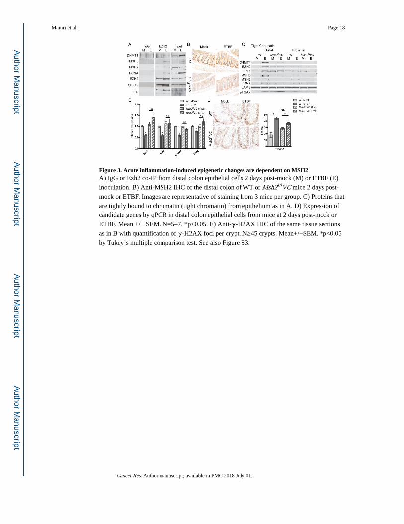
Figure 3. Acute inflammation-induced epigenetic changes are dependent on MSH2A) IgG or Ezh2 co-IP from distal colon epithelial cells 2 days post-mock (M) or ETBF (E)
inoculation. B) Anti-MSH2 IHC of the distal colon of WT or Msh2l/lVC mice 2 days post-
mock or ETBF. Images are representative of staining from 3 mice per group. C) Proteins that
are tightly bound to chromatin (tight chromatin) from epithelium as in A. D) Expression of
candidate genes by qPCR in distal colon epithelial cells from mice at 2 days post-mock or
ETBF. Mean +/− SEM. N=5–7. *p<0.05. E) Anti-γ-H2AX IHC of the same tissue sections
as in B with quantification of γ-H2AX foci per crypt. N≥45 crypts. Mean+/−SEM. *p<0.05
by Tukey’s multiple comparison test. See also Figure S3.
Maiuri et al. Page 18
Cancer Res. Author manuscript; available in PMC 2018 July 01.
Author M
anuscriptA
uthor Manuscript
Author M
anuscriptA
uthor Manuscript

Figure 4. Inflammation-induced tumorigenesis is increased in the distal colon of mice with altered Msh2 expressionA) PCA analysis of 16S microbiome sequencing of DNA from stool samples from WT/Min
or Msh2l/lVC/Min mice 8 weeks post mock or ETBF. B) Heatmap representing the
unsupervised hierarchical clustering of 65 OTUs found to be differentially abundant by one
pair-wise comparison (rows) in individual stool samples (columns) from indicated mice
treated as in A. C) ETBF abundance in stool relative to total bacterial DNA by qPCR.
Symbols represent data from individual mice. Horizontal line is mean +/− SEM. N≥13.
*p<0.05. D) Tukey box plots of tumor counts by cm in WT/Min or Msh2l/lVC/Min mice 8
weeks after mock or ETBF. N≥8. *p<0.05. E) Whole cell protein lysate from distal colon
epithelium or tumors from mice of the indicated genotypes 8 weeks post-mock or ETBF
inoculation, respectively, were blotted for the indicated proteins. See also Figure S4.
Maiuri et al. Page 19
Cancer Res. Author manuscript; available in PMC 2018 July 01.
Author M
anuscriptA
uthor Manuscript
Author M
anuscriptA
uthor Manuscript

Figure 5. Msh2 deficiency abrogates inflammation-induced epigenetic changes in tumorsA) Unsupervised hierarchical clustering of MBD-seq z-scores of regions with increased
DMRs in one of the tumor groups relative to mock epithelium (rows). Each column
corresponds to the indicated epithelium or tumor sample. The color of each cell reflects the
degree of methylation. B) Numbers of DMRs for the indicated tumor group relative to the
corresponding mock epithelium that overlap between comparisons. Green and red numbers
are hypomethylated and hypermethylated regions, respectively. C) MBD-seq data at
representative regions for indicated epithelium and tumors 8 weeks post-inoculation. D)
Expression of candidate genes by qRTPCR relative to WT/Min ETBF tumors. Mean +/−
SEM. N=5. *p<0.05. E) Total 5-mC content of DNA. Mean +/− SEM. N=3 mock, N=6
tumor. *p<0.05. See also Figure S5.
Maiuri et al. Page 20
Cancer Res. Author manuscript; available in PMC 2018 July 01.
Author M
anuscriptA
uthor Manuscript
Author M
anuscriptA
uthor Manuscript

Author M
anuscriptA
uthor Manuscript
Author M
anuscriptA
uthor Manuscript
Maiuri et al. Page 21
Table 1
Number of 500 bp regions from the MBD-seq data that are statistically different in the indicated comparisons.
Gains Losses Total
Min mock tumor X Min mock epithelium 6 700 706
Min ETBF epithelium X Min mock epithelium 40 265 305
Min ETBF tumor X Min mock epithelium 203 194 397
Cancer Res. Author manuscript; available in PMC 2018 July 01.

Author M
anuscriptA
uthor Manuscript
Author M
anuscriptA
uthor Manuscript
Maiuri et al. Page 22
Tab
le 2
Tum
ors
from
mic
e de
fici
ent i
n M
sh2
are
mic
rosa
telli
te in
stab
ility
(M
SI)
posi
tive.
Ani
mal
Gen
otyp
eE
TB
FU
1223
5[A
24]
aA
C09
6777
[A33
] a
AA
0030
63[A
23]
aA
C09
6777
[T27
] a
L24
372[
A27
] a
MSI
% b
403
Min
−−
++
−−
40
410
Min
−−
−+
−−
20
414
Min
−−
−+
−−
20
7M
in+
−−
−−
−20
7M
in+
−−
−−
−20
408
Min
+−
−−
−−
20
408
Min
+−
−−
−−
20
408
Min
+−
−−
−−
20
408
Min
+−
−−
−−
20
TR
Min
++
−+
+−
60
TR
Min
++
−−
−−
20
331
Msh
2l/lV
CM
in−
+no
dat
a+
++
80
322
Msh
2l/lV
CM
in−
++
−+
+80
322
Msh
2l/lV
CM
in−
++
−+
+80
322
Msh
2l/lV
CM
in−
+−
−+
+60
319
Msh
2l/lV
CM
in+
++
−+
+80
319
Msh
2l/lV
CM
in+
−+
−+
+60
319
Msh
2l/lV
CM
in+
++
−+
+80
320
Msh
2l/lV
CM
in+
−+
−+
+60
320
Msh
2l/lV
CM
in+
−+
−+
+60
320
Msh
2l/lV
CM
in+
−+
−+
+60
a + a
nd −
indi
cate
pos
itive
or
nega
tive
for
inst
abili
ty, r
espe
ctiv
ely,
for
the
give
n as
say.
b tum
ors
with
gre
ater
than
20%
of
test
ed lo
ci p
ositi
ve f
or in
stab
ility
are
MSI
+ (
bold
num
bers
).
Cancer Res. Author manuscript; available in PMC 2018 July 01.