mimickers of prostate cancer from adenosis to …iap-ad.org/lectures/23rd_congress_beirut/j...
TRANSCRIPT

12/7/2011
1
Mimickers of Prostate Cancer From
Adenosis to Xanthoma
Benign Mimickers of Well-Moderately
Differentiated Carcinoma
• Adenosis
• Atrophy
• Basal cell hyperplasia
• Nephrogenic adenoma
• HGPIN
• Radiation atypia

12/7/2011
2
Adenosis
(Atypical Adenomatous Hyperplasia)
Incidence
• TURP - 1.6%
• Needle - 0.8%
• Multifocal

12/7/2011
3

12/7/2011
4

12/7/2011
5
Diagnostic Criteria of Adenosis
Adenosis
Lobular
Small glands share cytoplasmic and
nuclear features with admixed larger
benign glands
Pale-clear cytoplasm
Medium sized nucleoli
Blue mucinous secretions rare
Corpora amylacea common
Basal cells present
Cancer
Haphazard growth pattern
Small glands differ in either nuclear or
cytoplasmic features from adjacent
benign glands
Occasionally amphophilic cytoplasm
Occasionally large nucleoli
Blue mucinous secretions common
Corpora amylacea rare
Basal cells absent
Features Shared in Adenosis and Cancer
• Crowded glands
• Crystalloids
• Medium sized nucleoli
• Scattered poorly formed glands and single cells
• Minimal infiltration at periphery

12/7/2011
6
Relation of Adenosis to Cancer
• No difference in risk of subsequent diagnosis of
cancer following TURP diagnosis as compared to
BPH
• “Adenosis is a mimicker of prostate cancer which is
not associated with an increased risk of cancer”
Atrophy
• Post-atrophic hyperplasia
• Partial atrophy
• Simple atrophy

12/7/2011
7
• Proliferative atrophy linked to prostate carcinogenesis
• Atrophy & inflammation associated with increased
proliferation.
• Gives rise to reactive oxygen and nitrogen species which in
the setting of increased turnover results in mutations.
• If atrophy is associated with cancer, it is not a proximate
cause.
• Proliferative atrophy seen in 47% of sextant biopsies
• Not associated with cancer on repeat biopsy
• Not typically even mentioned in pathology report unless
florid as a mimicker of cancer.

12/7/2011
8

12/7/2011
9
Partial Atrophy

12/7/2011
10

12/7/2011
11

12/7/2011
12
Basal Cell Hyperplasia
Basal Cell Hyperplasia in the
Peripheral Zone of the Prostate
• BCH in peripheral zone – 23% of whole prostates
• BCH on needle biopsy – 10.2%

12/7/2011
13

12/7/2011
14
Nephrogenic Adenoma
(Nephrogenic Metaplasia)
Nephrogenic Adenoma of the Prostatic Urethra:
A Mimicker of Prostate Adenocarcinoma
• Features mimicking prostate cancer:
– Presence of tubules, cords, and signet ring-like tubules
– Prominent nucleoli
– Muscle involvement
– Blue-tinged mucinous secretions (32%)
– Focal PSA (36%) and PSAP (50%)
– Negative staining for HMWCK in some cases

12/7/2011
15

12/7/2011
16

12/7/2011
17

12/7/2011
18
Nephrogenic Adenoma vs. Prostate Ca.
• Focal PSA and PSAP positivity in 1/3 of cases – Tends to not be diffusely strong
• Negative staining for HMWCK in 62%-75% of cases
• Cases with positive HMWCK rules out prostate cancer
• Positive AMACR (racemase) in 35%-58% of cases
• Positive PAX2 in NA and not in prostate cancer
PSA

12/7/2011
19
AMACR
HGPIN

12/7/2011
20

12/7/2011
21
PINATYP
High grade PIN with small focus of atypical glands. See
note:
Note: Adjacent to glands of high grade PIN are a few
small atypical glands. While these small glands may
represent a small focus of infiltrating cancer, we can
not exclude that they represent outpouching or
tangential sections off of the adjacent high grade PIN.

12/7/2011
22

12/7/2011
23
Radiation Change
Radiation Change in the Prostate
• RT affect in Benign Prostate - Differential Diagnosis
from Prostate Cancer
• RT results – Affect on Prognosis
– Positive for cancer (w/o treatment affect)
– Negative for cancer
– Positive for cancer (with treatment affect)
• More atypia in cases treated with IRT (seeds) than XRT
• No change in epithelial atypia over time in men treated with IRT. With XRT, less epithelial atypia in cases biopsies >48 months after treatment
• RT atypia may persist for a long time: Prominent RT atypia detected 72 months after IRT
• Some cases clinicians not aware of remote h/o of RT or do not relay this on to pathologists. Pathologists must be able to recognize RT atypia w/o relying on the clinician to provide this history

12/7/2011
24

12/7/2011
25

12/7/2011
26
Radiation Biopsy Results: Cancer with
Treatment Affect
Histologically cancer is seen, yet shows treatment effect
with degenerative features. Cancer is present, yet is it
viable?
Prognosis: Similar to cases with no cancer.
Do not grade radiated cancer with treatment effect.
Radiation Biopsy Results: Positive
Histologically, ordinary prostate cancer is seen, which
resembles non-radiated cancer.
If biopsy performed too soon (<12 months), can not tell
if the cancer is resistant or has not had enough time to
be destroyed by the treatment.
If biopsy is done >12 months following radiation,
indicates progression of cancer. Can assign a Gleason
grade.
Benign Mimickers of Moderately-Poorly
Differentiated Adenocarcinoma
• Nonspecific granulomatous prostatitis
• Paraganglia
• Urothelial carcinoma
• Xanthoma

12/7/2011
27
Nonspecific Granulomatous Prostatitis

12/7/2011
28

12/7/2011
29
Paraganglia of Prostate

12/7/2011
30
High Grade Prostatic Adenocarcinoma
vs.
Poorly Differentiated Urothelial Carcinoma

12/7/2011
31
Direct Invasion of UC Into the Prostate
(pT4): Invasive UC versus High Grade
Prostatic Adenocarcinoma
PROSTATE ADENOCARCINOMA
• Large solid sheets or cords of cells
• Uniform nuclear size & shape with prominent nucleoli, yet
advanced prostate cancer may be as pleomorphic as UC
• Microacinar formation
• Lacks stromal inflammation

12/7/2011
32
12/7/2011
33
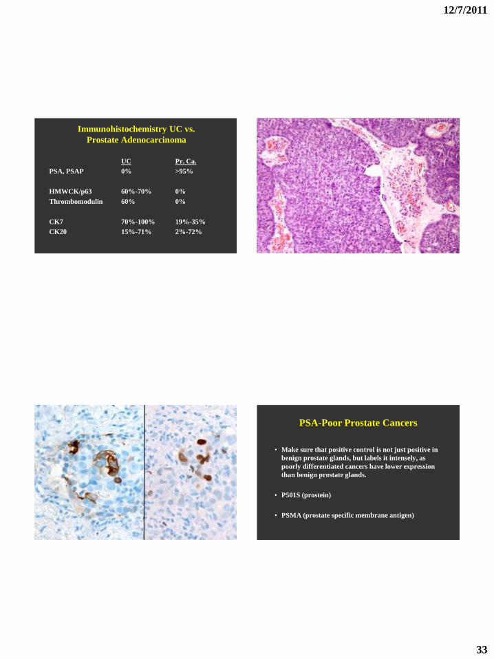
Immunohistochemistry UC vs.
Prostate Adenocarcinoma
UC Pr. Ca.
PSA, PSAP 0% >95%
HMWCK/p63 60%-70% 0%
Thrombomodulin 60% 0%
CK7 70%-100% 19%-35%
CK20 15%-71% 2%-72%
PSA-Poor Prostate Cancers
• Make sure that positive control is not just positive in
benign prostate glands, but labels it intensely, as
poorly differentiated cancers have lower expression
than benign prostate glands.
• P501S (prostein)
• PSMA (prostate specific membrane antigen)

12/7/2011
34
Recommended Panel
• PSA
• P501S
• PSMA
• NKX3.1
• Thrombomodulin
• p63
• HMWCK
• GATA3

12/7/2011
35
Prostatic Xanthoma

12/7/2011
36
Summary
Prostate biopsies and TURs are some of the most
difficult specimens to evaluate, in part due to the wide
range of mimickers of both moderately and poorly
differentiated adenocarcinoma of the prostate.
Recognition of these mimickers’ unique histologic
features can prevent an overdiagnosis of prostate
cancer.

12/7/2011
37