microsoft word - proposal 5 body · web viewneonatal sepsis is one of the most common cause of...
TRANSCRIPT
CHAPTER 1: BACKGROUND
Neonatal sepsis is one of the most common cause of neonatal morbidity and mortality. It is
estimated to cause 26% of all neonatal deaths worldwide (Lawn et al, 2005).It’s Incidence
varies from country to country but is much higher in developing countries (Kaistha et al,
2009) where it is responsible for about 30-50% of the total of neonatal deaths (Bang et al,
1999).It is one of the most common reasons for admission to neonatal units in developing
countries (Shitaye et al)
Neonatal sepsis refers to generalised bacterial infection documented by a positive blood
culture in the first 28 days of life. It encompasses various systemic infections of the new born
such as septicaemia, meningitis, pneumonia, arthritis, osteomyelitis and urinary tract
infections. Superficial infections like conjunctivitis and oral thrush are not usually included
under neonatal sepsis (Report of the national neonatal perinatal database, National
neonatology forum 2002-2003)
Sepsis can be classified into 2 types according to the age of onset; early onset sepsis and late
onset sepsis. Early onset sepsis presents within the first 72 hours of life and in severe cases
the neonate may be symptomatic at birth. The source of infection for early onset sepsis is
usually the maternal genital tract (Singh et al, 1994). Late onset sepsis presents after 72 hours
of age. It is usually nosocomial as a complication of neonatal intensive care or community
acquired. This classification is important as it helps in determining the most probable
organism and mode of transmission. This guides empiric treatment (Forfar and Arneil’s
textbook of paediatrics, 6th edition)
The spectrum of organisms that causes neonatal sepsis changes over time and varies from
region to region even within the same hospital. This is due to the changing pattern of
antibiotic use and changes in lifestyle (Shrestha et al, 2013).The local epidemiology of
2
neonatal sepsis should be constantly updated to detect changes in the pattern of causative
organisms and their susceptibility to various antibiotics. Early diagnosis and proper
management of neonatal sepsis by rational antimicrobial therapy and supportive care can
reduce mortality. Blood culture is the gold standard for diagnosis of sepsis but blood culture
reports are usually available after 48 to 72 hours. There is need to Identify the common
bacteria causing such infections in every hospital and their susceptibility patterns in order to
provide necessary information for timely intervention (Shrestha et al 2013).
Since the discovery of antimicrobial agents, microorganisms have developed resistance to
them though mechanisms such as mutations and increased enzyme production. Resistance to
commonly used antibiotics is an important problem worldwide.
3
CHAPTER TWO: LITERATURE REVIEW
2.1 Introduction
The number of neonatal patients at risk of acquiring nosocomial infections is increasing
because of the improved survival of very low birth weight infants and their need for invasive
monitoring and supportive care (Adams- Chapman et al, 2002)
The World Health organisation(WHO) reported in 2005 that over 70% of deaths in children
under age five occur within the first year of life and 40% occur within the first month (WHO,
World Health Report 2005. Make every mother and child count. Geneva: WHO;
2005.p.2005)
Neonatal infections currently cause 1.6 million deaths annually in developing countries.
Sepsis and meningitis are responsible for most of these deaths. (Vergnano et al, 2005)
According to the World Health Organisation (WHO) Global health Observatory Data
repository the neonatal mortality rate in Africa in 2012 was 32 per 1000 live births and 27 per
1000 live births in Asia. In the same report the Americas recorded a neonatal mortality rate of
8 per 1000 live births.
In the Kenya Demographic and Health Survey (KDHS) of 2008/2009 the neonatal mortality
rate stood at 31 per 1000 live births.
2.2 Causative bacteria
The causative organisms include a wide variety of gram positive and gram negative
organisms. These include staphylococcus aureus, coagulase negative staphylococcus
(CONS),Escherichia coli (Ecoli), Listeria monocytogenes, Klebsiella pneumoniae, Group B
4
streptococcus(GBS), Acinetobacter, Serratia, Pseudomonas, Haemophilus influenzae,
Enterobacter, Candida and anaerobes.
In many hospitals gram positive organisms cause upto 70% of nosocomial infections in
neonates (Patel et al, 2010)with coagulase negative Staphylococci accounting for more than
half of these(Van der Zwet WC et al, 2005). In developing countries gram negative
organisms may be far more prevalent as neonatal pathogens (Couto et al 2007).GBS is
generally rare or not seen at all although maternal recto-vaginal carriage rates of GBS may be
similar to those recorded in developed countries. In most of the African studies the incidence
of GBS is low with the exception of South Africa. In Asia GBS is also reported to be
extremely rare. Neonatal surveillance in developed countries generally identifies GBS and
Ecoli as the dominant early onset sepsis pathogens and CONS as the dominant late onset
sepsis pathogen followed by GBS and staphylococcus aureus. (Vergnano et al, 2005).
A retrospective study done in a neonatal intensive care unit in Australia by Sanghvi et al over
a 5 year period showed that CONS (38.8%), GBS (20.1 % )and gram negative bacilli (GNB)(
20.1%), were the common causes of sepsis. (Sanghvi et al,1996)
A retrospective review of 390 neonatal blood cultures carried out by Iregbu et al in the
department of Clinical Microbiology and Parasitology of a tertiary hospital in Nigeria in the
year 2006 showed gram positive cocci (GPC) and GNB in almost equal proportion.
Predominantly Klebsiella pneumoniae(86% of GNB) and Staphylococcus aureus (81% of
GPC) (Iregbu et al,2006).
In a prospective study done in a Kenya by Musoke et al over a 5 month period in the year
2000 the predominant organisms were gram negative(73.6 percent of isolates) with klebsiella
species topping the list at 31 percent.(Musoke et al,2000).

5
2.3 Risk Factors
Some of risk factors for neonatal sepsis include; prematurity or low birth weight, preterm
labour, premature or prolonged rupture of membranes, maternal chorioamnionitis, foetal
hypoxia, traumatic delivery, male gender and low socio-economic status.
2.4 Clinical features.
Neonates with sepsis may have nonspecific signs and symptoms and the initial manifestations
may have limited symptomatology. Some of the features include temperature instability,
hypotension, tachycardia, bradycardia, apnoea, respiratory distress, grunting, cyanosis,
irritability, lethargy, seizures, feeding intolerance, abdominal distension and jaundice (Nelson
Textbook of Paediatrics, 17th edition, 2004)
2.5 Diagnosis of neonatal sepsis
Neonatal sepsis is clinically diagnosed by a combination of clinical signs, nonspecific
laboratory tests and microbiologically confirmed by detection of bacteria in blood by culture
(Marchant et al, 2013)
Blood culture is the gold standard for diagnosis of septicaemia.
2.6 Antimicrobial management
There cannot be a single recommendation for the antibiotic regimen of neonatal sepsis for all
settings. The choice of antibiotics depends on the prevailing flora in the given unit and their
antimicrobial sensitivity. The decision to start antibiotics is based on clinical features and or
positive septic screen. (Sankar et al, 2008)
6
The antibiotic combination prescribed in most units is a penicillin (Benzyl penicillin,
ampicillin or cloxacillin) together with an aminoglycoside most commonly gentamicin
(Vergnano et al, 2005)
2.7 Antibiotic resistance
Resistance to antibiotics is a global problem. Reports of multiresistant bacteria causing
neonatal sepsis in developing countries are increasing and this may be explained by the wide
availability of over the counter antibiotics and the inappropriate use of broad-spectrum
antibiotics in the community. (Jyothi et al, 2013). At risk are unwell babies or premature
babies, those needing additional support such as ventilation, intravenous fluid or blood
products and those babies who stay in hospital for more than 48 hours (Al Rabea AA et al,
1998).Spread of resistant organisms in hospitals is a recognised problem although babies
admitted from the community may also carry resistant pathogens (Bhutta, 1996)
It is difficult to compare antibiotic resistance between countries because the epidemiology of
neonatal sepsis is extremely variable (Vergnano et al, 2005). Methicillin resistant
staphylococci, Extended spectrum beta lactamase (ESBL) and multidrug resistant gram
negative organisms represent the principal concern for clinicians who have to fight against
infections (Paolucci et al, 2012). Most gram negative bacteria are now resistant to ampicillin
and cloxacillin and many are becoming resistant to gentamicin (Vergnano et al, 2005).
A retrospective study carried out by Iregbu et al in the Department of Clinical Microbiology
and Parasitology of a tertiary hospital in Nigeria in the year 2006 showed that 89% of
staphylococcus aureus were sensitive to amoxicillin- clavulanic acid while 85%, 45%, 71%
and 64% were sensitive to cefuroxime, ciprofloxacin, chloramphenicol and erythromycin
respectively. The only three isolates tested against tetracycline were all susceptible to the
drug. The resistance to penicillin was 90%.Resistance to ceftazidime, ceftriaxone and
7
gentamicin were 71% ,64% and 60% respectively. The resistance of the isolated Klebsiella
pneumoniae to ceftazidime, ceftriaxone and cefotaxime was 85%, 87.5% and 94%
respectively. Resistance to amoxicillin and ampicillin-sulbactam was 100%, and 85% for
amoxicillin-clavulanic acid. All (100%) of the Klebsiella pneumoniae isolates tested against
imipenem were susceptible while 75% were susceptible to amikacin. (Iregbu et al, 2006)
A study done over a 5 month period in the year 2000 in a neonatal unit of a referral hospital
in Kenya by Musoke et al showed that resistance to gentamicin was 20%, chloramphenicol
23.6 %, and amoxicillin /ampicillin 66.3%, ceftazidime 19.1 %, and cefuroxime 21.3%
(Musoke et al,2000).
Infection with antibiotic resistant organisms results in delay in starting effective antibiotic
therapy, fewer possible treatment options and increased morbidity and mortality with
prolonged hospital stay and greater costs of hospitalisation (Patel et al, 2010). The slow pace
in the development of newer drugs and rapidity in resistance development are major areas of
concern. (Shah et al, 2012)
2.8 Supportive management.
The following supportive measures are recommended in the management of neonatal sepsis;
nursing in a thermo neutral environment to avoid hypo or hyperthermia, maintaining oxygen
in the normal range, intravenous fluids if hemodynamically unstable and corticosteroids for
adrenal insufficiency. Hyperbilirubinaemia should be monitored and treated with
phototherapy and or exchange transfusion.
8
2.9 Prevention of neonatal sepsis
Strategies to reduce rates of infection includes clean and safe deliveries, adherence to
universal precautions in all patient contact, strict postnatal cleanliness, early and exclusive
breastfeeding avoiding nursery overcrowding and limiting nurse to patient ratios(Haque et
al,2003, Simiyu 2003). Other measures include strict compliance to hand washing,
decreasing the number of venepunctures and heel pricks and providing education to nursery
personnel (Bang et al, 1999)
2.10 Complications
The short term complications of neonatal sepsis include respiratory failure, pulmonary
hypertension, cardiac failure, shock, renal failure, liver dysfunction and cerebral oedema.
Some of the long term complications include; developmental delays, sensory and
neurological dysfunction.

9
CHAPTER THREE: RESEARCH DEFINITION
3.1 Justification
Neonatal sepsis is a leading cause of morbidity and mortality in both developing and
developed countries. It is a life threatening emergency and delays in diagnosis and treatment
with appropriate antibiotics may have devastating consequences.
The pattern of causative organisms constantly changes and the emergence of resistant
bacteria has compounded the problem further. Knowledge of the aetiologic agents is
important and helps to reduce associated mortality in neonatal septicaemia. Longitudinal
surveillance should be carried out at regular intervals to describe the varied pathogens
causing neonatal sepsis as well as their changing antibiotic susceptibility pattern.
The result of this study is expected to guide therapy of neonatal sepsis at KNH.
3.2 Study questions
1. Which are the bacteria commonly causing neonatal sepsis at Kenyatta National hospital
and what is their susceptibility to commonly used antibiotics?
2. What are some of the risk factors for neonatal sepsis at Kenyatta National Hospital?
3.3 Objectives
Broad objective
To determine the bacterial aetiological agents of neonatal sepsis, risks associated with
acquisition and the susceptibility of these organisms to commonly used antimicrobial agents
at Kenyatta National Hospital.
10
Specific objectives
1.To determine the bacterial agents causing neonatal sepsis at Kenyatta National Hospital.
2.To describe the antibiotic susceptibility pattern of bacteria causing neonatal sepsis at
Kenyatta National Hospital.
3.To determine some of the associated risk factors for neonatal sepsis at Kenyatta National
Hospital.

11
CHAPTER FOUR: RESEARCH METHODOLOGY
4.1 Study site
The study was conducted at the Kenyatta National Hospital Medical Microbiology laboratory
and records department. KNH is located in Nairobi, the politico-administrative and economic
capital of Kenya. It caters for over 80,000 in-patients and over 500,000 out-patients annually.
About 75% of the patients treated as outpatients and inpatients are residents of Nairobi
through self-referral or referral from the public and private health facilities.
Neonates are reviewed in the Paediatric Filter Clinic (PFC). Those requiring admission are
admitted to the New Born Unit (NBU) or to the general paediatric wards because of the
limited space in the NBU. Others neonates are received from KNH labour ward and
maternity theatre.
4.2 Study design
It was a retrospective cross sectional study involving review of patients’ laboratory records
and files.
4.3 Sampling method
Convenience selection of positive blood culture reports was employed. Risk factors for
acquisition of sepsis were assessed through patients’ files at the medical records department.
4.4 Study population
Neonates admitted in KNH between the periods 1st January 2013 to 31st December 2013 with
reports showing bacterial growth from blood culture specimen.

12
4.5 Inclusion criteria
Positive blood culture reports of children less than or aged 28 days for the period 1st January
2013 to 31st December 2013.
4.6 Exclusion criteria
1. Reports of negative blood culture
2. Reports of patients > 28 days
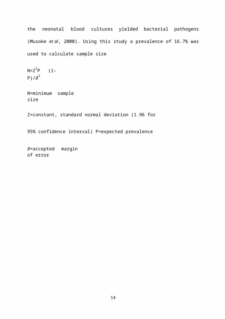
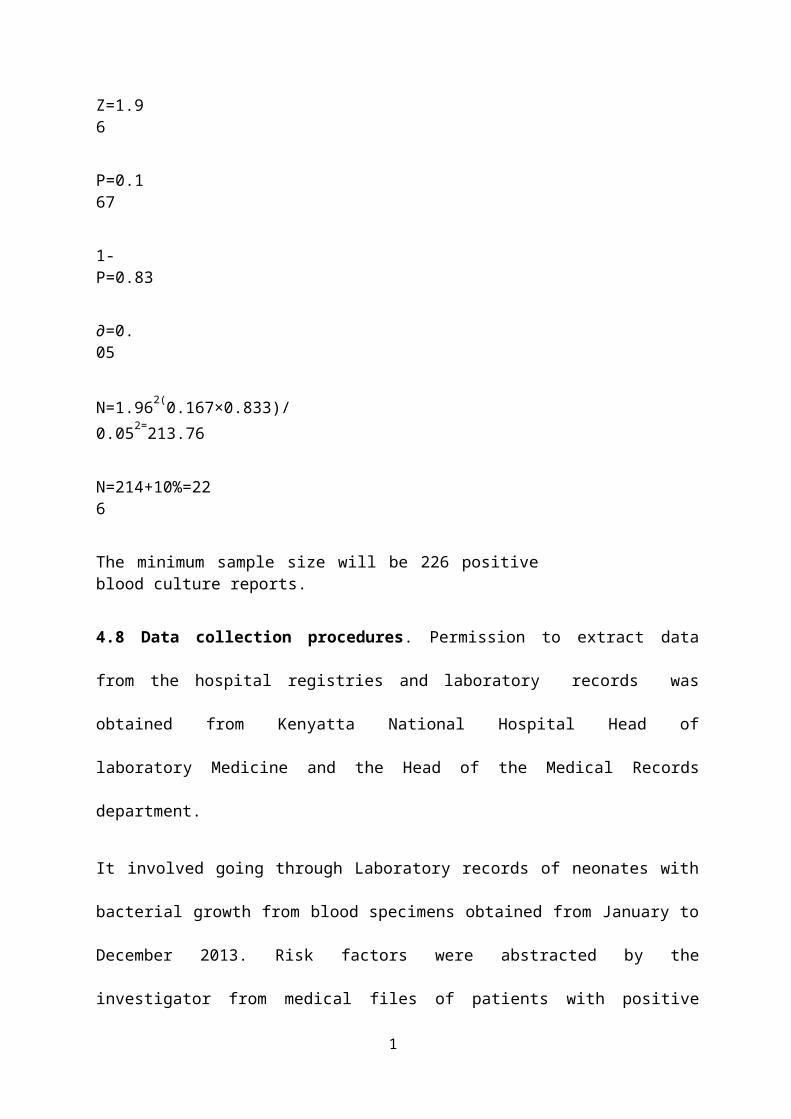
4.7 Sample size determination
The standard statistical approach to determination of sample size for a descriptive cross
sectional survey such as this one requires specification of the proportion (prevalence) of
neonatal sepsis; the desired level of confidence desired for the proportion estimate and a
tolerance error margin or width of the confidence interval (a measure precision of the
estimate) so that the necessary sample size is then calculable for a given precision level.
The fisher’s formula was hence used to estimate the sample size.
A study at Kenyatta National Hospital found that 16.7 % of the neonatal blood cultures
yielded bacterial pathogens (Musoke et al, 2000). Using this study a prevalence of 16.7% was
used to calculate sample size
N=Z2P (1-P)/∂2
N=minimum sample size
Z=constant, standard normal deviation (1.96 for 95% confidence interval)
P=expected prevalence
∂=accepted margin of error

13
Z=1.96
P=0.167
1-P=0.83
∂=0.05
N=1.962(0.167×0.833)/0.052=213.76
N=214+10%=226
The minimum sample size will be 226 positive blood culture reports.
4.8 Data collection procedures. Permission to extract data from the hospital registries and
laboratory records was obtained from Kenyatta National Hospital Head of laboratory
Medicine and the Head of the Medical Records department.
It involved going through Laboratory records of neonates with bacterial growth from blood
specimens obtained from January to December 2013. Risk factors were abstracted by the
investigator from medical files of patients with positive blood cultures. The medical files
were traced using the patient numbers on the blood culture reports.
A data collection form was used to collect data.
4.9 Data management and analysis.
Data was entered into password protected microsoft access and analysis done using
Statistical Package for Social sciences (SPSS) version 21.
14
4.10 Ethical consideration
Approval was sought from the Kenyatta National Hospital/University of Nairobi-Ethics
Review Committee (KNH/UoN-ERC).For confidentiality the patients’ laboratory records and
medical files were used in the confines of the KNH microbiology laboratory and the medical
records department.
The patients names were not included in the collection form. Only the investigator had access
to the laboratory records and medical files for the purposes of the study.
Raw data in form of filled forms, data stored in password protected computer, backup copies
in hard drives and compact disc will be destroyed at the end of the study.
4.11 Study limitations.
Incompletely filled patient laboratory and medical records. A number of files had incomplete
information with important variables missing.
Growth of contaminants due to improper aseptic techniques during blood specimen
collection.
4.12 Dissemination plan
The results of the study will be disseminated to the Paediatrics department of KNH, KNH
Microbiology Laboratory, University of Nairobi library and UNITID library.

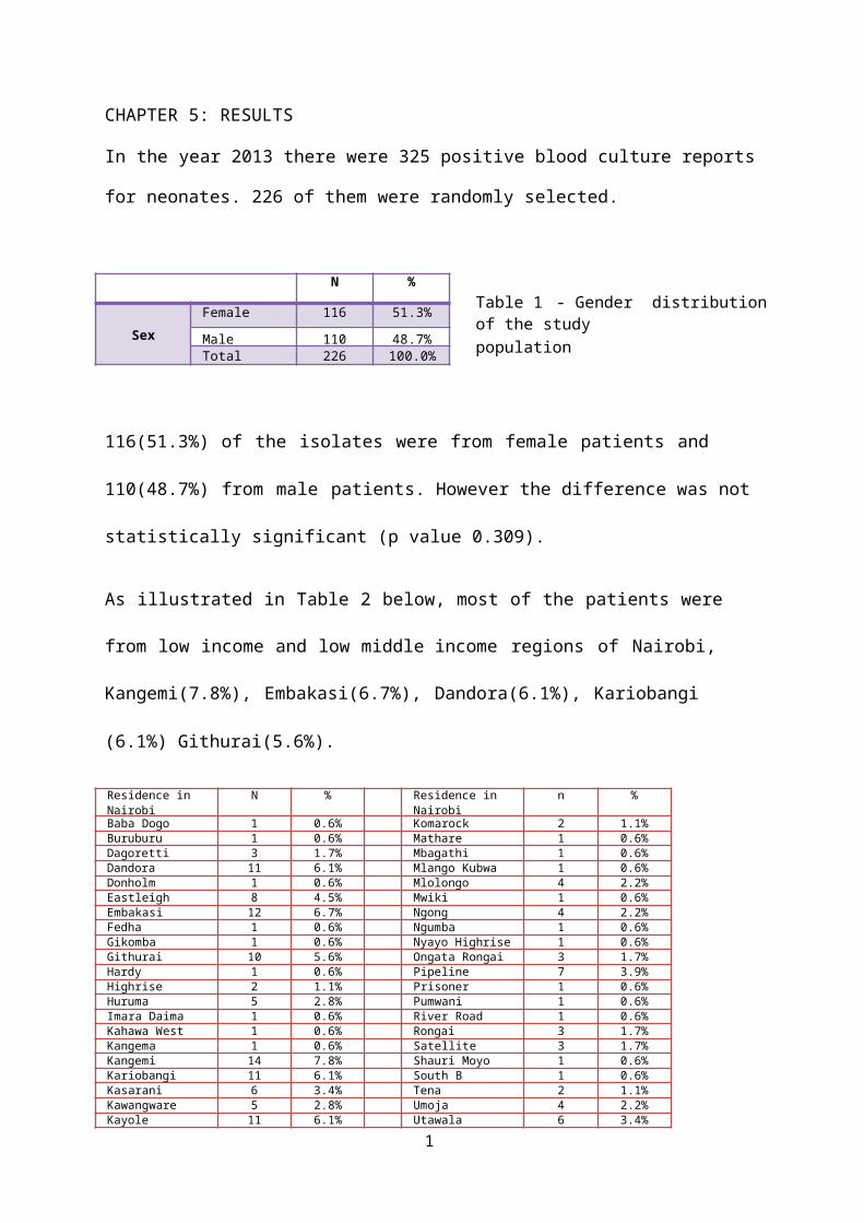
N %
SexFemale 116 51.3%
Male 110 48.7%Total 226 100.0%
15
CHAPTER 5: RESULTS
In the year 2013 there were 325 positive blood culture reports for neonates. 226 of them were
randomly selected.
Table 1 - Gender distribution of the studypopulation
116(51.3%) of the isolates were from female patients and 110(48.7%) from male patients.
However the difference was not statistically significant (p value 0.309).
As illustrated in Table 2 below, most of the patients were from low income and low middle
income regions of Nairobi, Kangemi(7.8%), Embakasi(6.7%), Dandora(6.1%), Kariobangi
(6.1%) Githurai(5.6%).
Residence inNairobi
N % Residence inNairobi
n %
Baba Dogo 1 0.6% Komarock 2 1.1%Buruburu 1 0.6% Mathare 1 0.6%Dagoretti 3 1.7% Mbagathi 1 0.6%Dandora 11 6.1% Mlango Kubwa 1 0.6%Donholm 1 0.6% Mlolongo 4 2.2%Eastleigh 8 4.5% Mwiki 1 0.6%Embakasi 12 6.7% Ngong 4 2.2%Fedha 1 0.6% Ngumba 1 0.6%Gikomba 1 0.6% Nyayo Highrise 1 0.6%Githurai 10 5.6% Ongata Rongai 3 1.7%Hardy 1 0.6% Pipeline 7 3.9%Highrise 2 1.1% Prisoner 1 0.6%Huruma 5 2.8% Pumwani 1 0.6%Imara Daima 1 0.6% River Road 1 0.6%Kahawa West 1 0.6% Rongai 3 1.7%Kangema 1 0.6% Satellite 3 1.7%Kangemi 14 7.8% Shauri Moyo 1 0.6%Kariobangi 11 6.1% South B 1 0.6%Kasarani 6 3.4% Tena 2 1.1%Kawangware 5 2.8% Umoja 4 2.2%Kayole 11 6.1% Utawala 6 3.4%Kibera 8 4.5% Uthiru 4 2.2%Kinoo 5 2.8% Zimmerman 6 3.4%Komarock 2 1.1%
Table 2 - Residence in Nairobi.

16
Organisms isolated from blood culture
As illustrated in figure 1 below there was a preponderance of gram negative organisms
116(51.3%) over gram positive organisms 96(42.5%).
Gram Positive 42.5%
Gram Negative 51.3%
0% 10% 20% 30% 40% 50% 60%
Figure 1 - Distribution of isolated organisms.
Coagulase negative staphylococcus was the most isolated organism (30.1%), followed by
Enterobacter spp (19.9%), Citrobacter spp(12.8%) and Klebsiella spp(11%) spp. Other less
frequently isolated organisms were Enterococcus spp(8.8%), Escherichia coli(7.1%),
Staphylococcus aureus(3.5%), Proteus spp(0.9%)
StaphylococcusAureus
4%
Enterococcus9%
EColi7%
Proteus1%
Micrococcus0%
Other Organisms1%
Klebsiella11%
Citrobacter14%
Coagulase NegativeStaphylococci
32%
Enterobacter21%
Figure 2 - Frequency of isolated organisms.

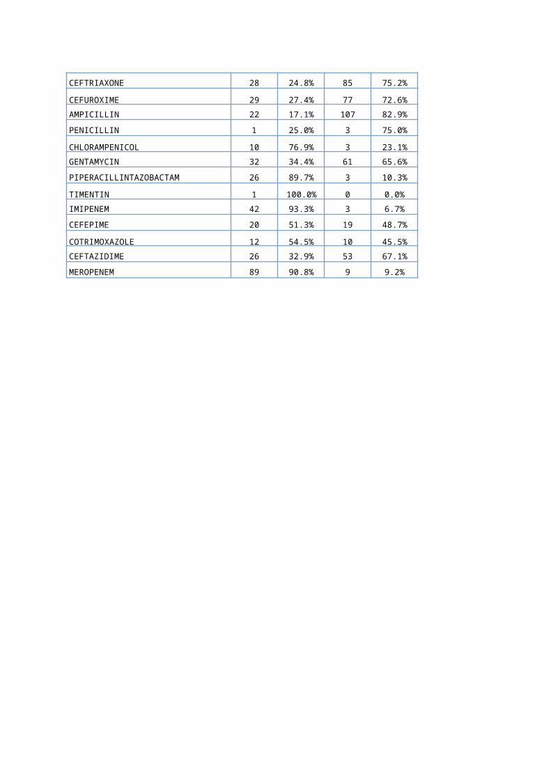
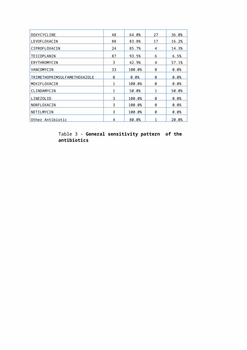
Antibiotic susceptibility patterns
The first line treatment of neonatal sepsis in Kenyatta hospital is a combination of penicillin
and gentamicin. As shown in table 3, out of the 4 organisms tested against penicillin 3 of
them showed resistance and 1 organism was susceptible. The resistance to gentamicin was
(65.6%). Resistance to other antibiotics was also high ceftriaxone (75.2%), cefuroxime
(72.6%), ampicillin (82.9%), ceftazidime (67.1%).
The was high sensitivity to vancomycin (100%), meropenem (90.8%), amikacin (87%),
piperacillin tazobactam (89.7%), teicoplanin (93.5%), levofloxacin (83.8%)
There was also high sensitivity to amikacin (87.0%), piperacillin tazobactam (89.7%)
imipenem (93.3%, meropenem (90.8%)
Sensitive Resistant
N % N %
AMOXY_CLAV 68 45.0% 83 55.0%
AMIKACIN 20 87.0% 3 13.0%
CEFTRIAXONE 28 24.8% 85 75.2%
CEFUROXIME 29 27.4% 77 72.6%
AMPICILLIN 22 17.1% 107 82.9%
PENICILLIN 1 25.0% 3 75.0%
CHLORAMPENICOL 10 76.9% 3 23.1%
GENTAMYCIN 32 34.4% 61 65.6%
PIPERACILLINTAZOBACTAM 26 89.7% 3 10.3%
TIMENTIN 1 100.0% 0 0.0%
IMIPENEM 42 93.3% 3 6.7%
CEFEPIME 20 51.3% 19 48.7%
COTRIMOXAZOLE 12 54.5% 10 45.5%
CEFTAZIDIME 26 32.9% 53 67.1%
MEROPENEM 89 90.8% 9 9.2%
DOXYCYCLINE 48 64.0% 27 36.0%
LEVOFLOXACIN 88 83.8% 17 16.2%
CIPROFLOXACIN 24 85.7% 4 14.3%
TEICOPLANIN 87 93.5% 6 6.5%
ERYTHROMYCIN 3 42.9% 4 57.1%
VANCOMYCIN 33 100.0% 0 0.0%
TRIMETHOPRIMSULFAMETHOXAZOLE 0 0.0% 0 0.0%
MOXIFLOXACIN 1 100.0% 0 0.0%
CLINDAMYCIN 1 50.0% 1 50.0%
LINEZOLID 3 100.0% 0 0.0%
NORFLOXACIN 3 100.0% 0 0.0%
NETILMYCIN 3 100.0% 0 0.0%
Other Antibiotic 4 80.0% 1 20.0%
Table 3 - General sensitivity pattern of the antibiotics
Resistance and susceptibility of selected organisms.
Coagulase negative staphylococci showed the highest resistance to ampicillin and
gentamicin(59.1%). Four out of the five organisms tested against cotrimoxazole were
sensitive. There was high sensitivity to vancomycin(100%), teicoplanin(94.9%),
levofloxacin(72.2%)
Coagulase Negative Staphylococci
Sensitive Resistant
N % N %
AMOXY_CLAV 34 68.0% 16 32.0%
AMIKACIN 1 100.0% 0 0.0%
CEFTRIAXONE 15 50.0% 15 50.0%
CEFUROXIME 20 62.5% 12 37.5%
AMPICILLIN 9 40.9% 13 59.1%
CHLORAMPENICOL 1 100.0% 0 0.0%
GENTAMYCIN 9 40.9% 13 59.1%
CEFOTAXIME 7 70.0% 3 30.0%
IMIPENEM 6 85.7% 1 14.3%
CEFEPIME 3 100.0% 0 0.0%
CEFOTAXIME1 1 100.0% 0 0.0%
COTRIMOXAZOLE 1 20.0% 4 80.0%
CEFTAZIDIME 8 44.4% 10 55.6%
MEROPENEM 10 66.7% 5 33.3%
DOXYCYCLINE 20 62.5% 12 37.5%
LEVOFLOXACIN 26 72.2% 10 27.8%
CIPROFLOXACIN 3 60.0% 2 40.0%
TEICOPLANIN 56 94.9% 3 5.1%
ERYTHROMYCIN 2 66.7% 1 33.3%
VANCOMYCIN 21 100.0% 0 0.0%
NETILMYCIN 3 100.0% 0 0.0%
Table 4 – Antimicrobial sensitivity pattern among CONS

20
Klebsiella spp showed high resistance to ceftriaxone(100%),cefuroxime(100%),
gentamicin(100%), cefepime(92.3%), ceftazidime(92.9%) but showed high sensitivity to
amikacin(100%), piperacillin tazobactam(100%), meropenem(100%). Although the number
of tested organisms were few.
Klebsiella
Sensitive Resistant
n % N %
AMOXY_CLAV 11 47.8% 12 52.2%
AMIKACIN 11 100.0% 0 0.0%
CEFTRIAXONE 0 0.0% 14 100.0%
CEFUROXIME 0 0.0% 18 100.0%
AMPICILLIN 2 10.5% 17 89.5%
PENICILLIN 0 0.0% 1 100.0%
CHLORAMPENICOL 1 100.0% 0 0.0%
GENTAMYCIN 0 0.0% 14 100.0%
CEFOTAXIME 0 0.0% 12 100.0%
PIPERACILLINTAZOBACTAM 11 100.0% 0 0.0%
TIMENTIN 0 0.0% 0 0.0%
IMIPENEM 2 100.0% 0 0.0%
CEFEPIME 1 7.7% 12 92.3%
CEFOTAXIME1 0 0.0% 5 100.0%
COTRIMOXAZOLE 5 100.0% 0 0.0%
CEFTAZIDIME 1 7.1% 13 92.9%
MEROPENEM 22 100.0% 0 0.0%
DOXYCYCLINE 5 83.3% 1 16.7%
LEVOFLOXACIN 8 100.0% 0 0.0%
CIPROFLOXACIN 10 100.0% 0 0.0%
NORFLOXACIN 3 100.0% 0 0.0%
Table 5 – Antimicrobial sensitivity pattern among Klebsiella spp
21
Enterobacter spp showed high sensitivity to levofloxacin(100%), meropenem((94.4%),
imipenem(94.7%) but showed high resistance to ampicillin(100%), ceftriaxone(94.3%),
cefuroxime(92.6%), cefotaxime(93.8%).
Enterobacter
Sensitive Resistant
n % N %
AMOXY_CLAV 9 29.0% 22 71.0%
AMIKACIN 3 50.0% 3 50.0%
CEFTRIAXONE 2 5.7% 33 94.3%
CEFUROXIME 2 7.4% 25 92.6%
AMPICILLIN 0 0.0% 33 100.0%
CHLORAMPENICOL 2 100.0% 0 0.0%
GENTAMYCIN 4 28.6% 10 71.4%
CEFOTAXIME 1 6.2% 15 93.8%
PIPERACILLINTAZOBACTAM 3 60.0% 2 40.0%
TIMENTIN 0 0.0% 0 0.0%
IMIPENEM 18 94.7% 1 5.3%
CEFEPIME 5 55.6% 4 44.4%
CEFOTAXIME1 0 0.0% 6 100.0%
COTRIMOXAZOLE 3 75.0% 1 25.0%
CEFTAZIDIME 4 25.0% 12 75.0%
MEROPENEM 17 94.4% 1 5.6%
DOXYCYCLINE 8 72.7% 3 27.3%
LEVOFLOXACIN 21 100.0% 0 0.0%
CIPROFLOXACIN 3 100.0% 0 0.0%
TEICOPLANIN 0 0.0% 1 100.0%
Table 6 – Antimicrobial sensitivity pattern among Enterobacter
22
Citrobacter spp showed high resistance to cefuroxime (100%), ampicillin (100%),
amoxyclav(82.6%), ceftriaxone(80%)
Citrobacter1
Sensitive Resistant
n % n %
AMOXY_CLAV 4 17.4% 19 82.6%
AMIKACIN 1 100.0% 0 0.0%
CEFTRIAXONE 3 20.0% 12 80.0%
CEFUROXIME 0 0.0% 12 100.0%
AMPICILLIN 0 0.0% 15 100.0%
CHLORAMPENICOL 0 0.0% 1 100.0%
GENTAMYCIN 3 25.0% 9 75.0%
CEFOTAXIME 0 0.0% 6 100.0%
PIPERACILLINTAZOBACTAM 8 88.9% 1 11.1%
TIMENTIN 1 100.0% 0 0.0%
IMIPENEM 9 90.0% 1 10.0%
CEFEPIME 5 62.5% 3 37.5%
COTRIMOXAZOLE 0 0.0% 0 0.0%
CEFTAZIDIME 6 33.3% 12 66.7%
MEROPENEM 22 91.7% 2 8.3%
DOXYCYCLINE 6 100.0% 0 0.0%
LEVOFLOXACIN 9 75.0% 3 25.0%
CIPROFLOXACIN 5 100.0% 0 0.0%
Table 7 – Antimicrobial sensitivity pattern among Citrobacter spp
23
CHAPTER 6: DISCUSSION, CONCLUSION AND RECOMMENDATIONS.
6.1 DISCUSSION
Neonatal sepsis is a leading cause of morbidity and mortality. The uncertainity surrounding
the clinical approach to treatment of neonatal septicaemia can be minimized by regular
epidemiological surveys of aetiologic agents and their antibiotic sensitivity patterns leading
to recognition of the most frequently encountered pathogens in a particular area. For effectual
management of septicaemia cases study of bacteriological profile along with the
antimicrobial sensitivity pattern plays a noteworthy role. (Agnihotri et al,2004).
Findings from this study showed that antimicrobial resistance is a problem in our setting.
There were more females affected (51.3%), than males (48.7%). Other studies have shown a
male predominance (Monjur et al 2010). This difference could be due to the fact that since
the study was a retrospective study, the study population was not systematically selected. It is
also possible that the males had a higher mortality rate though this was not explored in this
study.
This study showed that most of the patients were from low income and low middle income
regions of Nairobi. Low socioeconomic status is a risk factor for neonatal sepsis.
Other risk factors such as gestation at birth could not be assessed due to missing information
in many of the patients’ files.
Frequency and types of bacterial isolates.
There was a preponderance of gram negative organisms (51.3%) over gram positive
organisms (42.5%). This is comparable to a study done by Muhammad et al in the year 2010
which showed that gram negative organisms were more predominant (54.6%) than gram
positive organisms (45.4%)(Muhammad et al 2010.)

24
CoNS was the most commonly isolated organism (30.1%), followed by Enterobacter spp
(21%), Citrobacter spp(14%), Klebsiella spp(11%), Enterococcus spp(9%), Escherichia
coli(7%), Staphylococcus aureus (4%), Proteus spp(1%). This findings are comparable to
those of a study done by Lee et al in china in the year 2004, CONS (29%), Enterobacter
cloacae(17%)(Lee et al 2004). Until the 1970s CoNS was recognized as a contaminant. Since
then several studies have reported increasing incidence of infections due to CoNS (Mulat et
al 2013). However it is possible that CoNS isolates may in some cases represent
contaminants from skin.
Antibiotic susceptibility patterns
The antibiotic susceptibility was studied for all isolates causing neonatal sepsis. Resistance
was observed to be against commonly used antibiotics.
In this study the highest overall sensitivity was to vancomycin(100%).All 33 isolates tested
against vancomycin were sensitive. High sensitivity was also seen to teicoplanin(93.5%),
imipenem(93.3%), meropenem(90.8%), piperacillin tazobactam(89.7%), amikacin(87%). But
this drugs should not be used indiscriminately and be kept as reserve drugs otherwise
resistance to these drugs may develop thereby threatening the treatment of neonatal sepsis.
High sensitivity was also seen the quinolones ciprofloxacin(85.7%) and levofloxacin(83.8%)
but their use in children is restricted due to their side effects such as arthropathy.
High resistance was seen against ampicillin(82.9%), ceftriaxone(75.2%), cefuroxime(72.6%).
The resistance to gentamicin was 65.6%.This is in contrast to a study done by Musoke et al in
Kenya in the year 2000 in which the resistance to gentamicin was 20%. This could be due to
emergence of resistant strains due indiscriminate use of antibiotics for both prophylaxis and

25
treatment of sick neonates. Other aminoglycosides like amikacin not commonly used may be
recommended as alternatives or can be used in combination therapy.
Klebsiella spp showed high sensitivity to meropenem(100%), piperacillin tazobactam(100%),
Amikacin(100%). The resistance to ceftriaxone and cefuroxime and gentamicin was 100%
but the number of tested organisms were few.
Enterobacter spp showed high sensitivity to levofloxacin(100%), imipenem(94.7%) and
meropenem(94.4%).
This study has shown that the organisms are more sensitive to the more expensive antibiotics
such as meropenem, Teicoplanin. This poses a challenge since neonatal sepsis mostly affects
those of low socioecomic status who may not be able to afford these medications.
26
6.2CONCLUSION
Gram negative organisms (Enterobacter spp,Citrobacter spp,Klebsiella spp) and Coagulase
Negative Staphylococci are the leading cause of neonatal sepsis and most of them are
resistant to multiple antibiotics. Continuous surveillance for antibiotic susceptibility should
be done to look for resistance patterns.This is to inform on empirical and rational use of
antibiotics so as to tackle antimicrobial antibiotic resistance and to ensure effectiveness in
their use.

27
6.3 RECOMMENDATIONS
1. A prospective study needs to be carried out in KNH in order to properly assess the
risk factors for neonatal sepsis.
2. Regular antimicrobial audits and reviews of laboratory data (surveillance) should be
done so as to have proper documentation of drug resistance patterns and timely
updates of antibiotic formularies.
3. Health education should be provided to the public on the dangers of indiscriminate
use of antibiotics which is responsible for the emergence of resistance to the
commonly used antibiotics.

28
BUDGET AND JUSTIFICATION
Serial
Number
Item Unit Cost Number of Units Total Cost
Kshs
Stationary
1 A4 papers 500 4 2000
2 Punching machine 250 1 250
3 Box Files 100 4 400
4 Stapler 250 1 250
5 Documents Binding 2500 5 12,500
6 Documents Printing 20 250 5,000
Statistician 20,000
GRAND TOTAL 40,400

29
REFERENCES
1.Agnihotri N, Kaistha N, Gupta V- Antimicrobial susceptibility of isolates from neonatal
septicaemia- Jpn J infect Dis.2004 Dec; 57(6): 273-5.
2.Al-Rabea AA, Burwen DR, Eldeen MA, Fontaine RE, Tenover F, Jarvis WR- Klebsiella
pneumoniae blood stream infections in neonates in a hospital in the kingdom of Saudi
Arabia- Infect control Hosp. Epidemiol 1998; 19:674-9.
3.Bang AT, Bang RA, Baitule SB, Reddy MH, Deshmukh MD- Effect of home based
neonatal care and management of sepsis on neonatal mortality. Field trial in rural India.
Lancet 1999; 354:1955-61.
4.Berkley JA, Lowe BS, Mwangi I, Williams T, Bauni E, Mwarumba S, Ngetsa C, Slack MP,
Njenga S, Hart CA, Maitland K, English M, Marsh K, Scott JA- Bacteraemia among children
admitted to a rural hospital in Kenya. N Engl J med .2005 Jan 6 352(1) 39-47.
5.Bhutta ZA. Enterobacter sepsis in the newborn, a growing problem in Karachi. J Hosp.infec
1996; 34:211-16
6.Couto RC, Carvalho EA, Pedrosa TM, Pedroso ER, Neto MC, Biscione FM- A 10 year
prospective surveillance of nosocomial infections in a neonatal intensive care unit. Am J infec
Control. 2007 Apr; 35(3): 183-9
7. Forfar and Arneil’s textbook of paediatrics, 6th ed. Churchill livingstone, 2003, p. 336-
352.
8. Haque K, Macintosh HPN, Smyth RL et al editors. Infection and immunity in the newborn
Iregbu K C, Olufumilayo Y E , Iretiola B B. Bacteriological profile of neonatal septicaemia
in a tertiary hospital in Nigeria. Afr Health Sci. 2006 Sep; 6(3):151-4
9.Jyothi P, Basavaraj M C, Basavaraj PV- Bacteriological profile of neonatal septicaemia and
antibiotic susceptibility pattern of the isolates- J Nat Sci Biol Med. 2013 Jul;4(2):306-9 9.
Kaistha N, Mehta M, Singla N, Garq R, Chander J- Neonatal septicaemia isolates and

30
resistance patterns in a tertiary care hospital in North India. J infect Dev Ctries. 2009 Nov 13;
4(1):55-7
10. Kenya Demographic and Health Survey 2008/2009.
11. Lawn JE, Cousens S Zupan J- 4 million neonatal deaths, when? Where? why? Lancet.
2005; 365: 891-900
12.Lee NC, Chen SJ, Tang RB, Hwang BT-Neonatal bacteraemia in a neonatal intensive care
unit:-analysis of causative organisms and antimicrobial susceptibility. J Chin Med
Assoc.2004 Jan;67(1): 15-20
13.Marchant EA, Guilaine E B, Sadarangani M, Lavoie PM- Neonatal sepsis due to
coagulase negative staphylococci- Clin Dev Immunol. 2013; 586076
14. Monjur F, Rizwan F, Asaduzzaman M, Nasrin N, Ghosh N K, Apu AS, Haque F-
Antibiotic sensitivity pattern of causative organisms of neonatal septicaemia in an urban
hospital in Bangladesh. Indian J Med Sci. 2010 Jun;64(6): 265-71.
15.Muhammad Z, Ahmed A, Hayat U, Wazir MS, Rafiyatullah, Wagas H- Neonatal sepsis:
Causative bacteria and their resistance to antibiotics. J Ayub Med Coll Abbottabad 2010 Oct-
Dec; 22(4):33-6
16..Mulat D, Gizachew Y, Mucheye G, Alemayehu G, et al- Bacterial profile and
antimicrobial susceptibility pattern in septicaemia suspected patients attending Gondar
University Hospital, North West Ethiopia. BMC Res notes.2013; 6:283.
17. Musoke RN, Revathi G-Emergence of multidrug resistant gram negative organisms in a
neonatal unit and the therapeutic implications. J Trop Paediatr. 2000 Apr; 46(2): 86-91.
18.Paolucci M, Londini MP, Sambri V-How can the microbiologist help in diagnosing
neonatal sepsis? Int J Pediatr.2012:120139
31
19.Patel SJ, Saiman L. Antibiotic resistance in neonatal intensive care unit, pathogens,
mechanisms, clinical impact and prevention including antibiotic stewardship. Clinics in
perinatology. 2010; 37(3):547-563.
20. Report of the national neonatal perinatal database, National Neonatalology Forum 2002-
2003
21.Richard E M.B, Robert M Md, Kliegman, Hal B Md. Jenson. Editor infections of the
neonatal infant; Nelson textbook of Paediatrics, 17th ed, saunders. 2004.p. 624-640
22.Sanghvi KP, Tudehope DI. Neonatal bacterial sepsis in a neonatal intensive care unit: a 5
year analysis. J Paediatr Child Health. 1996 Aug; 32(4):333-8
23.Sankar MJ, Agarwal R, Deorari AK, Paul VK. Sepsis in the newborn. Indian J Pediatr.
2008 Mar; 75(3):261-
24. Singh M, Narang A, Bhakoo ON- Predictive Perinatal score in the diagnosis of neonatal
sepsis.Trop.Paediatr.1994 Dec; 40 (6): 365-81.
25.Simiyu DE, morbidity and mortality of neonates admitted in general paediatric wards at
Kenyatta National hospital. East Afr Med J.2003. 80(12):p.611-6.
26.Shah A J, Mulla S A, Revdiwala S B. Neonatal Sepsis: High antibiotic resistance of the
bacterial pathogens in a neonatal intensive care unit of a tertiary care hospital- J Clin
Neonatol.2012 Apr;1(2):72-5
27.Van der Zwet WC, Kaiser AM, Van Elburg RM, Berkhof J, Fetter WP, Parleuliet GA,
Vandenbroucke-Grauls CM- Nosocomial infections in a Dutch neonatal intensive care unit:
surveillance study with definitions for infection specifically adapted for neonates. Journal of
hospital infection. 2005; 61 (4):300-311.
28.Vergnano S, Sharland M, kazembe P, Mwansambo C, Heath PT-Neonatal sepsis an
international Perspective. Arch Dis Child Fetal Neonatal Ed, 2005: 90 F220-4.
32
29.WHO World health report 2005. Make every mother and child count. Geneva: WHO;
2005 .p.2005
30. WHO Global health Observatory Data repository 2012

<
33
APPENDICES
APPENDIX A: DATA COLLECTION FORM
TOPIC: IDENTIFICATION AND SUSCEPTIBILITY PATTERNS OF BACTERIAL
ISOLATES FOR NEONATES WITH SEPSIS AT KENYATTA NATIONAL
HOSPITAL.
DATE
DATA COLLECTION FORM NUMBER
A) SOCIO- DEMOGRAPHIC CHARACTERISTICS
AGE OF PATIENT DAYS
SEX MALE
FEMALE
RESIDENCE………………………………………….
B) RISK FACTOR ASSESMENT
AGE AT ONSET <72hours >72hours
GESTATION AT BIRTH weeks.
BIRTH WEIGHT KGS
MODE OF DELIVERY CAESERIAN SECTION NORMAL DELIVERY

34
PLACE OF DELIVERY; HEALTH FACILITY HOME DELIVERY
C) ORGANISM ISOLATED
Klebsiella
Enterobacter
Escherichia coli
Coagulase negative staphylococci
Enterococcus
Micrococcus
Citrobacter
Proteus
Staphylococcus aureus
Others……………………………….specify………………………

35
D) ANTIBIOTIC SUSCEPTIBILITY PATTERNS
ANTIBIOTIC SENSITIVE(S) RESISTANT(R)
AMOXY/CLAV
AMIKACIN
CEFTRIAXONE
CEFUROXIME
AMPICILLIN
PENICILLIN
CHLORAMPENICOL
GENTAMYCIN
CEFOTAXIME
PIPERACILLIN
TIMENTIN
IMIPENEM
CEFEPIME
CEFOTAXIME
COTRIMOXAZOLE
CEFTAZIDIME
MEROPENEM
OTHERS…….SPECIFY…………………