micro-aero- anaerobic - journal of bacteriology
TRANSCRIPT
THE MAINTENANCE OF AEROBIC, MICRO-AERO-PHILIC AND ANAEROBIC CONDITIONS
IN A PETRI DISH'
ABRAHAM CANTOR
Laboratories of Public Health and Preventive Medicine, School of Medicine,University of Pennsylvania
Received for publication June 7, 1940
INTRODUCTION
The method herein described is more than an anaerobic methodin that it permits the growth in one petri dish of aerobic, "micro-aerophilic" (see Discussion), and anaerobic organisms as isolatedcolonies readily available for subculturing. Such a method mayeliminate the necessity for making the expedient but ill-advisedchoice (Spaulding and Goode, 1939) of using only an aerobicagar-plate method in culturing clinical specimens.The basis of the method is the establishment within the or-
dinary petri dish of a dimension which corresponds to the depthof media in tubes. Hall (1929), in a survey of anaerobic methods,mentions the use of thin plates of isinglass on the surface ofplate cultures. The necessary conditions are established by anextension of this principle, together with the phenomenon ofnegative drift in oxidation-reduction potential in sterile mediaprotected against the ingress of oxygen (Dubos, 1929a).A drawback to the use of surface colony culture methods in
the anaerobic study of unknown mixed flora such as is encounteredin clinical specimens has been indicated by Hall (1920). Thisdrawback (considered in the Discussion) is not characteristic ofthe method to be described. Another important factor, themaintenance of a carbon dioxide concentration within the medium(Knight, 1938), is also considered.
1 Based on a dissertation presented to the faculty of the Graduate School inpartial fulfillment of the requirements for the degree of Doctor of Philosophy.
155
Dow
nloa
ded
from
http
s://j
ourn
als.
asm
.org
/jour
nal/j
b on
29
Janu
ary
2022
by
42.9
8.96
.40.

ABRAHAM CANTOR
EXPERIMENTAL
Watch glasses and glass discs. During the earlier phase of thework ordinary 75 mm. diameter watch glasses were used instandard 90 mm. petri dishes. Two 2 mm. by 2 mm. grooveswere ground into the edge of the watch glass at opposite pointsand a 4 to 5 mm. hole was ground through the center of the glass.The watch glass was then placed convex face up inside a petridish. The melted seeded agar which was poured into the dishentered the space between the watch glass and the bottom of thepetri dish by flowing through the grooves; the air escaped throughthe hole in the center of the glass and sufficient agar was used toprovide the usual aerobic layer above the watch glass. Thediffusion of oxygen into the solid medium through the air escapehole in the watch glass established a micro-aerophilic potentialzone adjacent to the hole, and the remainder of the agar underthe watch glass drifted to an anaerobic potential. Watch glassesof less convexity, used in the same manner but more satisfactorily,were 85 mm. diameter glasses cut out of 200 mm. diameter watchglasses. It was not necessary to grind grooves into the edgesof these glasses.The most satisfactory and herein recommended type of glass
disc was a 1 mm. thick and 80 mm. diameter flat disc.2 Supportsfor the disc were cut out of ordinary 1 mm. thick microscopeslides and cemented to the disc at three equidistant points nearits circumference. Two types of cement were used with not verysatisfactory results. They were Canada balsam and Insalute.2After autoclaving the Canada balsam cemented discs a fewtimes the supports came off. The Insalute was somewhat moresatisfactory for repeated autoclaving but contained free alkaliwhich had to be removed before using the discs. This disc isshown in an otherwise empty petri dish in figure 1. It is merelya 1 mm. high and 1 mm. thick glass platform, and 15 to 20 ml.of agar fill the space below the disc and cover the disc with anaerobic layer of agar. This disc was first used, as described abovefor the watch glass type, by pouring the melted agar into the
' Purchased from Scientific Equipment Company, Philadelphia, Pa.
156
Dow
nloa
ded
from
http
s://j
ourn
als.
asm
.org
/jour
nal/j
b on
29
Janu
ary
2022
by
42.9
8.96
.40.
MAINTENANCE OF MICROORGANISMS IN PETRI DISH 157
plate containing the disc, but in another preferred way for routinework. Plates were poured in the manner used in quantitativemethods for determining bacterial counts. That is, 1 ml. ofvarious dilutions -of pure cultures or clinical specimens waspipetted into sterile, flat-bottomed petri dishes which were atnot less than 250C. Twenty milliliters of sterile, melted, 450to 47TC. medium were added, and the contents of the plate mixedby rotation. Immediately thereafter a sterile disc was trans-ferred aseptically from the supply of discs (baked in groups offour or five in a petri dish) by picking it up with alcohol-flamedforceps, and lowering it into the melted agar. Slight pressureon the surface of the disc with the sterile forceps forced it belowthe agar, and a final rotation of the petri dish spread the meltedagar across the still-exposed face of the disc. The time neces-sary for the procedure, other than that required for the prepara-tion of the pour plate, was from five to fifteen seconds.A translucent white material, Plexiglass3 (1.5 mm. thick),
was cut into 80 mm. discs and used to determine the effect ofvariously distributed 4 mm. holes in the discs upon the potentialgradient produced in the bottom agar layers. These discs weresterilized by autoclaving because the dry heat temperature ofthe oven decomposed the plastic. Supports for the disc were cutfrom the same material and were attached to discs by drilling1 mm. holes through both and pressing headless thumbtacksthrough these holes. It was necessary to autoclave these discsupside down on a flat surface because the plastic softened inthe autoclave.
Culture media. One liter of 1.5 per cent agar was preparedwith distilled water plus phenolphthalein indicator. Two milli-liters of a 5 per cent solution of NaHCO3 were added to eachof ten 100 ml. (4 oz.) screw-cap medicine bottles. The agarwas then distributed in the bottles in 100 ml. portions. Fiveof the bottles were plugged with cotton. The remaining fivewere tightly closed with screw caps fitted with rubber liners. Theten bottles were then autoclaved at 15 pounds pressure (12000.)for twenty minutes. Upon removal from the autoclave the five
' Purchased from Rohm and Haas Company, Bristol, Pa.
Dow
nloa
ded
from
http
s://j
ourn
als.
asm
.org
/jour
nal/j
b on
29
Janu
ary
2022
by
42.9
8.96
.40.

ABRAHAM CANTOR
cotton-plugged bottles were alkaline to the phenolphthalein,indicating loss of CO2, and the five screw-capped bottles were acidto the phenolphthalein, indicating retention of CO2. One ofthe five screw-capped bottles was cooled to 500C., opened, and20 ml. poured into a petri dish. A sterile flat glass disc wasslipped into the plate. After fifteen to twenty hours the aerobicsurface layer was pink, indicating a loss of C02 from the medium,and the agar layer below the disc was colorless, indicating reten-tion of the CO2. The second of the five screw-capped bottleswas incubated after replacing the screw-cap with a cotton plug.After fifteen to twenty hours a narrow (3 to 4 mm.) pink layerat the top of the agar indicated loss of CO2 from that portionof the agar. The three remaining screw-capped bottles werereautoclaved after a forty-eight hour interval. All three re-mained colorless, or acid to phenolphthalein, further indicatingsatisfactory retention of the C02 in the bottles.
Bacto-Brain Liver Heart (Semi-Solid) medium (Difco Manual,1939) was used to grow the pure cultures of Clostridium species,and a 0.2 per cent glucose veal infusion broth was used for thebeta-hemolytic streptococcus. All pure culture and clinicalspecimen dilutions were prepared in peptone water (1 per centpeptone, 0.5 per cent NaCl, pH 7.2).The plating medium was Bacto Heart-Infusion Agar (Difco
Manual, 1939) + 0.1 per cent K2HPO4 + 0.001 per centCH2(SH)COONa (sodium thioglycollate, Brewer, 1940) + 0.02per cent NaHCO3 + 4 per cent fresh defibrinated horse blood,pH 7.5 to 7.6. This medium (hereinafter referred to as theheart-infusion blood-agar medium) was prepared in the usualmanner with the exception that the NaHCO8 was added to theempty 100 ml. bottles, followed by the addition of 100 ml. of the500 to 600C. otherwise complete medium. Immediately afteradding the melted medium the bottles were closed tight. Thescrew-caps were not loosened at any time thereafter until thetemperature of the autoclaved or remelted sterile agar was downto 450 to 50°C. and ready for the addition of the defibrinatedblood.The above described plating medium, heart-infusion agar,
158
Dow
nloa
ded
from
http
s://j
ourn
als.
asm
.org
/jour
nal/j
b on
29
Janu
ary
2022
by
42.9
8.96
.40.
MAINTENANCE OF MICRO6RGANISMS IN PETRI DISH 159
without the blood was subjected to the procedure describedfor demonstrating the retention of CO2 in screw-cap bottles.In this case the pH of the medium was determined before andafter autoclaving in screw-capped and in cotton plugged bottles.The same heart-infusion agar medium was prepared without
the sodium thioglycollate or blood, and with a 1:100,000 concen-tration of methylene blue (LaMotte Oxidation-Reduction Indi-cator). One-tenth per cent sodium thioglycollate was addedto half of the medium. After autoclaving, 20 ml. of each of thesemedia were poured into two sets of sterile petri dishes and a sterilePlexiglass disc was slipped into each plate.Three types of tubed media were used for subculturing the
anaerobic organisms. One was Bacto Heart-Infusion Broth(Difco Manual, 1939) + 0.1 per cent K2HPO4 + 0.02 per centNaHCO3 + 0.01 per cent CH2(SH)COONa + 0.05 per centglucose + 0.1 per cent agar, pH 7.7. The second anaerobicmedium was similar to the first with the exception that theK2HPO4 and the NaHCO3 were omitted and the pH was 7.6.The third was an aerobic medium, similar to the first with theexception that the two constituents which kept the depth of themedium anaerobic were omitted (sodium thioglycollate and agar),pH 7.6. The two anaerobic media were tubed in 7 ml. amountsin narrow (10 mm. diameter) tubes and the aerobic mediumwas tubed in 7 ml. amounts in wide (18 mm. diameter) tubes.The aerobic medium was prepared not less than twenty-fourhours before use to permit oxygen solution in the medium tocounteract the negative potential drift. Colonies which de-veloped only in the bottom agar layer in the primary plates ofclinical specimens were subcultured into the first of the twoanaerobic media. After good growth was evident in these tubes,equal volumes (0.05 ml.) of each culture were transferred to boththe aerobic and anaerobic media. In this way the anaerobicnature of the isolated colonies was further established.Pure cultures and clinical specimens. The organisms used to
test the preliminary methods were Clostridium sporogenes, Clos-tridium welchii, Clostridium tetani, and a beta-hemolytic strepto-coccus, Lancefield Group A, Strain 1685, Sharpe and Dohme,
Dow
nloa
ded
from
http
s://j
ourn
als.
asm
.org
/jour
nal/j
b on
29
Janu
ary
2022
by
42.9
8.96
.40.
ABRAHAM CANTOR
each in pure culture and in various combinations with eachother. Dilutions of the twenty to twenty-four hour cultureswere prepared in the peptone water. The Clostridium specieswere plated in 10-4 ml., 10-6 ml. and 10-8 ml. and the strepto-coccus in 10-6 ml., 10-7 ml., 10- ml. and 10-9 ml. amounts.
Dilutions of vaginal swab specimens from fourteen cases ofnonspecific vaginitis, two feces samples, one urine and two gum-line scraping specimens were plated. The flat 80 mm. diameter,1 mm. high type of glass disc was used in this latter phase of thework, in the previously recommended manner. The fresh feceswere plated in 10-4 grams, 10-6 grams, and 10- grams amountsand the urine in 1 ml., 10-2 ml., 10-4 ml., and 10-6 ml. volumes.The sterile wooden applicators used to obtain gum-line scrapings,and the vaginal swab specimens were rotated in 5 ml. of diluentof whichlO-2mI.,2 X 10-4ml.4 X 10-6ml., and8 X 10-ml.volumes were then plated. These plates were inverted andincubated in the usual manner. The aerobic colonies were notordinarily examined. After five days incubation at 370C. themicro-aerophilic and anaerobic colonies were examined andsubcultured into the tubed anaerobic medium.The colonies in the bottom layer of agar were made available
for picking by removing the aerobic layer of agar and the glassdisc in one 15 to 30 second manipulation. A scalpel, spatula,or stiff needle was used to cut down through the aerobic agar layerto the surface of the glass disc. This cut was made very close tothe edge of the disc and was then continued around the plateuntil a complete circle was cut into the aerobic agar layer. Apair of forceps was then used to lift the disc and aerobic agarlayer out of the plate, leaving the bottom agar layer in the plate.After the first few practice attempts the bottom agar layer wasrarely broken by this manipulation.
RESULTS
Watch gkisses and glass discs. The curvature of ordinary75 mm. diameter watch glasses made necessary the use of about40 ml. of agar in each plate. Moreover, the depth and variationin thickness of the agar layers increased the difficulty of picking
160
Dow
nloa
ded
from
http
s://j
ourn
als.
asm
.org
/jour
nal/j
b on
29
Janu
ary
2022
by
42.9
8.96
.40.

MAINTENANCE OF MICROORGANISMS IN PETRI DISH 161
isolated colonies. The curvature of watch glasses cut out of200 mm. glasses was less objectionable on this score, but the zoneof micro-aerophilic potential was not satisfactory, the glass didnot lend itself to as convenient manipulation as the flat disc,and there was the difficulty of cutting large convex glasses intosmaller glasses. It was found that placing the flat glass disc offcenter in the petri dish, that is with its edge against the side ofthe petri dish at one point (fig. 1), minimized the possibility of aslipping movement of the disc across the bottom agar layer whenremoving the aerobic agar layer and disc from the plate.
Culture media. The pH change in the heart infusion agarmedium autoclaved in cotton plugged bottles was from 7.5 to7.6 to 7.7 to 7.8. That is, the maximum possible pH changein the aerobic blood agar layer in the plates due to loss of CO2was not objectionable when 0.02 per cent NaHCO3 was usedin the medium.
After it was established that the negative potential drift inthe heart-infusion agar medium reduced indigo disulfonate aswell as methylene blue, the use of the former was discontinuedbecause the full and partial colors of methylene blue were easierto detect. The same medium plus blood was its own indicatorof negative potential drift. That is, a hollyhock color by reflectedlight or a mallow color by transmitted light (Webster's NewInternational Dictionary, 1939, Color Chart, No. 108 and No.107 respectively) appeared in the anaerobic area of the bottomagar layer (Lepper and Martin, 1929, 1930).Within twenty-four hours the aerobic agar layers in both the
heart-infusion agar medium without sodium thioglycollate andwith 0.1 per cent sodium thioglycollate were blue. However,in the medium without the sodium thioglycollate there was apartial restoration of the methylene blue color in a peripheral5 to 6 mm. zone, and no color in the remainder of the bottomagar layer. In the 0.1 per cent thioglycollate medium the entirebottom agar layer was colorless, up to the edge of the disc. Afterforty-eight hours the bottom agar layers appeared as shown infigure 2. That is, the medium not fortified with sodium thio-glycollate showed a wide (15 mm.) zone of methylene blue color
Dow
nloa
ded
from
http
s://j
ourn
als.
asm
.org
/jour
nal/j
b on
29
Janu
ary
2022
by
42.9
8.96
.40.
ABRAHAM CANTOR
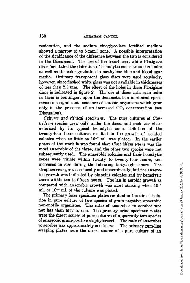
restoration, and the sodium thioglycollate fortified mediumshowed a narrow (5 to 6 mm.) zone. A possible interpretationof the significance of the difference between the two is consideredin the Discussion. The use of the translucent white Plexiglassdiscs facilitated the detection of hemolytic zones around coloniesas well as the color gradation in methylene blue and blood agarmedia. Ordinary transparent glass discs were used routinely,however, since flashed white glass was not available in thicknessesof less than 2.5 mm. The effect of the holes in these Plexiglassdiscs is indicated in figure 2. The use of discs with such holesin them is contingent upon the demonstration in clinical speci-mens of a significant incidence of aerobic organisms which growonly in the presence of an increased C02 concentration (seeDiscussion).
Cultures and clinical specimens. The pure cultures of Clos-tridium species grew only under the discs, and each was char-acterized by its typical hemolytic zone. Dilution of thetwenty-four hour cultures resulted in the growth of isolatedcolonies when as little as 10-7 ml. was plated. In the earlierphase of the work it was found that Clostridium tetani was themost anaerobic of the three, and the other two species were notsubsequently used. The anaerobic colonies and their hemolyticzones were visible within twenty to twenty-four hours, andincreased in size during the following forty-eight hours. Thestreptococcus grew aerobically and anaerobically, but the anaero-bic growth was indicated by pinpoint colonies and by hemolyticzones within ten to fifteen hours. The lag in aerobic growth ascompared with anaerobic growth was most striking when 10-7ml. or 108 ml. of the culture was plated.The primary feces specimen plates resulted in the direct isola-
tion in pure culture of two species of gram-negative anaerobicnon-motile organisms. The ratio of anaerobes to aerobes wasnot less than fifty to one. The primary urine specimen plateswere the direct source of pure cultures of apparently two speciesof anaerobic gram-positive staphylococci. The ratio of anaerobestQ aerobes was approximately one to two. The primary gum-linescraping plates were the direct source of a pure culture of an
162
Dow
nloa
ded
from
http
s://j
ourn
als.
asm
.org
/jour
nal/j
b on
29
Janu
ary
2022
by
42.9
8.96
.40.

MAINTENANCE OF MICROORGANISMS IN PETRI DISH 163
anaerobic, gram-negative, non-motile, filamentous, branching(by dark field examination), beta-hemolytic organism. The ratioof anaerobes to aerobes was not less than fifty to one. Theprimary vaginal swab specimen plates were, with an interestingexception, the direct source of pure cultures of anaerobic, gram-positive streptococci, and micro-aerophilic and anaerobic gram-negative, frequently bean-shaped, diplococci which formedoccasional short chains. Replated cultures of some of the abovespecies are shown in figure 3 and figure 4. The ratio of anaerobesto aerobes was not less than one hundred to one in eleven of thefourteen vaginal swab specimens. In the other three specimens,the approximate one to one ratio of anaerobes to aerobes wasapparently due to an increase in facultative gram-negative rods.
In one of the two feces specimens, and in two of the threevaginal swab specimens showing unusually high aerobic counts,an observation was made which has been previously reported(Topley and Wilson, 1938). The anaerobic agar layers in thelow dilution (overcrowded) plates of the above three specimenscontained a few beta-hemolytic colonies which were found tobe gram-positive streptococci. There was no evidence of similarlysharply defined and easily detected hemolytic zones in the aerobiclayers in the same overcrowded plates.One of the vaginitis patients was on two occasions the source
of a micro-aerophilic organism, shown in figure 3 in two dilutionsof a replated culture. The appearance of the growth in theprimary plates was identical with the appearance of the replatedculture as photographed.On two out of two occasions it was correctly anticipated that
the subculturing of single well-isolated anaerobic colonies wouldresult in the growth of two colony types. Such a prognosis with-out smear examination was indicated in one case by the numberof aerobic colonies in the aerobic agar layers in the same plates.The second observation was made in the case of the micro-aerophilic organism. That is, a similar concentration of well-isolated colonies of gram-negative cocci grew anaerobically inthe same primary plates. When the anaerobic gram-negativecocci were replated from subculture, the ring of micro-aerophilic
Dow
nloa
ded
from
http
s://j
ourn
als.
asm
.org
/jour
nal/j
b on
29
Janu
ary
2022
by
42.9
8.96
.40.
ABRAHAM CANTOR
growth appeared along with the growth of the anaerobes (seeDiscussion).The gram stain retention of the organisms was not affected
by the growth of the organisms in either of the two anaerobicmedia used for subculturing. However, some of the originallygram-negative cocci did retain the gram stain after three transferswhen decolorized with 95 per cent alcohol, and failed to retainit when decolorized with 25 per cent acetone and 75 per centalcohol. The micro-aerophile mentioned above was one ofthese. The organisms referred to as gram-negative diplococci,frequently bean-shaped, were, when not spherical, always char-acterized by their long axis lying parallel to the plane of theiradjacent faces.The lack of qualitative or quantitative correlation between the
bacterial flora indicated by direct-smear gram stain examinationof vaginitis specimens and the anaerobes isolated therefrom wasmore striking than anticipated.
All the anaerobes isolated were similar to types which, asindicated in Bergey's Manual of Determinative Bacteriology(1939), had been previously isolated from the same habitats.
DISCUSSION
The increasing use of screw-cap medicine bottles for the storageof bacteriological media (McCartney, 1933) makes generallyavailable the described routine method for the preparation ofagar media which are themselves a source of the carbon dioxidenecessary for the growth of many organisms. Directions for thepreparation of sterile solutions containing sodium bicarbonateusually call for a filtration method of sterilization (Parker, 1938).However, that instability of sodium bicarbonate (Merck Index,1940) which excludes heat sterilization in cotton-plugged con-tainers makes sodium bicarbonate a convenient source of carbondioxide in media. The decomposition of the sodium bicarbonatewhich takes place in the autoclave is followed by the reformationof the sodium bicarbonate as the medium cools and the carbondioxide trapped in the screw-cap bottle reacts with the sodiumcarbonate. The formation of compounds of carbon dioxide with
164
Dow
nloa
ded
from
http
s://j
ourn
als.
asm
.org
/jour
nal/j
b on
29
Janu
ary
2022
by
42.9
8.96
.40.

MAINTENANCE OF MICROORGANISMS IN PETRI DISH 165
peptones and proteins (Gortner, 1938) and the present lack ofunderstanding as to the role of carbon dioxide in bacterial metab-olism indicated that a test for the mechanical retention of carbondioxide was called for, rather than a titration for carbon dioxideor sodium bicarbonate as such. It may be necessary to considerthe unique role of sodium bicarbonate and carbon dioxide in rela-tion to hemoglobin and to red-cell permeability (Wright, 1938)before deciding that a medium fortified with sodium bicarbonatewill not result in the production of atypical hemolysis byorganisms.The investigation by Wilson (1931) which made carbon dioxide
rather than oxidation reduction potential the basis for explainingthe band phenomenon in the growth of Brucella aborts and theimportance of carbon dioxide in the growth of many organismsmakes pertinent a reconsideration of the incidence of what wereformerly considered micro-aerophiles. Until Wilson investigatedthe band phenomenon it was explained as an indication of themicro-aerophilic nature of the organism.The above question is of importance in a consideration of
media and methods for investigating unknown bacterial flora.If an organism grows only at an aerobic potential and at a carbondioxide concentration higher than that which can be maintainedat an agar medium-air interface, it grows in a band below theinterface in a tubed medium, or in a ring as shown in figure 3 inthis plating method. That is, the rate of oxygen penetration ofthe medium is greater than the rate of carbon dioxide escape fromthe medium, and it is this difference in rates which results in theestablishment of necessary conditions for growth. Reference atthis point to figure 2 and a description thereof in the Resultsindicate a method for decreasing the rate of oxygen penetrationof a medium. If this fortification of a medium with an excessof a reducing compound decreases the difference in the rates ofoxygen penetration of and carbon dioxide loss from the medium,then it may be well to consider the possibility that the growthof such organisms may be not merely delayed, but also preventedby the use of an excess of reducing compounds. There is thepossibility however that a lowering of the potential in media,
Dow
nloa
ded
from
http
s://j
ourn
als.
asm
.org
/jour
nal/j
b on
29
Janu
ary
2022
by
42.9
8.96
.40.
ABRAHAM CANTOR
particularly blood-enriched media, may also interfere with therate of loss of carbon dioxide from the medium (Wright, 1938.)
Hall (1920), in an article on anaerobic methods, reported twoobservations on the growth of what may have been micro-aero-philic organisms. A certain culture, grown by the deep tubemethod, was found to contain a second colony type which grewonly within a narrow range (5 mm.) at a constant depth beneaththe surface (3 cm.). He reported a similar instance upon cul-turing the nasal washings of a patients. These organisms wouldhave been overlooked in his surface method of cultivation, andthe surface methods of cultivation in use at the present timeare subject to the same criticism. That is, a uniform potentialis established at the medium surface. The frequency with whichorganisms may be encountered which can grow only in similarlyvery limited potential ranges can most conveniently be deter-mined by using a method which permits their isolated growthand subculturing simultaneously with that of the more faculta-tive organisms. This objection to surface culture methods isfurther considered and restated in Hall's review of anaerobicmethods (1929).
Another objection to the use of surface growth methods is afunction of the tendency to streak clinical specimens undilutedor in one dilution on enriched, non-inhibitory media. The evi-dence is not yet available to indicate that the anaerobic etiolog-ical agents responsible for various conditions are present in sucha ratio to the residual anaerobic flora as to make their surfaceisolation on streaked non-inhibitory media as likely to succeedas the poured agar plate method.
In so far as the mechanics of subculturing colonies from thesurface of a streaked plate versus the depth of a poured plate areconcerned, the subculturing of colonies from the 1 mm. thickbottom agar layer in the dish is more akin to the former than tothe latter. In those plates where the bottom agar layer wasovercrowded with anaerobic colonies, that is, in low dilutionplates which could not be used as a direct source of pure cul-tures, the agar layer was disrupted by gas. The bottom agarlayer was rarely split in this manner by well isolated colonies.
166
Dow
nloa
ded
from
http
s://j
ourn
als.
asm
.org
/jour
nal/j
b on
29
Janu
ary
2022
by
42.9
8.96
.40.
MAINTENANCE OF MICROORGANISMS IN PETRI DISH 167
The aerobic colonies on the surface agar layer may be examinedand picked as soon as they are properly developed and theplate then replaced in the incubator for whatever additional timeis necessary.
Spaulding and Goode (1939) mention the, fortunately onlyoccasional, appearance of Clostridium species in the twenty-fivehundred specimens they examined aerobically and anaerobically.This is mentioned here in light of an academic objection whichmay be made to the method described, that is, the possibilityof agar-glass interface spreading of motile anaerobes. The COs-tridium tetani culture used in this work first developed as isolatedcolonies and then, after the second day of growth, frequentlystarted to spread at the glass-agar interface. This seems, ifanything, an improvement over the spreading which takesplace with surface cultivation methods for growing Clostridiumtetani.The plating medium used in this work, basically similar to one
used in an investigation of non-spore bearing anaerobic organismsin feces (Eggerth, 1935), is not here recommended as best forgeneral use. It was used merely to demonstrate the method.In the case of one set of plates poured during the pure culturephase of the work, tryptose agar (Difco Manual, 1939) was usedas a base, as well as the heart-infusion base agar. The growth ofanaerobes took place in the bottom agar layer in low, but notin high dilution of the cultures in the tryptose agar, whereassatisfactory growth took place in the same high dilution in theinfusion base agar. In addition, there was a more striking differ-ence between poor aerobic and good anaerobic growth of highdilutions of the streptococcus in the tryptose agar base than inthe heart-infusion agar. These results with pure cultures areanalogous to the results reported by Spaulding and Goode(1939) for clinical specimens, when they compared an infusionbase medium with a meat extract medium in their survey, andanalogous to the pure culture work in tubed media reported byDubos (1929b).The nature of some of the anaerobes isolated from Trichomonas-
complicated vaginitis, specifically the gram-negative diplococci,
Dow
nloa
ded
from
http
s://j
ourn
als.
asm
.org
/jour
nal/j
b on
29
Janu
ary
2022
by
42.9
8.96
.40.
ABRAHAM CANTOR
tempts one to consider the possibility that the reported inferiorityof the vaginal aerobic plate culture method in the diagnosis ofsupposedly Trichomonas-complicated gonorrhea (King, Mascalland Price, 1936) may be explained by the fact that the organismsseen in some of the cases may not have been the typical Neisseriagonorrhoeae.
In extension and conclusion, the agar plate method hereindescribed maintains the gradient potentials present in tubedmedia, and the intermediate regions of that gradient are spreadover a larger area.
SUMMARY
1. A new method is described which makes possible the growthand subculturing of colonies of aerobic, micro-aerophilic, andanaerobic microorganisms in the same petri dish.
2. The method is based upon the negative potential drift insterile media and the counteracting of that drift by the restricteddiffusion of oxygen into the medium.
3. The maintenance of the necessary oxidation-reductionpotentials was tested by the use of oxidation-reduction potentialindicators, by the growth of stock cultures of facultative andanaerobic bacteria, and by the plating of gum-line scrapings,urine, feces, and vaginal swab specimens from cases of non-specific vaginitis.
4. A simple method is indicated for preparing agar mediawhich, after autoclaving, are a source of the carbon dioxide essen-tial for the growth of many organisms.
5. The simplicity of the method, the elimination of the neces-sity for conventional anaerobic apparatus, the elimination ofthe separate method for plating anaerobes, and the potentialgradient which makes possible the direct plating of organismwhich are micro-aerophilic upon primary isolation, may be takento indicate that the method is practicable for the clinicallaboratory.
The vaginitis specimens used in this investigation were madeavailable by the cooperation of the Gynecologic Clinic, Phila-delphia General Hospital.
168
Dow
nloa
ded
from
http
s://j
ourn
als.
asm
.org
/jour
nal/j
b on
29
Janu
ary
2022
by
42.9
8.96
.40.
MAINTENANCE OF MICROORGANISMS IN PETRI DISH 169
REFERENCESBERGEY, D. H., BREED, R. S., MURRAY, E. G. D., AND HITCHENS, A. P. 1939
Bergey's Manual of Determinative Bacteriology. The Williams &Wilkins Company, Baltimore.
BREWER, J. H. 1940 A clear liquid medium for the "aerobic" cultivation ofanaerobes. J. Bact., 39, 10.
Difoo Laboratories 1939 Manual of dehydrated culture media and reagents.Detroit, Michigan.
Dunos, RENE. 1929a Observations on the oxidation-reduction properties ofsterile bacteriological media, J. Exptl. Med., 49, 507-523.
Dumos, RENE 1929b The initiation of growth of certain facultative anaerobesas relative to oxidation-reduction processes in the medium. J. Exptl.Med., 49, 559-573.
EGGERTH, A. H. 1935 The gram positive non-spore bearing anaerobic bacilliof human feces. J. Bact., 30, 277-299.
GORTNER, R. A. 1938 Outlines of Biochemistry, p. 379. John Wiley and Sons,Inc., New York, 2nd Edition.
HALL, I. C. 1920 Practical methods in the purification of obligate anaerobes.J. Infectious Diseases, 27, 576-590.
HALL, I. C. 1929 A review of the development and application of physical andchemical principles in the cultivation of obligately anaerobic bacteria.J. Bact., 17, 255-301.
HEWITT, L. F. 1936 Oxidation-Reduction Potentials in Bacteriology and Bio-chemistry. London County Council, 4th edition.
KING, A. J., MASCALL, W. N., AND PRICE, I. N. 0. 1936 Trichomonas vaginalis;its incidence and coexistence with gonococcal infections. Lancet,2, 18-20.
KNIGHT, B. C. J. G. 1938 Bacterial Nutrition; Medical Research Council,Special report series, % 210, London.
LEPPER, E., AND MARTIN, C. J. 1929 The chemical mechanism exploited in theuse of cooked meat media for the cultivation of anaerobes. Brit. J.Exptl. Path., 10, 327.
LEPPER, E., AND MARTIN, C. J. 1930 The oxidation-reduction potential ofcooked meat media. Brit. J. Exptl. Path., 11, 137.
MCCARTNEY, J. E. 1933 Screw-capped bottles in the preparation and storageof culture media. Lancet, 2, 433.
The Merck Index 1940 P. 505, Merck and Company, Inc., Rahway, N. J., 5thedition.
PARKER, R. C. 1938 Methods of Tissue Culture, p. 37, Paul B. Hoeber, Inc.,New York.
SPAULDING, E. H., AND GOODE, W. G. 1939 Anaerobic cultivation as a routinebacteriologic procedure in the clinical laboratory. J. Lab. Clin.Med., 25, 305-314.
TOPLEY, W. W. C., AND WILSON, G. S. 1936 The Principles of Bacteriology andImmunity, p. 437. William Wood and Company, 2nd edition.
Webster's New International Dictionary 1939 Color Chart B, G. & C. MerriamCompany, Springfield, Mass.
WILSON, G. S. 1931 Growth of Brucella abortus (bovine type) in shake tubes.Brit. J. Exptl. Path., 12, 152-165.
WRIGHT, S. 1938 Applied Physiology, p. 484-488. Oxford University Press,New York, 6th edition.
Dow
nloa
ded
from
http
s://j
ourn
als.
asm
.org
/jour
nal/j
b on
29
Janu
ary
2022
by
42.9
8.96
.40.
170 ABRAHAM CANTOR
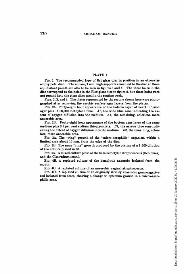
PLATE 1
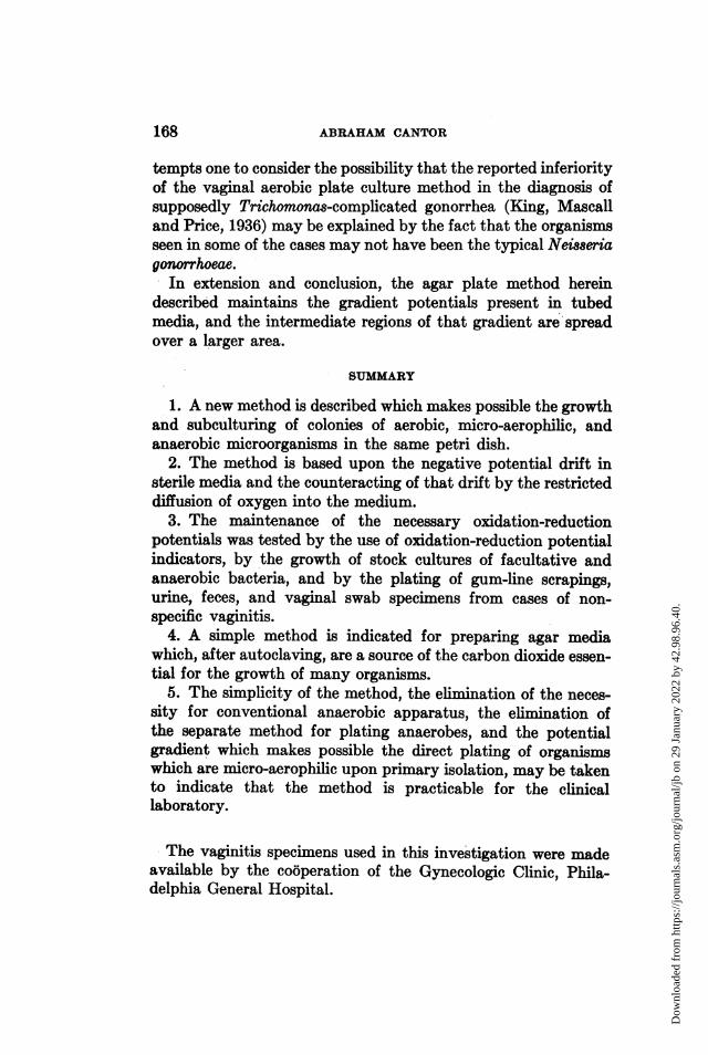
FIG. 1. The recommended type of flat glass disc in position in an otherwiseempty petri dish. The square, 1 mm. high supports cemented to the disc at threeequidistant points are also to be seen in figures 3 and 4. The three holes in thedisc correspond to the holes in the Plexiglass disc in figure 2, but these holes werenot ground into the glass discs used in the routine work.
FIGs. 2, 3, and 4. The plates represented by the sectors shown here were photo-graphed after removing the aerobic surface agar layers from the plates.
FIG. 2A. Forty-eight hour appearance of the bottom layer of heart infusionagar plus 1:100,000 methylene blue. Al, the wide blue zone indicating the ex-tent of oxygen diffusion into the medium. AS, the remaining, colorless, moreanaerobic area.
FIG. 2B. Forty-eight hour appearance of the bottom agar layer of the samemedium plus 0.1 per cent sodium thioglycollate. BR, the narrow blue zone indi-cating the extent of oxygen diffusion into the medium. B1, the remaining, color-less, more anaerobic area.
FIG. 3A. The "ring" growth of the "micro-aerophilic" organism within alimited area about 10 mm. from the edge of the disc.
FIG. 3B. The same "ring" growth produced by the plating of a 1:100 dilutionof the culture plated in 3A.
FIG. 4A. A mixed culture plate of the beta hemolytic streptococcus (2 colonies)and the Clostridium tetani.
FIG. 4B. A replated culture of the hemolytic anaerobe isolated from themouth.
FIG. 4C. A replated culture of an anaerobic vaginal streptococcus.FIG. 4D. A replated culture of an originally strictly anaerobic gram-negative
rod isolated from feces, showing a change to optimum growth in a micro-aero-philic zone.
Dow
nloa
ded
from
http
s://j
ourn
als.
asm
.org
/jour
nal/j
b on
29
Janu
ary
2022
by
42.9
8.96
.40.

JOURNAL OF BACTERIOLOGY, VOL. XLI
1 2_.t> ,, ,aw r, ... +ii,;.,.-,.Z.>-,r^g>^,.ril!,t';;6 ;' s 1;M
FaS 94j2¢ *^' 11-!5_
XiM ot°,:':D'S0F 11*-2uNt- X;gi i.# m-w 8 : .p rr3 tt<?;S t aii iE aiij,: j t
i !.,r.Sg.l Ma,, :...... _. . . .. _ E'''- :, jM- &'-' 1 x . ihi- * __'x''s.t1 !
_
iv__ t/B1 ' t"x ,{^ I-[ ! X ; J
!, XX ffi
]d SI_ gX ot...j,'' t.'i,"' 1..... ' m . .,^ . '-. ;r-; 9i lw w ,:h+.3,_
L * liiilL - 's-B C' D
__
3 4
(Abrahrm Cantor: Maintenance of Microorganisms in Petri Dish)
PLATE 1
e-
Dow
nloa
ded
from
http
s://j
ourn
als.
asm
.org
/jour
nal/j
b on
29
Janu
ary
2022
by
42.9
8.96
.40.