methods of study bone growth / orthodontic courses by indian dental academy
TRANSCRIPT

Methods of studying bone growth
INDIAN DENTAL ACADEMY
Leader in continuing dental education www.indiandentalacademy.com
www.indiandentalacademy.com

Bone growth ?
Bone = bone cells(osteoblasts ,osteocytes ,osteoclasts) +matrix (collagen + calcium hydroxyapatite 65-70%)
Woven boneLamellar bone (compact bone)Composite bone (cancellous bone)Bundle bone
Vitamin-D 9-11mg/dl CalcitoninPTH Serum Ca++
www.indiandentalacademy.com

Study Acquisition of knowledge by investigation.
Collection of data
Analysis of this data
Presentation of data
Explanation
Passion & controversy may have a place in discussion & interpretation but certainly not in study of rigorously collected biomedical &clinical data A. G. Petrovic
www.indiandentalacademy.com
Source of data
CensusRegistration of vital eventsHospital records & other health recordsSurvey of population
Health interview survey (opinion)Health examination surveyHealth record surveymailed question survey
www.indiandentalacademy.com
Types of dataOpinion :it is a clever guess .Observation :Done to see presence or
absence of certain phenomenaRating & ranking.Measurements :Most scientific approch
direct dataindirect dataderived data
www.indiandentalacademy.com

Methods of collecting dataCross sectional method :based on
single examination of a cross-section of population at one point in time .
Longitudinal method :observations period are repeated in the same population over a prolonged of time by means of follow up examination.
Semi-longitudinal method :www.indiandentalacademy.com

Cross-sectional method
ADVANTAGESeasy & quickless expensiveno attrition of samplevarious factors
acting at that time can be analyzed
can be repeated
DISADVANTAGESvariability with in
the samplelarger size of
samplefactors acting at
various time period cannot be analyzed
www.indiandentalacademy.com

Longitudinal method
ADVANTAGESmore specificfactors acting at
different time can be analyzed
less no of subjectsindividual
variations
DISADVANTAGESmore expensivedifficulty in
maintaining lab & data
more timeattrition of samplescannot be repeated
Eg Bolton Brush study ,Michigan study ,Iova child welfare studywww.indiandentalacademy.com

Semi-longitudinal methodADVANTAGESTries to combine advantages of both
longitudinal & cross-sectional method.
Data of 15 yrs of study obtained in 3 yrs
DISADVANTAGESNot as authentic as longitudinal study
www.indiandentalacademy.com
Approach for analysis of obtained data.
MEASUREMENT APPROACH
Here animals & humans are measured without inducing any change in them.
Dead/aliveLongitudinal/cross-
sectional
EXPERIMENTAL APPROACH
Here growth is manipulated and observations are made.
More detailed study.Mainly animal study.Longitudinal/cross-
sectionalwww.indiandentalacademy.com
Methods of presenting dataSimple tablesGraphs (special
curves)Charts
Bar charts Histograms Pie charts Pictograms
Diagrams (pictures)www.indiandentalacademy.com

Explanation concerning craniofacial growth in current literatureDeductive:logically explained consequence of certain
premises.
Deductivoprobabilistic: tries to relate various items explained by the deductive explanation with
certain assumptions.Forms basis of D/D &prognosis.Functional: Based on single assumption.Generally
Noncolinear relations are seen here . Phylogenetic:Growth trends explained based on
evolutionary trends.
www.indiandentalacademy.com

Methods of studying bone growth.
Mineralized sections Polarized light birefringence Fluorescent labels Micro radiography Auto radiography nuclear volume
morphometry Cell kinetics Micro electrodes Finite element modeling Vital staining Metallic implants
MEASUREMENT Craniometry Comparative
anatomy Anthropometry Radiology/Imaging
EXPERIMENTAL
www.indiandentalacademy.com

CraniometryMeasurement of skull of human skeleton.Broca (1875) defined landmarks &
instruments used for measurements. Congress of German anthropological
society held at Frankfurt in 1882.
Comparative anatomywww.indiandentalacademy.com

AnthropometryMeasurement of skeletal dimensions on
living individualsPhysical anthropology :Study of mans
biologic behavior in time and spaceSpecial instruments are usedADV :Longitudinal study
No harm to subjects
DISADV :Soft tissue error Operator error
www.indiandentalacademy.com

Radiology /ImagingConventional radiographs Nature and production of x-raysHow does it detect bone growth ?Films :composition
size: 22*35 24*40 32*41 57*76 mm 8*10” :Image formation ,developing & fixing
Intensifying screens (calcium tungstate & rare earth)
Grids (parallel ,focused &Potter Bucky grids.80-100lines/Inch)
www.indiandentalacademy.com
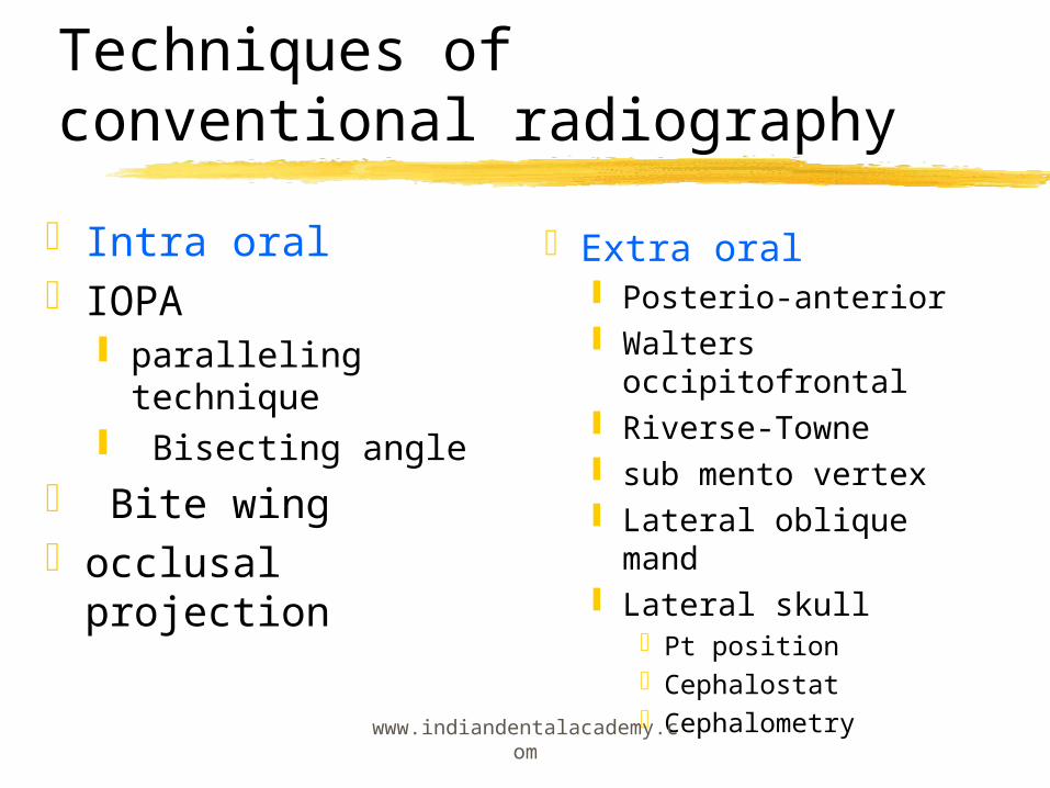
Techniques of conventional radiographyIntra oralIOPA
paralleling technique
Bisecting angle Bite wingocclusal projection
Extra oral Posterio-anterior Walters
occipitofrontal Riverse-Towne sub mento vertex Lateral oblique mand Lateral skull
Pt positionCephalostatCephalometry
www.indiandentalacademy.com

Broadbent-Bolton cephalometer
www.indiandentalacademy.com
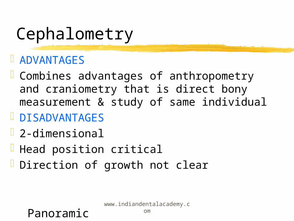
CephalometryADVANTAGESCombines advantages of anthropometry
and craniometry that is direct bony measurement & study of same individual
DISADVANTAGES2-dimensional Head position criticalDirection of growth not clear
Panoramic www.indiandentalacademy.com

Specialized radiographic techniqueDigital imaging (CCD -voltage-bits 0-255)
Direct digital radiography (R V G )Indirect digital radiographyDigital subtraction radiography Digitized image interpretation
Computed tomographyMagnetic resonanceRadionuclide imagingUltrasoundElectronic thermography
www.indiandentalacademy.com
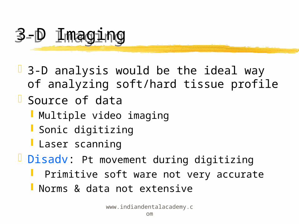
3-D Imaging3-D analysis would be the ideal way of
analyzing soft/hard tissue profileSource of data
Multiple video imaging Sonic digitizing Laser scanning
Disadv: Pt movement during digitizing Primitive soft ware not very accurate Norms & data not extensive
www.indiandentalacademy.com

Specific experimental method
Mineralized sections Polarized light Fluorescent labelsMicro radiographyAuto radiographynuclear volume morphometryCell kineticsMicro electrodes
Finite element modelingVital stainingMetallic implants
www.indiandentalacademy.com

Histopathological techniquePreparation of tissues for microscopySoft tissue embedded in paraffin
Fixationprocessing
colloidal embedding - hard tissues(decalcified)
Acid treatmentchelationHydrolysis
Ground sections- hard tissues(undecalcified)Frozen sections for immediate examination
www.indiandentalacademy.com

Mineralized sectionsCritical analysis of tissues as there is less
distortion during processingBoth inorganic mineral & organic matrix can
be studied100um -tissue level details25um-Enhanced cellular details
Bone labels quench rapidly Tissue density inadequate for microradiography
www.indiandentalacademy.com
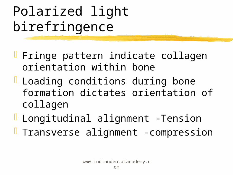
Polarized light birefringenceFringe pattern indicate collagen
orientation within boneLoading conditions during bone
formation dictates orientation of collagen
Longitudinal alignment -TensionTransverse alignment -compression
www.indiandentalacademy.com
Fluorescent labelsIn vivo administration of Cl binders act
as time markers of bone formationSix fluorescent bone labels are used
Tetracycline -bright yellow Calcein - green Xylenol- orange Alizarin- complexone red Demeclocyclin- gold Oxytetracycline- greenish yellow
www.indiandentalacademy.com

MicroradiographyHigher resolution images of polished 100um
mineralized sections obtained1 week primary mineralization8 months secondary mineralizationExperimental animals analyzed by both
microradiography & using fluoroscentlabels midfacial sutures PDL Alveolar process Mandibular condyle temporal fossa
www.indiandentalacademy.com

AutoradiographySpecific radioactive labels for proteins
carbohydrates ,& nucleic acids are injected at known intervals before sampling
Detected by coating histologic sections with nuclear track emulsion
3H thymidine labels DNA synthesis 3H Proline for bone matrix
www.indiandentalacademy.com

Nuclear volume morphometry
Cytomorphometric procedure for accessing the size of osteoblastic precursor cells
Mechanism of osteogenesis in orthodontically activated PDL
Preosteoblasts have larger nucleus than committed osteoprogenitor cells and their precursors
www.indiandentalacademy.com

Cell kineticsBy noting the -increase in nuclear
volume or labeling S-phase cells in vivo using Bromodeoxyuridine (BDU) cell movement & differentiation is noted
Generally done in PDL under normal conditions under metabolic stimuli mechanical stimuli
www.indiandentalacademy.com

MicroelectrodesTungsten or glass electrodes are
inserted atraumatically into PDL in live animals via gingival sulcus
changes in electric potential are notedWidened areas have a negative chargeCompressed areas have positive chargeThis coincides with the age old principle,
that bone forms near cathode & resorbs at anode
www.indiandentalacademy.com

Finite element modelingFinite element modeling is a branch of
mechanical engineering where in the stress & strain of mechanically loaded structures are analyzed.
Initial stress for periodontium are derived by assuming linear elastic properties
For complex tissues like periodontium with viscoelastic properties both solid & fluid mechanics must be considered
www.indiandentalacademy.com

Vital stainingReported initially by Belchier (1796)
& John Hunter where in they attributed staining to alizarin
This method reveals the site ,manner, amount , direction ,timing & duration of bone growth
Tetracycline stains in humanswww.indiandentalacademy.com

Metallic implantsMethod of study used extensively by
Prof Bjork & coworkers R D C Copenhagen
They gave new dimension to study of dentofacial growth.
Remodeling changes in the contours of jaws was better understood
Rotational pattern of jaw growthwww.indiandentalacademy.com

ConclusionTooth movement has
been possible because bone behaves dynamically
Better understanding of physiology of bone PDL interface is necessary
All these methods have given us the tool for further study it is up to us to use it
www.indiandentalacademy.com

ReferencesEnlow;Hand book of facial growth, W B Saunders Company,1982
Orbans:Oral histology &embryology,Delhi, C B S publishers,1990
Rakosi ,Jones & Graber:Colour atlas of orthodontic diagnosis,New York,Thieme medical publishers,1993
Farkas L G:Anthropometry of head &face, New York, Raven press,1994
Jacobson :Radiographic cephalometry,quintessence books,1995 www.indiandentalacademy.com

Goaz & White:Oral radiology, St Louis,C V Mosby, 1994K Park : Preventive &social medicine, Jabalpur , M/S Banarsidas Bhanot,1997Profitt W R:Contemporary orthodontics,St Louis,C V Mosby,1997Graber,Rakosi,Petrovic:Dentofacial orthopedics with functional appliances, 1997Graber Vanarsdall:Orthodontics current principles &technique, St Louis,C V Mosby,2000.
References ctd
Xwww.indiandentalacademy.com

Thank you
For more details please visit www.indiandentalacademy.com
www.indiandentalacademy.com