orthodontic retreatment: dental trauma and root resorptioncdn.intechweb.org/pdfs/24351.pdf ·...
TRANSCRIPT
10
Orthodontic Retreatment: Dental Trauma and Root Resorption
Pedro Marcelo Tondelli, Fabiana Akemy Kay, Osmar Aparecido Cuoghi and Marcos Rogério de Mendonça
Dental School of Araçatuba, UNESP – Universidade Estadual Paulista Brazil
1. Introduction
This chapter describes aspects related to orthodontic retreatment, care and control of root resorption. A clinical case is presented to illustrate the control forces on the teeth allows to perform orthodontic treatment in cases where a large root resorption for tooth movement associated with dental trauma. Even though both Dentistry and Orthodontics have presented continuous improvement, when one or more teeth are submitted to a dental trauma, the treatment of malocclusion can be completely altered (Malmgren & Malmgren, 2007). Previous detailed evaluation of the root morphology, at the beginning of the orthodontic treatment, is a requirement emphasized by Levander & Malmgren (1988). These authors advised a through examination of the root outline, observing the occurrence of any root resorptions, surface concavities and malformations, since teeth with these characteristics may be severely reabsorbed during treatment. When facing evident root resorptions, the treatment purposes must be reviewed (Malmgren et al., 1982). However, dental movement on traumatized teeth still receives inadequate attention by the specialized literature. Examining the related literature, many different publications about the topic can be found which do not contribute to the development of a safe protocol for clinical procedures. Kindelan et al., 2008, published a review about dental trauma and its influence on the management of orthodontic treatment, contributing with the currently best evidence on the topic. Recently, a research with questionnaires showed that about 40% of orthodontists were not acquainted with the recommendations for the orthodontic movement of traumatized teeth (Tondelli et al., 2010).
2. Dental trauma and orthodontics
Dentoalveolar trauma represents an emerging public health problem. Traumatic injuries to one or more teeth can alter remarkably an ongoing or planned orthodontic treatment. The clinical protocols for traumatized teeth are still uncertain, which increases the risks associated to orthodontic mechanics and influences directly the prognosis. In patients with trauma to the oral region, an appropriate treatment plan after the injury is mandatory for a good prognosis. For dentists and health care providers, knowledge and skills to manage cases of dentoalveolar trauma allied to a sound clinical judgment dictated by the conditions
www.intechopen.com
Principles in Contemporary Orthodontics
238
of a given traumatic situation are key factors for delivering the best care possible with more predictable outcomes (Andreasen, Andreasen & Andersson, 2007). People of all ages and types of malocclusions can suffer traumatic dental injuries, but children and preadolescents are the most frequently affected groups. Increased overjet is a risk factor for dentoalveolar trauma. This risk is 2-fold higher in individuals with 3 to 6 mm overjet and 3-fold higher in individuals with >6 mm overjet (Andreasen, Andreasen & Andersson, 2007). Maxillary incisor exposure and lack of lip seal predispose to traumatic injuries to the anterior oral region, with boys being affected twice as often as girls (Andreasen, Andreasen & Andersson, 2007). These characteristics are present in most patients that seek for orthodontic treatment. Information collected from clinical interview, clinical examination and radiographs are of great value for establishing an accurate diagnosis and case prognosis. In some cases, history of dentoalveolar trauma is not reported by patients or parents/caregivers, evolving to sequelae, such as root resorption or ankylosis, which may alter completely the treatment plan or even result in major damage to the patient, leading to legal proceedings or lawsuit. It is imperative that all patients be questioned about any previous dental trauma prior to
commencing on a course of orthodontic treatment. This will allow the orthodontist to
anticipate any potential complications which may occur and to carefully monitor the
traumatized tooth during orthodontic movement (Kindelan et al., 2008).
The relevance of this topic increases when the professional faces a traumatized tooth during the orthodontic treatment, a fact which may change the initial planning completely. Based on literature data, before undergoing orthodontic movement, a repairing period of 3 to 5 months is recommended for slight dental traumas such as concussion and subluxation, to repair the cement layer affected (Kindelan et al., 2008; Malmgren & Malmgren, 2007). In a severe trauma such as luxation, a period of 6 months to 1 year is recommended. In root fractured teeth, the recommended period is of 1 to 2 years and in reimplanted teeth at least 1 year (Kindelan et al., 2008; Malmgren & Malmgren, 2007). When a root fracture is detected, the repairing may be provided through a fusion of the fractured segment, and the tooth remains in its original size, or through interposition of conjunctive tissue between the fragments. In both cases, the teeth could be moved after a repairing period of 1 to 2 years, though, with the fragment separation, the movement would occur upon a tooth with a shorter root (Kindelan et al., 2008; Malmgren & Malmgren, 2007). A trimestrial radiographic control of the traumatized teeth was suggested, analyzing root
resorptions, the root outline and surface concavity. When a minimal root resorption is
detected during the orthodontic treatment, there is low risk of severe resorption in these
teeth, however, in greater resorption, treatment should be interrupted and the possibility of
discontinuity or limitation of procedures taken into account. Inflammatory root resorption
may be radiographically detected within 3 weeks after its initial and ankylosis within the
period of 2 months to 1 year (Brezniak & Wasserstein, 1993a; 1993b; Malmgren & Malmgren,
2007).
One of the protocols for intrusive luxation treatment includes the extrusive orthodontic
movement to traumatic tooth repositioning (Turley, Joiner & Hellstrom, 1984; Turley,
Crawford & Carrington, 1987). In cases of intrusive luxations of about 3mm and the teeth
present open apexes, it is possible to await spontaneous eruption, in case this does not
www.intechopen.com
Orthodontic Retreatment: Dental Trauma and Root Resorption
239
happen, orthodontic repositioning is indicated, observing the pulp vitality (Kinirons, 1998).
In teeth with complete root formation, the surgical or orthodontic repositioning must be
adopted as soon as possible, together with the endodontic treatment with calcium
hydroxide, otherwise the dental pulp will become necrotic (Malmgren & Malmgren, 2007).
When traumatically intruded teeth don’t present mobility, surgical luxation, previously to
orthodontic extrusion, would be helpful to enable the movement, without compromising
anchorage units, and promote their intrusion (Turley, Joiner & Hellstrom, 1984; Turley,
Crawford & Carrington, 1987).
The ankylosis results from severe periodontal injuries, mainly as a result of an intrusion or
avulsion. It can also develop following inflammatory resorption where the damage to the
cementum has resulted in a defect beyond the critical size for healing by favourable healing.
In these situations the root will slowly be replaced by bone, governed by the age of the
patient and speed of bone turnover, and eventually lost (Day et al., 2008; Malmgren &
Malmgren, 2007). There are different treatment options like periodontal regeneration,
surgical repositioning, distraction osteogenesis, build-up to the incisal level, extraction and
decoronation (Day et al., 2008).
Bauss et al. (2008) have conclude that previously traumatized maxillary incisors, and especially lateral incisors, with severe periodontal injuries have higher susceptibility to pulp necrosis during orthodontic intrusion than nontraumatized teeth. Some of the sequels which may occur due to dental trauma are: ankylosis, external root resorption, pulp necrosis, pulp obliteration, marginal bone loss, alveolar fracture, crown fracture, root fracture, concussion, subluxation, luxation, avulsion (Kindelan et al., 2008; Malmgren & Malmgren, 2007; Turley, Joiner & Hellstrom, 1984; Turley, Crawford & Carrington, 1987) According to Malmgren et al. (2007), there is no single explanation for the reasons that lead to the occurrence of root resorption, but dentoalveolar trauma is always implicated as an etiological factor. The authors refer to dental trauma as a predisposing condition to root resorption, which may be exacerbated by the orthodontic treatment. A literature review was not the scope of this paper, but to focus the current clinical evidence. A practical guide has been prepared summarizing the clinical and orthodontic protocols to be adopted by general dentists and orthodontists dealing with cases of dentoalveolar trauma in patients undergoing orthodontic care and patients with indication for further orthodontic therapy (Table 1).
Type of trauma
Clinical and radiographic findings
Clinical protocol Orthodontic
protocol
Source of information (#Reference)
Concussion
The tooth has increased sensitivity
to percussion without increased mobility or
displacement. No radiographic
abnormalities are observed
Monitoring of pulpal condition for at least 1 year
Wait 3-5 months to start orthodontic
movement, maintaining radiographic
control during 1 year after trauma.Use of mild and
intermittent forces
Brezniak & Wasserstein, 1993a; 1993b;
Kindelan et al., 2008;
Malmgren & Malmgren,
2007
www.intechopen.com

Principles in Contemporary Orthodontics
240
Type of trauma
Clinical and radiographic findings
Clinical protocol Orthodontic
protocol
Source of information (#Reference)
Subluxation
The tooth has increased sensitivity to percussion with increased mobility.
Bleeding from gingival crevice may
be noted. No radiographic abnormalities are
observed
Stabilization of the tooth with a
flexible splint for 2 weeks. Monitoring of pulpal condition for at least 1 year
Wait 3-5 months to start the
orthodontic movement, maintaining radiographic
control during 1 year after trauma.Use of mild and
intermittent forces
Brezniak & Wasserstein, 1993a; 1993b;
Kindelan et al., 2008;
Malmgren & Malmgren,
2007
Extrusive luxation
The tooth appears elongated and is
excessively mobile. Pulp
revascularization may occur, especially
in immature teeth. Radiographic
examination reveals increased periodontal
ligament space apically
Gentle repositioning of the
tooth into its socket,
stabilization of the tooth for 2 weeks using a flexible
splint, monitoring of pulpal condition and radiographic
control for 5 years.
Wait at least 6 months to start the
orthodontic movement.
Radiographic control should be performed every 3
months throughout the
orthodontic treatment.
Use of mild and intermittent forces.
The orthodontic treatment should be simplified if
necessary.
Brezniak & Wasserstein, 1993a; 1993b;
Kindelan et al., 2008;
Malmgren & Malmgren,
2007
Lateral Luxation
The tooth is buccally or lingually/palatally
displaced, is immobile, and
percussion gives a metallic sound.
Pulp revascularization
may occur, especially in immature teeth.
The periodontal ligament space is
widened in an occlusal radiographic
view.
Gentle repositioning of the tooth, stabilization of the tooth for 4
weeks using a flexible splint, monitoring of
pulpal condition and radiographic
control for 5 years.
Wait at least 6 months to start the
orthodontic movement.
Radiographic control should be performed every 3
months throughout the
orthodontic treatment.
Use of mild and intermittent forces.
The orthodontic treatment should be simplified if
necessary.
Brezniak & Wasserstein, 1993a; 1993b;
Kindelan et al., 2008;
Malmgren & Malmgren,
2007
www.intechopen.com
Orthodontic Retreatment: Dental Trauma and Root Resorption
241
Type of trauma
Clinical and radiographic findings
Clinical protocol Orthodontic
protocol
Source of information (#Reference)
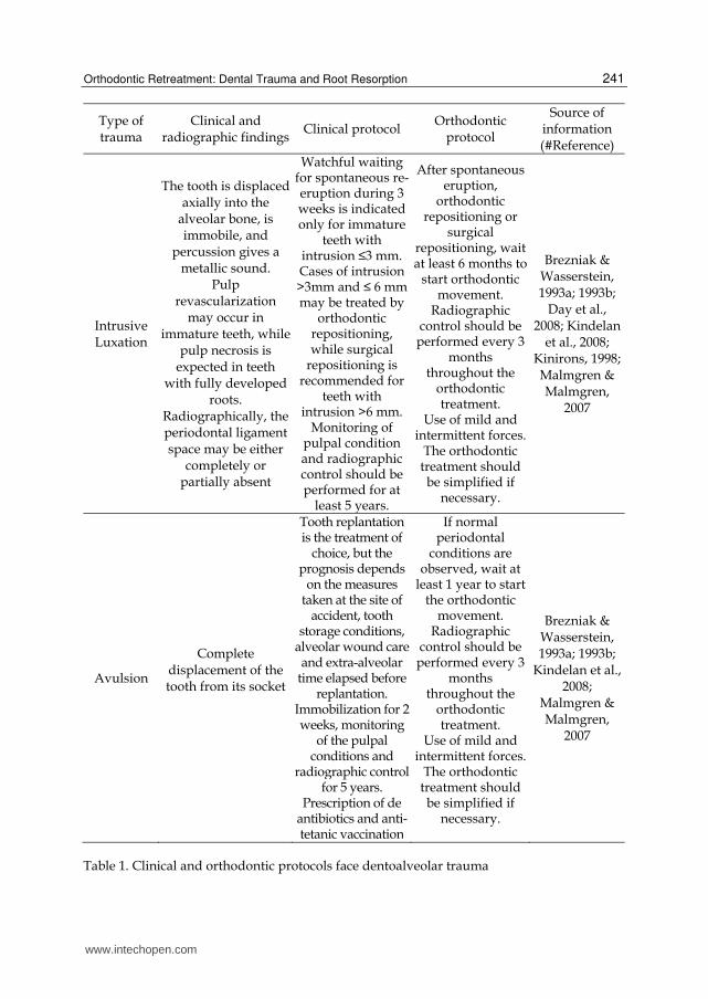
Intrusive Luxation
The tooth is displaced axially into the
alveolar bone, is immobile, and
percussion gives a metallic sound.
Pulp revascularization
may occur in immature teeth, while
pulp necrosis is expected in teeth
with fully developed roots.
Radiographically, the periodontal ligament space may be either
completely or partially absent
Watchful waiting for spontaneous re-eruption during 3 weeks is indicated only for immature
teeth with intrusion ≤3 mm. Cases of intrusion >3mm and ≤ 6 mm may be treated by
orthodontic repositioning, while surgical
repositioning is recommended for
teeth with intrusion >6 mm.
Monitoring of pulpal condition and radiographic control should be performed for at
least 5 years.
After spontaneous eruption,
orthodontic repositioning or
surgical repositioning, wait at least 6 months to
start orthodontic movement.
Radiographic control should be performed every 3
months throughout the
orthodontic treatment.
Use of mild and intermittent forces.
The orthodontic treatment should be simplified if
necessary.
Brezniak & Wasserstein, 1993a; 1993b;
Day et al., 2008; Kindelan
et al., 2008; Kinirons, 1998; Malmgren & Malmgren,
2007
Avulsion
Complete displacement of the tooth from its socket
Tooth replantation is the treatment of
choice, but the prognosis depends
on the measures taken at the site of
accident, tooth storage conditions,
alveolar wound care and extra-alveolar
time elapsed before replantation.
Immobilization for 2 weeks, monitoring
of the pulpal conditions and
radiographic control for 5 years.
Prescription of de antibiotics and anti-tetanic vaccination
If normal periodontal
conditions are observed, wait at
least 1 year to start the orthodontic
movement. Radiographic
control should be performed every 3
months throughout the
orthodontic treatment.
Use of mild and intermittent forces.
The orthodontic treatment should be simplified if
necessary.
Brezniak & Wasserstein, 1993a; 1993b;
Kindelan et al., 2008;
Malmgren & Malmgren,
2007
Table 1. Clinical and orthodontic protocols face dentoalveolar trauma
www.intechopen.com
Principles in Contemporary Orthodontics
242
3. Clinical considerations about orthodontic retreatment, root resorption and force control
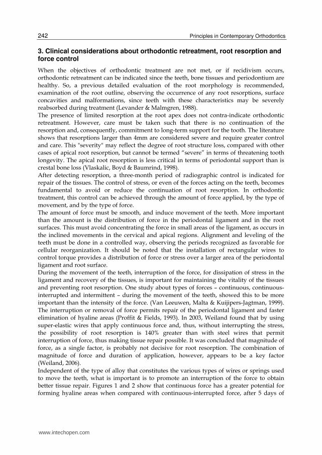
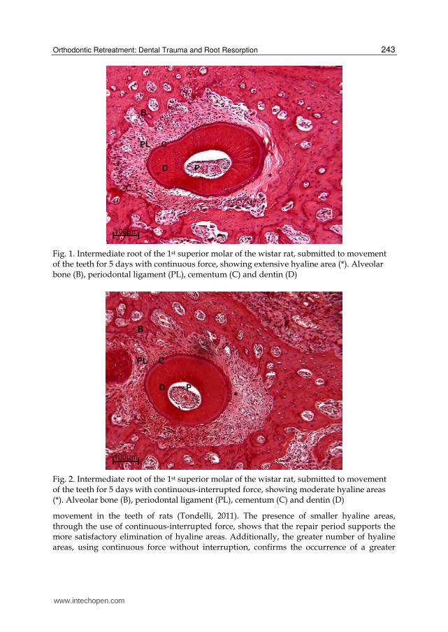
When the objectives of orthodontic treatment are not met, or if recidivism occurs, orthodontic retreatment can be indicated since the teeth, bone tissues and periodontium are healthy. So, a previous detailed evaluation of the root morphology is recommended, examination of the root outline, observing the occurrence of any root resorptions, surface concavities and malformations, since teeth with these characteristics may be severely reabsorbed during treatment (Levander & Malmgren, 1988). The presence of limited resorption at the root apex does not contra-indicate orthodontic retreatment. However, care must be taken such that there is no continuation of the resorption and, consequently, commitment to long-term support for the tooth. The literature shows that resorptions larger than 4mm are considered severe and require greater control and care. This "severity" may reflect the degree of root structure loss, compared with other cases of apical root resorption, but cannot be termed “severe” in terms of threatening tooth longevity. The apical root resorption is less critical in terms of periodontal support than is crestal bone loss (Vlaskalic, Boyd & Baumrind, 1998). After detecting resorption, a three-month period of radiographic control is indicated for repair of the tissues. The control of stress, or even of the forces acting on the teeth, becomes fundamental to avoid or reduce the continuation of root resorption. In orthodontic treatment, this control can be achieved through the amount of force applied, by the type of movement, and by the type of force. The amount of force must be smooth, and induce movement of the teeth. More important than the amount is the distribution of force in the periodontal ligament and in the root surfaces. This must avoid concentrating the force in small areas of the ligament, as occurs in the inclined movements in the cervical and apical regions. Alignment and leveling of the teeth must be done in a controlled way, observing the periods recognized as favorable for cellular reorganization. It should be noted that the installation of rectangular wires to control torque provides a distribution of force or stress over a larger area of the periodontal ligament and root surface. During the movement of the teeth, interruption of the force, for dissipation of stress in the ligament and recovery of the tissues, is important for maintaining the vitality of the tissues and preventing root resorption. One study about types of forces – continuous, continuous-interrupted and intermittent – during the movement of the teeth, showed this to be more important than the intensity of the force. (Van Leeuwen, Malta & Kuijipers-Jagtman, 1999). The interruption or removal of force permits repair of the periodontal ligament and faster elimination of hyaline areas (Proffit & Fields, 1993). In 2003, Weiland found that by using super-elastic wires that apply continuous force and, thus, without interrupting the stress, the possibility of root resorption is 140% greater than with steel wires that permit interruption of force, thus making tissue repair possible. It was concluded that magnitude of force, as a single factor, is probably not decisive for root resorption. The combination of magnitude of force and duration of application, however, appears to be a key factor (Weiland, 2006). Independent of the type of alloy that constitutes the various types of wires or springs used to move the teeth, what is important is to promote an interruption of the force to obtain better tissue repair. Figures 1 and 2 show that continuous force has a greater potential for forming hyaline areas when compared with continuous-interrupted force, after 5 days of
www.intechopen.com
Orthodontic Retreatment: Dental Trauma and Root Resorption
243
Fig. 1. Intermediate root of the 1st superior molar of the wistar rat, submitted to movement of the teeth for 5 days with continuous force, showing extensive hyaline area (*). Alveolar bone (B), periodontal ligament (PL), cementum (C) and dentin (D)
Fig. 2. Intermediate root of the 1st superior molar of the wistar rat, submitted to movement of the teeth for 5 days with continuous-interrupted force, showing moderate hyaline areas (*). Alveolar bone (B), periodontal ligament (PL), cementum (C) and dentin (D)
movement in the teeth of rats (Tondelli, 2011). The presence of smaller hyaline areas, through the use of continuous-interrupted force, shows that the repair period supports the more satisfactory elimination of hyaline areas. Additionally, the greater number of hyaline areas, using continuous force without interruption, confirms the occurrence of a greater
www.intechopen.com
Principles in Contemporary Orthodontics
244
period of stress in the periodontal ligament, resulting in greater potential for root resorption. In this way, the interruption of force could be recommended to reduce or prevent the continuation of resorptions.
4. Case report
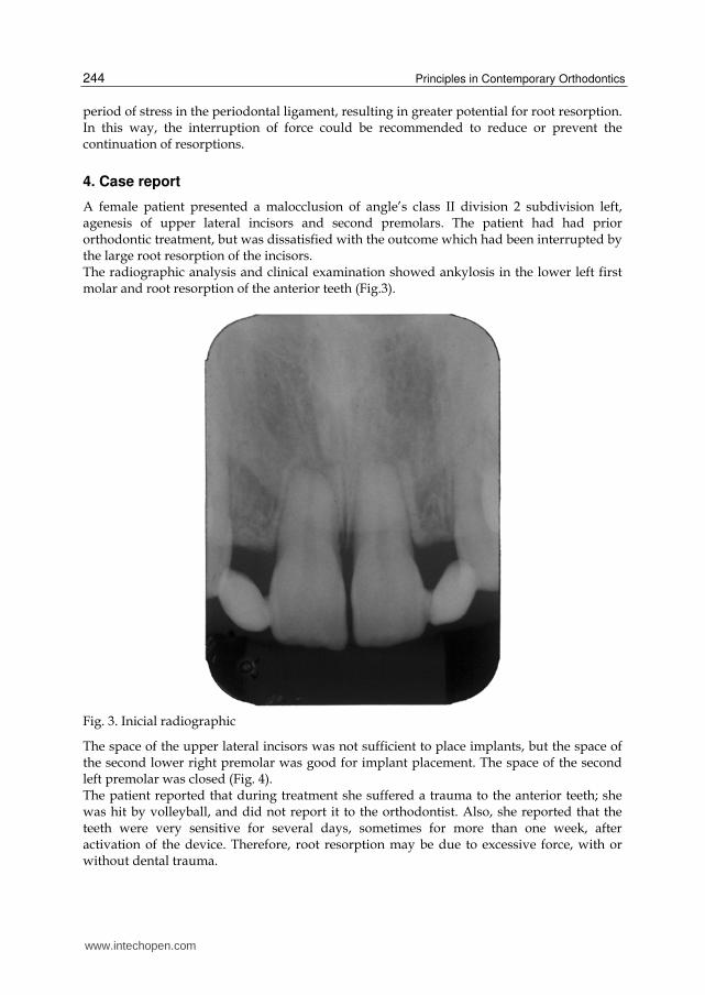
A female patient presented a malocclusion of angle’s class II division 2 subdivision left, agenesis of upper lateral incisors and second premolars. The patient had had prior orthodontic treatment, but was dissatisfied with the outcome which had been interrupted by the large root resorption of the incisors. The radiographic analysis and clinical examination showed ankylosis in the lower left first molar and root resorption of the anterior teeth (Fig.3).
Fig. 3. Inicial radiographic
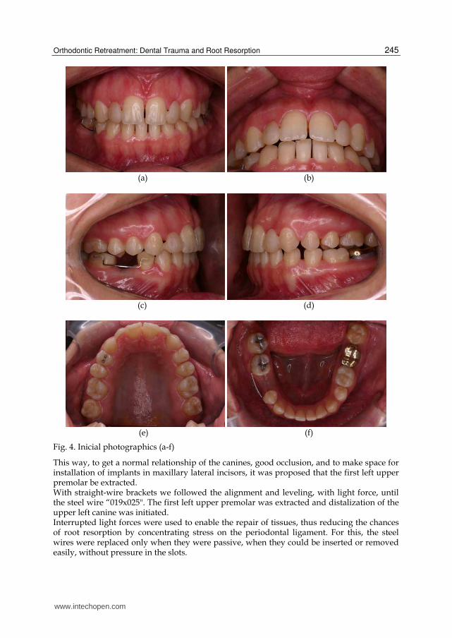
The space of the upper lateral incisors was not sufficient to place implants, but the space of the second lower right premolar was good for implant placement. The space of the second left premolar was closed (Fig. 4). The patient reported that during treatment she suffered a trauma to the anterior teeth; she was hit by volleyball, and did not report it to the orthodontist. Also, she reported that the teeth were very sensitive for several days, sometimes for more than one week, after activation of the device. Therefore, root resorption may be due to excessive force, with or without dental trauma.
www.intechopen.com
Orthodontic Retreatment: Dental Trauma and Root Resorption
245
(a) (b)
(c) (d)
(e) (f)
Fig. 4. Inicial photographics (a-f)
This way, to get a normal relationship of the canines, good occlusion, and to make space for installation of implants in maxillary lateral incisors, it was proposed that the first left upper premolar be extracted. With straight-wire brackets we followed the alignment and leveling, with light force, until the steel wire “019x025". The first left upper premolar was extracted and distalization of the upper left canine was initiated. Interrupted light forces were used to enable the repair of tissues, thus reducing the chances of root resorption by concentrating stress on the periodontal ligament. For this, the steel wires were replaced only when they were passive, when they could be inserted or removed easily, without pressure in the slots.
www.intechopen.com
Principles in Contemporary Orthodontics
246
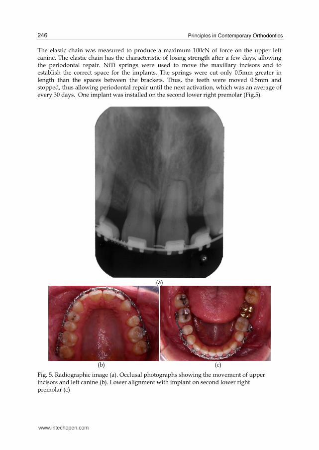
The elastic chain was measured to produce a maximum 100cN of force on the upper left canine. The elastic chain has the characteristic of losing strength after a few days, allowing the periodontal repair. NiTi springs were used to move the maxillary incisors and to establish the correct space for the implants. The springs were cut only 0.5mm greater in length than the spaces between the brackets. Thus, the teeth were moved 0.5mm and stopped, thus allowing periodontal repair until the next activation, which was an average of every 30 days. One implant was installed on the second lower right premolar (Fig.5).
(a)
(b) (c)
Fig. 5. Radiographic image (a). Occlusal photographs showing the movement of upper incisors and left canine (b). Lower alignment with implant on second lower right premolar (c)
www.intechopen.com
Orthodontic Retreatment: Dental Trauma and Root Resorption
247
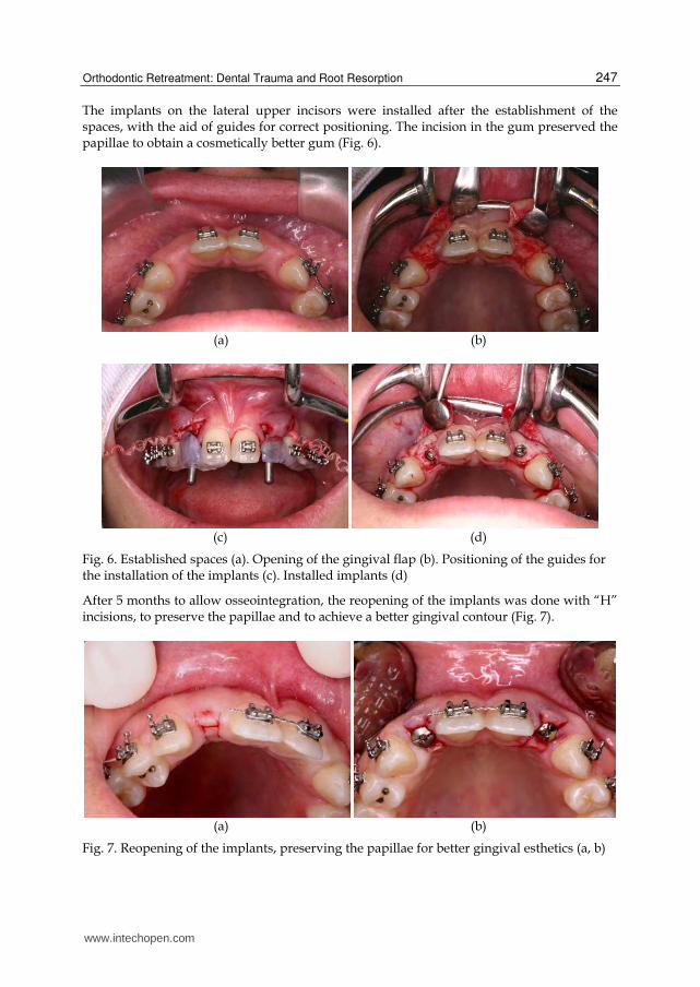
The implants on the lateral upper incisors were installed after the establishment of the spaces, with the aid of guides for correct positioning. The incision in the gum preserved the papillae to obtain a cosmetically better gum (Fig. 6).
(a) (b)
(c) (d)
Fig. 6. Established spaces (a). Opening of the gingival flap (b). Positioning of the guides for the installation of the implants (c). Installed implants (d)
After 5 months to allow osseointegration, the reopening of the implants was done with “H” incisions, to preserve the papillae and to achieve a better gingival contour (Fig. 7).
(a) (b)
Fig. 7. Reopening of the implants, preserving the papillae for better gingival esthetics (a, b)
www.intechopen.com
Principles in Contemporary Orthodontics
248
(a)
(b) (c)
(d) (e)
www.intechopen.com
Orthodontic Retreatment: Dental Trauma and Root Resorption
249
(f) (g)
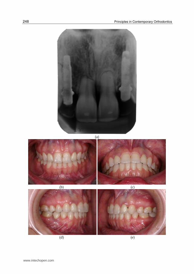
Fig. 8. Final radiographic (a).Orthodontic retreatment with esthetic, stable and functional occlusion (b-g)
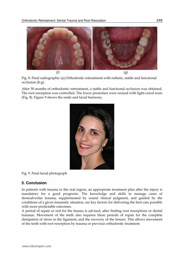
After 30 months of orthodontic retreatment, a stable and functional occlusion was obtained. The root resorption was controlled. The lower premolars were resized with light-cured resin (Fig. 8). Figure 9 shows the smile and facial harmony.
Fig. 9. Final facial photograph
5. Conclusion
In patients with trauma to the oral region, an appropriate treatment plan after the injury is mandatory for a good prognosis. The knowledge and skills to manage cases of dentoalveolar trauma, supplemented by sound clinical judgment, and guided by the conditions of a given traumatic situation, are key factors for delivering the best care possible with more predictable outcomes. A period of repair or rest for the tissues is advised, after finding root resorptions or dental traumas. Movement of the teeth also requires these periods of repair for the complete dissipation of stress in the ligament, and the recovery of the tissues. This allows movement of the teeth with root resorption by trauma or previous orthodontic treatment.
www.intechopen.com
Principles in Contemporary Orthodontics
250
6. References
Andreasen, J.O., Andreasen, F.M. & Andersson L. (2007). Textbook and Color Atlas of Traumatic Injuries to the Teeth, 4th ed. Oxford: Blackwell Munksgaard.
Bauss, O., Röhling, J., Sadat-Khonsari, R. & Kiliaridisd, S. (2008). Influence of orthodontic intrusion on pulpal vitality of previously traumatized maxillary permanent incisors. Am J Orthod Dentofacial Orthop 134:12-7.
Brezniak, N. & Wasserstein, A. (1993). Root resorption after orthodontic treatment: Part 1. Literature review. Am J Orthod Dentofacial Orthop 103:62-6 (a).
Brezniak, N. & Wasserstein, A. (1993). Root resorption after orthodontic treatment: Part 2. Literature review. Am J Orthod Dentofacial Orthop 103:138-46 (b).
Day, P.F., Kindelan, S.A., Spencer, J.R., Kindelan, J.D. & Duggal, M.S. (2008). Dental trauma: Part 2. Managing poor prognosis anterior teeth – treatment options for the subsequent space in a growing patient. J Orthod 35:144-55.
Kindelan, S.A., Day, P.F., Kindelan, J.D., Spencer, J.R. & Duggal, M.S. (2008). Dental trauma: an overview of its influence on the management of orthodontic treatment. Part 1. J Orthod 35:68–78.
Kinirons, M.J. (1998). Treatment of traumatically intruded permanent incisor teeth in children. Int J Paediat Dent 8:165-8.
Levander, E. & Malmgren, O. (1988). Evaluation of the risk of root resorption during orthodontic treatment: a study of upper incisors. Eur J Orthod 10(1):30-8.
Malmgren, O., Goldson, L., Hill, C., Orwin, A., Petrini, L. & Lundberg, M. (1982). Root resorption after orthodontic treatment of traumatized teeth. Am J Orthod 82(6):487-91.
Malmgren, O. & Malmgren, B. (2007). Orthodontic management of the traumatized teeth. In: Andreasen, J.O., Andreasen, F.M. & Andersson, L., editors. Textbook and color atlas of traumatic injuries to the teeth. 4th ed. Oxford: Blackwell Munksgaard; 2007. pp. 699-715.
Proffit, R.W. & Fields, H.W.J. (1993). Contemporary Orthodontics. 2 ed. St. Louis: Ed. Mosby. Tondelli, P.M., Mendonça, M.R., Cuoghi, O.A., Pereira, A.L.P. & Busato, M.C.A. (2010).
Knowledge on dental trauma and orthodontic tooth movement held by a group of orthodontists. Braz Oral Res 24(1):76-82.
Tondelli, P.M. (2011). Avaliação histomorfométrica da movimentação dentária induzida em ratos com força contínua, contínua interrompida e intermitente. [Tese]. Araçatuba: Univesidade Estadual Paulista – UNESP.
Turley, P.K., Joiner, M.W. & Hellstrom, S. (1984). The effect of orthodontic extrusion on traumatically intruded teeth. Am J Orthod 85:47-56.
Turley, P.K., Crawford, L.B. & Carrington, K.W. (1987). Traumatically intruded teeth. Angle Orthod 57:234-44.
Van Leeuwen, E.J., Malta, J.C. & Kuijipers-Jagtman, A.M. (1999). Tooth movement with light continuous and discontinuous forces in beagle dogs. Eur J Oral Scien 107;468-74.
Vlaskalic, V., Boyd, R.L. & Baumrind, S. (1998). Etiology and Sequelae of Root Resorption. Semin Orthod 4(2):124-131
Weiland, F. (2003). Constant versus dissipating forces in orthodontics: the effect on initial tooth movement and root resorption. Eur J Orthod 25;335-42.
Weiland, F. (2006). External root resorptions and orthodontic forces: correlations and clinical consequences. Prog Orthod 7(2):156-63.
www.intechopen.com
Principles in Contemporary OrthodonticsEdited by Dr. Silvano Naretto
ISBN 978-953-307-687-4Hard cover, 584 pagesPublisher InTechPublished online 25, November, 2011Published in print edition November, 2011
InTech EuropeUniversity Campus STeP Ri Slavka Krautzeka 83/A 51000 Rijeka, Croatia Phone: +385 (51) 770 447 Fax: +385 (51) 686 166www.intechopen.com
InTech ChinaUnit 405, Office Block, Hotel Equatorial Shanghai No.65, Yan An Road (West), Shanghai, 200040, China
Phone: +86-21-62489820 Fax: +86-21-62489821
Orthodontics is a fast developing science as well as the field of medicine in general. The attempt of this book isto propose new possibilities and new ways of thinking about Orthodontics beside the ones presented inestablished and outstanding publications available elsewhere. Some of the presented chapters transmit basicinformation, other clinical experiences and further offer even a window to the future. In the hands of the readerthis book could provide an useful tool for the exploration of the application of information, knowledge and beliefto some orthodontic topics and questions.
How to referenceIn order to correctly reference this scholarly work, feel free to copy and paste the following:
Pedro Marcelo Tondelli, Fabiana Akemy Kay, Osmar Aparecido Cuoghi and Marcos Roge ́rio de Mendonc ̧a(2011). Orthodontic Retreatment: Dental Trauma and Root Resorption, Principles in ContemporaryOrthodontics, Dr. Silvano Naretto (Ed.), ISBN: 978-953-307-687-4, InTech, Available from:http://www.intechopen.com/books/principles-in-contemporary-orthodontics/orthodontic-retreatment-dental-trauma-and-root-resorption