meningioma - abta.org · cancer, head trauma, or cell phone use may be risk factors for developing...
TRANSCRIPT
A M E R I C A N B R A I N T U M O R A S S O C I AT I O N
Meningioma
Meningioma
ACKNOWLEDGEMENTS
This publication is not intended as a substitute for professional medical advice and does not provide advice on treatments or conditions for individual patients. All health and treatment decisions must be made in consultation with your physician(s), utilizing your specific medical information. Inclusion in this publication is not a recommendation of any product, treatment, physician or hospital.
COPYRIGHT © 2017 ABTA
REPRODUCTION WITHOUT PRIOR WRITTEN PERMISSION
IS PROHIBITED
ABOUT THE AMERICAN BRAIN TUMOR ASSOCIATIONFounded in 1973, the American Brain Tumor
Association (ABTA) was the first national nonprofit
advocacy organization dedicated solely to brain tumor
research. For nearly 45 years, the ABTA has been
providing comprehensive resources that support the
complex needs of brain tumor patients and caregivers,
as well as the critical funding of research in the pursuit
of breakthroughs in brain tumor diagnosis, treatment
and care.
To learn more about the ABTA, visit www.abta.org.
We gratefully acknowledge Santosh Kesari, MD, PhD,
FANA, FAAN chair of department of translational neuro-
oncology and neurotherapeutics, and Marlon Saria,
MSN, RN, AOCNS®, FAAN clinical nurse specialist, John
Wayne Cancer Institute at Providence Saint John’s Health
Center, Santa Monica, CA; and Albert Lai, MD, PhD,
assistant clinical professor, Adult Brain Tumors, UCLA
Neuro-Oncology Program, for their review of this edition
of this publication.
3www.abta.org
AMERICAN BRAIN TUMOR ASSOCIATION
Meningioma
INTRODUCTIONAlthough meningiomas are considered a type of primary
brain tumor, they do not grow from brain tissue itself,
but instead arise from the meninges, three thin layers
of tissue covering the brain and spinal cord. These
tumors most commonly grow inward causing pressure
on the brain or spinal cord, but they may also grow
outward toward the skull, causing it to thicken. Most
meningiomas are benign, slow-growing tumors. Some
contain cysts (sacs of fluid), calcifications (mineral
deposits), or tightly packed bunches of blood vessels.
There are several systems used to name, or group,
these tumors. One system names meningiomas by the
type of cells in the tumor. Syncytial (or meningothelial)
meningiomas are the most common and feature
unusually plump cells. Fibroblastic meningiomas
feature long, thin shaped cells. Transitional
meningiomas contain both types of cells.
Another system uses the terms benign, atypical and
malignant (or anaplastic) to describe the overall grade
of meningiomas. In this system, benign meningiomas
contain easily recognized, well-differentiated
(resembling normal) cell types which tend to
grow slowly. Atypical tumors represent 10–15% of
meningiomas. They contain proliferating cells that may
AMERICAN BRAIN TUMOR ASSOCIATION4
be faster growing and more likely to grow back after
treatment, even after seemingly complete resection
(surgical removal). Therefore, these tumors must
be followed carefully for early signs of recurrence.
Malignant or “anaplastic” tumors are poorly
differentiated forms that often recur rapidly. Although
they are quite rare (1–3%), malignant meningiomas
can be highly aggressive and difficult to treat.
Another common practice is to attach the location
of the tumor to its name. For example, a parasagittal
meningioma is located near the sagittal sinus, a major
blood vessel at the top of the cerebral hemispheres. A
sphenoid ridge meningioma is found along the ridge
of bone behind the eyes and nose. Some meningiomas
can cause problems despite their benign nature, because
they are difficult to remove when they are located in
functionally sensitive or hard to reach areas. Depending
on the situation, stereotactic radiotherapy or radiosurgery
may be particularly helpful in some of these cases.
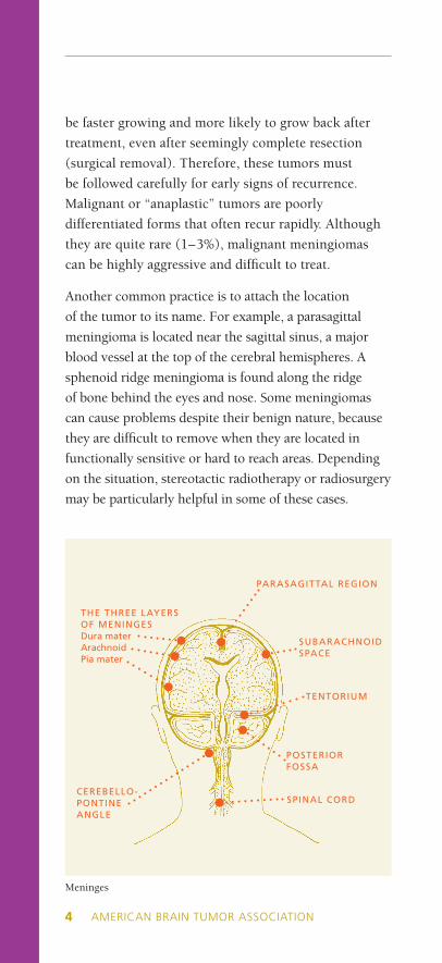
Meninges
THE THREE LAYERS OF MENINGESDura materArachnoidPia mater
TENTORIUM
PARASAGITTAL REGION
SUBARACHNOID SPACE
CEREBELLO-PONTINE ANGLE
POSTERIOR FOSSA
SPINAL CORD

MENINGIOMA
5www.abta.orgAMERICAN BRAIN TUMOR ASSOCIATION
INCIDENCEMeningiomas account for about one-third of all primary
brain tumors. They are most likely to be diagnosed in adults
older than 60 years of age, and the incidence appears to
increase with age. Meningiomas are rarely found in children.
They occur about twice as often in women as in men.
CAUSEAlthough the exact cause of meningioma is not known,
it has been associated with radiation exposure. The most
described genetic alteration is the loss of chromosome
22, normally involved in suppressing tumor growth. It
is seen in approximately 50% of patients with mutations
in the neurofibromatosis type 2 (NF-2) gene. Multiple
meningiomas occur in 5–15% of patients, particularly
those with NF-2. Meningiomas also frequently have extra
copies of the platelet-derived growth factor (PDFGR)
and epidermal growth factor receptors (EGFR), which
may contribute to the growth of these tumors. Other
genes linked to menigiomas include DAL1, SMO, AKT1,
TRAF7, and mTORC1.
Obesity (high body mass index), a history of breast
cancer, head trauma, or cell phone use may be risk
factors for developing meningioma, but the evidence is
inconclusive.
Some meningiomas have receptors that interact with the
sex hormones such as progesterone, androgen and less
commonly, estrogen. The expression of progesterone
receptor is seen most often in benign meningiomas, both
in men and women. The function of these receptors is
not fully understood, and thus, it is often challenging
for doctors to advise their female patients about the use
of hormones if they have a history of a meningioma.
Although the exact role of hormones in the growth of
meningiomas has not been determined, researchers have
observed that occasionally meningiomas may grow faster
during pregnancy.
AMERICAN BRAIN TUMOR ASSOCIATION6
If you have questions about using hormone replacement
therapy (HRT) during menopause, please discuss your
concerns with your doctors. Together, you can weigh the
benefits and risks in light of your individual health situation.
SYMPTOMSMeningiomas are usually slow growing and, may not
cause any symptoms until it is big enough to compress
adjacent structures. These tumors are most often found
in the coverings of the parasagittal/falcine region (near
the top of the brain) and the convexity (the outer
curve) of the brain. Other common sites include the
sphenoid ridge at the bottom of the brain, called the
skull base.
As the tumor grows, it may interfere with the normal
functions of the brain. The symptoms will depend on
the location of the tumor. Headache and weakness in
an arm or leg are the most common, although seizures,
personality change or visual problems may also occur.
Pain and loss of sensation or weakness in the arms or
legs are the most common symptoms of spinal cord
meningioma.
DIAGNOSISYour doctor will begin with a neurological
examination, followed by an MRI and/or a CT scan.
MR angiography (a MRI scan of the blood vessels)
or an arteriogram (a blood vessel X-ray) may be
performed to help the doctors plan an embolization,
a procedure to block the blood vessels in the tumor.
An octreotide scan may be helpful in distinguishing
meningiomas from other tumors. Used for tumors that
have an extensive blood supply, embolization may help
reduce bleeding during surgery.
If you have a tumor, these tests help your doctor
determine the location, size and probable type of
tumor. However, only an examination of a sample
MENINGIOMA
7www.abta.orgAMERICAN BRAIN TUMOR ASSOCIATION
of tumor tissue under a microscope confirms the exact
diagnosis. Such a tissue sample can only be obtained
through a surgical biopsy or excision.
TREATMENT
SURGERY Surgery is the primary treatment for meningiomas located
in an accessible area of the brain or spinal cord, although
some tumors may be inoperable. Another factor that
neurosurgeons consider is whether your vital organs
(heart, lungs, kidneys and liver) are strong enough to
withstand anesthesia and surgery.
The goals of surgery are to obtain tumor tissue for
diagnosis and to remove as much tumor as possible. If
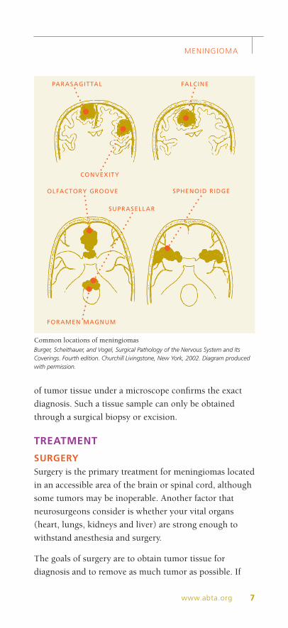
Common locations of meningiomasBurger, Scheithauer, and Vogel, Surgical Pathology of the Nervous System and Its Coverings. Fourth edition. Churchill Livingstone, New York, 2002. Diagram produced with permission.
PARASAGITTAL
CONVEXITY
FALCINE
SPHENOID RIDGE
SUPRASELLAR
OLFACTORY GROOVE
FORAMEN MAGNUM
AMERICAN BRAIN TUMOR ASSOCIATION8
the tumor cannot be removed, a biopsy to obtain a
sample of tumor tissue may be performed.
A computer program that combines different MR
images taken before surgery may be used to make a
three dimensional, or stereotactic, map of your brain.
This map helps the neurosurgeon plan the surgery
to remove as much of the tumor as possible while
avoiding parts of the brain that control vital functions.
During the operation, the surgeon may use stereotactic
imaging and instrument guiding technologies to
navigate through the brain. Occasionally, surgery is
performed within a specialized MRI (intraoperative
MRI), which allows the surgeon to view the tumor
during the operation and determine the extent of
tumor that is removed. High powered microscopes may
be used to help the surgeon to better see the tumor.
Ultrasonic aspirators are used to break up and suction
out parts of the tumor.
In cases where the tumor cannot be removed
completely, partial removal can help decrease
symptoms. Radiation may then be used to treat the
remaining tumor.
RADIATION Radiation therapy (external beam) may be used for
inoperable tumors, tumors that are not completely
removed in surgery, atypical and malignant tumors, or
recurrent tumors. There are different types of radiation,
which use various doses and schedules. Most forms of
radiation, however, are aimed at the tumor and a small
area around the tumor.
Conventional external beam radiation is “standard”
radiation given five days a week for five or six weeks.
Stereotactic radiation aims converged beams of
radiation at the tumor. Intensity modulated radiation
therapy, also called IMRT, conforms radiation beams
MENINGIOMA
9www.abta.orgAMERICAN BRAIN TUMOR ASSOCIATION
to the shape of the tumor. Additional information about
these forms of radiation therapy is available from our
office.
Stereotactic radiosurgery (SRS) utilizes numerous finely
focused beams of radiation to accurately administer
a single high-dose treatment to the tumor, while
minimizing the effects to adjacent normal tissue.
Therefore, despite the name, this is a noninvasive
procedure and there is no real “surgery” involved.
This may be particularly advantageous for patients
that are poor surgical candidates, have tumors in high-
risk regions of the brain, or have recurrences that are
no longer amenable to conventional forms of surgical
and radiation therapies. The disadvantages are that if
no surgery or biopsy is done, no tissue is obtained for
examination under the microscope; the technique may
only inhibit further growth, stabilizing – rather than
killing or removing – the tumor, and the technique is
limited to relatively small tumors, usually those that are
less than three centimeters in size.
For large tumors, or tumors located close to critical
structures, conventional or stereotactic radiotherapy
is often used instead. While stereotactic radiosurgery
(SRS) involves the use of a single large dose of focused
radiation, stereotactic radiotherapy, (SRT), involves the
administration of smaller doses of focused radiation
over a longer period of time (up to several weeks).
This reduces the potential for swelling or injury to
surrounding structures.
OTHER TREATMENTS Systemic therapy may be indicated for tumors that are
not surgically accessible or for patients in whom further
radiation is not possible. Some of these treatments are
offered in organized research studies called clinical trials.
Your doctor can determine if you are a candidate for
treatment in one of these trials.

AMERICAN BRAIN TUMOR ASSOCIATION10
Several other treatment approaches have or are being
explored:
• Hydroxyurea (used as a radiosensitizing drug in the
treatment of other types of tumors)
• Progesterone receptor inhibitors (eg. mifepristone)
• Somatostatin analogs (hormones that prevent the
release of growth hormones) (eg. octreotide)
• Targeted molecular agents (eg. everolimus)
• Epidermal growth factor receptor (EGFR) inhibitors
(eg. erlotinb)
• Platelet-derived growth factor receptor (PDGFR)
inhibitors (eg. imatinib)
• Vascular endothelial growth factors (VEGF)
inhibitors (eg. bevacizumab)
• Immunotherapy or the use of biological agents to
stimulate the immune system (eg. interferon alfa,
nivolumab)
There are also several drugs used to treat the symptoms
of a brain tumor. Steroids are used to decrease swelling,
or edema, around the tumor. Anti-seizure drugs control
seizures. Anti-nausea drugs prevent vomiting and help
control nausea. Additional suggestions for managing side
effects are offered on the ABTA website at www.abta.org.
WATCHFUL WAITING Depending on the location of the tumor, symptoms
caused by the tumor and sometimes patient preference,
some meningiomas may be carefully watched. Scans
will be recommended during the time of observation,
and it is very important to be sure those scans are
done. If your doctor suggests a course of observation,
remember that any new or changed symptoms should
be promptly reported to your doctor.
MENINGIOMA
11www.abta.orgAMERICAN BRAIN TUMOR ASSOCIATION 11www.abta.org
RECURRENCEMost meningiomas are benign and treatable with surgery.
However, brain tumors recur when all of the tumor
cells cannot be removed with surgery or killed with
other treatments. Over time, those cells multiply and
result in tumor regrowth. Your doctor can talk with you
about the chances of your tumor recurring. In general,
at five years following surgery, about 5% of completely
resected benign meningiomas, 30% of partially resected
benign meningiomas and 40% of atypical meningiomas
have recurred. Although rare, it is also possible that the
meningioma may recur as a more aggressive, or higher
grade, tumor.
Depending on your general health and the growth
characteristics of the tumor, repeat surgery and possibly
radiation therapy can be considered if the tumor recurs.
Focused forms of radiation therapy, such as stereotactic
radiotherapy or radiosurgery, may be repeated or used
following a history of conventional radiation therapy.
Treatments offered in clinical trials may also be used for
recurrent tumors.
RECOVERYAs with any brain tumor treatment, the length of recovery
time varies. The age and general health of the patient,
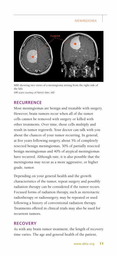
MRI showing two views of a meningioma arising from the right side of the falxMRI scans courtesy of Patrick Wen, MD
TUMOR
AMERICAN BRAIN TUMOR ASSOCIATION12
the location and size of the tumor, and the type of
treatment all affect the recovery time. Prior to your
surgery, ask your doctor what side effects you might
expect.
Muscle coordination or speech problems may occur
following surgery depending on the location of the
tumor; they are often temporary. During this healing
time, many brain tumor patients discover the benefits
of rehabilitative services. The goal of rehabilitative
medicine is to restore physical, vocational and
psychological functions. Services may include physical,
occupational and/or speech therapy to help reduce
some of the symptoms that may accompany a tumor
or treatment. Cognitive retraining – a memory training
method – is used to teach another part of the brain to
take over the tasks of the impaired portion. Visual aids
may be required for those with tumors near the optic
nerves. Support services that help patients and their
families live with a brain tumor diagnosis are just as
important. Call the ABTA’s CareLine at 800-886-ABTA
(2282) for help locating both rehabilitative and support
services in your area. For additional information,
access to ABTA’s online support community and to
view webinars, please visit www.abta.org.
PROGNOSISPeople diagnosed with a meningioma often have very
specific questions regarding their future. They may
want to know the risks involved in their surgery, the
need for follow-up care or additional treatments, if or
how the tumor might affect their life, and what the
chances are for their tumor recurring. Although the
medical term “prognosis” is usually associated with
malignant tumors, a “predication of outcome” may be
more applicable to a person with a meningioma.
We encourage you to ask your doctor these outcome
questions. They can respond to your concerns based
on your individual tumor. Your doctor can also

MENINGIOMA
13www.abta.orgAMERICAN BRAIN TUMOR ASSOCIATION
explain your treatment plan, the benefits and risks of
the treatment plan suggested for you, and what you can
expect in the future.
THE ABTA IS HERE FOR YOUYou don’t have to go through this journey alone. The
American Brain Tumor Association is here to help.
Visit us at www.abta.org to find additional brochures,
view free, educational webinars on demand, read about
research and treatment updates, connect with a support
community, join a local event and more.
We can help connect patients and caregivers with
information and resources that can help support them in
the brain tumor journey. Our team of caring professionals
are available. via email at [email protected] or via our
toll-free CareLine at 800-886-ABTA (2282).

AMERICAN BRAIN TUMOR ASSOCIATION14
NOTES/QUESTIONS
AMERICAN BRAIN TUMOR ASSOCIATION
AMERICAN BRAIN TUMOR ASSOCIATION PUBLICATIONS AND SERVICES
CARE & SUPPORTCareLine: 800-886-ABTA (2282)
Email: [email protected]
PUBLICATIONSAbout Brain Tumors: A Primer for Patients and Caregivers
Brain Tumors – A Handbook for the Newly Diagnosed*
Brain Tumor Dictionary*
Caregiver Handbook*
Returning to Work: Accessing Reasonable Accommodations*
Quick Guide to the Family and Medical Leave Act*
Tumor Types:
Ependymoma
Glioblastoma and Malignant Astrocytoma
Medulloblastoma
Meningioma
Metastatic Brain Tumors
Oligodendroglioma and Oligoastrocytoma
Pituitary Tumors
Treatments:
Chemotherapy
Clinical Trials
Conventional Radiation Therapy
Proton Therapy
Stereotactic Radiosurgery*
Steroids
Surgery
Most publications are available for download in Spanish. Exceptions are marked *
CLINICAL TRIALSTrialConnect®: www.abtatrialconnect.org or 877-769-4833
More brain tumor resources and information
are available at www.abta.org.
For more information contact:
CareLine: 800-886-ABTA (2282)
Email: [email protected]
Website: www.abta.org
Connect with us on social media:
Facebook.com/theABTA
Twitter.com/theABTA
To find out how you can get
more involved locally, contact
[email protected] or call
800-886-1281
8550 W. Bryn Mawr Avenue, Suite 550
Chicago, IL 60631
A M E R I C A N B R A I N T U M O R A S S O C I AT I O N
FGS0417