membrane-bound matrix metalloproteinase-8 on … · membrane-bound matrix metalloproteinase-8 on...
TRANSCRIPT

of September 11, 2018.This information is current as Collagenase and Serpinase
Inhibitor of Metalloproteinase-Resistant Polymorphonuclear Cells Is a Potent, TissueMetalloproteinase-8 on Activated Membrane-Bound Matrix
Steven D. ShapiroCaroline A. Owen, Zhuma Hu, Carlos Lopez-Otin and
http://www.jimmunol.org/content/172/12/7791doi: 10.4049/jimmunol.172.12.7791
2004; 172:7791-7803; ;J Immunol
Referenceshttp://www.jimmunol.org/content/172/12/7791.full#ref-list-1
, 20 of which you can access for free at: cites 45 articlesThis article
average*
4 weeks from acceptance to publicationFast Publication! •
Every submission reviewed by practicing scientistsNo Triage! •
from submission to initial decisionRapid Reviews! 30 days* •
Submit online. ?The JIWhy
Subscriptionhttp://jimmunol.org/subscription
is online at: The Journal of ImmunologyInformation about subscribing to
Permissionshttp://www.aai.org/About/Publications/JI/copyright.htmlSubmit copyright permission requests at:
Email Alertshttp://jimmunol.org/alertsReceive free email-alerts when new articles cite this article. Sign up at:
Print ISSN: 0022-1767 Online ISSN: 1550-6606. Immunologists All rights reserved.Copyright © 2004 by The American Association of1451 Rockville Pike, Suite 650, Rockville, MD 20852The American Association of Immunologists, Inc.,
is published twice each month byThe Journal of Immunology
by guest on September 11, 2018
http://ww
w.jim
munol.org/
Dow
nloaded from
by guest on September 11, 2018
http://ww
w.jim
munol.org/
Dow
nloaded from

Membrane-Bound Matrix Metalloproteinase-8 on ActivatedPolymorphonuclear Cells Is a Potent, Tissue Inhibitor ofMetalloproteinase-Resistant Collagenase and Serpinase1
Caroline A. Owen,2* Zhuma Hu, † Carlos Lopez-Otin,‡ and Steven D. Shapiro*
Little is known about the cell biology or the biologic roles of polymorphonuclear cell (PMN)-derived matrix metalloproteinase-8(MMP-8). When activated with proinflammatory mediators, human PMN release only�15–20% of their content of MMP-8 (�60ng/106 cells) exclusively as latent pro-MMP-8. However, activated PMN incubated on type I collagen are associated with peri-cellular collagenase activity even when bathed in serum. PMN pericellular collagenase activity is attributable to membrane-boundMMP-8 because: 1) MMP-8 is expressed in an inducible manner in both pro- and active forms on the surface of human PMN; 2)studies of activated PMN from mice genetically deficient in MMP-8 (MMP-8�/�) vs wild-type (WT) mice show that membrane-bound MMP-8 accounts for 92% of the MMP-mediated, PMN surface type I collagenase activity; and 3) human membrane-boundMMP-8 on PMN cleaves types I and II collagens, and�1-proteinase inhibitor, but is substantially resistant to inhibition by tissueinhibitor of metalloproteinase-1 (TIMP-1) and TIMP-2. Binding of MMP-8 to the PMN surface promotes its stability becausesoluble MMP-8 hast1/2 � 7.5 h at 37°C, but membrane-bound MMP-8 retains>80% of its activity after incubation at 37°C for18 h. Studies of MMP-8�/� vs WT mice given intratracheal LPS demonstrate that 24 h after intratracheal LPS, MMP-8�/� micehave 2-fold greater accumulation of PMN in the alveolar space than WT mice. Thus, MMP-8 has an unexpected, anti-inflammatory role during acute lung injury in mice. TIMP-resistant, active MMP-8 expressed on the surface of activated PMN islikely to be an important form of MMP-8, regulating lung inflammation and collagen turnover in vivo. The Journal of Immu-nology, 2004, 172: 7791–7803.
M atrix metalloproteinase-8 (MMP-8;3 collagenase-2,EC 3.4.24.34) along with MMP-1 and MMP-13 arethe major members of the interstitial collagenase sub-
group of the MMP family of zinc-dependent, neutral proteinases.Interstitial collagens (types I-III) are major structural componentsof the extracellular matrix (ECM). They are composed of threepolypeptide chains arranged in a rigid triple helix conformation,rendering them resistant to degradation by proteinases other thanthe interstitial collagenases. Interstitial collagenases mediate theinitial and rate-limiting step in interstitial collagen degradation bycleaving the three collagen polypeptide chains at a single locusthree-fourths of the distance of the collagen molecule from its
N-terminal end (1). The three-fourth and one-fourth fragmentsgenerated denature spontaneously, and the denatured collagenfragments (gelatins) are susceptible to further cleavage by gelati-nases (MMP-2 and MMP-9) and to a lesser extent by other MMPs(including MMP-8) and serine proteinases (2). Although MMP-8cleaves all three interstitial collagens, it differs from MMP-1 inthat it cleaves type I collagen at a higher rate than type III collagen.MMP-8 also degrades type II collagen, the predominant collagenof cartilage, at a higher rate than MMP-1 (3). MMP-8 can alsocleave nonmatrix proteins such as serpins, bradykinin, angiotensinI, and substance P (4, 5).
The biologic roles of MMP-8 in vivo are currently uncertain. Asa result of its known catalytic activities, MMP-8 is believed to beinvolved in wound healing and tissue remodeling during inflam-mation. In addition, MMP-8 has been implicated in the pathogen-esis of several chronic inflammatory diseases characterized by ex-cessive influx and activation of polymorphonuclear cell (PMN),including cystic fibrosis (6), rheumatoid arthritis (7), periodontaldisease (8), and chronic skin wounds (9). Recent data from Balbinet al. (10) demonstrate that MMP-8 has an unexpected role in vivoin protecting male mice from the development of skin tumors in achemical carcinogenesis model. This study showed that mice de-ficient in MMP-8 (MMP-8�/� mice) have important abnormalitiesin their inflammatory response to chemical carcinogens. The lackof MMP-8 hampers the early PMN recruitment in response tochemical carcinogens, due in part to the loss of MMP-8-mediatedcleavage and activation of LPS-induced CXC chemokine. How-ever, once inflammation is established in MMP-8�/� mice, it isabnormally sustained (by unknown mechanisms), creating a morefavorable environment for tumor initiation. Moreover, bone mar-row transplantation experiments confirmed that PMN were thesource of MMP-8 promoting protection against tumor develop-ment in this model.
*Division of Pulmonary and Critical Care Medicine, Brigham and Women’s Hospital,Harvard Medical School, Boston, MA 02115; †Department of Internal Medicine,University of Utah Health Sciences Center, Salt Lake City, UT 84132; and ‡Depar-tamento de Bioquı́mica y Biologı́a Molecular, Instituto Universitario de Oncologia,Universidad de Oviedo, Oviedo, Spain
Received for publication October 3, 2003. Accepted for publication April 9, 2004.
The costs of publication of this article were defrayed in part by the payment of pagecharges. This article must therefore be hereby marked advertisement in accordancewith 18 U.S.C. Section 1734 solely to indicate this fact.1 This work was supported by U.S. Public Health Service Grants HL63137 (toC.A.O.), a Career Investigator Award from the Massachusetts Thoracic Society (toC.A.O.), and grants from the European Union and Comision Interministerial de Cien-cia y Tecnologia (to C.L.-O.).2 Address correspondence and reprint requests to Dr. Caroline A. Owen, Division ofPulmonary and Critical Care Medicine, Brigham and Women’s Hospital, 905 ThornBuilding, 75 Francis Street, Boston, MA 02115. E-mail address: [email protected] Abbreviations used in this paper: MMP, matrix metalloproteinase; �1-PI, �1-pro-teinase inhibitor; APMA, 4-aminophenylmercuric acetate; BAL, bronchoalveolar la-vage; ECM, extracellular matrix; HLE, human leukocyte elastase; IT, intratracheal;McaPLGLDpaAR, (7-methoxycoumarin-4-yl)-acetyl-Pro-Leu-Gly-Leu-(3-[2,4-dini-trophenyl]-L2,3-diamino-propionyl)-Ala-Arg-NH2; MPO, myeloperoxidase; PAF,platelet-activating factor; PMN, polymorphonuclear cell; TIMP, tissue inhibitor ofmetalloproteinase; WBC, white blood cell; WT, wild type.
The Journal of Immunology
Copyright © 2004 by The American Association of Immunologists, Inc. 0022-1767/04/$02.00
by guest on September 11, 2018
http://ww
w.jim
munol.org/
Dow
nloaded from

PMN are the main source of MMP-8 in humans. However,MMP-8 is also expressed at lower levels by chondrocytes (11),rheumatoid synovial fibroblasts (12), activated macrophages,smooth muscle cells, and endothelial cells (13). PMN-derivedMMP-8 differs from interstitial collagenases expressed by othercells in that it is not synthesized de novo by mature PMN. Rather,MMP-8 is expressed during the myelocyte stage of development ofPMN precursors in the bone marrow (14), and it is stored as alatent enzyme (pro-MMP-8, Mr 85 kDa) within the specific gran-ules of PMN (15). Pro-MMP-8 is rapidly released from activatedPMN undergoing degranulation (16, 17), and is then activated viathe cysteine switch mechanism to yield the active form of theenzyme (Mr �65 kDa (18)). Activation can be achieved in vitro byorganomercurials (19), serine proteinases (20, 21), MMP-3 (22),and reactive oxygen species (18). However, it is not clear howpro-MMP-8 is activated or how it retains its activity in vivo, be-cause tissues contain highly effective inhibitors of MMP-8, includ-ing tissue inhibitors of MMP (TIMPs).
We have shown that activated PMN are associated with strikingpericellular type collagenase activity even when incubated in thepresence of TIMPs. In addition, MMP-8 is expressed on the cellsurface of activated PMN, and this form of the proteinase cancontribute to potent, TIMP-resistant pericellular collagenase andserpinase activity. The binding of MMP-8 to the PMN surfacepromotes its stability and preserves its activity in the extracellularspace in the presence of TIMPs. Surprisingly, our studies of MMP-8�/� mice in a model of acute lung injury demonstrate thatMMP-8 has an anti-inflammatory role in the lung. Our data indi-cate that the TIMP-resistant form of MMP-8 activity expressed onthe surface of human PMN is likely to contribute in importantways to its anti-inflammatory and interstitial collagen-degradingactivities in the lung and other tissues in vivo.
Materials and MethodsMaterials
Goat anti-rabbit F(ab�)2-Alexa Fluor 546 and 488, goat anti-murineF(ab�)2-Alexa Fluor 546, and phalloidin-Alexa 546 were obtained fromMolecular Probes (Eugene, OR). Human pro-MMP-8, human TIMP-1, hu-man TIMP-2, polyclonal rabbit anti-human MMP-8 IgG (AB8115), murineanti-human MMP-9, and the kit for measuring type II collagenase activitywere purchased from Chemicon International (Temecula, CA). FITC-con-jugated type I collagen was obtained from Elastin Products (Owensville,MO). Purified myeloperoxidase (MPO) was obtained from Athens Re-search (Athens, GA). Quenched FITC-conjugated gelatin and type I col-lagen were obtained from Molecular Probes. The (7-methoxycoumarin-4-yl)-acetyl-Pro-Leu-Gly-Leu-(3-[2,4-dinitrophenyl]-L2,3-diamino-propionyl)-Ala-Arg-NH2 (McaPLGLDpaAR) was purchased fromCalbiochem Novabiochem (San Diego, CA). RS113456 and RS104210were generously provided by Roche Bioscience (Palo Alto, CA). MMP-8ELISA kits were purchased from R&D Systems (Minneapolis, MN). BDBiocoat Matrigel Invasion Chambers were obtained from BD Labware(Bedford, MA). All other reagents were purchased from Sigma-Aldrich (St.Louis, MO).
Quantitation of MMP-8 in PMN lysates and culturesupernatants
Extracts of human PMN were prepared at 5 � 106/ml in PBS containing0.1% (v/v) Triton X-100. Cell extracts and cell-free supernatants fromunstimulated and activated PMN were assayed for total human MMP-8using a commercially available immunoassay (R&D Systems, Minneapo-lis, MN). This ELISA kit measures both pro- and active MMP-8, but doesnot detect purified human MMP-9 or human leukocyte elastase.
Human PMN isolation and activation
Human PMN (�95% pure) were isolated from peripheral blood of healthydonors (23). Human PMN were suspended at 107/ml in HBSS containing1 mM Ca2�, 1 mM Mg2�, and 10 mM HEPES (pH 7.4), then incubated for30 min at 37°C with or without platelet-activating factor (PAF) (10�6 to10�10 M), TNF-� (1–1000 U/ml), LPS from Escherichia coli 0111:B4
(1–1000 ng/ml), fMLP (10�6 to 10�12 M), or PMA (100 ng/ml). PMNwere also incubated at 37°C for 15 min with optimal concentrations of LPS(100 ng/ml), PAF (10�8 M), TNF-� (100 U/ml), or cytochalasin B (5�g/ml), then optimally activated with fMLP (10�8 M for 30 min). Toterminate assays, cells and supernatant samples were separated followingcentrifugation (300 � g for 3 min). For immunostaining experiments, PMNwere fixed for 3 min at 4°C in PBS (pH 7.4) containing 3% paraformal-dehyde and 0.5% glutaraldehyde (24).
Type I collagenase activity associated with activated PMN
LabTec chamber slides (Nunc, Naperville, IL) were coated with FITC-conjugated type I collagen (1 mg/ml) for 2 h at 37°C. HBSS, autologousserum, or autologous serum containing 20 �M RS104210, or 20 �M 4-(2-aminoethyl)benzylsulfonyl fluoride, or 20 �M leupeptin was added to thewells (300 �l/well), followed by human PMN (2 � 105 cells in 20 �l ofHBSS). The cells were activated with PAF (10�8 M) for 5 min; then 10�8
M fMLP was added and the chambers were incubated at 37°C for 3 h in ahumidified atmosphere of 5% CO2. Cells adherent to the slides were fixedfor 3 min at 4°C using PBS containing 3% (w/v) paraformaldehyde and0.5% (v/v) glutaraldehyde. The chamber slides were dismantled, and theslides were mounted in PBS containing 25% (v/v) glycerol and 250 �g/mlp-phenylenediamine, and then examined by bright field and incident lightepifluorescence microscopy. Images of the cells were captured using achilled charge-coupled device camera and MetaMorph software (UniversalImaging, West Chester, PA).
Isolation of PMN undergoing directed migration
We used BD Biocoat Matrigel Invasion Chambers (a modified Boydenchamber assay, with upper and lower chambers separated by an 8-�mmicroporous, polyethylene terephthalate membrane coated with a uniformlayer of Matrigel Matrix, a prototype basement membrane (25)). The lowerchambers were filled with medium alone (RPMI 1640 containing 10 mMHEPES and 1% human serum albumin, pH 7.4), or medium containing10�7 M fMLP. Human PMN (8 � 106 in 2 ml of medium) were placed inthe upper chambers, and the chambers were incubated for 3 h at 37°C in ahumidified atmosphere of 5% CO2. PMN that migrated through the mem-brane were immunostained for cell surface-bound MMP-8.
Immunofluorescence staining of human PMN for MMP-8,quantitative image analysis, and confocal microscopy
PMN were incubated at 4°C with polyclonal rabbit anti-human MMP-8(AB8115) or rabbit IgG, as a control (both at 1 �g/106 cells), followed bygoat anti-rabbit IgG conjugated to Alexa 546. AB8115 was raised againsta peptide sequence in the hinge region of MMP-8. Western blot analysisconfirmed that AB8115 recognizes both pro-MMP-8 and active MMP-8,but has no cross-reactivity for purified MMPs-9, -12, -1, or -2; humanleukocyte elastase; cathepsin G; or proteinase 3. PMN were then washedtwice in HBSS, incubated for 2 h at 4°C with goat anti-rabbit F(ab�)2 AlexaFluor 546, and then washed twice in HBSS. The PMN were examined byincident light fluorescence microscopy. Cell surface immunofluorescencewas quantified using MetaMorph image analysis software, and the datawere corrected for nonspecific staining, as described previously (24).Briefly, MetaMorph software was used to quantify the integrated fluores-cence of 150–200 cells in each group in arbitrary fluorescence units. Tocorrect for nonspecific staining, for each condition, the mean fluorescenceintensity of cells stained with the nonimmune control Ab was subtractedfrom each of the integrated fluorescence values for cells incubated with theanti-MMP-8 Ab. The results for cell surface MMP-8 expressed by acti-vated PMN were expressed as a percentage of cell surface MMP-8 ex-pressed by unstimulated PMN.
Cell surface localization of MMP-8 was confirmed by analyzing cellsdouble immunostained with: 1) rabbit anti-MMP-8 (AB8115) and goatanti-rabbit IgG conjugated to Alexa 488; 2) murine anti-human MMP-9and goat anti-murine IgG conjugated to Alexa 546. Cells were analyzedwith a Leica TCSNT confocal laser-scanning microscope (Leica, Exton,PA) fitted with air-cooled argon and krypton lasers. Fluorescent confocalmicrographs were recorded under dual fluorescent imaging mode in whichcells were simultaneously exposed to 488 and 568 nm light attenuated byan acusto tunable optical filter. A band pass (530 � 30 nm) filter was usedto select light emitted from the Alexa 488-labeled MMP-8, and a long-pass590-nm filter was used to detect the Alexa 546-labeled MMP-9.
Activated human PMN were also immunostained with Alexa 488 forsurface MMP-8 using AB8115 as the primary Ab, then fixed, as describedabove. Cells were then permeabilized using 250 �g/ml lysophosphatidylcholine in PBS for 30 min at 4°C. The cells were then immunostained forintracellular F-actin using phalloidin conjugated to Alexa 546, and them
7792 POTENT TIMP-RESISTANT MMP-8 ACTIVITY ON SURFACE OF PMN
by guest on September 11, 2018
http://ww
w.jim
munol.org/
Dow
nloaded from
examined by incident light fluorescence microscopy, as described previ-ously (27).
Immunoblot analysis of MMP-8 in PMN plasma membranes
Peripheral blood PMN (6 � 108) were optimally activated (primed for 15min with 10�8 M PAF, then activated for 30 min with 10�8 M fMLP).Viable cells were subjected to nitrogen cavitation and Percoll gradientcentrifugation to separate the granule-free plasma membrane fraction (�band), the azurophil granule faction (� band), and the specific granulefraction (� bands), and then the purity and total protein content of theplasma membrane fraction were quantified, as described previously (28).Aliquots of the plasma membrane fraction and the specific granule factionwere solubilized by incubation with 0.05% Brij35 for 1 h at 4°C, followedby centrifugation at 50,000 � g for 15 min. Plasma membrane samples (25�g of total protein), along with purified, 4-aminophenylmercuric acetate(APMA)-activated MMP-8, solubilized specific granules, and cell-free su-pernatant samples from activated PMN, were subjected to 12% SDS-PAGE. The proteins were transferred to Immuno-Blot polyvinylidene di-fluoride membrane (100 V for 90 min in 12.5 mM Tris buffer (pH 8.3)containing 96 mM glycine and 20% (v/v) methanol), and the membraneswere blocked with 3% nonfat milk in PBS containing 0.1% Tween 20, thenprobed with polyclonal rabbit anti-human MMP-8 (AB8115). Binding ofAb was detected with HRP-coupled goat anti-rabbit secondary Ab and anOpti-4CN chromogenic detection system (Bio-Rad, Hercules, CA). Theintensities of the bands generated were quantified using Scion Image � 4.02(Scion, Frederick, MD).
Pro-MMP-8 activation
Purified human pro-MMP-8 (1 �M) was incubated at 37°C for 2–4 h with2 mM APMA. Complete activation of pro-MMP-8 was confirmed by anal-ysis of samples on 15% SDS-PAGE. Activated MMP-8 was active sitetitrated using TIMP-2 and McaPLGLDpaAR as the substrate.
Murine PMN isolation and activation
Mice genetically deficient in MMP-8 (MMP-8�/�) were generated by re-placing exons 2, 3, and 4 with a phosphoglycerate kinase-neomycin fusiongene and homologous recombination (10). PMN (�85% pure) were iso-lated from the bone marrow of MMP-8�/� or wild-type (WT) mice in thesame genetic background (mixed 129/SvEv � C57BL/6) by positive se-lection for Ly-6-G using immunomagnetic beads (29). Extracts of murinePMN were prepared in PBS containing 0.1% (v/v) Triton X-100, and an-alyzed for MMP-8 � 12% SDS-PAGE, followed by Western blot analysisusing AB8115 as the primary Ab. Murine PMN were also activated at 37°Cfor 15 min with PAF (10�7 M), followed by fMLP (10�7 M for 30 min),then fixed, and immunostained for cell surface MMP-8 using AB8115 orrabbit IgG as the primary Abs, followed by goat anti-rabbit conjugated toAlexa 488. Cell surface MMP-8 expression on murine PMN was quantifiedusing MetaMorph, as described above.
Catalytic activity of MMP-8 on the surface of murine PMN
PMN from MMP-8�/� and WT mice were activated and fixed (as de-scribed above), washed, and resuspended in Tris assay buffer (0.05 M Triscontaining 0.15 M NaCl and 0.02 M CaCl2, pH 7.4). Cells from bothgenotypes (106 cells/assay) were incubated in triplicate with and without1,10-phenanthroline (1 mM, a synthetic inhibitor of MMPs) at 37°C forvarying times in the presence of 1.8 �M McaPLGLDpaAR. Total cellsurface-bound MMP activity was quantified as the 1,10-phenanthroline-inhibitable, McaPLGLDpaAR-hydrolyzing activity in cell-free supernatantsamples by fluorometry (F2500 fluorescence spectrophotometer; Hitachi,Tokyo, Japan; �ex 328 nm, �em 393 nm). To quantify PMN cell surfacegelatinase and type I collagenase activities, activated PMN from MMP-8�/� and WT mice were preincubated in triplicate for 30 min at 37°C with1 mM PMSF (to inactivate cell surface serine proteinases having gelatinaseactivity (2)) with and without 1 mM 1,10-phenanthroline. Cells were in-cubated at 37°C for 3 h with 50 �g/ml quenched FITC-conjugated gelatin(3 � 106 cells/assay) or for 18 h with 50 �g/ml quenched FITC-conjugatedtype I collagen (4 � 106 cells/assay), and MMP-mediated gelatinase andtype I collagenase activities were quantified as the 1,10-phenanthroline-inhibitable cleavage of substrates in supernatant fluids by fluorometry (�ex
490 nm, �em 520 nm).
Cell surface type I collagenase activity on activatedhuman PMN
PMN were activated for 15 min with 10�8 M PAF for 15 min, then for 30min with 10�8 M fMLP, and then fixed to prevent release of intracellular
proteinases (29, 30). PMN were then incubated with 20 �g of bovine typeI collagen with or without 2 � 107 PMN at 37°C for up to 20 h, and thenreduced cell-free supernatant samples were analyzed using 12% SDS-PAGE and Coomassie brilliant blue G250 staining.
Catalytic activity of human membrane-bound MMP-8 on PMN
Activated human PMN express other proteinases on their surface that cancleave MMP-8 substrates other than type I collagen (24, 29, 31, 32). Tostudy the activity of human membrane-bound MMP-8 against MMP-8 sub-strates other than interstitial collagens, we studied exogenous humanMMP-8 bound to the surface of unstimulated PMN. This strategy permitsus to load MMP-8 onto the PMN surface, and to quantify the amount boundfor studies of its catalytic activity and efficiency. Proteinases that are en-dogenously expressed at low levels on the surface of unstimulated PMNwere first inactivated (29, 31). PMN were then incubated at 4°C with ac-tivated, soluble MMP-8 (1 �g/106 cells), then fixed (using PBS containing3% paraformaldehyde and 0.5% glutaraldehyde (pH 7.4) for 3 min at 4°C)to prevent release of proteinases from the cells during subsequent activityassays (29, 31). Cell surface-bound MMP-8 was quantified by incubatingthe following in Tris assay buffer at 37°C for 30 min with 1.8 �M McaPL-GLDpaAR: 1) MMP-8 bound to PMN (2 � 106 cells/assay); 2) controlPMN (which had no MMP-8 bound to their surface; 2 � 106 cells/assay);and 3) assay standards of soluble, active site-titrated, APMA-activatedMMP-8 (3–400 ng/assay). Cleavage of the substrate was quantified in cell-free supernatant fluids by fluorometry. The amount of MMP-8 bound to thesurface of PMN was calculated by interpolation from the standard curve forsoluble, active MMP-8, and expressed as ng MMP-8 activity bound per 106
cells.
kcat/KM determinations
Hydrolysis of 1.5 �M McaPLGLDpaAR by soluble MMP-8 or exogenousMMP-8 bound to PMN (both at 31–500 pM in 250 �l of Tris assay buffer)was monitored over 6 min at 25°C as the increase in fluorescence (�ex 328nm, �em 393 nm (33)). A substrate concentration of 1.5 �M fulfills theconditions of [S] �� KM to permit the direct determination of kcat/KM
under first order conditions.
Cleavage of interstitial collagens and serpins by membrane-bound MMP-8
Bovine type I collagen (20 �g) was incubated at 37°C for 4–20 h with orwithout: 1) human MMP-8 bound to PMN (2.5 � 107 cells); 2) humanMMP-8 bound to PMN (2.5 � 107 cells) that had been preincubated with10 �M RS113456 (a general, hydroxamate inhibitor of MMPs); or 3) 500ng of soluble, APMA-activated MMP-8. Cell-free supernatant sampleswere reduced and analyzed by 12% SDS-PAGE and Coomassie brilliantblue G250 staining. A commercial kit was used to assess whether humanmembrane-bound MMP-8 has type II collagenase activity. Human MMP-8bound to PMN (2 � 106 cells), control PMN (2 � 106 cells), and assaystandards of soluble, active site-titrated MMP-8 (25–800 ng) were incu-bated for 3 h at 42°C with FITC-conjugated type II collagen (100 �g) inTris buffer. Cell-free supernatant samples were assayed for type II colla-genase activity by fluorometry (�ex 490 nm, �em 520 nm), as recommendedby the kit manufacturer. The type II collagenase activity expressed bymembrane-bound MMP-8 and control PMN was converted from arbitraryfluorescence units to ng of soluble MMP-8 activity per 106 PMN by in-terpolation from the standard curve generated for soluble, active MMP-8.
MMP-8 bound to PMN (8 � 106 cells), control PMN (8 � 106 PMN),or soluble MMP-8 (125 ng) were also incubated for 8 h at 37°C with 6 �gof purified �1-proteinase inhibitor (�1-PI) in Tris assay buffer. Reducedcell-free supernatant samples were analyzed by 15% SDS-PAGE and Coo-massie brilliant blue G250 staining. To assess whether the catalytic activityassociated with MMP-8 bound to PMN was due to release or leakage ofproteinases from PMN, PMN that bound MMP-8 to their surface were alsoincubated in Tris buffer alone, and MMP activity was assayed in cell-freesupernatant samples using McaPLGLDpaAR.
Effect of fixatives on the catalytic activity ofmembrane-bound MMP-8
PMN were fixed for 3 min at 4°C using PBS containing 3% paraformal-dehyde and 0.5% glutaraldehyde (pH 7.4). Cells were then washed threetimes in 0.05 M Tris containing 0.15 M NaCl and 0.02 M CaCl2 (pH 7.4).To assess the effects of fixatives on the catalytic activity of membrane-bound MMP-8, we incubated PMN at 4°C for 2 h with activated, solubleMMP-8 (1 �g/106 cells), washed the cells twice, and then divided the cellsinto two equal aliquots. One aliquot was fixed, as described above; theother was incubated without fixatives. Both groups were washed three
7793The Journal of Immunology
by guest on September 11, 2018
http://ww
w.jim
munol.org/
Dow
nloaded from

times in PBS, and then incubated in Tris assay buffer at 37°C for 30 minwith 1.8 �M McaPLGLDpaAR. Cleavage of the substrate was quantifiedin cell-free supernatants in arbitrary fluorescence units by fluorometry, asdescribed above. The results for fixed membrane-bound MMP-8 were ex-pressed as a percentage of the activity associated with nonfixed, mem-brane-bound MMP-8.
Stability of soluble vs membrane-bound MMP-8
Pro-MMP-8 was activated with APMA; then an aliquot of active MMP-8was bound to the surface of unstimulated PMN, and the amount bound toan aliquot of the cells was quantified, using McaPLGLDpaAR. Aliquots(50 ng) of the remaining soluble MMP-8 and membrane-bound MMP-8were then incubated in duplicate for 30 min at 37°C in Tris assay buffercontaining 1.8 �M McaPLGLDpaAR, and initial reaction velocities werequantified by fluorometry. Additional 50-ng aliquots of each form of theproteinases were incubated at 37°C in duplicate in Tris assay buffer withoutsubstrate for 2, 4, 6, and 18 h; then McaPLGLDpaAR was added, andreaction velocities were measured, as described above. The results wereexpressed as a percentage of the initial reaction velocity for each form ofthe proteinase.
Susceptibility of membrane-bound MMP-8 to inhibition
Soluble MMP-8 (10 nM) or MMP-8 bound to PMN (10 nM) were incu-bated at 37°C for 30 min in Tris buffer with and without: 1) 1 mM 1,10-phenanthroline; 2) 0.1 nM-1 �M RS113456; 3) 15.6 nM-1 �M TIMP-1; 4)15.6 nM-1 �M TIMP-2; or 5) 1 mM PMSF. Membrane-bound MMP-8 wasalso preincubated for 30 min with 1 mM PMSF, then incubated for 30 minwith 200 nM TIMP-1. Residual MMP-8 activity was quantified in cell-freesupernatant samples using 1.8 �M McaPLGLDpaAR. IC50 values weredetermined by nonlinear regression analysis using SigmaStat (SPSS,Chicago, IL).
Studies of LPS-mediated acute lung injury in mice
Male MMP-8�/� and WT mice aged 10–16 wk, both in mixed SvEv129 �C57BL/6 genetic background, were anesthetized with ketamine (35 mg/kg)and xylazine (5 mg/kg), then given 10 �g of LPS from E. coli 0111:B4 in30 �l of endotoxin-free PBS, or 30 �l of PBS alone by the intratracheal(IT) route as a control. After 24 h, mice were euthanized by CO2 narcosis,and then bronchoalveolar lavage (BAL) was performed using 8 � 0.5 mlof sterile PBS. The BAL cell and supernatant fractions were separated bycentrifugation (500 � g for 3 min), then total and differential white bloodcell (WBC) counts were performed on the BAL cell fraction. Extracts ofthe BAL cell fraction were prepared using PBS containing 0.1% TritonX-100. MPO activity was quantified in triplicate in BAL cell extracts, asdescribed previously (34), using assay standards of purified MPO.
Statistics
Data are expressed as mean � SEM or mean � SD. The results for pairedand unpaired data were compared using Student’s t test for parametric dataand the Mann-Whitney rank sum test for nonparametric data; p values�0.05 were considered significant.
ResultsQuantitation of MMP-8 contained within and released byhuman PMN
Freshly isolated human peripheral blood PMN contain �60 ng ofMMP-8/106 cells (Table I). As expected, PMN freely release min-imal amounts of MMP-8 when incubated without agonists. How-ever, when PMN are optimally activated with proinflammatorystimuli, they freely release only 15–20% of their cellular content ofMMP-8 as a soluble proteinase (Table I). When cells are activatedwith pharmacologic mediators (cytochalasin B and PMA), 50–70% of the PMN content of MMP-8 is freely released from PMNas a soluble proteinase. Previously, we detected no release of ac-tive forms of MMPs from PMN activated with biologic mediatorsusing McaPLGLDpaAR, a sensitive, general MMP substrate (29).All of the MMP-8 released from PMN activated with biologicmediators is in the latent pro-MMP-8 form because Western blotanalysis of supernatant samples from PAF- and fMLP-activatedPMN detected a single band with Mr �85 kDa (Fig. 4, lane 4).However, activated human PMN migrating upon fluorescent type
I collagen are associated with striking pericellular collagenase ac-tivity (the dark areas on the fluorescent background indicated bythe open arrows), even though autologous serum that contains �Mconcentrations of TIMPs (35) was used as the bathing medium(Fig. 1B). In the absence of cells, there was no degradation of thefluorescent collagen substrate (Fig. 1A). The type I collagenaseactivity associated with PMN was substantially abrogated by add-ing a hydroxamate MMP inhibitor to the serum (Fig. 1C), butaddition of PMSF or leupeptin (inhibitors of serine and cysteineproteinases) to the serum had no effect (data not shown). Together,these data indicate that although activated PMN release minimalquantities of MMP-8 exclusively in the latent, proenzyme form,they express potent pericellular collagenase activity even in thepresence of micromolar concentrations of TIMPs present in serum.Although this pericellular collagenase activity is TIMP resistant, itcan be substantially inhibited by a low Mr MMP inhibitor.
Inducible expression of MMP-8 on the cell surface of PMN
We tested the hypothesis that PMN pericellular collagenase activ-ity shown in Fig. 1B is mediated by MMP-8, which expressed onthe PMN cell surface. To test this hypothesis, we immunostainednonpermeabilized PMN for surface-bound MMP-8. Epifluores-cence microscopy of immunostained cells showed that unstimu-lated human PMN express minimal amounts of MMP-8 on theircell surface (Fig. 2A). Activated PMN have intense, focal cell sur-face staining for MMP-8 (Fig. 2B). Cells immunostained with acontrol primary Ab have no cell surface staining (Fig. 2C). Exam-ination of the cells by confocal microscopy not only confirmed cellsurface localization of MMP-8, but also demonstrated that MMP-8is strikingly colocalized with MMP-9 (Fig. 2D), another enzymethat is expressed on the surface of activated PMN (29). In addition,MMP-8 and MMP-9 are often both localized to the leading edge ofpolarized PMN (arrows, Fig. 2D).
To assess the relationship between MMP-8 localized to the cellsurface and intracellular events occurring during PMN activation,we immunostained activated PMN for membrane-bound MMP-8,then permeabilized the cells and stained them for intracellular F-actin (polymerized actin). MMP-8 was frequently localized to ar-eas of the PMN plasma membrane that were adjacent to areas ofcytoplasm containing F-actin (data not shown). The lack of com-plete colocalization of surface-bound MMP-8 and intracellular F-actin is likely to be due to the different time courses for actinpolymerization and degranulation in PMN; actin polymerization israpid, reversible, and precedes PMN degranulation (36).
We also quantified the effects of various agonists on PMN sur-face expression of MMP-8 using immunofluorescence staining and
Table I. Quantitation of the cellular content and release of MMP-8 byhuman PMN
Condition MMP-8 (ng/106 PMN)
PMN total cell contenta 59.9 � 14.4c
No agonistsb 3.0 � 1.2PAF � fMLPb 9.6 � 1.3TNF-� � fMLPb 11.2 � 0.9Cytochalasin B � fMLPb 32.1 � 6.9PMAb 42.6 � 10.1
a Total cellular MMP-8 was quantified in cell extracts of freshly isolated PMNfrom healthy donors by ELISA (n 6).
b PMN from the same donors were incubated at 37°C with and without agonists,as described in Materials and Methods, and MMP-8 was quantified in cell-free su-pernatant samples by ELISA.
c Data are mean � SD.
7794 POTENT TIMP-RESISTANT MMP-8 ACTIVITY ON SURFACE OF PMN
by guest on September 11, 2018
http://ww
w.jim
munol.org/
Dow
nloaded from

quantitative image analysis, as described in Materials and Meth-ods. Activation of PMN with LPS, TNF-�, PAF, and fMLP in-duced concentration-dependent increases in cell surface expressionof MMP-8, which were modest (2- to 4-fold), but statistically sig-nificant (Fig. 3, A–D). These agonists also induced additive (up to6-fold) increases in cell surface MMP-8 expression on PMN whencompared with cells incubated with agonist alone (Fig. 3E). Theeffects of agonists were also time dependent (data not shown). Theoptimal incubation time for activation of the cells with fMLP was30 min, which resulted in 3- to 4-fold increases in cell surfaceMMP-8 expression (Fig. 3D). After longer incubation times withfMLP, cell surface MMP-8 expression decreased (data not shown),but was still modestly (but statistically significantly) greater thanthat on unstimulated PMN 3 h after the addition of fMLP (Fig.3F, �).
To assess whether MMP-8 is expressed on the PMN cell surfaceduring physiologic processes, we quantified MMP-8 expression onthe cell surface of PMN undergoing directed migration. PMN mi-grating through Matrigel Invasion Chambers for 3 h in response toa gradient of fMLP expressed �5-fold more cell surface MMP-8than cells undergoing random migration in the absence of fMLP(Fig. 3F, f), and �3-fold more cell surface MMP-8 than PMNincubated for 3 h in suspension with the same concentration offMLP (Fig. 3F, �). These data indicate that cell surface expressionof MMP-8 is up-regulated in a persistent manner on PMN under-going directed migration in vitro.
Forms of MMP-8 expressed in plasma membranes ofactivated PMN
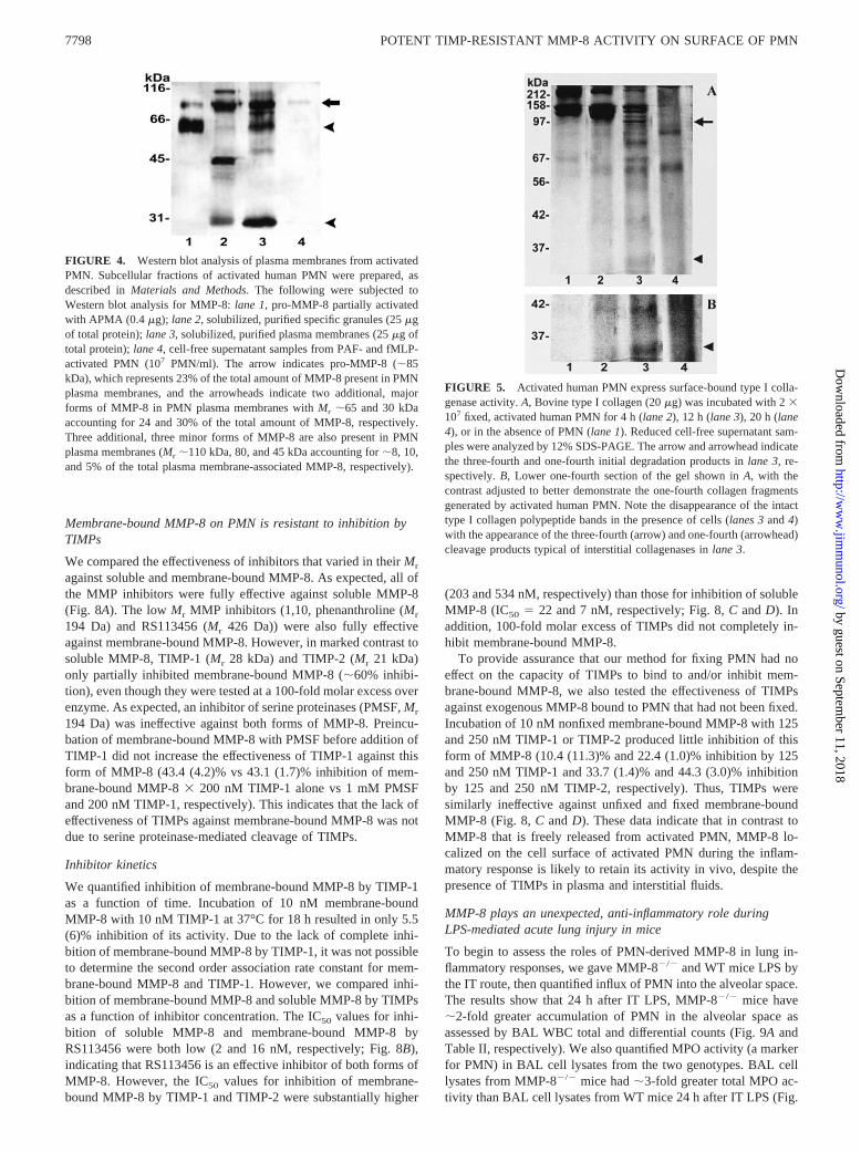
To compare intracellular and the two extracellular forms of PMN-derived MMP-8, we performed Western blot analysis for MMP-8in cell-free supernatants from stimulated PMN, and in PMNplasma membranes and PMN-specific granules isolated by subcel-lular fractionation of PMN (Fig. 4). In PMN-specific granules,pro-MMP-8 is a major form of MMP-8 (lane 2). The specific gran-ules of PMN contain additional forms of MMP-8 having Mr �110,�40, and �30 kDa, as reported previously (15, 16). MMP-8,which is freely released from stimulated PMN as a soluble pro-teinase, is exclusively in the 85-kDa proenzyme form (lane 4).PMN plasma membranes contain three major forms of MMP-8that differ in Mr (lane 3), including forms having the same Mr aspro-MMP-8 (85 kDa) and active MMP-8 (65 kDa), and a 30-kDaform, each representing 23, 24, and 30% of the total amount ofMMP-8 present in PMN plasma membranes, respectively. Threeadditional minor forms of MMP-8 are present in PMN plasmamembranes having Mr 110, 80, and 46 kDa and representing 8, 10,and 5% of the total membrane-associated MMP-8, respectively.The 46- and 30-kDa forms are likely to be proteolytically pro-cessed, inactive forms of MMP-8 because soluble MMP-8 under-goes proteolytic processing, generating forms with lower Mr thathave lost catalytic activity (37).
Activated human PMN express cell surface collagenase activity
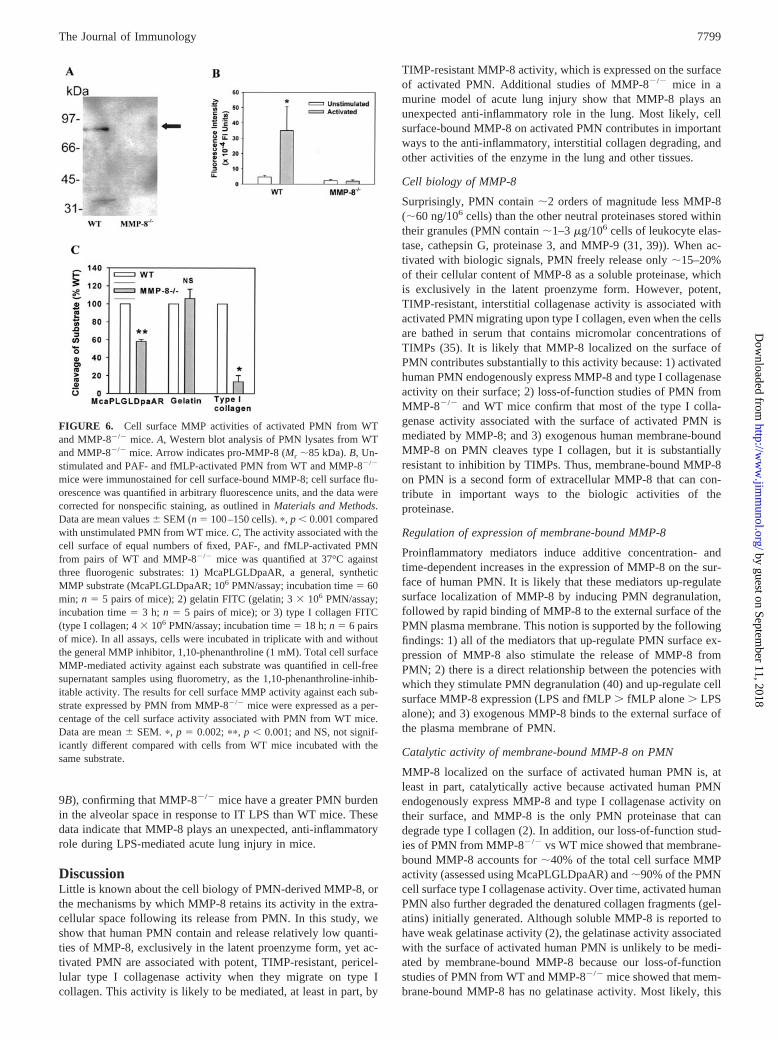
To assess whether membrane-bound MMP-8 on PMN is catalyti-cally active, we tested whether human PMN that are optimallyactivated to induce surface localization of MMP-8 express surface-bound type I collagenase activity, because MMP-8 is the onlyPMN proteinase that degrades type I collagen (2). Activated PMNexpress surface-bound proteinase activity that generates the three-fourth (Fig. 5A, lane 3) and one-fourth (Fig. 5B, lane 3) fragmentstypical of interstitial collagenases within 12 h. After 20 h, therewas complete degradation of intact type I collagen, as well assubstantial degradation of the gelatins generated (Fig. 5A, lane 4).Thus, activated human PMN express interstitial collagenase (andgelatinase) activity on their cell surface.
Catalytic activity of membrane-bound MMP-8 on PMN
Our goal was to determine whether MMP-8 localized on the sur-face of PMN is catalytically active, and to assess whether it hassimilar substrate specificity and catalytic efficiency as solubleMMP-8. However, it is not possible to study MMP-8, which isendogenously expressed on the surface of human PMN to assessits catalytic activity and efficiency against substrates other thaninterstitial collagens because: 1) activated PMN express other pro-teinases on their surface that cleave MMP-8 substrates other thaninterstitial collagen (24, 29, 31, 32); and 2) currently, there are noinhibitors available that are specific for MMP-8. We therefore usedtwo complementary strategies to assess the catalytic activity and
FIGURE 1. PMN are associated with pericellular collagenase activityeven in the presence of serum. Chamber slides were coated with FITC-conjugated type I collagen, and either autologous serum (A and B) or au-tologous serum containing 20 �M RS104210, a low m.w., synthetic, gen-eral MMP inhibitor (C), was added as the bathing medium. The samenumber of PMN was added to B and C, but not to A. The cells wereactivated for 5 min with PAF (10�7 M); then 10�7 M fMLP was added andthe chamber slides were incubated at 37°C for 3 h. The cells were thenexamined by incident light fluorescence microscopy. Areas of pericellularcollagen degradation generated by cells as they migrate upon the collagensubstrate are the dark areas on the background fluorescent collagen sub-strate (open arrows). The yellow arrows indicate cells remaining adherentto the substrate that are visible because they autofluoresce and/or ingestfluorescent substrate. Note in B, that as the substrate undergoes extensivepericellular degradation, cells detach from the chamber slides, and fewercells remain adherent at the end of the assay than in C, in which proteolysisof type I collagen is markedly reduced by the addition of RS104210 to theserum. Magnification: �200.
7795The Journal of Immunology
by guest on September 11, 2018
http://ww
w.jim
munol.org/
Dow
nloaded from

efficiency of membrane-bound MMP-8 on PMN: 1) a loss-of-func-tion strategy in which we compared the cell surface proteolyticactivities expressed by activated PMN from MMP-8�/� vs WTmice against potential MMP-8 substrates; and 2) a model cell sys-tem in which we studied the activities of exogenous humanMMP-8 bound to the surface of unstimulated human PMN.
Loss-of-function studies
We first confirmed that PMN from MMP-8�/� mice do not containMMP-8, by Western blot analysis (Fig. 6A), and that activatedPMN from WT mice express MMP-8 in an inducible manner ontheir cell surface (Fig. 6B). We then compared the capacity of cellsfrom both genotypes to hydrolyze MMP-8 substrates. PMN fromMMP-8�/� mice expressed �58, 100, and 8% of the surface-bound, MMP-mediated activity against McaPLGLDpaAR (a gen-eral, synthetic MMP substrate), gelatin, and type I collagen, re-spectively (Fig. 6C). These data indicate that MMP-8 is present onthe surface of activated murine PMN in a catalytically active form.In addition, membrane-bound MMP-8 on murine PMN accountsfor �40% of the total cell surface MMP activity, �90% of the cell
surface type I collagenase activity on activated murine PMN, butnone of the PMN surface gelatinase activity.
Catalytic activity, efficiency, and stability of exogenous humanmembrane-bound MMP-8
To assess the catalytic activity, efficiency, and stability of humanmembrane-bound MMP-8 in isolation and in a quantitative man-ner, we studied exogenous, active MMP-8 bound to the surface ofunstimulated PMN (which have had no detectable proteinase ac-tivity on their cell surface). We first quantified the amount ofMMP-8 that bound to the surface of PMN using McaPLGLDpaARand assay standards of soluble, active site-titrated MMP-8. PMNbound 60.2 (12.7) ng of MMP-8 activity per 106 PMN, whereascontrol cells (which had no MMP-8 bound to their surface) had nodetectable activity (n 7). When soluble MMP-8 and membrane-bound MMP-8 were incubated with McaPLGLDpaAR, the twoforms of the proteinase produced similar, progressive cleavage ofthe substrate over time (Fig. 7A). This activity was not due torelease, leakage, or detachment of proteinases from the cells, be-cause cell-free supernatant fluids from cells incubated in bufferalone had no detectable activity against McaPLGLDpaAR (n 4).
FIGURE 2. Cell surface expression ofMMP-8 on unstimulated and activated PMN.PMN were incubated in the absence of ago-nists (A), or primed for 15 min with 10�8 MPAF, then activated for 30 min with 10�8 MfMLP (B–D). Cells were fixed, then immu-nostained with anti-MMP-8 (A, B, and D), orwith rabbit IgG (C), as a control. Represen-tative microscopic fields were examined bothby phase-contrast microscopy (A–C, leftpanels) and by incident light fluorescencemicroscopy (A–C, right panels). D, To con-firm cell surface localization of MMP-8,nonpermeabilized, activated PMN were alsodouble immunostained with Alexa 488 forsurface-bound MMP-8 (D, left panel) andwith Alexa 546 for surface-bound MMP-9(D, middle panel), and then examined byconfocal microscopy (D). Dual excitation ofthe fluorophores (D, right panel) demon-strates substantial colocalization of both en-zymes on the PMN cell surface (yellow over-lay). Arrow indicates MMP-8 and MMP-9colocalized on the leading edge of a polar-ized PMN. Magnification: �1000.
7796 POTENT TIMP-RESISTANT MMP-8 ACTIVITY ON SURFACE OF PMN
by guest on September 11, 2018
http://ww
w.jim
munol.org/
Dow
nloaded from

Membrane-bound MMP-8 on PMN also cleaves type I collageninitially at a single locus, generating three-fourth (�98-kDa) andone-fourth length (34-kDa) collagen fragments typical of solubleinterstitial collagenases (Fig. 7B, lane 4). Cleavage of type I col-lagen by membrane-bound MMP-8 on PMN (lane 4) was as effi-cient as that by soluble MMP-8 (lane 2) because both forms ofMMP-8 produced almost complete disappearance of the intact typeI polypeptide chains. RS113456 (a synthetic, hydroxamate MMPinhibitor) completely inhibited the type I collagenase activity as-sociated with membrane-bound MMP-8 (Fig. 7B, lane 5). Thisactivity was all cell associated because supernatant samples fromstimulated PMN contain no type I collagenase activity (Fig. 7B,lane 6). Membrane-bound MMP-8 also cleaves type II collagen.MMP-8 bound to the surface of 106 PMN expressed type II col-lagenase activity equivalent to �100 ng of soluble, active MMP-8(Fig. 7C). As reported previously for soluble MMP-8 (38), mem-brane-bound MMP-8 also cleaves �1-PI at a single locus within itsactive site loop, generating two cleavage products having Mr �50kDa (Fig. 7D), and �4 kDa (data not shown). Both forms ofMMP-8 produced substantial cleavage of �1-PI. Control PMN didnot cleave any of the biologic substrates tested (Fig. 7, A–D).
Catalytic efficiency of membrane-bound MMP-8
We compared the saturation kinetic constants (kcat/KM) for solubleand membrane-bound MMP-8. Each form of the proteinase wasincubated with McaPLGLDpaAR under first order conditions, andinitial reaction velocities were measured over 6 min. The kcat/KM
values for soluble and membrane-bound MMP-8 were very similar
(14,800 M�1s�1, n 5; 10,100 M�1s�1, n 3, respectively),indicating that soluble and membrane-bound MMP-8 cleave thissubstrate with similar catalytic efficiency.
In the above experiments, we studied exogenous membrane-bound MMP-8 that had been fixed onto the surface of PMN toprevent detachment of the enzyme from the cell surface. To assesswhether our fixation process has any effect on the catalytic activityof membrane-bound MMP-8, we compared initial reaction veloc-ities of equimolar amounts of exogenous membrane-boundMMP-8 that had been either fixed or not fixed onto the PMNsurface. Fixed membrane-bound MMP-8 expressed 86.1% of theactivity of nonfixed proteinase (n 6 experiments), indicating thatour fixation process has little effect on the catalytic activity ofsurface-bound MMP-8.
Stability of soluble vs membrane-bound MMP-8
We measured reaction velocities of both forms of the enzyme be-fore and after incubation at 37°C for varying times, using McaPL-GLDpaAR as the substrate (Fig. 7E). Soluble MMP-8 rapidly lostactivity over time (t1/2 at 37°C 7.5 h). In marked contrast, mem-brane-bound MMP-8 retained close to 100% of its activity after6 h, and �80% of its activity after 18 h.
Together, these data indicate that soluble MMP-8 and mem-brane-bound MMP-8 on PMN have a similar spectrum of catalyticactivity, and similar catalytic efficiency against McaPLGLDpaAR.However, membrane-bound MMP-8 is more catalytically stablethan soluble MMP-8.
FIGURE 3. Proinflammatory mediatorsup-regulate cell surface expression of MMP-8on PMN. A–D, PMN were incubated at 37°Cfor 30 min with or without varying concentra-tions of LPS (A), TNF-� (B), PAF (C), andfMLP (D). E, �, PMN incubated for 30 min at37°C with or without 100 ng/ml LPS, 10�8 MPAF, 100 U/ml TNF-�, or 10�8 M fMLP. f,PMN, which were primed for 15 min with thesame concentrations of LPS, PAF, or TNF-�,then activated for 30 min with 10�8 M fMLP.Cell surface MMP-8 expression was quantifiedby immunofluorescence staining and imageanalysis. The data were corrected for nonspe-cific staining and expressed as a percentage ofthe mean integrated fluorescence of the un-stimulated PMN. Data are mean values �SEM (n 100–150 cells). A–C, �, p � 0.001compared with unstimulated cells. D, �, p 0.018; ��, p � 0.001 compared with unstimu-lated cells. E, �, p � 0.001 compared withunstimulated cells; ��, p � 0.001 comparedwith cells incubated with agonists alone. F, f,PMN undergoing random migration over 3 h inresponse to buffer alone in the lower wells(random), or directed migration in response to10�7 M fMLP in the lower wells of MatrigelInvasion Chambers. �, PMN incubated in sus-pension for 3 h at 37°C with or without 10�7
M fMLP. Cell surface MMP-8 was quantified,as outlined above. The data (mean � SEM) areexpressed as a percentage of the mean inte-grated fluorescence of unstimulated PMN in-cubated in suspension (n 150–200 cells). �,p � 0.001 when compared with unstimulatedPMN in suspension; ��, p � 0.001 comparedwith all other conditions.
7797The Journal of Immunology
by guest on September 11, 2018
http://ww
w.jim
munol.org/
Dow
nloaded from
Membrane-bound MMP-8 on PMN is resistant to inhibition byTIMPs
We compared the effectiveness of inhibitors that varied in their Mr
against soluble and membrane-bound MMP-8. As expected, all ofthe MMP inhibitors were fully effective against soluble MMP-8(Fig. 8A). The low Mr MMP inhibitors (1,10, phenanthroline (Mr
194 Da) and RS113456 (Mr 426 Da)) were also fully effectiveagainst membrane-bound MMP-8. However, in marked contrast tosoluble MMP-8, TIMP-1 (Mr 28 kDa) and TIMP-2 (Mr 21 kDa)only partially inhibited membrane-bound MMP-8 (�60% inhibi-tion), even though they were tested at a 100-fold molar excess overenzyme. As expected, an inhibitor of serine proteinases (PMSF, Mr
194 Da) was ineffective against both forms of MMP-8. Preincu-bation of membrane-bound MMP-8 with PMSF before addition ofTIMP-1 did not increase the effectiveness of TIMP-1 against thisform of MMP-8 (43.4 (4.2)% vs 43.1 (1.7)% inhibition of mem-brane-bound MMP-8 � 200 nM TIMP-1 alone vs 1 mM PMSFand 200 nM TIMP-1, respectively). This indicates that the lack ofeffectiveness of TIMPs against membrane-bound MMP-8 was notdue to serine proteinase-mediated cleavage of TIMPs.
Inhibitor kinetics
We quantified inhibition of membrane-bound MMP-8 by TIMP-1as a function of time. Incubation of 10 nM membrane-boundMMP-8 with 10 nM TIMP-1 at 37°C for 18 h resulted in only 5.5(6)% inhibition of its activity. Due to the lack of complete inhi-bition of membrane-bound MMP-8 by TIMP-1, it was not possibleto determine the second order association rate constant for mem-brane-bound MMP-8 and TIMP-1. However, we compared inhi-bition of membrane-bound MMP-8 and soluble MMP-8 by TIMPsas a function of inhibitor concentration. The IC50 values for inhi-bition of soluble MMP-8 and membrane-bound MMP-8 byRS113456 were both low (2 and 16 nM, respectively; Fig. 8B),indicating that RS113456 is an effective inhibitor of both forms ofMMP-8. However, the IC50 values for inhibition of membrane-bound MMP-8 by TIMP-1 and TIMP-2 were substantially higher
(203 and 534 nM, respectively) than those for inhibition of solubleMMP-8 (IC50 22 and 7 nM, respectively; Fig. 8, C and D). Inaddition, 100-fold molar excess of TIMPs did not completely in-hibit membrane-bound MMP-8.
To provide assurance that our method for fixing PMN had noeffect on the capacity of TIMPs to bind to and/or inhibit mem-brane-bound MMP-8, we also tested the effectiveness of TIMPsagainst exogenous MMP-8 bound to PMN that had not been fixed.Incubation of 10 nM nonfixed membrane-bound MMP-8 with 125and 250 nM TIMP-1 or TIMP-2 produced little inhibition of thisform of MMP-8 (10.4 (11.3)% and 22.4 (1.0)% inhibition by 125and 250 nM TIMP-1 and 33.7 (1.4)% and 44.3 (3.0)% inhibitionby 125 and 250 nM TIMP-2, respectively). Thus, TIMPs weresimilarly ineffective against unfixed and fixed membrane-boundMMP-8 (Fig. 8, C and D). These data indicate that in contrast toMMP-8 that is freely released from activated PMN, MMP-8 lo-calized on the cell surface of activated PMN during the inflam-matory response is likely to retain its activity in vivo, despite thepresence of TIMPs in plasma and interstitial fluids.
MMP-8 plays an unexpected, anti-inflammatory role duringLPS-mediated acute lung injury in mice
To begin to assess the roles of PMN-derived MMP-8 in lung in-flammatory responses, we gave MMP-8�/� and WT mice LPS bythe IT route, then quantified influx of PMN into the alveolar space.The results show that 24 h after IT LPS, MMP-8�/� mice have�2-fold greater accumulation of PMN in the alveolar space asassessed by BAL WBC total and differential counts (Fig. 9A andTable II, respectively). We also quantified MPO activity (a markerfor PMN) in BAL cell lysates from the two genotypes. BAL celllysates from MMP-8�/� mice had �3-fold greater total MPO ac-tivity than BAL cell lysates from WT mice 24 h after IT LPS (Fig.
FIGURE 4. Western blot analysis of plasma membranes from activatedPMN. Subcellular fractions of activated human PMN were prepared, asdescribed in Materials and Methods. The following were subjected toWestern blot analysis for MMP-8: lane 1, pro-MMP-8 partially activatedwith APMA (0.4 �g); lane 2, solubilized, purified specific granules (25 �gof total protein); lane 3, solubilized, purified plasma membranes (25 �g oftotal protein); lane 4, cell-free supernatant samples from PAF- and fMLP-activated PMN (107 PMN/ml). The arrow indicates pro-MMP-8 (�85kDa), which represents 23% of the total amount of MMP-8 present in PMNplasma membranes, and the arrowheads indicate two additional, majorforms of MMP-8 in PMN plasma membranes with Mr �65 and 30 kDaaccounting for 24 and 30% of the total amount of MMP-8, respectively.Three additional, three minor forms of MMP-8 are also present in PMNplasma membranes (Mr �110 kDa, 80, and 45 kDa accounting for �8, 10,and 5% of the total plasma membrane-associated MMP-8, respectively).
FIGURE 5. Activated human PMN express surface-bound type I colla-genase activity. A, Bovine type I collagen (20 �g) was incubated with 2 �107 fixed, activated human PMN for 4 h (lane 2), 12 h (lane 3), 20 h (lane4), or in the absence of PMN (lane 1). Reduced cell-free supernatant sam-ples were analyzed by 12% SDS-PAGE. The arrow and arrowhead indicatethe three-fourth and one-fourth initial degradation products in lane 3, re-spectively. B, Lower one-fourth section of the gel shown in A, with thecontrast adjusted to better demonstrate the one-fourth collagen fragmentsgenerated by activated human PMN. Note the disappearance of the intacttype I collagen polypeptide bands in the presence of cells (lanes 3 and 4)with the appearance of the three-fourth (arrow) and one-fourth (arrowhead)cleavage products typical of interstitial collagenases in lane 3.
7798 POTENT TIMP-RESISTANT MMP-8 ACTIVITY ON SURFACE OF PMN
by guest on September 11, 2018
http://ww
w.jim
munol.org/
Dow
nloaded from
9B), confirming that MMP-8�/� mice have a greater PMN burdenin the alveolar space in response to IT LPS than WT mice. Thesedata indicate that MMP-8 plays an unexpected, anti-inflammatoryrole during LPS-mediated acute lung injury in mice.
DiscussionLittle is known about the cell biology of PMN-derived MMP-8, orthe mechanisms by which MMP-8 retains its activity in the extra-cellular space following its release from PMN. In this study, weshow that human PMN contain and release relatively low quanti-ties of MMP-8, exclusively in the latent proenzyme form, yet ac-tivated PMN are associated with potent, TIMP-resistant, pericel-lular type I collagenase activity when they migrate on type Icollagen. This activity is likely to be mediated, at least in part, by
TIMP-resistant MMP-8 activity, which is expressed on the surfaceof activated PMN. Additional studies of MMP-8�/� mice in amurine model of acute lung injury show that MMP-8 plays anunexpected anti-inflammatory role in the lung. Most likely, cellsurface-bound MMP-8 on activated PMN contributes in importantways to the anti-inflammatory, interstitial collagen degrading, andother activities of the enzyme in the lung and other tissues.
Cell biology of MMP-8
Surprisingly, PMN contain �2 orders of magnitude less MMP-8(�60 ng/106 cells) than the other neutral proteinases stored withintheir granules (PMN contain �1–3 �g/106 cells of leukocyte elas-tase, cathepsin G, proteinase 3, and MMP-9 (31, 39)). When ac-tivated with biologic signals, PMN freely release only �15–20%of their cellular content of MMP-8 as a soluble proteinase, whichis exclusively in the latent proenzyme form. However, potent,TIMP-resistant, interstitial collagenase activity is associated withactivated PMN migrating upon type I collagen, even when the cellsare bathed in serum that contains micromolar concentrations ofTIMPs (35). It is likely that MMP-8 localized on the surface ofPMN contributes substantially to this activity because: 1) activatedhuman PMN endogenously express MMP-8 and type I collagenaseactivity on their surface; 2) loss-of-function studies of PMN fromMMP-8�/� and WT mice confirm that most of the type I colla-genase activity associated with the surface of activated PMN ismediated by MMP-8; and 3) exogenous human membrane-boundMMP-8 on PMN cleaves type I collagen, but it is substantiallyresistant to inhibition by TIMPs. Thus, membrane-bound MMP-8on PMN is a second form of extracellular MMP-8 that can con-tribute in important ways to the biologic activities of theproteinase.
Regulation of expression of membrane-bound MMP-8
Proinflammatory mediators induce additive concentration- andtime-dependent increases in the expression of MMP-8 on the sur-face of human PMN. It is likely that these mediators up-regulatesurface localization of MMP-8 by inducing PMN degranulation,followed by rapid binding of MMP-8 to the external surface of thePMN plasma membrane. This notion is supported by the followingfindings: 1) all of the mediators that up-regulate PMN surface ex-pression of MMP-8 also stimulate the release of MMP-8 fromPMN; 2) there is a direct relationship between the potencies withwhich they stimulate PMN degranulation (40) and up-regulate cellsurface MMP-8 expression (LPS and fMLP � fMLP alone � LPSalone); and 3) exogenous MMP-8 binds to the external surface ofthe plasma membrane of PMN.
Catalytic activity of membrane-bound MMP-8 on PMN
MMP-8 localized on the surface of activated human PMN is, atleast in part, catalytically active because activated human PMNendogenously express MMP-8 and type I collagenase activity ontheir surface, and MMP-8 is the only PMN proteinase that candegrade type I collagen (2). In addition, our loss-of-function stud-ies of PMN from MMP-8�/� vs WT mice showed that membrane-bound MMP-8 accounts for �40% of the total cell surface MMPactivity (assessed using McaPLGLDpaAR) and �90% of the PMNcell surface type I collagenase activity. Over time, activated humanPMN also further degraded the denatured collagen fragments (gel-atins) initially generated. Although soluble MMP-8 is reported tohave weak gelatinase activity (2), the gelatinase activity associatedwith the surface of activated human PMN is unlikely to be medi-ated by membrane-bound MMP-8 because our loss-of-functionstudies of PMN from WT and MMP-8�/� mice showed that mem-brane-bound MMP-8 has no gelatinase activity. Most likely, this
FIGURE 6. Cell surface MMP activities of activated PMN from WTand MMP-8�/� mice. A, Western blot analysis of PMN lysates from WTand MMP-8�/� mice. Arrow indicates pro-MMP-8 (Mr �85 kDa). B, Un-stimulated and PAF- and fMLP-activated PMN from WT and MMP-8�/�
mice were immunostained for cell surface-bound MMP-8; cell surface flu-orescence was quantified in arbitrary fluorescence units, and the data werecorrected for nonspecific staining, as outlined in Materials and Methods.Data are mean values � SEM (n 100–150 cells). �, p � 0.001 comparedwith unstimulated PMN from WT mice. C, The activity associated with thecell surface of equal numbers of fixed, PAF-, and fMLP-activated PMNfrom pairs of WT and MMP-8�/� mice was quantified at 37°C againstthree fluorogenic substrates: 1) McaPLGLDpaAR, a general, syntheticMMP substrate (McaPLGLDpaAR; 106 PMN/assay; incubation time 60min; n 5 pairs of mice); 2) gelatin FITC (gelatin; 3 � 106 PMN/assay;incubation time 3 h; n 5 pairs of mice); or 3) type I collagen FITC(type I collagen; 4 � 106 PMN/assay; incubation time 18 h; n 6 pairsof mice). In all assays, cells were incubated in triplicate with and withoutthe general MMP inhibitor, 1,10-phenanthroline (1 mM). Total cell surfaceMMP-mediated activity against each substrate was quantified in cell-freesupernatant samples using fluorometry, as the 1,10-phenanthroline-inhib-itable activity. The results for cell surface MMP activity against each sub-strate expressed by PMN from MMP-8�/� mice were expressed as a per-centage of the cell surface activity associated with PMN from WT mice.Data are mean � SEM. �, p 0.002; ��, p � 0.001; and NS, not signif-icantly different compared with cells from WT mice incubated with thesame substrate.
7799The Journal of Immunology
by guest on September 11, 2018
http://ww
w.jim
munol.org/
Dow
nloaded from

gelatinase activity is mediated by the activities of other proteinaseshaving gelatinase activity expressed on the surface of activated PMN,including MMP-9 (29) and human leukocyte elastase (HLE) (24).
To further assess the catalytic activity, catalytic efficiency, andsusceptibility to inhibition of membrane-bound MMP-8 by TIMPsin isolation and in a quantitative manner, we studied exogenousMMP-8 bound to the cell surface of unstimulated PMN. Mem-brane-bound MMP-8 has similar substrate specificity as the solu-ble form of MMP-8 cleaving McaPLGLDpaAR, types I and IIinterstitial collagens, and a serpin (�1-PI). Like soluble MMP-8,membrane-bound MMP-8 generates one-fourth and three-fourth
fragments of the constituent polypeptide chains of type I collagen,and cleaves �1-PI at a single locus within its reactive site loop,which inactivates the inhibitor (38). MMP-8 localized on the sur-face of PMN also has catalytic efficiency similar to that of solubleMMP-8, as determined by the similar kcat/KM values for the twoforms of MMP-8 when tested against McaPLGLDpaAR. All of theactivities associated with the cells were due to MMP-8 bound tothe PMN surface because: 1) control PMN (which had no MMP-8bound to their surface) had no significant activity against any ofthe substrates tested; and 2) no proteinase activity was detected incell-free supernatant fluids from cells that bound MMP-8.
FIGURE 7. Human exogenous MMP-8 bound to PMN can degrade synthetic and biologic substrates. A, McaPLGLDpaAR. Cleavage of McaPLGLD-paAR by soluble MMP-8 (50 ng, squares), membrane-bound MMP-8 on PMN (50 ng, circles), or control cells incubated without MMP-8 (triangles) wasquantified by fluorometry. Data are mean � SD. B, Type I collagen. Bovine type I collagen was incubated for 18 h at 37°C either alone (lane 1) or withthe following: 1) soluble, active MMP-8 (lane 2); 2) control PMN (lane 3); 3) MMP-8 bound to PMN (lane 4); 4) MMP-8 bound to PMN with 10 �MRS113456 (lane 5); and 5) cell-free supernatants from PMN that bound MMP-8 (lane 6). Reduced cell-free supernatant samples were analyzed by 12%SDS-PAGE. �, The band corresponding to soluble, active MMP-8 that is present only in lane 2. Arrows indicate the three-fourth and one-fourth initialcleavage products of the polypeptide chains of type I collagen in lanes 2 and 4. C, Type II collagen. We incubated PMN that bound MMP-8 to their surfaceor control PMN (106 cells/assay), along with assay standards of soluble, active site-titrated MMP-8 (25–800 ng) with FITC-conjugated type II collagenfor 4 h. Cleavage of the substrate by soluble MMP-8 and membrane-bound MMP-8 was quantified in cell-free supernatant samples in arbitrary fluorescenceunits using fluorometry. The results for cell-associated activity against type II collagen were converted to ng equivalents of soluble MMP-8 activity byinterpolation from the standard curve for soluble MMP-8 activity (y-axis). Data are mean values � SD (n 5 donors). �, p � 0.001 compared with controlcells. D, �1-PI. Purified human �1-PI was incubated at 37°C alone (lane 1) or with soluble MMP-8 (lane 2), control PMN (lane 3), or MMP-8 bound toPMN (lane 4). Reduced cell-free supernatant samples were analyzed by 20% SDS-PAGE. The arrow indicates intact �1-PI, and the arrowhead indicatesthe 50-kDa cleavage product. The 4-kDa cleavage product is not visible on the gel. E, Stability of soluble and membrane-bound MMP-8. Soluble MMP-8(circles) and membrane-bound MMP-8 (squares) were incubated at 37°C in buffer for varying times (x-axis), and then initial reaction velocities weremeasured over 30 min after addition of McaPLGLDpaAR. The results were expressed as percentage of initial reaction velocities, and the t1/2 for solubleMMP-8 (7.5 h) was calculated by nonlinear regression analysis. Data are mean � SEM; n 4.
7800 POTENT TIMP-RESISTANT MMP-8 ACTIVITY ON SURFACE OF PMN
by guest on September 11, 2018
http://ww
w.jim
munol.org/
Dow
nloaded from

Soluble MMP-8 and membrane-bound MMP-8 differ markedlyin their stability at 37°C. Although soluble MMP-8 has a short t1/2
at 37°C due to autoproteolysis (37), MMP-8 localized on the sur-face of PMN retains �80% of its activity even after incubation at37°C for 18 h. The mechanism by which binding of MMP-8 to thePMN plasma membrane prevents loss of its activity is unclear.However, the binding of pro-MMP-9 to insoluble substrates in-duces a conformational change in the enzyme leading to proen-zyme activation (41), and it is possible that the binding of MMP-8to the PMN cell surface induces a conformational change that lim-its its autoproteolysis.
Susceptibility of membrane-bound MMP-8 to inhibition
The pericellular collagenase activity endogenously expressed byactivated human PMN migrating upon type I collagen is resistantto inhibition by TIMPs present in serum, but is substantially ab-rogated by a low Mr MMP inhibitor. Our studies of individualMMP inhibitors tested against human membrane-bound MMP-8showed that there is an indirect relationship between inhibitor sizeand its effectiveness against this form of MMP-8. In particular, theIC50 values for inhibition of membrane-bound MMP-8 by TIMP-1and -2 were high (200–500 nM), and there was incomplete inhi-bition of membrane-bound MMP-8 � 100-fold molar excess ofTIMPs. This further supports the notion that membrane-boundMMP-8 contributes to the pericellular collagenase activity associ-ated with activated PMN. In addition, the data suggest that sterichindrance is most likely the mechanism by which membrane-bound MMP-8 on PMN evades inhibition by physiologic inhibi-tors. Large, globular TIMPs may form complexes less readily withMMP-8 that is sterically confined on the surface of PMN than with
soluble MMP-8. This possibility is the focus of ongoing studies inour laboratory.
Although MMP-8 localized on the PMN surface can contributeto the pericellular collagenase activity associated with PMN mi-grating upon type I collagen, it is also possible that the generationof sequestered microenvironments by cells adherent to type I col-lagen contributes to this activity. Tight adherence of the PMN tothe substrate could create a compartment into which diffusion ofTIMPs is impaired, because �1-PI diffuses poorly into compart-ments generated when PMN adhere to fibronectin (42). However,degradation of type I collagen in this protected microenvironmentis likely to be mediated by membrane-bound MMP-8, rather thanMMP-8 freely released by PMN into this compartment. This con-cept is supported by our Western blot analysis of cell-free super-natants from stimulated PMN showing that MMP-8 is releasedfrom PMN exclusively in its latent proenzyme form. In addition,we have shown previously that soluble MMP activity is not de-tectable in culture supernatants from activated PMN even whencells are incubated in inhibitor-free buffers (29). Our data indicatethat while TIMPs can effectively control the activity of MMP-8that is freely released from stimulated PMN, they are ineffectiveinhibitors of MMP-8 localized on the PMN surface. Membrane-bound MMP-8 on PMN is thus likely to substantially retain itsactivity in the extracellular space at sites of inflammation.
Mechanisms of activation of membrane-bound MMP-8
MMP-8 is expressed on the surface of PMN, at least in part, in acatalytically active form because: 1) activated human and murinePMN have substantial type I collagenase activity associated with
FIGURE 8. Membrane-bound MMP-8 on PMN is substantially resistant to inhibition by TIMPs. A total of 10 nM soluble MMP-8 (f) or 10 nMmembrane-bound MMP-8 on PMN (�) was incubated for 30 min with and without: 1) 1,10-phenanthroline (o-Phen,1 mM); 2) RS113456 (1 �M); 3)TIMP-1 (1 �M); 4) TIMP-2 (1 �M); or 5) PMSF (1 mM). Residual MMP-8 activity was quantified in cell-free supernatant samples using McaPLGLD-paAR. Data are mean values � SEM (n 5). �, p 0.004; ��, p � 0.001 compared with soluble MMP-8 incubated with the same inhibitor. B–D,Concentration dependence of inhibition of soluble vs membrane-bound MMP-8. A total of 10 nM soluble MMP-8 (open symbols) or 10 nM membrane-bound MMP-8 (filled symbols) was incubated for 30 min at 37°C with or without varying concentrations of RS113456 (B), TIMP-1 (C), or TIMP-2 (D);then residual MMP-8 activity was quantified in cell-free supernatant samples using McaPLGLDpaAR. IC50 values were calculated by nonlinear regressionanalysis. Data are mean values � SD.
7801The Journal of Immunology
by guest on September 11, 2018
http://ww
w.jim
munol.org/
Dow
nloaded from

their cell surface; and 2) Western blot analysis of plasma mem-branes from activated PMN contains forms of MMP-8 that havethe same Mr as active MMP-8 (�65 kDa). Proteinases (such ascathepsin G, MMP-3, and tryptase (20, 21)) and reactive oxygenspecies including oxidants generated by the MPO system (43) canactivate pro-MMP-8 in vitro. Thus, it is possible that soluble pro-teinases and oxidants released by PMN and other cells during theinflammatory response activate pro-MMP-8 after its binding to thePMN surface in vivo. It is noteworthy in this respect that cathepsinG and MPO are expressed on the surface of activated PMN (30,44, 45). Thus, membrane-bound cathepsin G and MPO could ac-tivate membrane-bound pro-MMP-8 in a juxtacrine manner. In ad-dition, MT6-MMP is also constitutively present on the surfacePMN, and is shed from the cell surface as a soluble proteinasewhen PMN are activated (32). MT6-MMP has been shown to ac-tivate pro-MMP-9 and -2 in vitro (32), but it is not known whetherit can activate soluble or membrane-bound pro-MMP-8. Themechanisms by which pro-MMP-8 binds to and is activated on thesurface of PMN will be a focus of future studies in our laboratory.
Biologic roles of membrane-bound MMP-8
Our in vitro studies suggest that MMP-8 localized on the surfaceof activated PMN contributes to the pericellular ECM-degradingactivities of activated PMN in vivo. PMN express other ECM-degrading proteinases on their cell surface, including serine pro-teinases (24, 31, 46), MMP-9 (29), and MT6-MMP (32). Amongthese proteinases, only MMP-8 and HLE have interstitial collage-nase activity, and HLE can only degrade type III collagen at a rateof only �1% of that of the interstitial collagenases (47). Thus, theexpression of MMP-8 on the surface of activated PMN adds potentinterstitial collagenase (and serpinase) activity to the pericellular,proteolytic armamentarium of the activated PMN. Surface-boundMMP-8 on PMN may locally degrade interstitial collagens duringinflammation and wound healing. Another biologic role of cellsurface-bound MMP-8 on PMN may be to degrade serpins in thelocal environment of PMN because membrane-bound MMP-8 caninactivate �1-PI. It is noteworthy that agonists that up-regulateMMP-8 localization on the surface of PMN also induce cell sur-face serine proteinase expression (30, 31, 44). Pericellular serpi-nase activity mediated by membrane-bound MMP-8 on PMN maypotentiate the activities of serine proteinases coordinately ex-pressed on the surface of activated PMN during inflammation. Fi-nally, our studies of MMP-8�/� vs WT mice indicate that MMP-8
has an unexpected, anti-inflammatory role in the lung during LPS-mediated acute lung injury in mice by down-regulating the alve-olar PMN burden. It is noteworthy in this respect that recent stud-ies of MMP-8�/� vs WT mice in a chemical carcinogenesis modelin the skin have shown that MMP-8 up-regulates early PMN ac-cumulation in the skin (via cleavage and activation of LPS-inducedCXC chemokine). At later time points in this model, MMP-8 hasan anti-inflammatory activity in the skin associated with a lessfavorable environment for tumor initiation, but the mechanism forthis is not known (10). Our future goals are to investigate whetherPMN-derived MMP-8 cleaves and inactivates PMN chemokine(s),up-regulates anti-inflammatory mediators in the lung, and/or pro-motes clearance of PMN from the alveolar space. Thus, studies ofMMP-8�/� mice have identified unexpected roles for MMP-8 inregulating inflammation in different organs. Moreover, TIMP-re-sistant MMP-8, which is expressed on the surface of activatedPMN during inflammatory responses, is a bioactive form of theenzyme that is likely to contribute in important ways to its anti-inflammatory and other activities in vivo because it is a catalyti-cally efficient and stable, but TIMP-resistant form of theproteinase.
Conclusions
MMP-8 localized on the surface of PMN is likely to be an impor-tant bioactive form of the proteinase in vivo. During the inflam-matory response, proinflammatory mediators can rapidly inducethe expression of MMP-8 on the surface of PMN as they migratethrough tissues. Binding of MMP-8 to the PMN surface not onlyleads to activation of pro-MMP-8, but also focuses, stabilizes, andpreserves the collagenase and serpinase activities of the enzyme inthe pericellular environment of PMN, even in the presence ofTIMPs. The plasma membrane of PMN regulates the temporal andspatial localization of MMP-8 and other neutral proteinases ofPMN, and thereby coordinates their activities in the pericellularenvironment of PMN. Although membrane-bound MMP-8 cancontribute to physiologic processes of PMN, it also has the capac-ity to mediate tissue injury if its expression is excessive, pro-longed, or inappropriate. Thus, MMP-8-mediated proteolyticevents occurring at the PMN surface are likely to be criticallyimportant in physiologic and pathologic processes.
AcknowledgmentsWe thank Roche Bioscience for the kind gifts of RS113456 and RS104210.We thank Jean Lai, BioImaging Laboratory, Harvard School of PublicHealth (Boston, MA), for performing the confocal microscopic analysis ofimmunostained PMN.
References1. Gross, J., and Y. Nagai. 1965. Specific degradation of the collagen molecule by
tadpole collagenolytic enzyme. Proc. Natl. Acad. Sci. USA 54:1197.2. Owen, C. A., and E. J. Campbell. 1999. The cell biology of leukocyte-mediated
proteolysis. J. Leukocyte Biol. 65:137.
FIGURE 9. MMP-8 plays an anti-inflammatory role during LPS-medi-ated acute lung injury in mice. MMP-8�/� and WT control mice weregiven 10 �g of LPS (n 10 in each genotype) by the IT route, or ITendotoxin-free PBS as a control (n 4 in each genotype). A, After 24 h,the mice were euthanized, BAL was performed, and total WBC countswere performed. �, p 0.017 compared with WT mice given IT LPS. B,MPO activity was also quantified in cell extracts of BAL cells, as a markerfor PMN. Data are mean (SD). �, p 0.029 compared with WT mice givenIT LPS.
Table II. BAL differential WBC counts after IT LPS or IT PBS
Genotype, Treatmenta% PMN
Mean (SD)% MACS
Mean (SD)
WT, IT PBS 0 (0) 100 (0)MMP-8�/�, IT PBS 5 (4) 95 (4)WT, IT LPS 93 (1) 7 (1)MMP-8�/�, IT LPS 92 (2) 8 (2)
a WT or MMP-8�/� mice were given 10 �g of LPS (n 10) or endotoxin-freePBS (n 4) by the IT route. After 24 h, BAL was performed, and differential countswere performed on the WBC fraction.
7802 POTENT TIMP-RESISTANT MMP-8 ACTIVITY ON SURFACE OF PMN
by guest on September 11, 2018
http://ww
w.jim
munol.org/
Dow
nloaded from

3. Jeffrey, J. J. 2001. Interstitial collagenases. In Matrix Metalloproteinases.W. C. Parks and R. P. Mecham, eds. Academic Press, San Diego, pp. 15–42.
4. Knauper, V., A. Osthues, Y. A. DeClerck, K. E. Langley, J. Blaser, andH. Tschesche. 1993. Fragmentation of human polymorphonuclear-leukocyte col-lagenase. Biochem. J. 291:847.
5. Diekmann, O., and H. Tschesche. 1994. Degradation of kinins, angiotensins andsubstance P by polymorphonuclear matrix metalloproteinases MMP 8 and MMP9. Braz. J. Med. Biol. Res. 27:1865.
6. Power, C., C. M. O’Connor, D. Macfarlane, S. O’Mahoney, K. Gaffney, J. Hayes,and M. X. Fitzgerald. 1994. Neutrophil collagenase in sputum from patients withcystic fibrosis. Am. J. Respir. Crit. Care Med. 150:818.
7. Matsuki, H., N. Fujimoto, K. Iwata, V. Knauper, Y. Okada, and T. Hayakawa.1996. A one-step sandwich enzyme immunoassay for human matrix metallopro-teinase 8 (neutrophil collagenase) using monoclonal antibodies. Clin. Chim. Acta1996:129.
8. Lee, W., S. Aitken, J. Sodek, and C. A. McCulloch. 1995. Evidence of a directrelationship between neutrophil collagenase activity and periodontal tissue de-struction in vivo: role of active enzyme in human periodontitis.J. Periodontal Res. 30:23.
9. Nwomeh, B. C., H.-X. Liang, I. K. Cohen, and D. R. Yager. 1999. MMP-8 is thepredominant collagenase in healing wounds and nonhealing ulcers. J. Surg. Res.81:189.
10. Balbin, M., A. Fueyo, A. M. Tester, A. M. Pendas, A. S. Pitiot, A. Astudillo,C. M. Overall, S. D. Shapiro, and C. Lopez-Otin. 2003. Loss of collagenase-2confers increased skin tumor susceptibility to male mice. Nat. Genet. 35:252.
11. Cole, A. A., S. Chubinskaya, B. Schumacher, K. Huch, G. Szabo, J. Yao,K. Mikecz, K. A. Hasty, and K. E. Kuettner. 1996. Chondrocyte matrix metal-loproteinase-8: human articular chondrocytes express neutrophil collagenase.J. Biol. Chem. 271:11023.
12. Hanemaaijer, R., T. Sorsa, Y. T. Konttinen, Y. Ding, M. Sutinen, H. Visser,V. W. vanHinsbergh, T. Helaakoski, T. Kainulainen, H. Ronka, et al. 1997. Ma-trix metalloproteinase-8 is expressed in rheumatoid synovial fibroblasts and en-dothelial cells: regulation by tumor necrosis factor-� and doxycycline. J. Biol.Chem. 272:31504.
13. Herman, M. P., G. K. Sukhova, P. Libby, N. Gerdes, N. Tang, D. B. Horton,M. Kilbride, R. E. Breitbart, M. Chun, and U. Schonbeck. 2001. Expression ofneutrophil collagenase (matrix metalloproteinase-8) in human atheroma: a novelcollagenolytic pathway suggested by transcriptional profiling. Circulation104:1899.
14. Cowland, J. B., and N. Borregaard. 1999. The individual regulation of granuleprotein mRNA levels during neutrophil maturation explains the heterogeneity ofneutrophil granules. J. Leukocyte Biol. 66:989.
15. Murphy, G., J. J. Reynolds, U. Bretz, and M. Baggiolini. 1977. Collagenase is acomponent of the specific granules of human neutrophil leukocytes. Biochem. J.162:195.
16. Hasty, K. A., M. S. Hibbs, A. H. Kang, and C. L. Mainardi. 1986. Secreted formsof human neutrophil collagenase. J. Biol. Chem. 261:5645.
17. Balbin, M., A. Fueyo, V. Knauper, A. M. Pendas, J. M. Lopez, M. G. Jimenez,G. Murphy, and C. Lopez-Otin. 1998. Collagenase 2 (MMP-8) expression inmurine tissue-remodeling processes: analysis of its potential role in postpartuminvolution of the uterus. J. Biol. Chem. 273:23959.
18. Springman, E. B., E. L. Angleton, H. Birkedal-Hansen, and H. E. Van Wart.1990. Multiple modes of activation of latent human fibroblast collagenase: evi-dence for the role of a Cys73 active-site zinc complex in latency and a “cysteineswitch” mechanism for activation. Proc. Natl. Acad. Sci. USA 87:364.
19. Blaser, J., V. Knauper, A. Osthues, H. Reinke, and H. Tschesche. 1991. Mercurialactivation of human polymorphonuclear leukocyte procollagenase. Eur. J. Bio-chem. 202:1223.
20. Knauper, V., S. Kramer, H. Reinke, and H. Tschesche. 1990. Characterizationand activation of procollagenase from human polymorphonuclear leukocytes.Eur. J. Biochem. 189:295.
21. Gruber, B. L., L. B. Schwartz, N. S. Ramamurthy, A. M. Irani, andM. J. Marchese. 1988. Activation of latent rheumatoid synovial collagenase byhuman mast cell tryptase. J. Immunol. 140:3936.
22. Knauper, V., S. M. Wilhelm, P. K. Seperack, Y. A. DeClerck, K. E. Langley,A. Osthues, and H. Tschesche. 1993. Direct activation of human neutrophil pro-collagenase by recombinant stromelysin. Biochem. J. 295:581.
23. Boyum, A. 1963. Isolation of mononuclear cells and granulocytes from humanblood: isolation of mononuclear cells by one centrifugation and of granulocytesby combining centrifugation and sedimentation at 1 g. Scand. J. Clin. Lab. Invest.21(Suppl. 97):77.
24. Owen, C. A., M. A. Campbell, P. L. Sannes, S. S. Boukedes, and E. J. Campbell.1995. Cell-surface-bound elastase and cathepsin G on human neutrophils: a
novel, non-oxidative mechanism by which neutrophils focus and preserve cata-lytic activity of serine proteinases. J. Cell Biol. 131:775.
25. Betsuyaku, T., J. M. Shipley, Z. Liu, and R. M. Senior. 1999. Neutrophil emi-gration in the lungs, peritoneum, and skin does not require gelatinase B.Am. J. Respir. Cell Mol. Biol. 20:1303.
26. Shipley, J. M., R. L. Wesselschmidt, D. K. Kobayashi, T. J. Ley, andS. D. Shapiro. 1996. Metalloelastase is required for macrophage-mediated pro-teolysis and matrix invasion in mice. Proc. Natl. Acad. Sci. USA 93:3942.
27. Owen, C. A., M. A. Campbell, S. S. Boukedes, and E. J. Campbell. 1994. Mono-cytes recruited to sites of inflammation express a distinctive pro-inflammatory (P)phenotype. Am. J. Physiol. 267:L786.
28. Borregaard, N., J. M. Heiple, E. R. Simons, and R. A. Clark. 1983. Subcellularlocalization of the b-cytochrome component of the human neutrophil microbici-dal oxidase: translocation during activation. J. Cell Biol. 97:52.
29. Owen, C. A., Z. Hu, B. Barrick, and S. D. Shapiro. 2003. Inducible expression ofTIMP-resistant matrix metalloproteinase-9 on the cell surface of neutrophils.Am. J. Respir. Cell Mol. Biol. 29:283.
30. Owen, C. A., M. A. Campbell, S. S. Boukedes, and E. J. Campbell. 1997. Cy-tokines regulate membrane-bound leukocyte elastase on neutrophils: a novelmechanism for effector activity. Am. J. Physiol. (Lung Cell. Mol. Physiol.).272:L385.
31. Campbell, E. J., M. A. Campbell, and C. A. Owen. 2000. Bioactive proteinase 3on the cell surface of human neutrophils: quantification, catalytic activity, andsusceptibility to inhibition. J. Immunol. 165:3366.
32. Kang, T., J. Yi, A. Guo, X. Wang, C. Overall, W. Jiang, R. Elde, N. Borregaard,and D. Pei. 2001. Subcellular distribution and cytokine/chemokine-regulated se-cretion of leukolysin/MT6-MMP/MMP-25 in neutrophils. J. Biol. Chem.276:21960.
33. Knight, C. G., F. Willenbrock, and G. Murphy. 1992. A novel coumarin-labelledpeptide for sensitive continuous assays of the matrix metalloproteinases. FEBSLett. 296:263.
34. Owen, C. A., M. A. Campbell, S. S. Boukedes, R. A. Stockley, andE. J. Campbell. 1994. A discrete subpopulation of human monocytes expresses aneutrophil-like pro-inflammatory (P) phenotype. Am. J. Physiol. 267:L775.
35. Ricou, B., L. Nicod, S. Lacraz, H. G. Welgus, P. M. Suter, and J.-M. Dayer. 1996.Matrix metalloproteinases and TIMP in acute respiratory distress syndrome.Am. J. Respir. Crit. Care Med. 154:346.
36. Henson, P. M., J. E. Henson, C. Fittschen, D. L. Bratton, and D. W. H. Riches.1992. Degranulation and secretion by phagocytic cells. In Inflammation: BasicPrinciples and Clinical Correlates. J. I. Gallin, I. M. Goldstein, andR. Snyderman, eds. Raven Press, New York, pp. 511–539.
37. Knauper, V., A. J. P. Docherty, B. Smith, H. Tschesche, and G. Murphy. 1997.Analysis of the contribution of the hinge region of human neutrophil collagenase(HNC, MMP-8) to stability and collagenolytic activity by alanine scanning mu-tagenesis. FEBS Lett. 405:60.
38. Sires, U. I., G. Murphy, H. G. Welgus, and R. M. Senior. 1994. Matrilysin ismuch more efficient than other metalloproteinases in the proteolytic inactivationof �1-antitrypsin. Biochem. Biophys. Res. Commun. 204:613.
39. Atkinson, J. J., and R. M. Senior. 2003. Matrix metalloproteinase-9 in lung re-modeling. Am. J. Respir. Cell Mol. Biol. 28:12.
40. Fittschen, C., R. A. Sandhaus, G. S. Worthen, and P. M. Henson. 1988. Bacteriallipopolysaccharide enhances chemoattractant-induced elastase secretion by hu-man neutrophils. J. Leukocyte Biol. 43:547.
41. Bannikov, G. A., T. V. Karelina, I. E. Collier, B. L. Marmer, and G. I. Goldberg.2002. Substrate binding of gelatinase B induces its enzymatic activity in thepresence of intact propeptide. J. Biol. Chem. 277:16022.
42. Campbell, E. J., and M. A. Campbell. 1988. Pericellular proteolysis by neutro-phils in the presence of proteinase inhibitors: effects of substrate opsonization.J. Cell Biol. 106:667.
43. Claesson, R., M. Karlsson, Y. Y. Zhang, and J. Carlsson. 1996. Relative role ofchloramines, hypochlorous acid, and proteases in the activation of human poly-morphonuclear leukocyte collagenase. J. Leukocyte Biol. 60:598.
44. Owen, C. A., M. A. Campbell, S. S. Boukedes, and E. J. Campbell. 1995. In-ducible binding of cathepsin G to the cell surface of neutrophils: a mechanism formediating extracellular proteolytic activity of cathepsin G. J. Immunol. 155:5803.
45. Pryzwansky, K. B., E. K. MacRae, J. K. Spitznagel, and M. H. Cooney. 1979.Early degranulation of human neutrophils: immunocytochemical studies of sur-face and intracellular phagocytic events. Cell 18:1025.
46. Vassalli, J.-D., A. P. Sappino, and D. Belin. 1991. The plasminogen activator/plasmin system. J. Clin. Invest. 88:1067.
47. Mainardi, C. L., D. L. Hasty, J. M. Seyer, and A. H. Kang. 1980. Specific cleav-age of human type III collagen by human polymorphonuclear leukocyte elastase.J. Biol. Chem. 255:12006.
7803The Journal of Immunology
by guest on September 11, 2018
http://ww
w.jim
munol.org/
Dow
nloaded from