melanoma and other skin cancers · age the relationship between the incidence of melanoma and age...
TRANSCRIPT

TABLE OF CONTENTS
EpidemiologyEtiology and risk factorsSigns and symptomsDiagnosisPathologyStaging and prognosisTreatmentInitial treatment ofnonmelanoma skin cancerSuggested reading
CANCER MANAGEMENT: ONLINE EDITION
Melanoma and Other Skin Cancers
By Mary S. Brady, MD , Aradhana Kaushal, MD , Christine Ko, MD , Keith Flaherty, MD | 1 2 2 3
2011t1014å1 Division of Surgery, Memorial Sloan-Kettering Cancer Center Radiation Oncology Branch, National Cancer Institute2
Division of Hematology/Oncology, Massachusetts General Hospital3
Skin cancer is the single most common form of cancer, accountingfor more than 75% of all cancer diagnoses. More than 1 millioncases of squamous cell and basal cell carcinomas are diagnosedannually, with a lifetime risk of more than one in five. The vastmajority of skin cancers can be cured with surgery alone. Resectionis the mainstay of therapy, even for skin cancer involving regionallymph nodes or, in some cases, more distant metastatic sites.
Sun exposure is the predominant risk factor for squamous cell andbasal cell skin cancer, and is the only known environmental riskfactor for melanoma. Genetic susceptibility to melanoma clearlyvaries across the population and correlates to a large degree withlight skin, hair, and eye color. Melanoma contributes to 75% ofdeaths from skin cancer. An estimated 123,590 new cases of melanoma will be diagnosed in the UnitedStates in 2011 with 53,360 noninvasive (in situ) and 70,230 invasive cases, and nearly 8,790 resulting indeath. Death from melanoma increased by 5.8% in the United States between 1990 and 2005 due to anincrease in melanoma deaths in men, although there was a slight decrease in mortality from melanomain women. The lifetime risk of melanoma for Caucasians is 1 in 39 for men and 1 in 58 for women inthe United States. The 5-year survival was 92% of patients diagnosed with melanoma in the UnitedStates between 1996 and 2004, an increase of 10% compared with the years 1975 through 1977. This isalmost certainly due to an increase in early detection.
The superficial nature of melanoma and other skin cancers supports campaigns to raise public awarenessand healthcare provider expertise in detecting skin cancers at the earliest possible stage. High-riskgroups, for whom screening efforts might make the largest impact, are older men and families withnumerous cases of melanoma and older. Screening initiatives would be less likely to benefit the 10% ofmelanomas that are nonpigmented or arise from the choroid of the eyes or mucosal surfaces.
SKIN CANCERS
EPIDEMIOLOGY
SKIN LESIONS
Back to Top
Vol. No. October 14, 2011
http://www.cancernetwork.com/cancer-management/moles-melanomas/article/10165/1802671 1
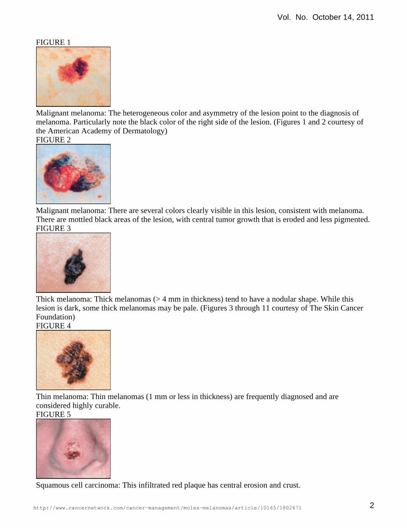
FIGURE 1
Malignant melanoma: The heterogeneous color and asymmetry of the lesion point to the diagnosis ofmelanoma. Particularly note the black color of the right side of the lesion. (Figures 1 and 2 courtesy ofthe American Academy of Dermatology)FIGURE 2
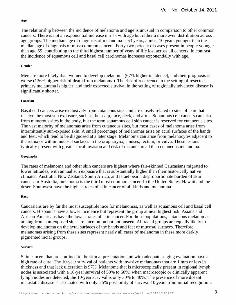
Malignant melanoma: There are several colors clearly visible in this lesion, consistent with melanoma.There are mottled black areas of the lesion, with central tumor growth that is eroded and less pigmented.FIGURE 3
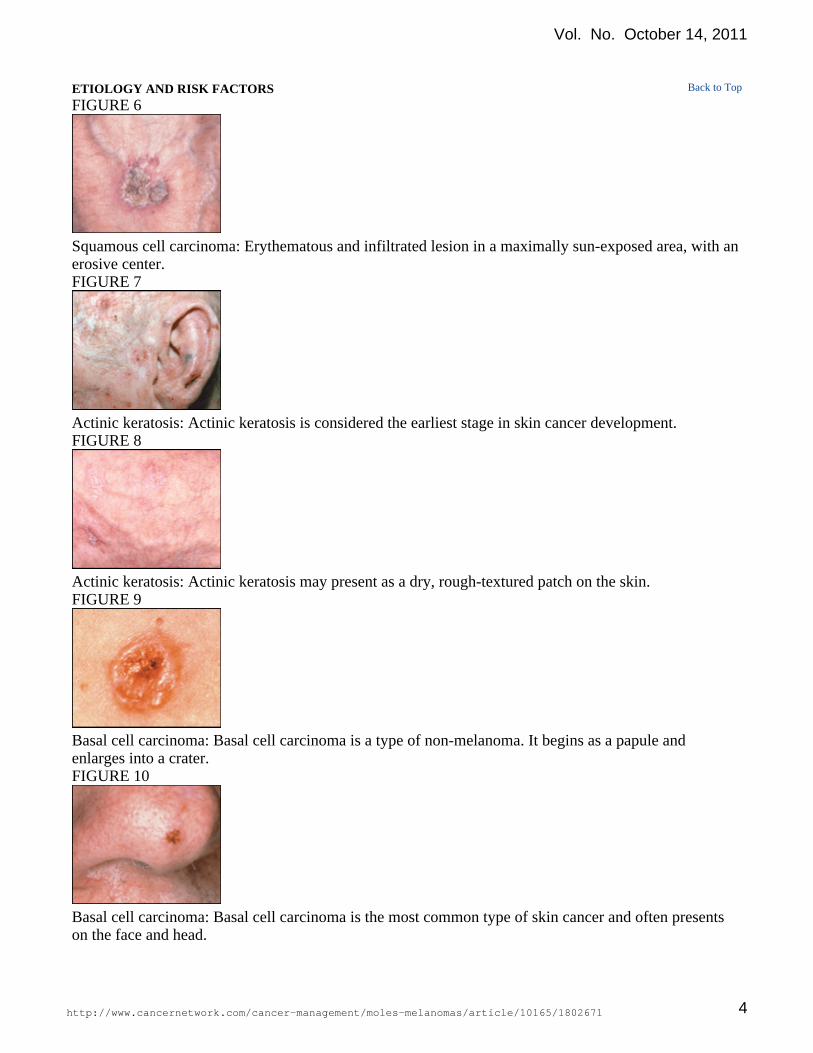
Thick melanoma: Thick melanomas (> 4 mm in thickness) tend to have a nodular shape. While thislesion is dark, some thick melanomas may be pale. (Figures 3 through 11 courtesy of The Skin CancerFoundation)FIGURE 4
Thin melanoma: Thin melanomas (1 mm or less in thickness) are frequently diagnosed and areconsidered highly curable.FIGURE 5
Squamous cell carcinoma: This infiltrated red plaque has central erosion and crust.
Vol. No. October 14, 2011
http://www.cancernetwork.com/cancer-management/moles-melanomas/article/10165/1802671 2
Age
The relationship between the incidence of melanoma and age is unusual in comparison to other commoncancers. There is not an exponential increase in risk with age but rather a more even distribution acrossage groups. The median age of diagnosis of melanoma is 53 years, almost 10 years younger than themedian age of diagnosis of most common cancers. Forty-two percent of cases present in people youngerthan age 55, contributing to the third highest number of years of life lost across all cancers. In contrast,the incidence of squamous cell and basal cell carcinomas increases exponentially with age.
Gender
Men are more likely than women to develop melanoma (67% higher incidence), and their prognosis isworse (136% higher risk of death from melanoma). The risk of recurrence in the setting of resectedprimary melanoma is higher, and their expected survival in the setting of regionally advanced disease issignificantly shorter.
Location
Basal cell cancers arise exclusively from cutaneous sites and are closely related to sites of skin thatreceive the most sun exposure, such as the scalp, face, neck, and arms. Squamous cell cancers can arisefrom numerous sites in the body, but the term squamous cell skin cancer is reserved for cutaneous sites.The vast majority of melanomas arise from cutaneous sites, but most cases of melanoma arise fromintermittently sun-exposed skin. A small percentage of melanomas arise on acral surfaces of the handsand feet, which tend to be diagnosed at a later stage. Melanoma can arise from melanocytes adjacent tothe retina or within mucosal surfaces in the oropharynx, sinuses, rectum, or vulva. These lesionstypically present with greater local invasion and risk of distant spread than cutaneous melanoma.
Geography
The rates of melanoma and other skin cancers are highest where fair-skinned Caucasians migrated tolower latitudes, with annual sun exposure that is substantially higher than their historically nativeclimates. Australia, New Zealand, South Africa, and Israel bear a disproportionate burden of skincancer. In Australia, melanoma is the third most common cancer. In the United States, Hawaii and thedesert Southwest have the highest rates of skin cancer of all kinds and melanoma.
Race
Caucasians are by far the most susceptible race for melanomas, as well as squamous cell and basal cellcancers. Hispanics have a lower incidence but represent the group at next highest risk. Asians andAfrican-Americans have the lowest rates of skin cancer. For those populations, cutaneous melanomasarising from sun-exposed sites are uncommon but not unseen. All racial groups are equally likely todevelop melanoma on the acral surfaces of the hands and feet or mucosal surfaces. Therefore,melanomas arising from these sites represent nearly all cases of melanoma in these more darklypigmented racial groups.
Survival
Skin cancers that are confined to the skin at presentation and with adequate staging evaluation have ahigh rate of cure. The 10-year survival of patients with invasive melanomas that are 1 mm or less inthickness and that lack ulceration is 97%. Melanoma that is microscopically present in regional lymphnodes is associated with a 10-year survival of 50% to 60%; when macroscopic or clinically apparentlymph nodes are detected, the 10-year survival is only 30% to 40%. The presence of more distantmetastatic disease is associated with only a 5% possibility of survival 10 years from initial recognition.
Vol. No. October 14, 2011
http://www.cancernetwork.com/cancer-management/moles-melanomas/article/10165/1802671 3
ETIOLOGY AND RISK FACTORSFIGURE 6
Squamous cell carcinoma: Erythematous and infiltrated lesion in a maximally sun-exposed area, with anerosive center.FIGURE 7
Actinic keratosis: Actinic keratosis is considered the earliest stage in skin cancer development.FIGURE 8
Actinic keratosis: Actinic keratosis may present as a dry, rough-textured patch on the skin.FIGURE 9
Basal cell carcinoma: Basal cell carcinoma is a type of non-melanoma. It begins as a papule andenlarges into a crater.FIGURE 10
Basal cell carcinoma: Basal cell carcinoma is the most common type of skin cancer and often presentson the face and head.
Back to Top
Vol. No. October 14, 2011
http://www.cancernetwork.com/cancer-management/moles-melanomas/article/10165/1802671 4

FIGURE 11
Basal cell carcinoma: Basal cell carcinoma may crust and bleed. Metastasis is rare.Genetic predisposition
Although there are families in which melanoma can occur with high likelihood, an underlying geneticpredisposition can only be found in 3% of all cases. The pedigrees have been identified because of theirhigh likelihood of a mutation carrier developing melanoma. Lower-penetrance genotypes remain to beelucidated. Nonetheless, the identification of the genes responsible for familial melanoma hascontributed greatly to the understanding of the molecular pathophysiology of melanoma.
The clinical observations that patients with multiple dysplastic nevi were at greater risk of developingmelanoma and that many such patients came from families with multiple affected individuals providedthe first insight into a melanoma progression model that might be accelerated based on inborn geneticabnormalities. Two highly related genes were discovered to harbor germline mutations in roughly 50%of melanoma pedigrees: and . enocodes two products via alternate splicing ofCDKN2A CDK4 CDKN2Amessenger RNA: p16INK4A and p14ARF. Each of these tumor suppressor genes exerts an inhibitoryeffect on cell cycle progression.
Xeroderma pigmentosum is a rare inherited disorder in which DNA repair mechanisms arecompromised, particularly in response to ultraviolet (UV) light. Mutations in genes through XP A Ghave been identified as the underlying molecular event. Squamous cell and basal cell carcinomas andmelanoma are prevalent in this population and at a young age. The near-complete penetrance ofmelanoma in these patients emphasizes the critical balance between UV-induced DNA damage andrepair in risk for skin cancer. As DNA damage repair is mediated by a complex network of sensor andeffector proteins, variability in the function of this system almost certainly underlies the variability inrisk among the fair-skinned population.
Genetic variability in the melanocortin-1 receptor has been clearly implicated in pigmentation(MC1R)of skin and hair and, more recently, in melanoma predisposition. It has been known for decades thatmelanoma is more prevalent among fair-skinned individuals with red or blond hair. Furthermore,blond-haired individuals with an inability to tan are at substantially greater risk of developing melanomathan blond-haired individuals who tan readily. Polymorphisms, distinct from mutations, in appearMC1Rto account for skin and hair color differences among Caucasians. It appears that individuals withmelanocortin receptors that have a muted response to increased melanocortin expression following sunexposure suffer the greatest UV-induced genetic damage, leading to a greater risk of melanoma.
Exposure
Even the inheritance of and mutations is insufficient to lead to melanoma in all carriers.CDKN2A CDK4It is clear that multiple genetic changes are required to give rise to invasive disease. UV damage is thebest-described modifiable risk factor for melanoma, as well as squamous cell and basal cell skin cancers.It is believed that the acquired or somatic genetic changes that give rise to melanoma occur as aconsequence of UV-induced genetic damage.
Vol. No. October 14, 2011
http://www.cancernetwork.com/cancer-management/moles-melanomas/article/10165/1802671 5

Epidemiologic data relate the risk of melanoma most closely to a connection between cumulative sunexposure, severe sun burns, or sun exposure during childhood, depending on the study. Thedisagreement between studies likely stems from methodologic differences in obtaining a sun exposurehistory, a heterogeneous effect of sun exposure and risk depending on the underlying geneticcomposition of the study population, or both. It has been clarified that melanoma arising onintermittently sun-exposed skin (such as the trunk) has its peak incidence among younger individualsand declines severely with increasing age. On the other hand, melanoma arising from chronicallysun-damaged skin (such as the face, neck, and upper extremities) has the highest incidence in olderindividuals. With the rise in popularity of indoor tanning salons, data indicate that those who use themare at higher risk of melanoma than those who are sun-exposed. There is little dispute regarding thecausal link between sun exposure or tanning salon use and risk of melanoma; however, there isdisagreement regarding the constituents of light (UV-A or UV-B) that contribute most to geneticdamage. Laboratory studies support a connection for both and suggest that prevention strategies musttake the entire UV light spectrum into account.
Prevention
With the incidence of melanoma still rising, it is clear that primary prevention efforts have not yet takenhold. The only approach firmly rooted in evidence is to minimize sun exposure. The use ofsun-protective clothing appears to be the next best strategy. There are conflicting data regarding theprotective effect of sunscreens for melanoma, although there is no controversy regarding their ability toprevent squamous cell and basal cell carcinomas. Protection against UV-A has been a long-standingfeature of widely available suncreens, whereas UV-B protection has more recently been engineered intoall mainstream products. It is possible that the more widespread of these wide-spectrum sunscreens willprovide more meaningful protective effects over the coming decades.
Immunosuppression
There is incontrovertible evidence linking immunosuppression and squamous cell skin cancer. Thisincreased risk applies to patients with acquired immunodeficiency syndrome as well as transplantrecipients on chronic immunosuppressive medications. The risk of developing primary melanoma in thesetting of immunosuppression is less well established, but there is some evidence that patients who havea history of melanoma are more likely to develop disease recurrence in the setting ofimmunosuppression.
Nevi
Patients with numerous benign nevi (small, regularly shaped, and uniformly pigmented moles) are atincreased risk of melanoma, as are patients with relatively few dysplastic nevi (large, irregularly shaped,and heterogeneously pigmented moles). Patients in either group may have a fivefold increased risk ofdeveloping melanoma compared with those with few benign nevi or without dysplastic nevi. However,it is critical to recognize that these preexisting moles, rather than precursor lesions, represent a riskfactor for melanoma in most cases. The vast majority of dysplastic nevi do not give rise to melanoma.There seems to be little value in resecting every nevus that appears dysplastic on clinical grounds.Surveying the skin regularly for new or changing moles has been most widely adopted strategy foreducating patients and healthcare providers. In patients older than 25 to 30 years, new mole formationwarrants examination by a provider who is comfortable making the diagnosis of melanoma.
Congenital nevi
Vol. No. October 14, 2011
http://www.cancernetwork.com/cancer-management/moles-melanomas/article/10165/1802671 6
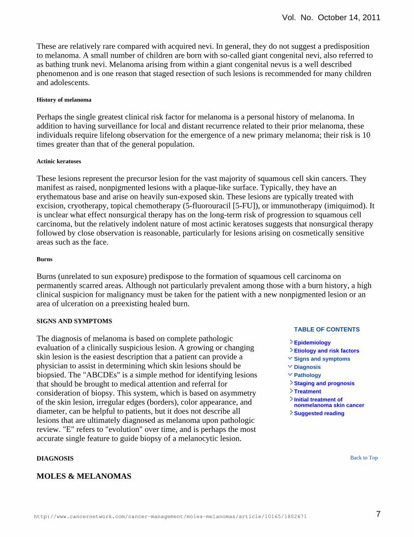
TABLE OF CONTENTS
EpidemiologyEtiology and risk factorsSigns and symptomsDiagnosisPathologyStaging and prognosisTreatmentInitial treatment ofnonmelanoma skin cancerSuggested reading
These are relatively rare compared with acquired nevi. In general, they do not suggest a predispositionto melanoma. A small number of children are born with so-called giant congenital nevi, also referred toas bathing trunk nevi. Melanoma arising from within a giant congenital nevus is a well describedphenomenon and is one reason that staged resection of such lesions is recommended for many childrenand adolescents.
History of melanoma
Perhaps the single greatest clinical risk factor for melanoma is a personal history of melanoma. Inaddition to having surveillance for local and distant recurrence related to their prior melanoma, theseindividuals require lifelong observation for the emergence of a new primary melanoma; their risk is 10times greater than that of the general population.
Actinic keratoses
These lesions represent the precursor lesion for the vast majority of squamous cell skin cancers. Theymanifest as raised, nonpigmented lesions with a plaque-like surface. Typically, they have anerythematous base and arise on heavily sun-exposed skin. These lesions are typically treated withexcision, cryotherapy, topical chemotherapy (5-fluorouracil [5-FU]), or immunotherapy (imiquimod). Itis unclear what effect nonsurgical therapy has on the long-term risk of progression to squamous cellcarcinoma, but the relatively indolent nature of most actinic keratoses suggests that nonsurgical therapyfollowed by close observation is reasonable, particularly for lesions arising on cosmetically sensitiveareas such as the face.
Burns
Burns (unrelated to sun exposure) predispose to the formation of squamous cell carcinoma onpermanently scarred areas. Although not particularly prevalent among those with a burn history, a highclinical suspicion for malignancy must be taken for the patient with a new nonpigmented lesion or anarea of ulceration on a preexisting healed burn.
SIGNS AND SYMPTOMS
The diagnosis of melanoma is based on complete pathologicevaluation of a clinically suspicious lesion. A growing or changingskin lesion is the easiest description that a patient can provide aphysician to assist in determining which skin lesions should bebiopsied. The "ABCDEs" is a simple method for identifying lesionsthat should be brought to medical attention and referral forconsideration of biopsy. This system, which is based on asymmetryof the skin lesion, irregular edges (borders), color appearance, anddiameter, can be helpful to patients, but it does not describe alllesions that are ultimately diagnosed as melanoma upon pathologicreview. "E" refers to "evolution" over time, and is perhaps the mostaccurate single feature to guide biopsy of a melanocytic lesion.
DIAGNOSIS
MOLES & MELANOMAS
Back to Top
Vol. No. October 14, 2011
http://www.cancernetwork.com/cancer-management/moles-melanomas/article/10165/1802671 7
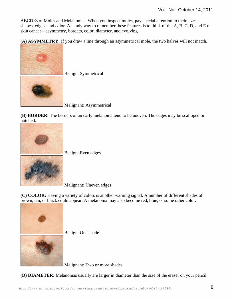
ABCDEs of Moles and Melanomas: When you inspect moles, pay special attention to their sizes,shapes, edges, and color. A handy way to remember these features is to think of the A, B, C, D, and E ofskin cancer—asymmetry, borders, color, diameter, and evolving.
If you draw a line through an asymmetrical mole, the two halves will not match.(A) ASYMMETRY:
Benign: Symmetrical
Malignant: Asymmetrical
The borders of an early melanoma tend to be uneven. The edges may be scalloped or(B) BORDER:notched.
Benign: Even edges
Malignant: Uneven edges
Having a variety of colors is another warning signal. A number of different shades of(C) COLOR:brown, tan, or black could appear. A melanoma may also become red, blue, or some other color.
Benign: One shade
Malignant: Two or more shades
Melanomas usually are larger in diameter than the size of the eraser on your pencil(D) DIAMETER:
Vol. No. October 14, 2011
http://www.cancernetwork.com/cancer-management/moles-melanomas/article/10165/1802671 8

(1/4 inch or 6 mm), but they may sometimes be smaller when first detected.
Benign: Smaller than 6 mm
Malignant: Larger than 6 mm
Any change in a mole—in size, shape, color, elevation, or another trait, or any new(E) EVOLVING:symptom such as bleeding, itching, or crusting—points to suspicion and possible danger.
Benign
Malignant
People at high risk of developing melanoma are those who have:
• A family history of melanoma, or who have had a melanoma in the past• Unusual moles on the skin, or changing moles• Fair skin, light hair and eye color, and who sunburn easily or tan with difficulty• A record of painful or blistering sunburns as children or in their teenage years• Indoor occupations and outdoor recreational habits.Early diagnosis of melanoma
Beyond prevention, early detection is the next priority in limiting the life-threatening potential ofmelanoma. Although tumor invasion into the dermis (the definition of invasive melanoma) cannot beassessed clinically, there are hallmarks of melanoma that can be appreciated by visual inspection. The"ABCD" algorithm, promoted by the ACS, has not proved to be a particularly effective method ofeducating the general population, but healthcare providers may be better suited to implementing thissystem in clinical practice. (See the color atlas following this chapter.) 'A' indicates gross asymmetry ofa pigmented lesion. 'B' refers to irregular or indistinct borders. 'C' regards color, which, in the case ofmelanoma, refers to black, dark brown, or blue color for newly formed lesions. In the case of preexistingmoles, the loss of pigmentation in a portion of the lesion can indicate regression, which is worrisome formelanoma. Additionally, a focus of darker pigmentation within a preexisting mole is grounds forconcern. 'D', or diameter of 6 mm or greater, raises concern for melanoma, whereas smaller lesions arerarely indicative of invasive lesions. None of these single criteria can be considered as grounds for
Vol. No. October 14, 2011
http://www.cancernetwork.com/cancer-management/moles-melanomas/article/10165/1802671 9

performing a biopsy, rather lesions that satisfy multiple criteria warrant either close observation orbiopsy of the most abnormal portion. Not included in the ABCD system is the papular or nodularcharacteristic of most melanomas. If melanoma arises within a preexisting mole, the raised element ofan otherwise flat or macular lesion should be considered suspicious for melanoma. Some primarymelanomas do not produce melanin and therefore lack the classic appearance.
Atypical nevi
These lesions represent a risk factor for melanoma in that individual lesions can occasionally progress toinvasive melanoma. More commonly, the presence of dysplastic nevi suggests an individual at risk formelanoma formation at other sites. The clinical definition of atypical nevi has never been formallyestablished but generally refers to the presence of one or two of the ABCD features. Cutaneousphotography has been routinely incorporated in the follow-up of patients with multiple clinicallydysplastic nevi in specialized pigmented lesion clinics. Full-body cutaneous photography is increasinglyavailable to patients and provides an objective baseline from which to judge change of preexistinglesions or the appearance of new lesions. In general, excision of all atypical nevi is impractical and doesnot adequately address the risk that patients face of developing melanoma in sites where no precursorlesion is found.
Nodular melanoma
This represents the most difficult subset of melanomas to diagnose at an early stage. These lesions aremore rapidly proliferative than typical melanomas and are generally not pigmented. Furthermore, theyare generally raised early in their development and have symmetric and well-demarcated boundaries.Thus, they do not meet the ABCD criteria. Nodular melanomas account for approximately 10% ofmelanoma cases but account for a disproportionate percentage of fatalities. The most useful clinical ruleto apply in the assessment of lesions that have these features is that the de novo appearance of suchlesions over a short time (months) warrants consideration of biopsy. As the features of nodularmelanoma are common to some benign skin lesions, as well as basal cell carcinoma, the yield ofbiopsies for such lesions may be relatively low. Nonetheless, heightened awareness of this small butlethal subset of melanoma is needed.
Nonmelanoma skin cancers
Classically, basal cell skin cancers are nodular and have a pearly, nonpigmented surface. Squamous cellcarcinomas typically have a scaly, nonpigmented surface and can occasionally be ulcerated. Both typesare generally slow to evolve. In most cases, squamous cell carcinomas are believed to arise from actinickeratoses. The differentiation between the two is based largely on size, with actinic keratoses being lessthan 1 cm in diameter. The appearance of nodularity or induration in preexisting keratinized lesionsraises concern about possible tumor invasion into the dermis and warrants biopsy.
Skin examination
Examination of the entire body should be performed for any patient who presents with numerousbenign-appearing nevi, any dysplastic nevi, or a history of even a single melanoma. The entire skin is atrisk in these individuals and must be investigated for the appearance of new or changing lesions.Patients who fit into any of these risk groups should be instructed to perform monthly skinself-examination. Two mirrors are required to adequately examine the back if a partner is not availableto assist in examining the back longitudinally.
Skin examination should be performed in a well-lit room with the patient completely disrobed. Forpatients with numerous moles, full-body cutaneous photography is extremely helpful to provide an
Vol. No. October 14, 2011
http://www.cancernetwork.com/cancer-management/moles-melanomas/article/10165/1802671 10

objective baseline from which to judge change. For patients with only a few atypical moles, close-upphotographs of those lesions may facilitate careful evaluation of those lesions while the remainder of theskin is surveyed for new lesions. A focused method for examining individual lesions, dermoscopyemploys low-level magnification of the epidermis with tangential light applied to a liquid-skin interface.Examination of pigmented lesions with dermoscopy allows more precise visualization of patterns ofpigmentation than is possible with the unaided eye. This method requires some degree of training andthe availability of appropriate equipment. When regression of part or all of a preexisting mole issuspected, a Wood's lamp can be helpful in bringing out the contrast in pigmentation between normallypigmented surrounding skin and an area where immunologic destruction of melanocytes has occurred.
Lymph node examination
This should be performed in all patients who are suspected of having invasive melanoma or largesquamous cell or basal cell carcinomas. The closest lymph node basin is the most essential area toexamine. The presence of a palpable lymph node mandates biopsy of the suspicious node, generally withfine needle aspiration (FNA). If the result of FNA is negative, a formal lymph node biopsy should bepursued for a clinically suspicious node. Whereas sentinel lymph node biopsy (SLNB) is offered tootherwise well patients with primary melanomas 1 mm or greater in depth or thin melanomas withulceration, a high mitotic count, or Clark's level IV, palpable lymphadenopathy with histologic evidenceof melanoma or nonmelanoma skin cancer warrants proceeding directly to a regional lymph nodedissection.
Biopsy techniques
is recommended for any lesion that is suspected of being melanoma, squamous cellExcisional biopsycancer, or basal cell carcinoma. This is especially true for lesions that can be entirely excised withoutconcern over causing an unacceptable cosmetic result. Local anesthesia is generally all that is required.Narrow margins of 1 to 2 mm are sufficient when making an initial diagnosis of skin cancer. The biopsyshould extend to the subcutaneous tissue to provide an adequate estimate of the depth of invasion,particularly for melanoma. Biopsy specimens should be placed in formalin and submitted for expertpathologic preparation including embedding in . Careful attentionparaffin(Drug information on paraffin)to documenting the site of the biopsy is essential to subsequent care.
is an acceptable alterative for large lesions, especially when located on the face, neck,Incisional biopsyor distal extremities. In the case of possible melanoma, the most abnormal-appearing area should bebiopsied. As with excisional biopsies, the biopsy should include subcutaneous tissue to allow anestimation of thickness. A punch biopsy, of sufficient diameter to encompass the mostabnormal-appearing area, is the preferred method. Should the biopsy identify a melanocytic lesion withdysplasia or atypia, the entire lesion should be removed, when possible, with radial margins of 1 to 2mm.
For lesions that are suspicious for melanoma, every attempt should be made toTechniques to avoidpreserve the ability to assess involvement of margins and to perform immunohistochemistry of theprimary tumor. The latter is critically important in borderline cases, for which the diagnosis ofmelanoma is uncertain. Thus, shave biopsies and serial thin section techniques (such as Mohs surgery)are strictly contraindicated. Frozen biopsy assessment is inadequate for the diagnosis of melanoma anddoes not allow adequate material for review in difficult pathologic cases.
PATHOLOGY
Nonmelanoma skin cancers
Back to Top
Vol. No. October 14, 2011
http://www.cancernetwork.com/cancer-management/moles-melanomas/article/10165/1802671 11

TABLE OF CONTENTS
EpidemiologyEtiology and risk factorsSigns and symptomsDiagnosisPathologyStaging and prognosis
Microstaging
Regional lymph nodeinvolvement
Melanoma of unknown primary
Clinical and pathologic staging
Other prognostic factors
TreatmentInitial treatment ofnonmelanoma skin cancerSuggested reading
The two most common skin cancers are basal and squamous cell carcinomas. Both of these skin cancersarise predominately on sun-exposed areas and may be considered in the differential diagnosis withmelanoma. The diagnosis of these skin cancers should be made after pathologic analysis of the skinspecimen.
STAGING AND PROGNOSIS OF MELANOMA
A great deal of information is available regarding factors thatcorrelate with clinical outcome in patients with melanoma. Inpatients with clinically localized disease, the most importantprognostic factors are Breslow's thickness, mitotic rate, ulceration,and SLN status. Overall, however, 85% of melanoma patientspresent with clinically normal lymph nodes. In clinicallynode-negative patients, most investigators have found themicroscopic degree of invasion of the melanoma, or microstaging,to be of critical importance in predicting outcome ( and ; Tables 1 2
).Figure 1
Microstaging
Primary melanoma
Three microscopic characteristics of primary melanoma are nowincorporated into the new AJCC staging system for melanoma, aseach has a significant contribution in predicting risk of regional lymph node involvement and long-termrisk of metastatic disease and death.
First described by Alexander Breslow, this method of describing tumor thicknessBreslow's thicknessmeasures from the top of the granular layer of the epidermis to the deepest contiguous tumor cell at thebase of the lesion using a micrometer in the microscope eyepiece. It is the primary determinant of Tstaging and has the highest prognostic value of any primary tumor characteristic. In the 2010 AJCC 7thEdition Cancer Staging Manual, Clark's level was removed from stage groupings, as it is not predictiveof outcome when the three cardinal features are considered.
TABLE 1 TNM staging for cutaneous melanoma
Vol. No. October 14, 2011
http://www.cancernetwork.com/cancer-management/moles-melanomas/article/10165/1802671 12
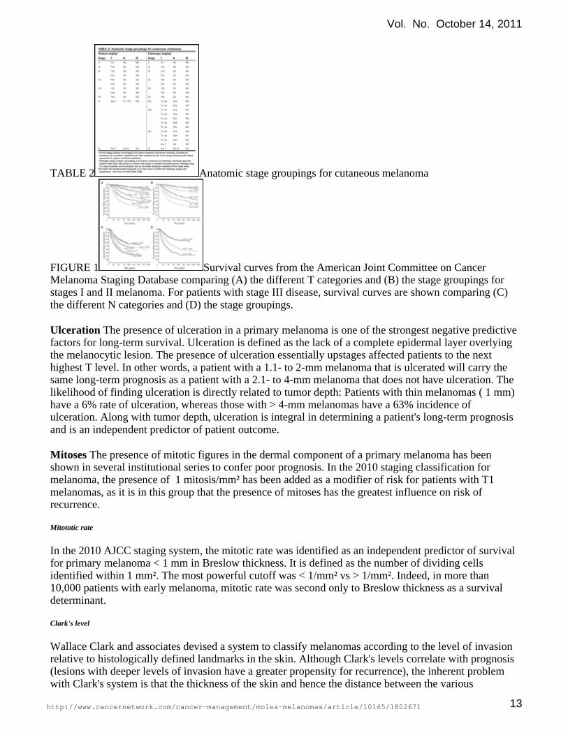
TABLE 2 Anatomic stage groupings for cutaneous melanoma
FIGURE 1 Survival curves from the American Joint Committee on CancerMelanoma Staging Database comparing (A) the different T categories and (B) the stage groupings forstages I and II melanoma. For patients with stage III disease, survival curves are shown comparing (C)the different N categories and (D) the stage groupings.
The presence of ulceration in a primary melanoma is one of the strongest negative predictiveUlcerationfactors for long-term survival. Ulceration is defined as the lack of a complete epidermal layer overlyingthe melanocytic lesion. The presence of ulceration essentially upstages affected patients to the nexthighest T level. In other words, a patient with a 1.1- to 2-mm melanoma that is ulcerated will carry thesame long-term prognosis as a patient with a 2.1- to 4-mm melanoma that does not have ulceration. Thelikelihood of finding ulceration is directly related to tumor depth: Patients with thin melanomas ( 1 mm)have a 6% rate of ulceration, whereas those with > 4-mm melanomas have a 63% incidence ofulceration. Along with tumor depth, ulceration is integral in determining a patient's long-term prognosisand is an independent predictor of patient outcome.
The presence of mitotic figures in the dermal component of a primary melanoma has beenMitosesshown in several institutional series to confer poor prognosis. In the 2010 staging classification formelanoma, the presence of 1 mitosis/mm² has been added as a modifier of risk for patients with T1melanomas, as it is in this group that the presence of mitoses has the greatest influence on risk ofrecurrence.
Mitototic rate
In the 2010 AJCC staging system, the mitotic rate was identified as an independent predictor of survivalfor primary melanoma < 1 mm in Breslow thickness. It is defined as the number of dividing cellsidentified within 1 mm². The most powerful cutoff was < 1/mm² vs > 1/mm². Indeed, in more than10,000 patients with early melanoma, mitotic rate was second only to Breslow thickness as a survivaldeterminant.
Clark's level
Wallace Clark and associates devised a system to classify melanomas according to the level of invasionrelative to histologically defined landmarks in the skin. Although Clark's levels correlate with prognosis(lesions with deeper levels of invasion have a greater propensity for recurrence), the inherent problemwith Clark's system is that the thickness of the skin and hence the distance between the various
Vol. No. October 14, 2011
http://www.cancernetwork.com/cancer-management/moles-melanomas/article/10165/1802671 13

landmark dermal layers varies greatly in different parts of the body. When initially described, the Clark'slevel was a standard way to stage patients with melanoma and predict outcome. Over the years, it hasproven much less reliable than Breslow thickness, and the 2002 AJCC staging system was used only forlesions < 1 mm. In the 2010 AJCC staging system, the mitotic index has replaced the Clark's level instaging lesions < 1 mm. The Clark's level is only used when mitotic index is unavailable for lesions < 1mm.
Regional lymph node involvement
In patients with intermediate-risk melanoma (1 to 4 mm in thickness), lymph node involvement is thestrongest prognostic indicator in the staging of melanoma. Patients with nodal involvement at the timeof their diagnosis have significantly decreased survival compared with node-negative patients. There is adirect relationship between the depth of invasion of the primary lesion and the potential for lymph nodeinvolvement. Among node-positive patients, the prognosis is more favorable in those with microscopicas opposed to macroscopic or clinically apparent disease. In addition, the increased number of nodesinvolved is associated with decreased survival in both microscopically detected and clinically apparentregional nodal metastasis. The worst prognosis occurs in patients with large, matted regional lymphnodes, whose outcome is similar to that of patients with stage IV disease.
In the 2010 version of the AJCC staging system, there was no change in nodal basin staging.Importantly, despite reports from some centers that a low burden of disease in the regional node predictsno additional nodal metastasis, the current staging system does not identify a minimal nodal tumorburden that would be considered essentially negative.
Melanoma of unknown primary
Melanoma of unknown primary (MUP) occurs in up to 5% of patients who present to tertiary cancerreferral centers. Lymphadenopathy is the most common clinical presentation, followed by identificationof visceral metastases and cutaneous nodules. Patients should be evaluated by a dermatologist andshould undergo a complete physical examination, including an anorectal and genital evaluation. A CTscan of the chest, abdomen, and pelvis and/or a whole-body PET scan are useful for staging patientswith MUP. In the absence of other sites of disease, surgical resection of the metastatic lesion (orcomplete regional lymphadenectomy) is appropriate. In the absence of symptoms, an ophthalmologicconsultation is probably not warranted unless liver metastases are the only site of disease, given thepropensity for ocular melanoma to metastasize to this site.
Single dermal nodules with no identifiable primary lesion are typically treated in a fashion similar tothat of primary melanomas, with wide local excision and regional nodal evaluation SLNB if appropriate.The prognosis for patients who present with MUP is similar to that for somewhat more favorablepatients with metastatic disease from a known primary (AJCC M1a).
Clinical and pathologic staging
Back to Top
Back to Top
Back to Top
Vol. No. October 14, 2011
http://www.cancernetwork.com/cancer-management/moles-melanomas/article/10165/1802671 14
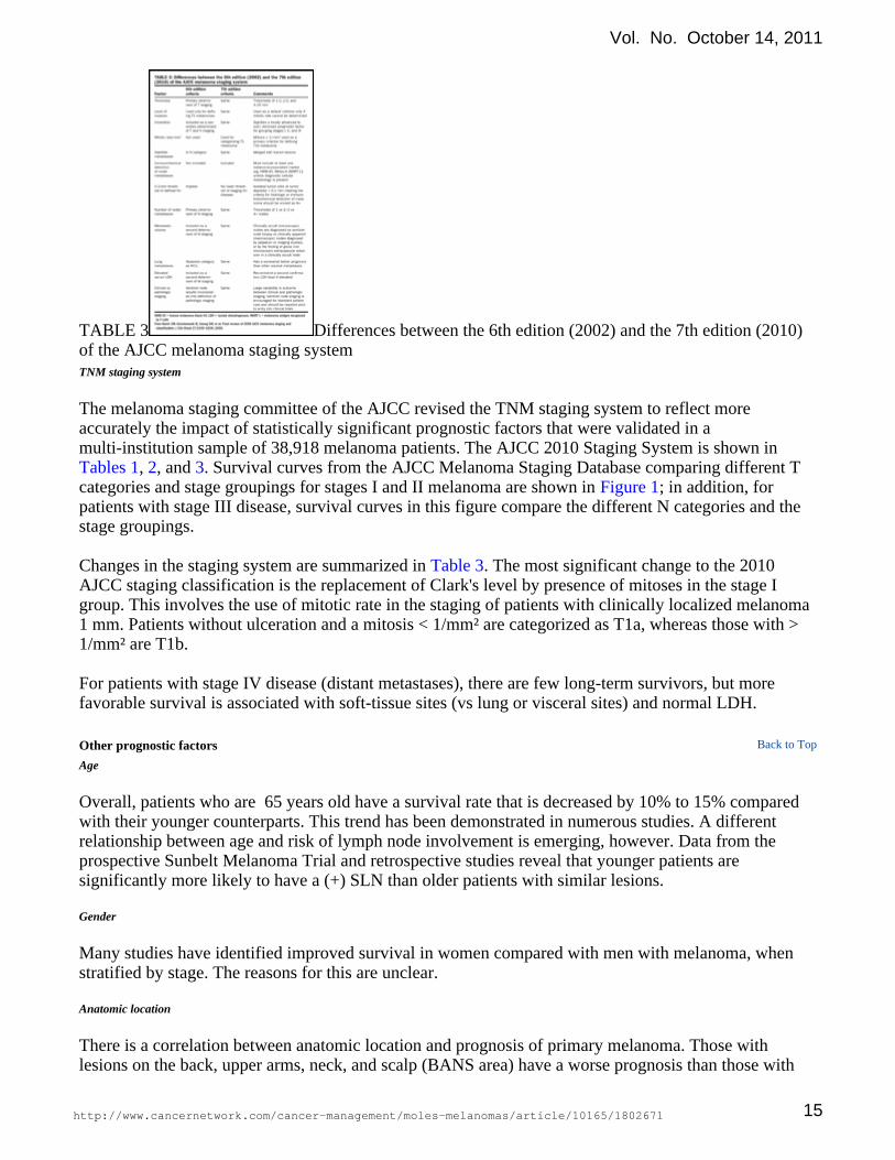
TABLE 3 Differences between the 6th edition (2002) and the 7th edition (2010)of the AJCC melanoma staging systemTNM staging system
The melanoma staging committee of the AJCC revised the TNM staging system to reflect moreaccurately the impact of statistically significant prognostic factors that were validated in amulti-institution sample of 38,918 melanoma patients. The AJCC 2010 Staging System is shown in
, , and . Survival curves from the AJCC Melanoma Staging Database comparing different TTables 1 2 3categories and stage groupings for stages I and II melanoma are shown in ; in addition, forFigure 1patients with stage III disease, survival curves in this figure compare the different N categories and thestage groupings.
Changes in the staging system are summarized in . The most significant change to the 2010Table 3AJCC staging classification is the replacement of Clark's level by presence of mitoses in the stage Igroup. This involves the use of mitotic rate in the staging of patients with clinically localized melanoma 1 mm. Patients without ulceration and a mitosis < 1/mm² are categorized as T1a, whereas those with >1/mm² are T1b.
For patients with stage IV disease (distant metastases), there are few long-term survivors, but morefavorable survival is associated with soft-tissue sites (vs lung or visceral sites) and normal LDH.
Other prognostic factors
Age
Overall, patients who are 65 years old have a survival rate that is decreased by 10% to 15% comparedwith their younger counterparts. This trend has been demonstrated in numerous studies. A differentrelationship between age and risk of lymph node involvement is emerging, however. Data from theprospective Sunbelt Melanoma Trial and retrospective studies reveal that younger patients aresignificantly more likely to have a (+) SLN than older patients with similar lesions.
Gender
Many studies have identified improved survival in women compared with men with melanoma, whenstratified by stage. The reasons for this are unclear.
Anatomic location
There is a correlation between anatomic location and prognosis of primary melanoma. Those withlesions on the back, upper arms, neck, and scalp (BANS area) have a worse prognosis than those with
Back to Top
Vol. No. October 14, 2011
http://www.cancernetwork.com/cancer-management/moles-melanomas/article/10165/1802671 15

lesions on the extremities. Men are more likely to develop truncal melanomas, whereas women are mostlikely to develop melanoma on their extremities.
Desmoplastic melanoma
Desmoplastic melanomas represent a less common but clinically distinct spindle cell variant ofmelanoma with dense fibrosis and frequent neurotropism. Clinically, they are raised, firm nodules thatare amelanotic in up to 40% of patients, commonly leading to a delay in diagnosis.They are usually deeplesions but have a more favorable prognosis than do conventional melanomas of similar Breslowthickness. The association of desmoplastic melanoma with local recurrence may be due to the fact thatmany of these lesions are misdiagnosed and undertreated, with regard to an appropriate radial margin ofexcision.
Additionally, although many desmoplastic melanomas are deeply invasive at the time of diagnosis, theyare less likely to involve regional lymph node basins. Patients with "pure" desmoplastic melanoma areextremely unlikely to harbor regional nodal metastasis, and SLN biopsy is unlikely to provide usefulinformation. Patients with "mixed" desmoplastic melanoma, containing a component of conventionalmelanoma, have a risk of regional nodal metastasis that is approximately half that of patients withconventional melanoma, and SLN mapping should be considered in these patients.
Despite the fact that these lesions are often relatively thick at presentation, patients with desmoplasticmelanomas have survival rates that are favorable compared with patients who have conventionalmelanomas of a similar depth.
Angiolymphatic invasion
Defined as invasion of tumor cells into the wall and/or lumen of vessels or lymphatics of the dermis ordeeper structures, angiolymphatic invasion is uncommon in melanoma. However, this finding is clearlyassociated with more aggressive tumors. Multiple large studies have shown worsened long-term survivaland more frequent lymph node involvement in patients with angioinvasive melanomas. Patients withvascular invasion in their primary melanoma have a threefold risk of lymph node involvement and areduction in 5-year survival by as much as 50% compared with matched patients without vascularinvasion.
Regression
The finding of regression represents a host immune response to invasive melanoma. Areas whereinvasive cells may have once existed are replaced by inflammatory reaction and fibrosis, which maymake it impossible to determine the precise depth of the initial lesion histologically. There is continuingdebate regarding the prognostic importance of regression; however, many clinicians believe that thinlesions that show signs of regression should be given higher consideration for surgical nodal staging,given the fact that the initial lesion may have originally been more deeply invasive. Although somestudies have shown that regressed lesions have a higher propensity for lymph node metastases thannonregressed primary tumors of the same thickness, this finding has not been universally observed.Indeed, a recent retrospective single-institution study demonstrated no increased risk of SLN metastasisin patients with regressed melanoma, suggesting that SLNB should not be performed for lesions thatwould otherwise not be considered for the procedure based on the presence of regression.
Tumor-infiltrating lymphocytes (TILs)
Vol. No. October 14, 2011
http://www.cancernetwork.com/cancer-management/moles-melanomas/article/10165/1802671 16

TABLE OF CONTENTS
EpidemiologyEtiology and risk factorsSigns and symptomsDiagnosisPathologyStaging and prognosisTreatment
Surgical treatment ofcutaneous melanoma
TNM staging system
Surgical treatment ofnoncutaneous melanoma
Adjuvant therapy for melanoma
Treatment of advancedmelanoma
Future directions
Initial treatment ofnonmelanoma skin cancerSuggested reading
Variable numbers of TILs are observed in melanoma. Tumors with a high number of TILs should carryan improved prognosis, presumably because of the active host response to tumor. In fact, many studieshave shown this tendency. The absence of TILs in the primary melanoma has been associated with ahigher risk of positive SLNs in a recent, large, single-institution study.
TREATMENT
Surgical treatment of cutaneous melanoma
Excision of primary lesion
It was recognized over a century ago that tumor cells could extendwithin the skin for several centimeters beyond the visible borders ofa melanoma, so that the risk of local recurrence relates to the widthof normal skin excised around the primary tumor. Only much morerecently was it realized that the thickness of the primary tumorinfluenced the likelihood of contiguous spread and that not allmelanomas require the same excision margin. This realizationprompted a number of randomized trials to determine the optimalexcision margins for melanomas of different Breslow thicknesses.
Initially, a "one-size-fits-all" approach of taking a 5-cm marginaround all invasive melanomas was adopted. With such widemargins, skin grafts were usually required for reconstruction.Modern melanoma surgical care is based on level I medicalevidence, and many large, prospective randomized trials have beenconducted to determine the appropriate radial margin for cutaneousmelanoma based on Breslow thickness.
A randomized trial conducted by the WHO found that when a 1-cm margin of normal skin was takenaround a melanoma < 1 mm thick, the local recurrence rate was exceedingly low (< 1%), and patientsurvival was just as good as if 3-cm margins were taken. For melanomas 1 to 2 mm in thickness, patientsurvival was the same for both margins of excision, but the local recurrence rate was higher with the1-cm margin (3.3% after 10-year follow-up).
The Intergroup Melanoma Trial compared 2- vs 4-cm margins for all cutaneous melanomas of the trunkor proximal extremity between 1 and 4 mm in thickness. Most patients in this trial had melanomas lessthan or equal to 2 mm in thickness. In the Intergroup trial, both local recurrence and survival were thesame regardless of whether 2- or 4-cm margins were obtained. Skin grafts were less frequent andhospital stays shorter with the narrower margin.
A recent trial conducted in the United Kingdom addressed patients with primary melanoma greater thanor equal to 2 mm in depth. Patients who underwent a 1-cm radial margin of excision had a higher risk oflocal/regional recurrence than those who underwent excision with 3-cm margins.
Current recommendations
Based on these important studies, it is possible to make rational recommendations for excision marginsfor melanoma patients.
• Patients with melanoma less than or equal to 1 mm should undergo excision of skin andsubcutaneous tissue for a radial margin of 1 cm.
Vol. No. October 14, 2011
http://www.cancernetwork.com/cancer-management/moles-melanomas/article/10165/1802671 17

• Patients with melanoma between 1 and 2 mm should undergo excision of skin and subcutaneoustissue for a radial margin of 1 to 2 cm.
• Patients with melanoma greater than 2 mm in depth should undergo a wide excision of skin andsubcutaneous tissue of 2 to 3 cm.
• When the anatomic location of the primary tumor precludes excision of the margin (eg, on theface), at least 1 cm should be taken when feasible.
TNM staging system
Clinically apparent lymphadenopathy
Melanoma patients with clinically enlarged nodes and no evidence of distant disease (AJCC stage IIIB)should undergo complete regional lymphadenectomy. The first step in evaluating palpable nodes isgenerally FNA. A negative or inadequate sample FNA may be repeated, with image guidance ifnecessary, or may lead directly to an open node biopsy, followed by complete lymphadenectomy in theevent of a positive frozen section or touch-prep cytologic determination of metastasis.
Clinically normal nodes
The surgical management of clinically normal nodes is determined by the characteristics of the primarylesion. A direct relationship between thickness of the primary lesion and nodal involvement has longbeen recognized. When the depth of the primary is unknown because of the biopsy technique or otherfactors, consideration should be given to SLN mapping.
SLNB is the primary method for regional nodal staging. All potentially involved basins should initiallybe examined as part of a thorough history and physical examination.
Patients with thin melanomas (< 1 mm Breslow depth) have a low risk of occult nodalThin melanomasinvolvement (< 5%). Some patients with melanomas 0.76 to 1 mm have a high enough risk of lymphnode involvement to justify consideration of SLNB in addition to wide excision.
When ulceration is present, consideration should be given to SLNB. In addition, a high mitotic count ( 1) should prompt consideration of regional nodal basin staging.
Risk of nodal metastasis rises significantly with increasing depthIntermediate-thickness melanomasof invasion. Patients with a 1-mm thick melanoma have approximately a 10% chance of nodalinvolvement, whereas those with a 4-mm melanoma have a 35% to 40% risk of nodal metastases. Forthese reasons, wide excision of the primary tumor is generally accompanied by SLNB for staging of thenodal basin. Patients who have evidence of metastasis in the SLN are offered completionlymphadenectomy as a standard approach. An alternative approach of wide excision alone withobservation and serial physical examination of nodal basins has been advocated by some clinicians andhas not been shown to reduce long-term survival if complete lymph node dissection is performed at theidentification of clinically positive nodes. This approach is justified in a recently completed prospectiverandomized clinical trial of wide excision and SLN mapping compared with wide excision and nodalbasin observation. There was no evidence that SLN mapping contributed to an overall survival benefit,but SLN status was a powerful predictor of outcome. The use of SLNB vs observation continues to bean area of intense debate. An prospective clinical trial evauating the need for completionlymphadenectomy in SLN (+) patients is ongoing (Multicenter Selective Lymphadenectomy Trial II).
Regional lymphadenectomy
Back to Top
Vol. No. October 14, 2011
http://www.cancernetwork.com/cancer-management/moles-melanomas/article/10165/1802671 18

The identification and management of nodal disease have evolved significantly over the past 20 years.Historically, complete lymph node dissections were performed on all patients with intermediatethickness melanomas, in the belief that doing so would lead to a survival benefit. This was based on theobservation that there was an approximately 20% survival advantage to patients found to havemicroscopic involvement of lymph nodes at "elective" lymph node dissection when compared withpatients who had clinically apparent nodal metastasis and underwent a "therapeutic" lymph nodedissection. With elective node dissection, however, a significant percentage (80% to 85%) of patientsunderwent complete lymphadenectomy only to find that their nodal basin was free of disease. Moreimportantly, a series of prospective randomized clinical trials failed to demonstrate a survival advantagefor patients undergoing elective lymphadenectomy over wide excision only. For this reason, the standardapproach prior to the use of SLN mapping was wide excision alone and clinical observation of the nodalbasin.
SLN mapping is performed by injecting radiolabeled colloid into the dermis surrounding the primarymelanoma. The radiotracer migrates via the lymphatics to the regional nodal basin(s), which isvisualized with a gamma camera. The patient is then taken to the operating room, where a hand-heldgamma probe is used to identify the sentinel node(s). This technique, in combination with the use ofvital blue dye, has led to a success rate of SLN identification exceeding 97% at experienced centers.
The intuitive hope that SLN mapping and biopsy followed by completion lymphadenectomy wouldresult in improved survival was not borne out in a recently reported prospective randomized clinical trial(Multicenter Selective Lymphadenectomy Trial I). Even in the absence of a proven survival benefit,however, the staging advantages of SLNB are sufficiently compelling to justify its routine use in healthypatients with melanomas at a significant risk of nodal involvement.
Deep melanomas harbor nodal metastases in up to 60% of patients; surgicalThick melanomasevaluation of the nodal basin may be performed, as most studies suggest that SLN status retainsprognostic status in patients with deep primary melanoma. Unless the information will direct furthermanagement, however, it is unlikely to be of benefit for these patients. In addition, a completionlymphadenectomy for those at high risk of local and regional recurrence may subject the patients toundue morbidity. The lymphedema associated with complete node dissection may complicate treatmentof subsequent recurrences in patients with deep melanoma of the extremity.
Surgical treatment of noncutaneous melanoma
Ocular melanomas
These lesions generally do not have access to lymphatic channels, so the surgical principles outlinedpreviously do not apply. These lesions have a unique propensity to metastasize hematogeneously, oftento the liver after a long relapse-free interval. Advances in understanding the biology of ocularmelanomas may lead to adjuvant approaches different from therapies now under investigation forcutaneous primaries.
A diagnosis of ocular melanoma with no evidence of distant disease signifies that a decision must bemade as to whether or not the eye can be spared. Some small melanomas situated peripherally in theretina can be excised with minimal loss of vision, but most cannot. For larger lesions, treatment optionsare enucleation (total removal of the eye) or implanted radiotherapy with a radioactive gold plaque fittedto the back of the eyeball immediately behind the tumor. A multi-institution, randomized trialcomparing implanted radiotherapy with enucleation for local disease control and overall survival wascompleted by the COMSG; it appears that both techniques provide similar outcomes for all sizes oftumors.
Melanomas of the anus and vulva
Back to Top
Vol. No. October 14, 2011
http://www.cancernetwork.com/cancer-management/moles-melanomas/article/10165/1802671 19

These "mucosal" melanomas are notoriously difficult to cure and are almost always associated with apoor outcome. The surgical management of anal melanoma is controversial, but most surgeons prefer awide excision, when possible, over an abdominoperineal resection (APR). In the past, an APR was morecommonly used for patients with less-advanced disease who were viewed as potentially curable. Thisalmost certainly explains the reported association of APR with long-term survivors. More recently, APRis reserved for patients with bulky disease or recurrent disease that is not amenable to wide excision,which is favored for patients with more localized/potentially curable disease. Not surprisingly, morerecent studies demonstrate no survival advantage to APR, which appears to be equivalent to wideexcision.
Radical resection is also less commonly performed for patients with vulvar melanoma, who also have ahigh risk of relapse and death. Function-preserving resection and nodal basin staging in the setting ofrelatively early disease are appropriate, but, as in patients with anal primaries, preemptive nodal stagingin patients with advanced primary tumors is unlikely to impart a benefit to those whose nodal basin canbe followed clinically and radiologically.
Nasal sinuses or nasopharyngeal melanomas
Melanomas arising in the nasal or nasopharyngeal mucosa should be widely excised to include adjacentbony structures, if needed. As in patients with anal or vulvar melanoma, node dissection is reserved forpatients who have proven nodal involvement. Radiotherapy should be considered for patients whoseprimary tumor cannot be fully removed from this site with adequate margins or as adjuvant therapy inpatients unlikely to be controlled with surgical resection alone.
Adjuvant therapy for melanoma
Interferon -2b (IFN-; Intron A) was approved for use in patients with deep primary melanoma orresected stage III or IV melanoma in 1995. In the ECOG trial that led to FDA approval, interferon -2bwas administered intravenously at 20 mU/m² for 5 consecutive days every 7 days for 4 weeks, duringthe "induction" phase. For a subsequent 48 weeks, 10 mU/m² was administered by subcutaneousinjection on alternate days for a total of three doses every 7 days in the "maintenance" phase.
This "high-dose" regimen was compared with observation. A statistically significant improvement inoverall survival (OS) was demonstrated with interferon, compared with the observation arm;relapse-free survival (RFS) was also improved. Three-quarters of the interferon patients experiencedsevere toxicities, most commonly fatigue, asthenia, fever, depression, and elevated liver transaminaselevels. A quality-of-life analysis found that the toxicity associated with this regimen was largelycompensated for by the prevention of disease relapse.
Due to uncertainty regarding the optimal dose and schedule of interferon, a second trial was initiatedcomparing the high-dose regimen and a low-dose regimen (3 mU by subcutaneous injection 3 timesweekly for 24 months) as well as observation. No significant improvement in OS for either thehigh-dose or low-dose arm was observed compared with observation; RFS was improved by 22% in thehigh-dose arm compared with observation. A pooled analysis of E1684 and E1690, with longerfollow-up for both trials, revealed a continued, statistically significant impact on RFS but not on OS.
High-dose interferon has been consistently shown to improve RFS compared with either observation organglioside Gm²/keyhole limpet hemocyanin (Gm²-KLH) vaccination but does not clearly confer an OSadvantage. A recent prospective randomized trial conducted by the HCOG compared outcome inpatients treated with high-dose interferon as an adjuvant for 1 month vs 1 year and found no differencein OS or disease-free survival; however, this trial was of insufficient size to firmly conclude that 1
Back to Top
Vol. No. October 14, 2011
http://www.cancernetwork.com/cancer-management/moles-melanomas/article/10165/1802671 20

month of interferon therapy is truly equivalent to the full-year regimen with regard to RFS. Due to thesignificant toxicity associated with 1 year of high-dose interferon and the lack of an OS advantage,consensus is lacking regarding the use of interferon in the adjuvant setting.
Intermediate-dose regimens with interferon have been evaluated in several trials; they have lessconsistently demonstrated an RFS advantage compared with observation and have never resulted in asurvival advantage. A recent trial conducted by the EORTC demonstrated no survival advantage forpatients undergoing 13 months or 25 months of intermediate-dose adjuvant interferon after resection ofstage IIB (deep primary melanoma) or stage III nodal disease when compared with observation.
The EORTC reported the results of a prospective randomized trial of observation vs pegylatedinterferon- for 5 years. Again, there was an advantage to treatment in terms of RFS but no OS advantagefor patients with high-risk resected melanoma. Nearly one-third of the patients in the treated armdiscontinued therapy due to toxicity. This regimen has received FDA approval. Induction therapyconsists of 6 g/kg weekly for 2 months, followed by 3 g/kg weekly provided that patients can maintaingood performance status. If performance is compromised, then the dose is reduced to 2 g/kg, and againto 1 g/kg as needed.
Adjuvant chemotherapy
Single-agent chemotherapy or combination chemotherapy regimens have not been systematicallyevaluated for the adjuvant treatment of melanoma because of the low response rates seen in patientswith advanced disease. The largest randomized trial compared an IV administered regimen of
(BiCNU; 80 mg/m²) every 4 weeks, carmustine(Drug information on carmustine) dactinomycin(Drug (Cosmegen, 10 g/kg), and vincristine (1.0 mg/m²) every 2 weeks for 6information on dactinomycin)
months with observation among patients with resected stage III or IV melanoma. A significantimprovement in RFS, but not OS, was observed in this small study. A randomized trial comparing
vs observation failed to demonstrate an improvement indacarbazine(Drug information on dacarbazine)either RFS or OS.
There is no widely accepted adjuvant therapy following resection of metastatic melanoma. Therefore,observation remains a standard of care for patients in this setting, and some investigational therapies arebeing evaluated in comparison to placebo in light of the significant percentage of patients who choosenot to pursue interferon therapy.
Neoadjuvant therapy for resectable stage IIIC or IV melanoma remains an investigational approach.Neoadjuvant biochemotherapy (cisplatin, dacarbazine, ,vinblastine(Drug information on vinblastine)interleukin-2 [IL-2], and interferon) was evaluated in a small study of patients with stage III melanoma.In the 50 patients with measurable disease, the response rate was 26%. High-dose interferon, however,produced a 55% clinical response and 15% pathologic response when used in a neoadjuvant setting in asmall group of patients with clinically apparent nodal metastasis. Currently available chemotherapyregimens for melanoma are not sufficiently active to support use in neoadjuvant therapy.
Ipilimumab (Yervoy) is a monoclonal antibody that blocks CTLA-4 mediated down regulation of T cellresponse. Yervoy has recently been approved for use in patients with stage IV melanoma following aprospective randomized trial demonstrating an improvement in OS in ipilimumab-treated stage IVpatients. An EORTC trial is accruing patients following lymphadenectomy for stage IIIA and Bmelanoma to determine whether adjuvant ipilimumab improves survival.
Radiation therapy
Vol. No. October 14, 2011
http://www.cancernetwork.com/cancer-management/moles-melanomas/article/10165/1802671 21

In vitro data from the 1970s demonstrated radiation resistance among melanoma cell lines and led to thereluctance to use radiation therapy in the treatment of melanoma. More modern data have shown thatmany melanomas are sensitive to radiation, but the disinclination toward use of radiation therapy formelanoma continues.
Another unanswered question is the proper fraction size to use in melanoma. In a large series of patientsstudied by Overgaard and colleagues, the investigators found that the response rate of metastaticmelanoma lesions was dependent on the fraction size. The complete response rate was 57% whenfractions greater than 4 Gy were used, compared with 24% for fractions less than 4 Gy. RTOG 83-05 isthe only randomized trial that compared hypofractionated (8 Gy for 4 fractions) vs conventional (2.5 Gyfor 20 fractions) schedules of radiation therapy. In this study, they noted no difference in the completeand partial responses. Though a wide spectrum of fraction sizes have been used, this does not precludethe use of radiation but emphasizes the need for more research to better understand and create aneffective fractionation schedule.
Radiation therapy can provide some benefit and should be considered in the palliative treatment ofmelanoma metastatic to bone or other symptomatic sites, but its use in other settings remainscontroversial.
Adjuvant radiation treatment
Radiation treatment has been increasingly employed in the postoperative setting to improve locoregionaltumor control in a select group of patients at "high risk" for local or regional recurrence. "High risk" hasbeen variably defined but includes patients with large, matted nodes, extranodal extension of tumor,inadequate margins due to anatomic constraints, head and neck site, and multiple local/regionalrecurrences. The 5-year regional control has ranged from 87% to 94%, and the 5-year OS has rangedfrom 36% to 46% with the addition of radiation to surgery. Single institutions have reported that localrecurrence is reduced to 6% to 11% when postoperative radiotherapy is used for high-risk lesions.Serious complication rates after radiation therapy were low, which is likely secondary to the superficialnature of the treated volumes.
A prospective randomized trial (ANZMTG 01.02/TROG 02.01) was reported at the 2011 meeting ofASCO. It evaluated the impact of adjuvant radiotherapy (RT) on regional control (primary objective) inhigh-risk melanoma patients. High-risk patients were those with 1 parotid, 2 cervical or axillary, or 3groin nodes; extranodal spread of tumor; or maximum metastatic node diameter 3 cm in the neck oraxilla or 4 cm in the groin. The investigators reported an improvement in regional control with adjuvantRT. Adjuvant RT is an option for patients with isolated regional recurrence of melanoma who areconsidered to be at high risk of further regional recurrence after lymphadenectomy. The study wasdesigned to determine the effects of RT on regional recurrence, survival, morbidity, and quality of life(QOL). At a median follow-up of 27 months, adjuvant RT improved regional control in melanomapatients at high risk for relapse after lymphadenectomy (hazard ratio [HR] = 1.77; 95% confidenceinterval (CI) = 1.02-3.08; = .041) but did not affect survival (2.6 years in the RT group and 3.9 yearsPin the observation group; = .14).P
At the Royal Prince Alfred Hospital, 143 patients with melanoma metastases to the parotid and/orcervical lymph nodes were treated with surgery alone or surgery and postoperative radiation therapy.The patients who received radiation therapy had more aggressive features such as at least two positivelymph nodes and extracapsular extension; there was a trend toward higher regional control in the groupthat received surgery and postoperative RT (94%) than with surgery alone (91%, = .065).P
Vol. No. October 14, 2011
http://www.cancernetwork.com/cancer-management/moles-melanomas/article/10165/1802671 22

In a retrospective study of 615 patients at Roswell Park and MD Anderson who had advanced regionalnodal metastasis, adjuvant radiotherapy was associated with improved regional control when comparedwith surgery alone. The risk of long-term lymphedema is known to be high in patients with inguinallymph node metastases; thus, it would be prudent to consider a higher threshold for treatment of thesepatients. In a review on the use of RT in malignant melanoma, Stevens and McKay offered thefollowing recommendations for postoperative RT after regional lymph node dissection:
• Multiple involved nodes (more than one for parotid, three to four in other regions)
• Any involved node greater than 3 to 4 cm in maximum diameter
• Extranodal spread
• Incomplete dissection
• Recurrence after previous lymph node dissection (no previous radiation therapy)
In November 2009, results of the ANZMTG/TROG 02.01 trial were reported at the annual ASTROmeeting. Patients were eligible for the trial if they had involvement of at least one parotid node, at leasttwo cervical or axillary nodes, or at least three groin nodes; extranodal extension; or a minimummetastatic node diameter of 3 cm in the neck or axilla or of 4 cm in the groin. Patients were randomizedto observation after surgery or to adjuvant radiation (48 Gy in 20 fractions). At a median follow-up of 38months, the rate of lymph node recurrence was 68% in the group having surgery and external-beam RTand 80% in the group having surgery alone. The radiation treatment compliance rate was 79%. Therewas a higher rate of acute toxicity (dermatitis and pain) with RT, but no grade 4 toxicity was seen.
Vaccines and other immunotherapy
These remain an investigational approach and are being tested among patients with resected stages II,III, and IV melanoma. The goal of this immunotherapy is to reverse immune tolerance of microscopicresidual melanoma following surgery through the amplification of melanoma-specific cytolytic T cells.This approach is distinct from conventional immunization against pathogens to which the host is naive,through which long-lasting, antibody-mediated immunity is sought by induction of memory B cells.
Melanoma peptide vaccines are being intensely evaluated in clinical trials. Peptides have been selectedfrom two classes for clinical development: melanocyte-specific proteins and so-called cancer-testisantigens. The latter are proteins that have a restricted spatiotemporal expression and are not detectable inadult tissues outside the immunologic sanctuary site of the testes. The most widely studied peptides arederived from gp-100, tyrosinase, melan-A (all melanocyte-specific), and and melanomaNY-ESO-1antigen-encoding gene ( ) proteins (cancer-testis antigens). Preliminary evidence suggests that thisMAGEapproach may successfully engender specific and potent immunologic responses to the proteinfragments being administered. Protection against disease recurrence in patients at high risk followingsurgery (adjuvant therapy) has not yet been demonstrated. However, a recent prospective randomizedtrial reported a survival advantage for patients with stage IV melanoma treated with a peptide vaccine(gp100:209-217). Patients with metastatic melanoma were randomized to receive a multivalent peptidevaccine and high-dose IL-2 vs high-dose IL-2 alone. This approach may prove promising in the adjuvantsetting.
Treatment of advanced melanoma
Surgery
Back to Top
Vol. No. October 14, 2011
http://www.cancernetwork.com/cancer-management/moles-melanomas/article/10165/1802671 23
Surgical resection is a reasonable approach to treatment of the patient with isolated metastasticmelanoma, or with limited sites of disease. Surgical resection is associated with better outcome than notreatment or treatment with traditional chemotherapeutic regimens, although patient selection makesconclusions about these reports difficult. All studies to date are retrospective reviews. A number ofretrospective studies have shown the validity of resecting pulmonary metastases in patients withmelanoma, and more recent studies have shown similar survival rates for patients undergoing resectionof limited number and sites of disease (usually up to four) for metastases to distant lymph nodes, skinand subcutaneous tissue, and lungs. Patients with isolated metastases in the liver, adrenal gland, brain,and gastrointestinal tract may undergo resection, but the survival of these patients may be less favorable.
Surgical resection may also be an important component of palliation. Bleeding or obstruction fromsmall-intestine metastases may be managed by resection. Although palliative resection can provideshort-term effective relief of patient symptoms, numerous studies have shown that incomplete resectiondoes nothing to enhance the length of survival.
Single-agent chemotherapy
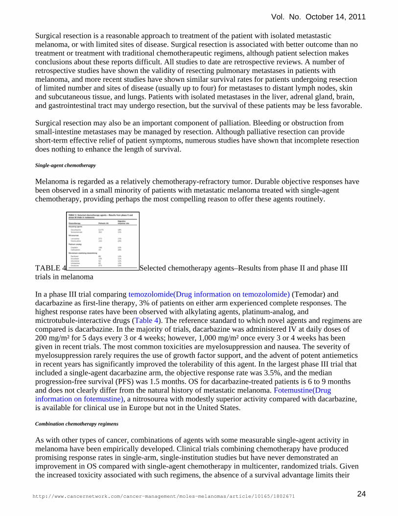
Melanoma is regarded as a relatively chemotherapy-refractory tumor. Durable objective responses havebeen observed in a small minority of patients with metastatic melanoma treated with single-agentchemotherapy, providing perhaps the most compelling reason to offer these agents routinely.
TABLE 4 Selected chemotherapy agents–Results from phase II and phase IIItrials in melanoma
In a phase III trial comparing (Temodar) andtemozolomide(Drug information on temozolomide)dacarbazine as first-line therapy, 3% of patients on either arm experienced complete responses. Thehighest response rates have been observed with alkylating agents, platinum-analog, andmictrotubule-interactive drugs ( ). The reference standard to which novel agents and regimens areTable 4compared is dacarbazine. In the majority of trials, dacarbazine was administered IV at daily doses of200 mg/m² for 5 days every 3 or 4 weeks; however, 1,000 mg/m² once every 3 or 4 weeks has beengiven in recent trials. The most common toxicities are myelosuppression and nausea. The severity ofmyelosuppression rarely requires the use of growth factor support, and the advent of potent antiemeticsin recent years has significantly improved the tolerability of this agent. In the largest phase III trial thatincluded a single-agent dacarbazine arm, the objective response rate was 3.5%, and the medianprogression-free survival (PFS) was 1.5 months. OS for dacarbazine-treated patients is 6 to 9 monthsand does not clearly differ from the natural history of metastatic melanoma. Fotemustine(Drug
, a nitrosourea with modestly superior activity compared with dacarbazine,information on fotemustine)is available for clinical use in Europe but not in the United States.
Combination chemotherapy regimens
As with other types of cancer, combinations of agents with some measurable single-agent activity inmelanoma have been empirically developed. Clinical trials combining chemotherapy have producedpromising response rates in single-arm, single-institution studies but have never demonstrated animprovement in OS compared with single-agent chemotherapy in multicenter, randomized trials. Giventhe increased toxicity associated with such regimens, the absence of a survival advantage limits their
Vol. No. October 14, 2011
http://www.cancernetwork.com/cancer-management/moles-melanomas/article/10165/1802671 24
consideration as standard therapies. These treatments include ,cisplatin(Drug information on cisplatin)vinblastine, and dacarbazine (CVD) and the Dartmouth regimen (cisplatin, carmustine, dacarbazine, and
). Inpatient treatment is standard for these regimens, and thetamoxifen(Drug information on tamoxifen)severity of myelosuppression often requires growth factor support not needed for single-agentchemotherapies. Given that longevity is not impacted for these patients who have such a short lifeexpectancy, the quality-of-life detriment associated with combination therapy cannot be justified.
Tamoxifen
Based on anecdotes of tumor regression associated with single-agent tamoxifen and laboratory evidencesuggesting synergy with chemotherapy, clinical trials were undertaken combining tamoxifen withindividual chemotherapy agents or combination regimens. As with other combination chemotherapytrials, single-arm trials of regimens containing tamoxifen appeared promising. However, in randomizedtrials in which the same chemotherapy regimen was administered to all patients, and tamoxifen givenonly to patients in the experimental arm, no evidence of clinical benefit was observed with the additionof tamoxifen.
Biologic therapies
IL-2 was approved by the FDA as a treatment for metastatic melanoma on the basis of durable completeand partial remissions associated with the "high-dose" regimen. This regimen requires patients to be inexcellent overall health to tolerate the physiologic stress of 5 days of inpatient therapy. The standarddose is 200,000 U/m² repeated every 8 hours, for a maximum of 14 doses. This treatment is followed bya treatment break of 10 to 14 days and readmission for another course of therapy. Patients whodemonstrate some degree of tumor regression are offered additional courses of therapy. Typicaltoxicities include fever, malaise, hypotension complicated by renal dysfunction, elevated levels of livertransaminases, and mood alterations. Clinical expertise among physicians and nurses is required tosafely and effectively administer this therapy. As a consequence, the use of high-dose IL-2 has largelybeen restricted to high-volume referral centers.
Interferon- has demonstrated single-agent activity in metastatic melanoma that is comparable to resultswith chemotherapy, including occasional durable responses. A randomized trial comparing interferon-with dacarbazine in metastatic disease has never been conducted. Given the significant toxicityassociated with chronic administration of interferon, its use in the metastatic setting is limited.
Biochemotherapy regimens
The distinct mechanism of action of biologic agents and evidence of single-agent activity have ledinvestigators to combine these agents with chemotherapy for the treatment of metastatic melanoma.Given the toxicity associated with high-dose IL-2, it is not possible to safely administer chemotherapyconcurrently. However, lower-dose IL-2 regimens can be safely coadministered with combinationchemotherapy. Likewise, the high-dose interferon regimen that is the current standard therapy foradjuvant treatment of stages II and III melanoma cannot be easily combined with chemotherapy,whereas this is possible with modified schedules of interferon.
The first biochemotherapy regimens tested in phase II trials and then subsequently in large, randomizedphase III trials combined interferon- with dacarbazine. Despite early evidence suggesting a substantiallyhigher response rate associated with the combination, definitive phase III trials failed to identify asurvival advantage to this regimen over treatment with dacarbazine alone.
More intensive regimens combining chemotherapy with biologic agents, requiring inpatientadministration, have more recently been evaluated. A regimen containing cisplatin, vinblastine,
Vol. No. October 14, 2011
http://www.cancernetwork.com/cancer-management/moles-melanomas/article/10165/1802671 25
dacarbazine, with concurrent interferon and IL-2, was evaluated in several phase II trials, a small,single-institution phase III trial, and a multicenter phase III trial in comparison to multiagentchemotherapy without the biologic agents. Although PFS appeared modestly superior to that of thechemotherapy backbone alone, no improvement in OS could be demonstrated. Likewise, a trial in whichthe contribution of IL-2 to a regimen of cisplatin, dacarbazine, and interferon- was isolated failed todemonstrate a significant improvement in outcome.
CTLA-4 blockade
Ipilimumab (Yervoy) is a promising new agent for the treatment of patients with metastatic melanoma.Ipilimumab is a monoclonal antibody against CTLA-4 (cytotoxic T-lymphocyte–associated molecule-4).It has been used in patients with metastatic melanoma and renal cell carcinoma refractory to otherimmunotherapies. The overall response rate is 10%, with a significant percentage of patients having adurable complete response.
The efficacy of ipilimumab is due to its ability to block the normal activity of CTLA-4, a molecule thattempers the immune system's response to antigen. Although the exact antitumor activity ofanti–CTLA-4 is unclear, it is thought that by inhibiting the activity of CTLA-4, the threshold for a fullimmune response to antigen may be lowered, and an immune response against tumor antigens isfacilitated.
In 2010, investigators reported results of the first phase III trial comparing ipilimumab combined withGP-100 peptide vaccine to vaccine alone or ipilimumab alone. This trial demonstrated OS benefit forboth of the ipilimumab-containing arms, compared with peptide vaccine alone. The rates of OS at 12months for ipilimumab plus GP-100 and ipilimumab alone were 44% and 46%, compared with 25% forpeptide vaccine alone. The rates of OS at 24 months for the ipilimumab-containing arms were 22% and24%, compared with 14% for peptide vaccine alone. These data confirm a durable benefit associatedwith this novel immunotherapy in patients with metastatic melanoma who had failed to respond to oneprior therapy. A second, randomized, phase III trial compared ipilimumab combined with dacarbazinevs dacarbazine alone among 502 metastatic melanoma patients. Survival rates at 1 year (47.3% vs36.3%, respectively), 2 years (28.5% vs 17.9%), and 3 years (20.8% vs 12.2%) confirmed thatipilimumab can confer a long-term survival benefit to a subpopulation of patients treated.
Targeted therapy
The discovery of somatic genetic mutations in melanoma, underlying aberrant signal transduction inmelanoma cells, has provided leads for the development of molecularly targeted therapy. The MAPkinase pathway, which is activated in the vast majority of melanomas due to mutations in ,BRAF, NRASand , has been the focus of most clinical investigations of signal transduction (kinase) inhibitors.c-kit
inhibition is an appealing strategy in patients with cutaneous melanoma, as 50% to 65% ofBRAFpatients have tumors with an activating mutation. Despite this, sorafenib (Nexavar), a first-generation B
targeting drug, was disappointing and ineffective. Vemurafenib (Zelboraf; formerly known asRAFPLX4032) is a more specific target, and response rates in phase I, II and III clinical trials haveBRAFranged from 50% to 80%. In a phase III trial among 675 patients with -mutated metastaticBRAFmelanoma that had not previously been treated in the metastatic setting, patients were randomized toreceive vemurafenib or dacarbazine. OS was the primary endpoint and was reached, with an HR of 0.37in favor of the vemurafenib group. This improvement in survival, in addition to the high early responserate, has established BRAF inhibition as an additional standard therapy in the treatment of metastaticmelanoma. mutation testing is now commerically available and can be conducted usingBRAFparaffin-embedded tumor tissue.
Vol. No. October 14, 2011
http://www.cancernetwork.com/cancer-management/moles-melanomas/article/10165/1802671 26

Activating mutations in the c-kit receptor were recently identified in patients with acral, mucosal, andlentigo maligna melanoma. The successful development of c-kit– targeted therapies in gastrointestinalcarcinoma, where mutations in the same gene are found, has provided the opportunity for immediateinvestigation of these oral agents in patients with metastatic melanoma harboring mutations. Earlyc-kitreports suggests that a subset of mutant melanomas respond to inhibitors of the signaling portion ofc-kitthis receptor.
Palliative radiation therapy
Unfortunately, many patients with malignant melanoma will present with CNS metastases. Thesepatients can be symptomatic, with complaints of headache, seizures, motor loss, or impaired mentation;these symptoms can be relieved with the use of whole-brain radiation therapy (WBRT). WBRT has beenused with multiple different fractionation schemes, although none has been found to be superior tostandard fractionation (300 cGy x 10). These patients have a median survival of 2.2 to 4.9 months withthe addition of WBRT, with local tumor control rates in the brain in the 70% to 80% range.
Stereotactic radiosurgery (SRS) involves the use of a large, single fraction of radiation to a limited targetvolume and rapid dose falloff outside the target. A standard linear accelerator is used to deliver onedose, usually between 15 and 24 Gy. Most centers that perform SRS have an upper size limit for themetastatic lesion (approximately 4 cm). Proponents of this treatment have shown less cognitive decline,due to the limited area of radiation. There has been a small prospective series of patients with one tothree brain metastases from melanoma, renal cell carcinoma, and sarcoma treated with SRS alone; theywere found to have a 32% intracranial recurrence rate outside the SRS volume, suggesting there is aneed for WBRT.
RTOG 95-08 was a randomized trial that evaluated addition of SRS to WBRT in patients with one tothree brain metastases (including those with metastatic melanoma). Both treatment modalities producedan improvement in Karnofsky Performance Status (KPS) at 6-month follow-up, with no difference inmental status or neurologic death. Patients with only one brain metastasis who underwent WBRT andSRS had a survival advantage (median survival time, 6.5 vs 4.9 months; = .0393). There is aPrandomized phase III trial currently undergoing accrual for WBRT after local treatment of brainmetastases in melanoma patients (trial registration Australia and New Zealand Clinic Trial Registry[ANZCTR] #ACTRN12607000512426). The primary end point is the proportion of patients with distantintracranial failure as determined by MRI assessment at 12 months. Secondary end points include:survival, quality of life, performance status, and neurocognitive function. The results of this trial shouldprovide evidence that will aid in treatment decision making.-
Patients with recurrent disease or metastasis at multiple sites may require palliative radiation therapy.Common examples include skin metastases that have bleeding and fungation, painful bony metastases orlymph node metastases that push against soft tissue, bony metastases, neurologic compromise, andspinal cord compression from vertebral metastases. These patients may experience improvement inquality of life with a short course of radiation treatment. The role of stereotactic body radiation therapy(SBRT) has been gaining favor to treat both metastatic and definitive disease sites. Recently there hasbeen further investigation into the use of spinal SBRT (sSBRT) for spinal metastatic disease (sMET). Aspatients being treated for sMET with sSBRT can be widely varied, an RPA analysis has been performedfor these metastatic patients. The investigators analyzed the survival based on histologies: median OSfor favorable, radioresistant (renal cell carcinoma, melanoma, sarcoma) and other histologies were 14months, 11.2 months, 7.3 months respectively ( = .02). In the study they performed a recursivePpartitioning analysis (RPA) that results in three classes of patients. (RPA, initially described by RTOG,is a decision-making tool that can be used to divide patients into groups based on the predicted length ofsurvival.) RPA Class 1 is defined as time from primary diagnosis (TPD) > 30 months and KPS > 70;Class 2 is defined as TPD > 30 months and KPS 70 or a TPD 30 months and age < 70 years; and Class
Vol. No. October 14, 2011
http://www.cancernetwork.com/cancer-management/moles-melanomas/article/10165/1802671 27

TABLE OF CONTENTS
EpidemiologyEtiology and risk factorsSigns and symptomsDiagnosisPathologyStaging and prognosisTreatmentInitial treatment ofnonmelanoma skin cancerSuggested reading
3 is TPD 30 months and age 70 years. With this RPA, they found that the median OS for class 1 was21.1 months, for class 2 8.7 months, and for class 3 was 2.4 months. The use of this RPA may help todirect which metastatic patients may benefit from the use of sSBRT.
Future directions
In addition to the previously mentioned trials in active research, multiple studies are being performed atinstitutions across the United States, some of which are assessing the use of radiosensitizers. TheUniversity of Utah is performing a phase II study looking at the response rate of measurable melanomalesions with the use of cisplatin, a known radiomodifier, with RT. The MD Anderson Cancer Center isperforming a phase I trial investigating the toxicity of using docetaxel(Taxotere)/temozolomide/cisplatin in patients with melanoma. There is still much to be understoodabout RT for patients with melanoma and how best to use it to its maximal efficacy in this subset ofpatients.
INITIAL TREATMENT OF NONMELANOMA SKIN CANCER
Surgery
Most nonmelanoma skin cancers can be conservatively excisedwith much narrower margins than are required for cutaneousmelanomas. Excision margins of 0.5 to 1.0 cm are adequate formost nonrecurrent basal cell and squamous cell cancers and yieldlocal recurrence rates under 5%, provided that histologicallynegative margins are achieved. For most tumors in most anatomicsites, these excision margins can be achieved using standardsurgical techniques with local anesthesia and primary closure.
Radiation therapy
RT is effective for the treatment of squamous cell and basal cell skin cancers, and is associated with alikelihood of cure similar to that of surgery. However, this practice is only considered standard in areasof the body where surgery would be disfiguring or debilitating. In instances in which negative marginscannot be obtained without an unacceptable cosmetic result, adjuvant RT to treat microscopic residualdisease appears effective. This practice is also typically extended to Merkel cell skin cancers andsarcomas of the skin. The use of RT to the surgical bed when negative margins have been obtained ismore controversial and is not well supported by data.
Topical and intralesional therapy
Topical therapy is a consideration for patients with large areas of skin affected by numerous or recurrentsquamous cell or basal skin cancers. Immunocompromised patients and occasional patients withextremes of cumulative lifetime sun exposure are candidates for topical 5-FU or imiquimod(Drug
. These therapies are generally not offered to patients who are otherwiseinformation on imiquimod)good candidates for surgical resection. 5-FU is an antimetabolite chemotherapy that is effective ineradicating both actinic keratoses and squamous cell skin cancers. Following treatment, patientstypically develop significant erythema in treated skin lasting for several weeks until the treated skin hasadequately regenerated its epithelium. Imiquimod stimulates the activity of antigen-presenting cells inthe skin when applied topically, which engenders an immune response in treated tumors. Completeregression of treated tumors is typically achieved. However, patients who are offered this therapy aretypically at high risk for local recurrence and appearance of new lesions elsewhere, and thus requirelifelong surveillance and, potentially, repeated treatment.
Back to Top
Vol. No. October 14, 2011
http://www.cancernetwork.com/cancer-management/moles-melanomas/article/10165/1802671 28

Intralesional therapy for unresectable or recurrent basal cell skin cancers has been investigated. The onlyreadily available agent that has been employed for this purpose is interferon-. Comparative trials havenot been conducted with intralesional vs topical therapies.
Management of recurrent nonmelanoma skin cancer
complicates approximately 5% of cases of nonmelanoma skin cancer; if possible,Local recurrencesurgical resection should be performed. In areas of skin where surgery would result in an unacceptablecosmetic result, RT can be offered. The vast majority of recurrent nonmelanoma skin cancers can becured with re-excision or RT.
Among nonmelanoma skin cancers, Merkel cell carcinoma is theRegional lymph node involvementmost likely to be associated with regional lymph node involvement. The prognosis of patients withlymph node involvement appears to be significantly worse than that of patients with localized disease.Thus, SLNB is justified as a staging procedure in patients with primary Merkel cell carcinoma.Squamous cell skin cancer is less likely to involve regional lymph nodes (5% of all cases), and SLNB isgenerally reserved for patients with primary tumors that are large, deeply invasive, or poorlydifferentiated. Lymph nodes should be evaluated with fine-needle aspiration to determine the need forlymph node dissection. Basal cell carcinoma infrequently spreads to regional lymph nodes.
occur infrequently in squamous cell carcinoma (2% of all cases) and generally areDistant metastaseslimited to patients who are immunocompromised. Fewer than 1 in 1,000 cases of basal cell carcinomadevelop metastatic disease. Therefore, clinical trials in these patients have not been conducted.Combination chemotherapy regimens employed for squamous cell carcinoma from other sites aretypically employed for patients with metastatic squamous cell carcinoma of the skin, although responsesappear to be infrequent and usually short-lived.
SUGGESTED READING
ON SKIN CANCERS
. Atlanta, Georgia, American Cancer Society,American Cancer Society: Cancer Facts & Figures 20112011.
Final version of 2009 AJCC melanoma staging andBalch CM, Gershenwald JE, Soong SW, et al:classification. J Clin Oncol 27:6199–6206, 2009.
Melanoma in the older person. Oncology 18:1187–1196,Swetter SM, Geller AC, Kirkwood JM:2004.
: Management of cutaneous melanoma. N Engl J Med 351:998–1012,Tsao H, Atkins MB, Sober AJ2004.
ON BIOLOGY AND EPIDEMIOLOGY OF MELANOMA
Relationship between age and other prognostic factors in melanoma. Am JChao C, McMasters KM:Oncol Rev 3:446–454, 2004.
Distinct sets of genetic alterations in melanoma. N Engl JCurtin JA, Fridlyand J, Kgeshita T, et al:Med 353:2135–2147, 2005.
Chemoprevention of melanoma. Curr Oncol Rep 6:406–413, 2004.Demierre MF, Merlino G:
Back to Top
Vol. No. October 14, 2011
http://www.cancernetwork.com/cancer-management/moles-melanomas/article/10165/1802671 29

Melanoma genomics. Curr Oncol Rep 6:401–405, 2004.Kashani-Sabet M:
A natural history of melanomas and dysplastic nevi:Tucker MA, Fraser MC, Goldstein AM, et al:An atlas of lesions in melanoma-prone families. Cancer 94:3192–3209, 2002.
ON PREVENTION
Reduced melanoma after regular sunscreen use:Green AC, Williams GM, Logan V, et al:Randomized trial follow-up. J Clin Oncol 29:257-263, 2011.
ON SURGICAL TREATMENT AND IMAGING
The benefits of adjuvant radiation therapy afterAgrawal S, Kane JM, Guadagnolo A, et al:therapeutic lymphadenectomy for clinically advanced, high risk, lymph node-metastatic melanoma.Cancer 115:5836–5844, 2009.
Low recurrence rate after surgery for dermatofibrosarcomaDuBay D, Cimmino V, Lowe L, et al:protuberans: A multidisciplinary approach from a single institution. Cancer 100:1008–1016, 2004.
Desmoplastic melanoma: A pathologically andHawkins WG, Busam KJ, Ben-Porat L, et al:clinically distinct form of cutaneous melanoma. Ann Surg Oncol 12:207–213, 2005.
Serial follow-up and the prognostic significance ofKammula US, Ghossein R, Bhattacharya S, et al:reverse transcriptase-polymerase chain reaction—Staged sentinel lymph nodes from melanoma patients.J Clin Oncol 22:3989–3996, 2004.
Sentinel lymph node biopsy for melanoma:McMasters KM, Reintgen DS, Ross MI, et al:Controversy despite widespread agreement. J Clin Oncol 19:2851–2855, 2001.
How morphometric analysis of metastatic loadMijnhout GS, Hoekstra OS, van Lingen A, et al:predicts the (un)usefulness of PET scanning: The case of lymph node staging in melanoma. J ClinPathol 56:283–286, 2003.
Sentinel-node biopsy or nodal observation inMorton DL, Thompson JF, Cochran AJ, et al:melanoma. N Engl J Med 355:1307–1317, 2006.
Malignant melanoma: Current state of primary and adjuvant treatment. CritPawlik TM, Sondak VK:Rev Oncol Hematol 45:245–264, 2003.
The sentinel lymph node statusStatius Muller MG, van Leeuwen PA, de Lange-de Klerk ES, et al:is an important factor for predicting clinical outcome in patients with stage I or II cutaneous melanoma.Cancer 91:2401–2408, 2001.
Excision margins in high-risk malignant melanoma.Thomas JM, Newton-Bishop J, A'Hern R, et al:N Engl J Med 350:757–766, 2004.
ON ADJUVANT THERAPY
Regional radiotherapy as adjuvant treatment for head and neckAng KK, Byers RM, Peters LJ, et al:malignant melanoma: Preliminary results. Arch Otolaryngol Head Neck Surg 116:169–172, 1990.
Vol. No. October 14, 2011
http://www.cancernetwork.com/cancer-management/moles-melanomas/article/10165/1802671 30

Postoperative radiotherapy for cutaneous melanoma of the headAng KK, Peters LJ, Weber RS, et al:and neck region. Int J Radiat Oncol Biol Phys 30:795–798, 1994.
Radiotherapy for cutaneous malignant melanoma: Rationale and indications.Ballo MT, Ang KK:Oncology 18:99–108, 2004.
Melanoma metastatic to cervical lymph nodes: CanBallo MT, Garden AS, Myers JN, et al:radiotherapy replace formal dissection after local excision of nodal disease? Head Neck 27:718–721,2005.
A critical assessment of adjuvant radiotherapy forBallo MT, Zagars GK, Gershenwald JE, et al:inguinal lymph node metastases from melanoma. Ann Surg Oncol 11:1079–1084, 2004.
Elective radiotherapy provides regional control for patientsBonnen MD, Ballo MT, Myers JN, et al:with cutaneous melanoma of the head and neck. Cancer 100:383–389, 2004.
Survival by radiation therapy oncology group recursiveBuchsbaum JC, Suh JH, Lee SY, et al:partitioning analysis class and treatment modality in patients with brain metastases from malignantmelanoma: A retrospective study. Cancer 94:2265–2272, 2002.
A pooled analysis of Eastern Cooperative Oncology GroupKirkwood JM, Manola J, Ibrahim J, et al:and Intergroup trials of adjuvant high-dose interferon for melanoma. Clin Cancer Res 10:1670–1677,2004.
Dispelling the myths surrounding radiotherapy for treatment of cutaneousStevens G, McKay MJ:melanoma. Lancet Oncol 7:575–583, 2006.
ON SYSTEMIC THERAPY
Improved survival with vemurafenib in melanoma withChapman PB, Hauschild A, Robert C et al:BRAF V600E mutation. N Engl J Med 2011; 364: 2507-2516.
Ipilimumab plus dacarbazine for previously untreatedRobert C, Thomas L, Bondarenko I et al:metastatic melanoma. N Engl J Med 2011; 364: 2517-2526.
Multidisciplinary management of brain metastases. Oncologist 12:884–898,Eichler AF, Loeffler JS:2007.
Mutation-driven drug development in melanoma. Curr OpinFlaherty KT, Hodi FS, Bastian BC:Oncol 22:178–183, 2010.
Inhibition of mutated, activated BRAF in metastaticFlaherty KT, Puzanov I, Kim KB, et al:melanoma. N Engl J Med 363:809–819, 2010.
A pooled analysis of Eastern Cooperative Oncology GroupKirkwood JM, Manola J, Ibrahim J, et al:and Intergroup trials of adjuvant high-dose interferon for melanoma. Cancer Res 10:1670–1677, 2004.
Overview of melanoma vaccines and promising approaches.Panelli MC, Wang E, Monsurro V, et al:Curr Oncol Rep 6:414–420, 2004.
Vol. No. October 14, 2011
http://www.cancernetwork.com/cancer-management/moles-melanomas/article/10165/1802671 31

Current developments in cancer vaccines and cellularRibas A, Butterfield LH, Glaspy JA, et al:immunotherapy. J Clin Oncol 21:2415–2432, 2003.
Shedding light on immunotherapy for cancer. N Engl J Med 350:1461–1463, 2004.Rosenberg SA:
Cancer regression in patients with metastatic melanoma after the transferRosenberg SA, Dudley ME:of autologous antitumor lymphocytes. Proc Natl Acad Sci 101(suppl 2):14639–14645, 2004.
Phase I/II study of ipilimumab for patients with metastaticWeber JS, O'Day S, Urba W, et al:melanoma. J Clin Oncol 26:5950–5956, 2008.
ON METASTATIC DISEASE TREATMENT
Whole brain radiation therapy with or withoutAndrews DW, Scott CB, Sperduto PW, et al:stereotactic radiosurgery boost for patients with one to three brain metastases: Phase III results of theRTOG 9508 randomised trial. Lancet 363:1665–1672, 2004.
Abbreviations in this chapter
ACS = American Cancer Society; AJCC = American Joint Committee on Cancer; ANZMTG =Australia and New Zealand Melanoma Trials Group; COMSG = Collaborative Ocular Melanoma StudyGroup; DCOG = Dermatologic Cooperative Oncology Group; ECOG = Eastern Cooperative OncologyGroup; EORTC = European Organisation for Research and Treatment of Cancer; FDA = US Food andDrug Administration; HCOG = Hellenic Cooperative Oncology Group; RTOG = Radiation TherapyOncology Group; TROG = Trans-Tasman Radiation Oncology Group; WHO = World HealthOrganization
Vol. No. October 14, 2011
http://www.cancernetwork.com/cancer-management/moles-melanomas/article/10165/1802671 32