melanoma – clinical, dermatoscopical, and ...melanoma – superficial spreading melanoma (ssm),...
TRANSCRIPT
�ACTA DERMATOVENEROLOGICA CROATICA
Acta Dermatovenerol Croat 2014;22(1):1-12 REVIEW
Melanoma – Clinical, Dermatoscopical, and Histopathological Morphological Characteristics
Mirna Šitum1,3, Marija Buljan1,3, Maja Kolić1, Majda Vučić2,4
�Department of Dermatovenereology and 2 Ljudevit Jurak Department of Pathology, Sestre milosrdnice University Hospital Centre; 3University of Zagreb, School of Dental Medicine; 4University of Zagreb, School of Medicine, Zagreb, Croatia
Corresponding author:
Marija Buljan, MD, PhD
Department of Dermatovenereology
Sestre milosrdnice University Hospital Centre
Vinogradska cesta 29
10 000 Zagreb
Croatia
Received: July 15, 2013
Accepted: October 12, 2013
SUMMARY Melanoma is one of the most malignant skin tumors with constantly rising incidence worldwide, especially in fair-skinned populations. Melanoma is usually diagnosed at the average age 50, but, nowdays is also diagnosed more frequently in younger adults, and very rarely in childhood. There is no unique or specific clinical presentation of a melanoma. The clinical presentation of melano-mas varies depending on the anatomic localization and the type of growth, i.e., the histopathological type of the cancer. There are four major histopathological types of melanoma – superficial spreading melanoma, nodular melanoma, lentigo maligna melanoma, and acral lentiginous melanoma. Although dermatoscopy is a very use-ful tool in early melanoma detection, dermatoscopical features of melanomas are also variable. Therefore, experience and education in dermatoscopy is crucial in the evaluation of skin tumors. Differ-ential diagnosis of melanomas includes a wide range of benign and malignant skin lesions, due to their clinical presentation and resem-blance to various dermatological entities. In this review we present the most important aspects of clinical, dermatoscopical, and histo-pathological features of melanomas.
KeY woRDS: melanoma, clinical characteristics, histopathological characteristics, dermatoscopy
IntRoDUCtIonMelanoma is a malignant tumor which evolves
from melanocytes and is one of the most aggressive malignant tumors of the skin and mucosa. It is char-acterized by a high tendency of early lymphatic and hematogenous spreading accompanied with lower local aggressiveness. During the last four decades, a continuous increase in the incidence of melanoma (from 3 to 8% per year) has been registered world-wide, with the highest incidence in Australia, where melanoma is the fourth most frequent malignant tu-
mor (1). In Western European countries, the survival of patients with melanoma has been prolonged in recent years, which can be explained by melanoma detection at an earlier stage. The incidence and mor-tality rates of melanoma in Croatia have been in-creasing by 140% and 50% respectively during the last two decades, which is in accordance with world trends (2,3). Melanoma represents approximately 3% of all malignancies in Croatia. According to the lat-est available data from the Croatian National Cancer
2 ACTA DERMATOVENEROLOGICA CROATICA
Registry, there were 555 newly diagnosed melanoma patients in 2010 (260 women and 295 men). The melanoma incidence rate in Croatia was 12.6/100000 (11.4/100000 for women and 13.8/100000 for men) in 2010 (4), whereas the mortality rate of melano-ma was 0.39/100000 (0.37/100000 for women and 0.40/100000 for men) (5).
In the epidemiological study from the Croatian Referral Centre for Melanoma which included more than 700 melanoma patients, the mean tumor thick-ness at the time of diagnosis was 2.24 mm (6). Mela-nomas usually occur on skin intermittently exposed to the sun, with predilection for shoulders and back in men and lower extremities in women. Melanoma is usually diagnosed at the average age of 50. How-ever, during recent decades, melanoma is more often diagnosed in young adults at the age of 25-40, and sometimes, though rarely, in childhood. The etiology of melanoma includes intrinsic risk factors (family his-tory of melanoma, previous melanoma or non-mela-noma skin cancer, type and number of nevi, skin type, and immunosuppression) and environmental factors, most importantly UV radiation (7,8).
Clinical, dermatoscopical, and histopatho-logical morphological characteristics of melanoma There is no typical clinical presentation of mela-
noma. The major clinical finding is a pigmented skin lesion, showing clinically important changes over a period of time (months or years). Every skin lesion changing in color, shape, or size, with irregular bor-ders, or associated with a burning feeling, itching or pain, should be closely evaluated. The above men-tioned changes are part of the “ABCDEFG rule” which is very important in clinical examination of every pigment lesion (A– asymmetry, B– border, C– color, D– diameter, E– elevation or evolution, F– feeling, G– growth). The clinical presentation of melanoma var-ies depending on the anatomic localization and the type of tumor growth, namely the histopathological type. There are four major histopathological types of melanoma – superficial spreading melanoma (SSM), nodular melanoma (NM), lentigo maligna melanoma (LMM), and acral lentiginous melanoma (ALM).
In everyday practice, the “ugly duckling sign” is a very useful clinical diagnostic tool for detecting suspicious lesions. This method is based on the ob-servation that each person with multiple nevi has a relatively specific “profile” of nevi (morphologically predominant type of nevi in an individual). Therefore, a lesion that looks different than other surrounding lesions should be considered suspicious (the “ugly duckling”) and more closely examined or excised.
In melanomas, both clinical presentation and dermatoscopical features are extremely variable. Melanomas have dermatoscopical features indicat-ing their melanocytic origin, such as pigment (mela-nocytic) network, aggregated brown or black glob-ules, and site-specific features (e.g. parallel patterns on palms and soles, follicular openings on facial skin, etc.) (9). However, in some cases, a melanoma may be clinically and dermatoscopically relatively featureless, and in such cases even experienced dermatologists may have difficulty recognizing the melanoma.
The 3-point checklist is a dermatoscopical algorithm developed as a screening method to prevent those with little training from misdiagnosing melanoma and to improve their skills. The following three criteria are especially important in distinguishing a melanoma from other pigmented skin lesions: dermatoscopi-cal (not necessarily clinical) asymmetry in color and structure, atypical pigment networks, and blue-white structures (10). It is recommended that all lesions with a positive test (3-point checklist score of 2 or 3) should be excised and histopathologically analyzed. There are several other dermatoscopical algorithms which may be helpful in the evaluation of skin lesions, including pattern analysis, the ABCD rule, the Menzies method, the Seven-point checklist, and the CASH algorithm.
Superficial spreading melanoma (SSM) SSM is the most common type of melanoma in
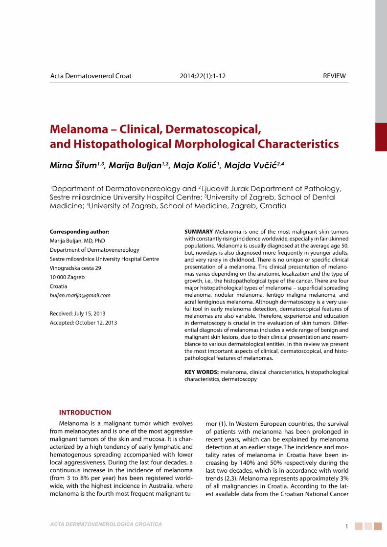
the Caucasian population, accounting for 70-80% of all melanomas (11). It is usually diagnosed at 30 to 50 years of age, and occurs more often in women. This type of melanoma can arise in any anatomic local-ization, most frequently on the trunk in men and on lower extremities in women. Patients usually report changes having taken place in the pigmented lesion within the last 1 to 5 years. Initially, in its horizontal growth phase, SSM presents as a light brown to black colored macule, with irregular borders. In the vertical growth phase, the surface is rough or papillomatous and may be ulcerated (Figure 1.).
Figure 1. Superficial spreading melanoma on the back of a 58-year-old man, showing a variety of col-ors, irregular borders, asymmetry, and a partially el-evated and rough surface.
Šitum et al. Acta Dermatovenerol CroatMelanoma 2014;22(1):1-12
3ACTA DERMATOVENEROLOGICA CROATICA
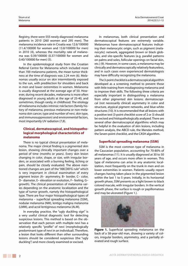
Dermatoscopy of an SSM usually shows one or more of the following dermatoscopical features: a blue-white veil (histopathologically corresponding to melanin in the mid-dermis with overlying epi-dermal orthokeratosis), multiple brown dots (corre-sponding to suprabasal epidermal malignant mela-nocytes representing the pagetoid spread of SSM), pseudopods and radial streaming (corresponding to confluent radial nests of atypical melanocytes at the dermoepidermal junction), scar-like depigmenta-tion or white milky areas (areas of tumor regression), peripheral black dots and/or globules (representing malignant melanocytes found at or near the stratum corneum), multiple colors, a broad and atypical net-work (rete ridges filled with malignant melanocytes), focal sharply cut-off borders, crystalline structures or chrysalis (which can be observed with polarized dermatoscopy only, corresponding to an altered stromal matrix), and irregular vascular structures (12) (Figure 2).
nodular melanoma (nM) Nodular melanoma is the second most com-
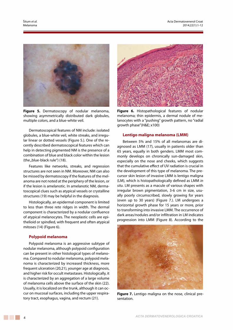
mon type of melanoma, accounting for 15–30 % of all melanomas (17). It is usually diagnosed in people between 40 and 50 years of age, equally frequent in both sexes. The most common localizations are the trunk, head and neck. Evolution of the lesion is usu-ally brief – the lesion develops in just a few months to 2 years prior to diagnosis. This type of melanoma is considered to be more aggressive than the SSM and often develops rapidly. It is usually darker than the SSM, is well circumscribed, and presents as a uni-formly colored nodule with or without ulceration. NM often features distinct pigmentation and a glossy sur-face, which enables clinical recognition of this type of melanoma (Figure 4). Due to a very brief horizontal growth phase and early onset of the vertical growth phase, NM is usually diagnosed at an advanced stage (16). Even very small lesions of NM have metastatic potential.
Figure 2. Dermatoscopical presentation of SSM; ab-solute asymmetry in shape and color, a broad and atypical network, focal sharply cut-off borders, mul-tiple colors, a blue-white veil, and crystalline struc-tures.
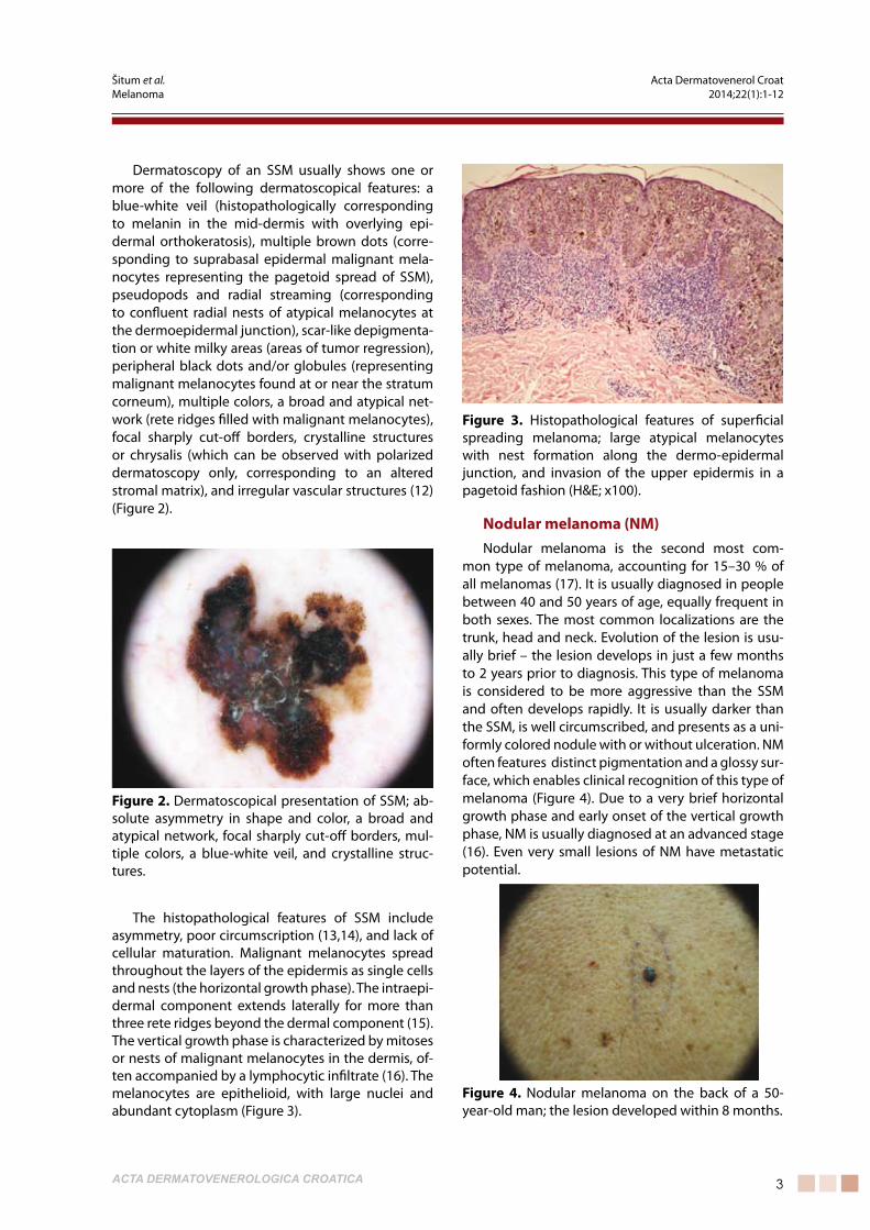
The histopathological features of SSM include asymmetry, poor circumscription (13,14), and lack of cellular maturation. Malignant melanocytes spread throughout the layers of the epidermis as single cells and nests (the horizontal growth phase). The intraepi-dermal component extends laterally for more than three rete ridges beyond the dermal component (15). The vertical growth phase is characterized by mitoses or nests of malignant melanocytes in the dermis, of-ten accompanied by a lymphocytic infiltrate (16). The melanocytes are epithelioid, with large nuclei and abundant cytoplasm (Figure 3).
Figure 3. Histopathological features of superficial spreading melanoma; large atypical melanocytes with nest formation along the dermo-epidermal junction, and invasion of the upper epidermis in a pagetoid fashion (H&E; x100).
Figure 4. Nodular melanoma on the back of a 50-year-old man; the lesion developed within 8 months.
Šitum et al. Acta Dermatovenerol CroatMelanoma 2014;22(1):1-12
4 ACTA DERMATOVENEROLOGICA CROATICA
Dermatoscopical features of NM include: isolated globules, a blue-white veil, white streaks, and irregu-lar linear or dotted vessels (Figure 5.). One of the re-cently described dermatoscopical features which can help in detecting pigmented NM is the presence of a combination of blue and black color within the lesion (the „blue-black rule“) (18).
Features like networks, streaks, and regression structures are not seen in NM. Moreover, NM can also be missed by dermatoscopy if the features of the mel-anoma are not noted at the periphery of the lesion, or if the lesion is amelanotic. In amelanotic NM, derma-toscopical clues such as atypical vessels or crystalline structures (19) may be helpful in the diagnosis.
Histologically, an epidermal component is limited to less than three rete ridges in width. The dermal component is characterized by a nodular confluence of atypical melanocytes. The neoplastic cells are epi-thelioid or spindled, with frequent and often atypical mitoses (14) (Figure 6).
Polypoid melanoma Polypoid melanoma is an aggressive subtype of
nodular melanoma, although polypoid configuration can be present in other histological types of melano-ma. Compared to nodular melanoma, polypoid mela-noma is characterized by increased thickness, more frequent ulceration (20,21), younger age at diagnosis, and higher risk for occult metastases. Histologically, it is characterized by an aggregation of a large volume of melanoma cells above the surface of the skin (22). Usually, it is localized on the trunk, although it can oc-cur on mucosal surfaces, including the upper respira-tory tract, esophagus, vagina, and rectum (21).
Lentigo maligna melanoma (LMM)Between 5% and 15% of all melanomas are di-
agnosed as LMM (17), usually in patients older than 65 years, equally in both genders. LMM most com-monly develops on chronically sun-damaged skin, especially on the nose and cheeks, which suggests that the cumulative effect of UV radiation is crucial in the development of this type of melanoma. The pre-cursor skin lesion of invasive LMM is lentigo maligna (LM), which is histopathologically defined as LMM in situ. LM presents as a macule of various shapes with irregular brown pigmentation, 3-6 cm in size, usu-ally poorly circumscribed, slowly growing for years (even up to 30 years) (Figure 7.). LM undergoes a horizontal growth phase for 15 years or more, prior to transforming into invasive LMM. The occurrence of dark areas/nodules and/or infiltration in LM indicates progression into LMM (Figure 8). According to the
Figure 5. Dermatoscopy of nodular melanoma, showing asymmetrically distributed dark globules, multiple colors, and a blue-white veil.
Figure 6. Histopathological features of nodular melanoma; thin epidermis, a dermal nodule of me-lanocytes with a “pushing” growth pattern, no “radial growth phase”(H&E; x100)
Figure 7. Lentigo maligna on the nose, clinical pre-sentation.
Šitum et al. Acta Dermatovenerol CroatMelanoma 2014;22(1):1-12
�ACTA DERMATOVENEROLOGICA CROATICA
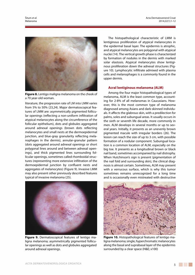
literature, the progression rate of LM into LMM varies from 5% to 50% (23,24). Major dermatoscopical fea-tures of LMM are: asymmetrically pigmented follicu-lar openings (reflecting a non-uniform infiltration of atypical melanocytes along the circumference of the follicular epithelium), dots and globules aggregated around adnexal openings (brown dots reflecting melanocytes and small nests at the dermoepidermal junction, and blue-gray granularity reflecting mela-nophages in the dermis), annular-granular pattern (dots aggregated around adnexal openings or short polygonal lines around and between adnexal open-ings), and thick pigmented lines surrounding fol-licular openings, sometimes called rhomboidal struc-tures (representing more extensive infiltration of the dermoepidermal junction by confluent nests and aggregates of melanocytes) (Figure 9). Invasive LMM may also present other previously described features typical of invasive melanoma (25).
The histopathological characteristic of LMM is lentiginous proliferation of atypical melanocytes in the epidermal basal layer. The epidermis is atrophic, and atypical melanocytes are polygonal with atypical nuclei (14). The vertical growth phase is characterized by formation of nodules in the dermis with marked solar elastosis. Atypical melanocytes show lentigi-nous proliferation down the adnexal structures (Fig-ure 10). Lymphocytic infiltrate admixed with plasma cells and melanophages is a commonly found in the upper dermis.
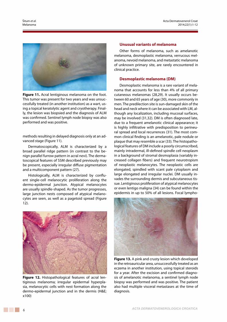
Acral lentiginous melanoma (ALM) Among the four major histopathological types of
melanoma, ALM is the least common type, account-ing for 2-8% of all melanomas in Caucasians. How-ever, this is the most common type of melanoma diagnosed among Asians and dark-skinned individu-als. It affects the glabrous skin, with a predilection for palms, soles and subungual areas. It usually occurs in the sixth or seventh life decade, more commonly in men. ALM develops in several months or up to sev-eral years. Initially, it presents as an unevenly brown pigmented macule with irregular borders (26). The lesion can reach the size of up to 3 cm, with possible formation of a nodular component. Subungual loca-tion is a common location of ALM, especially on the big toe. It presents as a longitudinal brown or black nail band, sometimes accompanied by nail distrophy. When Hutchinson’s sign is present (pigmentation of the nail fold and surrounding skin), the clinical diag-nosis is almost certain. Sometimes, ALM may present with a verrucous surface, which is why this tumor sometimes remains unrecognized for a long time and is occasionally even mistreated with destructive
Figure 8. Lentigo maligna melanoma on the cheek of a 70 year-old woman.
Figure 9. Dermatoscopical features of lentigo ma-ligna melanoma; asymmetrically pigmented follicu-lar openings as well as dots and globules aggregated around adnexal openings.
Figure 10. Histopathological features of lentigo ma-ligna melanoma; single, hyperchromatic melanocytes along the basal and suprabasal layer of the epidermis surrounded by a clear space (H&E; x100)
Šitum et al. Acta Dermatovenerol CroatMelanoma 2014;22(1):1-12
� ACTA DERMATOVENEROLOGICA CROATICA
methods resulting in delayed diagnosis only at an ad-vanced stage (Figure 11).
Dermatoscopically, ALM is characterized by a broad parallel ridge pattern (in contrast to the be-nign parallel furrow pattern in acral nevi). The derma-toscopical features of SSM described previously may be present, especially irregular diffuse pigmentation and a multicomponent pattern (27).
Histologically, ALM is characterized by conflu-ent single-cell melanocytic proliferation along the dermo-epidermal junction. Atypical melanocytes are usually spindle-shaped. As the tumor progresses, large junction nests composed of atypical melano-cytes are seen, as well as a pagetoid spread (Figure 12).
Unusual variants of melanomaOther forms of melanoma, such as amelanotic
melanoma, desmoplastic melanoma, verrucous mel-anoma, nevoid melanoma, and metastatic melanoma of unknown primary site, are rarely encountered in clinical practice.
Desmoplastic melanoma (DM)Desmoplastic melanoma is a rare variant of mela-
noma that accounts for less than 4% of all primary cutaneous melanomas (28,29). It usually occurs be-tween 60 and 65 years of age (30), more commonly in men. The predilection site is sun-damaged skin of the head and neck where it can be associated with LM, al-though any localization, including mucosal surfaces, may be involved (31,32). DM is often diagnosed late, due to a frequent amelanotic clinical appearance; it is highly infiltrative with predisposition to perineu-ral spread and local recurrences (31). The most com-mon clinical finding is an amelanotic, pale nodule or plaque that may resemble a scar (33). The histopatho-logical features of DM include a poorly circumscribed, mainly intradermal, ill-defined spindle cell neoplasm in a background of stromal desmoplasia (variably in-creased collagen fibers) and frequent neurotropism of neoplastic melanocytes. The neoplastic cells are elongated, spindled with scant pale cytoplasm and large elongated and irregular nuclei. DM usually in-vades the surrounding dermis and subcutaneous tis-sue. Lentiginous proliferation of atypical melanocytes or even lentigo maligna (34) can be found within the epidermis in up to 50% of all lesions. Focal lympho-
Figure 12. Histopathological features of acral len-tiginous melanoma; irregular epidermal hyperpla-sia, melanocytic cells with nest formation along the dermo-epidermal junction and in the dermis (H&E; x100)
Figure 11. Acral lentiginous melanoma on the foot. This tumor was present for two years and was unsuc-cessfully treated (in another institution) as a wart, us-ing a topical keratolytic agent and cryotherapy. Final-ly, the lesion was biopsied and the diagnosis of ALM was confirmed. Sentinel lymph node biopsy was also performed and was positive.
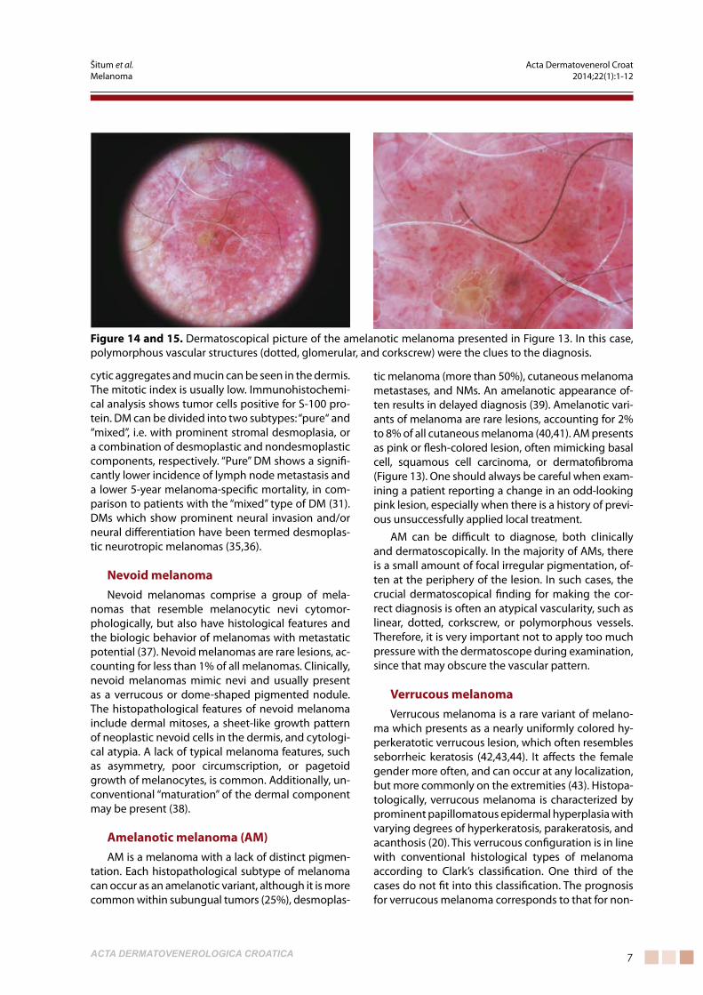
Figure 13. A pink and crusty lesion which developed in the retroauricular area, unsuccessfully treated as an eczema in another institution, using topical steroids for a year. After the excision and confirmed diagno-sis of amelanotic melanoma, a sentinel lymph node biopsy was performed and was positive. The patient also had multiple visceral metastases at the time of diagnosis.
Šitum et al. Acta Dermatovenerol CroatMelanoma 2014;22(1):1-12
�ACTA DERMATOVENEROLOGICA CROATICA
cytic aggregates and mucin can be seen in the dermis. The mitotic index is usually low. Immunohistochemi-cal analysis shows tumor cells positive for S-100 pro-tein. DM can be divided into two subtypes: “pure“ and “mixed”, i.e. with prominent stromal desmoplasia, or a combination of desmoplastic and nondesmoplastic components, respectively. “Pure” DM shows a signifi-cantly lower incidence of lymph node metastasis and a lower 5-year melanoma-specific mortality, in com-parison to patients with the “mixed” type of DM (31). DMs which show prominent neural invasion and/or neural differentiation have been termed desmoplas-tic neurotropic melanomas (35,36).
nevoid melanomaNevoid melanomas comprise a group of mela-
nomas that resemble melanocytic nevi cytomor-phologically, but also have histological features and the biologic behavior of melanomas with metastatic potential (37). Nevoid melanomas are rare lesions, ac-counting for less than 1% of all melanomas. Clinically, nevoid melanomas mimic nevi and usually present as a verrucous or dome-shaped pigmented nodule. The histopathological features of nevoid melanoma include dermal mitoses, a sheet-like growth pattern of neoplastic nevoid cells in the dermis, and cytologi-cal atypia. A lack of typical melanoma features, such as asymmetry, poor circumscription, or pagetoid growth of melanocytes, is common. Additionally, un-conventional “maturation” of the dermal component may be present (38).
Amelanotic melanoma (AM)AM is a melanoma with a lack of distinct pigmen-
tation. Each histopathological subtype of melanoma can occur as an amelanotic variant, although it is more common within subungual tumors (25%), desmoplas-
tic melanoma (more than 50%), cutaneous melanoma metastases, and NMs. An amelanotic appearance of-ten results in delayed diagnosis (39). Amelanotic vari-ants of melanoma are rare lesions, accounting for 2% to 8% of all cutaneous melanoma (40,41). AM presents as pink or flesh-colored lesion, often mimicking basal cell, squamous cell carcinoma, or dermatofibroma (Figure 13). One should always be careful when exam-ining a patient reporting a change in an odd-looking pink lesion, especially when there is a history of previ-ous unsuccessfully applied local treatment.
AM can be difficult to diagnose, both clinically and dermatoscopically. In the majority of AMs, there is a small amount of focal irregular pigmentation, of-ten at the periphery of the lesion. In such cases, the crucial dermatoscopical finding for making the cor-rect diagnosis is often an atypical vascularity, such as linear, dotted, corkscrew, or polymorphous vessels. Therefore, it is very important not to apply too much pressure with the dermatoscope during examination, since that may obscure the vascular pattern.
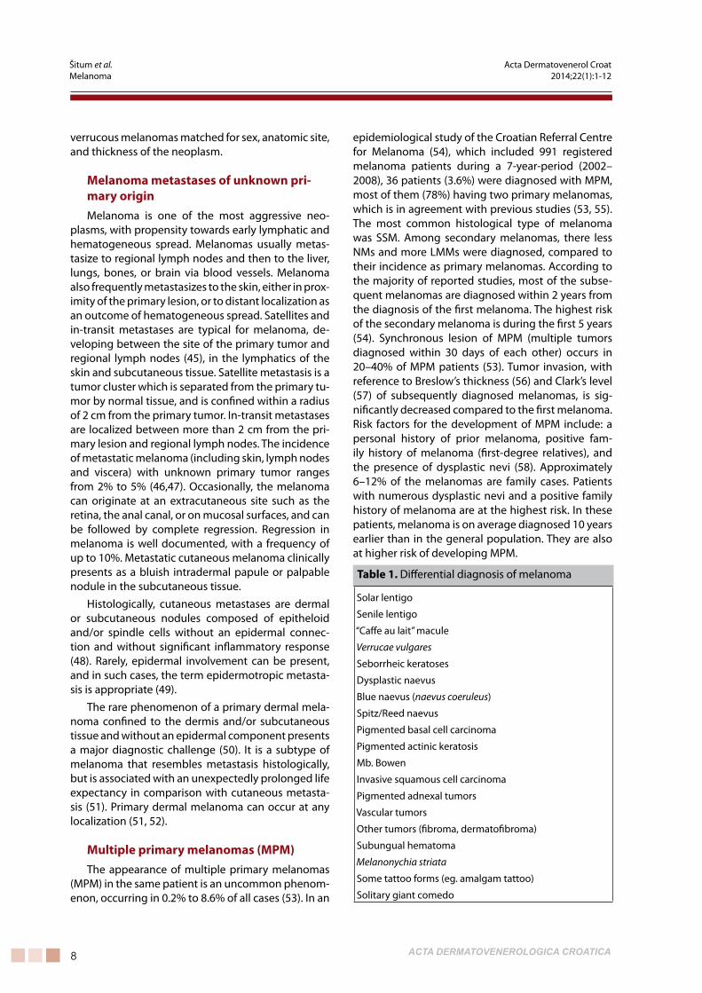
Verrucous melanomaVerrucous melanoma is a rare variant of melano-
ma which presents as a nearly uniformly colored hy-perkeratotic verrucous lesion, which often resembles seborrheic keratosis (42,43,44). It affects the female gender more often, and can occur at any localization, but more commonly on the extremities (43). Histopa-tologically, verrucous melanoma is characterized by prominent papillomatous epidermal hyperplasia with varying degrees of hyperkeratosis, parakeratosis, and acanthosis (20). This verrucous configuration is in line with conventional histological types of melanoma according to Clark’s classification. One third of the cases do not fit into this classification. The prognosis for verrucous melanoma corresponds to that for non-
Figure 14 and 15. Dermatoscopical picture of the amelanotic melanoma presented in Figure 13. In this case, polymorphous vascular structures (dotted, glomerular, and corkscrew) were the clues to the diagnosis.
Šitum et al. Acta Dermatovenerol CroatMelanoma 2014;22(1):1-12
� ACTA DERMATOVENEROLOGICA CROATICA
verrucous melanomas matched for sex, anatomic site, and thickness of the neoplasm.
Melanoma metastases of unknown pri-mary originMelanoma is one of the most aggressive neo-
plasms, with propensity towards early lymphatic and hematogeneous spread. Melanomas usually metas-tasize to regional lymph nodes and then to the liver, lungs, bones, or brain via blood vessels. Melanoma also frequently metastasizes to the skin, either in prox-imity of the primary lesion, or to distant localization as an outcome of hematogeneous spread. Satellites and in-transit metastases are typical for melanoma, de-veloping between the site of the primary tumor and regional lymph nodes (45), in the lymphatics of the skin and subcutaneous tissue. Satellite metastasis is a tumor cluster which is separated from the primary tu-mor by normal tissue, and is confined within a radius of 2 cm from the primary tumor. In-transit metastases are localized between more than 2 cm from the pri-mary lesion and regional lymph nodes. The incidence of metastatic melanoma (including skin, lymph nodes and viscera) with unknown primary tumor ranges from 2% to 5% (46,47). Occasionally, the melanoma can originate at an extracutaneous site such as the retina, the anal canal, or on mucosal surfaces, and can be followed by complete regression. Regression in melanoma is well documented, with a frequency of up to 10%. Metastatic cutaneous melanoma clinically presents as a bluish intradermal papule or palpable nodule in the subcutaneous tissue.
Histologically, cutaneous metastases are dermal or subcutaneous nodules composed of epitheloid and/or spindle cells without an epidermal connec-tion and without significant inflammatory response (48). Rarely, epidermal involvement can be present, and in such cases, the term epidermotropic metasta-sis is appropriate (49).
The rare phenomenon of a primary dermal mela-noma confined to the dermis and/or subcutaneous tissue and without an epidermal component presents a major diagnostic challenge (50). It is a subtype of melanoma that resembles metastasis histologically, but is associated with an unexpectedly prolonged life expectancy in comparison with cutaneous metasta-sis (51). Primary dermal melanoma can occur at any localization (51, 52).
Multiple primary melanomas (MPM)The appearance of multiple primary melanomas
(MPM) in the same patient is an uncommon phenom-enon, occurring in 0.2% to 8.6% of all cases (53). In an
epidemiological study of the Croatian Referral Centre for Melanoma (54), which included 991 registered melanoma patients during a 7-year-period (2002–2008), 36 patients (3.6%) were diagnosed with MPM, most of them (78%) having two primary melanomas, which is in agreement with previous studies (53, 55). The most common histological type of melanoma was SSM. Among secondary melanomas, there less NMs and more LMMs were diagnosed, compared to their incidence as primary melanomas. According to the majority of reported studies, most of the subse-quent melanomas are diagnosed within 2 years from the diagnosis of the first melanoma. The highest risk of the secondary melanoma is during the first 5 years (54). Synchronous lesion of MPM (multiple tumors diagnosed within 30 days of each other) occurs in 20–40% of MPM patients (53). Tumor invasion, with reference to Breslow’s thickness (56) and Clark’s level (57) of subsequently diagnosed melanomas, is sig-nificantly decreased compared to the first melanoma. Risk factors for the development of MPM include: a personal history of prior melanoma, positive fam-ily history of melanoma (first-degree relatives), and the presence of dysplastic nevi (58). Approximately 6–12% of the melanomas are family cases. Patients with numerous dysplastic nevi and a positive family history of melanoma are at the highest risk. In these patients, melanoma is on average diagnosed 10 years earlier than in the general population. They are also at higher risk of developing MPM.
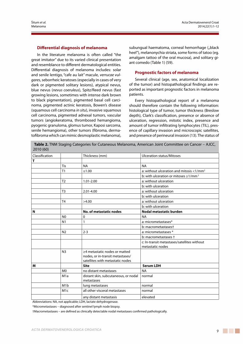
table 1. Differential diagnosis of melanoma
Solar lentigo
Senile lentigo
“Caffe au lait” macule
Verrucae vulgares
Seborrheic keratoses
Dysplastic naevus
Blue naevus (naevus coeruleus)
Spitz/Reed naevus
Pigmented basal cell carcinoma
Pigmented actinic keratosis
Mb. Bowen
Invasive squamous cell carcinoma
Pigmented adnexal tumors
Vascular tumors
Other tumors (fibroma, dermatofibroma)
Subungual hematoma
Melanonychia striata
Some tattoo forms (eg. amalgam tattoo)
Solitary giant comedo
Šitum et al. Acta Dermatovenerol CroatMelanoma 2014;22(1):1-12
�ACTA DERMATOVENEROLOGICA CROATICA
Differential diagnosis of melanomaIn the literature melanoma is often called “the
great imitator” due to its varied clinical presentation and resemblance to different dermatological entities. Differential diagnosis of melanoma includes: solar and senile lentigo, “cafe au lait” macule, verrucae vul-gares, seborrheic keratoses (especially in cases of very dark or pigmented solitary lesions), atypical nevus, blue nevus (nevus coeruleus), Spitz/Reed nevus (fast growing lesions, sometimes with intense dark brown to black pigmentation), pigmented basal cell carci-noma, pigmented actinic keratosis, Bowen’s disease (squamous cell carcinoma in situ), invasive squamous cell carcinoma, pigmented adnexal tumors, vascular tumors (angiokeratoma, thrombosed hemangioma, pyogenic granuloma, glomus tumor, Kaposi sarcoma, senile hemangioma), other tumors (fibroma, derma-tofibroma which can mimic desmoplastic melanoma),
subungual haematoma, corneal hemorrhage („black heel“), melanonychia striata, some forms of tatoo (eg. amalgam tattoo of the oral mucosa), and solitary gi-ant comedo (Table 1) (59).
Prognostic factors of melanomaSeveral clinical (age, sex, anatomical localization
of the tumor) and histopathological findings are re-ported as important prognostic factors in melanoma patients.
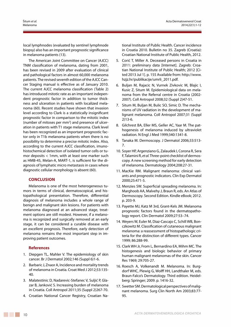
Every histopathological report of a melanoma should therefore contain the following information: histological type of tumor, tumor thickness (Breslow depth), Clark’s classification, presence or absence of ulceration, regression, mitotic index, presence and amount of tumor infiltrating lymphocytes (TIL), pres-ence of capillary invasion and microscopic satellites, and presence of perineural invasion (13). The status of
Classification Thickness (mm) Ulceration status/Mitosest
Tis NA NAT1 ≤1.00 a: without ulceration and mitosis <1/mm2
b: with ulceration or mitoses ≥1/mm 2
T2 1.01-2.00 a: without ulcerationb: with ulceration
T3 2.01-4.00 a: without ulcerationb: with ulceration
T4 >4.00 a: without ulcerationb: with ulceration
n no. of metastatic nodes nodal metastatic burdenN0 0 NAN1 1 a: micrometastases*
b: macrometastases†N2 2-3 a: micrometastases *
b: macrometastases †c: In-transit metastases/satellites without metastatic nodes
N3 ≥4 metastatic nodes or matted nodes, or in-transit metastases/ satellites with metastatic nodes
M Site Serum LDHM0 no distant metastases NAM1a distant skin, subcutaneous, or nodal
metastasesnormal
M1b lung metastases normalM1c all other visceral metastases normal
any distant metastasis elevatedAbbreviations: NA, not applicable; LDH, lactate dehydrogenase.*Micrometastases – diagnosed after sentinel lymph node biopsy.†Macrometastases – are defined as clinically detectable nodal metastases confirmed pathologically.
table 2. TNM Staging Categories for Cutaneous Melanoma, American Joint Committee on Cancer – AJCC, 2010 (60)
Šitum et al. Acta Dermatovenerol CroatMelanoma 2014;22(1):1-12
�0 ACTA DERMATOVENEROLOGICA CROATICA
local lymphnodes (evaluated by sentinel lymphnode biopsy) also has an important prognostic significance in melanoma patients (13).
The American Joint Committee on Cancer (AJCC) TNM classification of melanoma, dating from 2001, has been revised in 2009 after evaluation of clinical and pathological factors in almost 60,000 melanoma patients. The revised seventh edition of the AJCC Can-cer Staging manual is effective as of January 2010. The current AJCC melanoma classification (Table 2) has introduced mitotic rate as an important indepen-dent prognostic factor in addition to tumor thick-ness and ulceration in patients with localized mela-noma (60). Recent studies have shown that invasion level according to Clark is a statistically insignificant prognostic factor in comparison to the mitotic index (number of mitoses per mm2) and presence of ulcer-ation in patients with T1 stage melanoma. Clark level has been recognized as an important prognostic fac-tor only in T1b melanoma patients when there is no possibility to determine a precise mitotic index. Also, according to the current AJCC classification, imuno-histochemical detection of isolated tumor cells or tu-mor deposits < 1mm, with at least one marker such as HMB-45, Melan-A, MART-1, is sufficient for the di-agnosis of lymphatic micro metastasis in cases where diagnostic cellular morphology is absent (60).
ConCLUSIonMelanoma is one of the most heterogeneous tu-
mors in terms of clinical, dermatoscopical, and his-topathological presentation. Therefore, differential diagnosis of melanoma includes a whole range of benign and malignant skin lesions. For patients with melanoma diagnosed at an advanced stage, treat-ment options are still modest. However, if a melano-ma is recognized and surgically removed at an early stage, it can be considered a curable disease with an excellent prognosis. Therefore, early detection of melanoma remains the most important step in im-proving patient outcomes.
References1. Diepgen TL, Mahler V. The epidemiology of skin
cancer. Br J Dermatol 2002;146 (Suppl 6)1-6.
2. Barbaric J, Znaor A, Incidence and mortality trends of melanoma in Croatia. Croat Med J 2012;53:135-40.
3. Malatestinic D, Nadarevic-Stefanec V, Suljić P, Gla-zar B, Janković S. Increasing burden of melanoma in Croatia. Coll Antropol 2011;35 (Suppl 2)267-70.
4. Croatian National Cancer Registry, Croatian Na-
tional Institute of Public Health. Cancer incidence in Croatia 2010. Bulletin no 35. Zagreb (Croatia): Croatian National Institute of Public Health, 2012.
5. Corić T, Miller A. Deceased persons in Croatia in 2011: preliminary data [Internet]. Zagreb: Croa-tian National Institute of Public Health; 2012 [Ci-ted 2013 Jul 1]. p. 155 Available from: http://www.hzjz.hr/publikacije/umrli_2011.pdf.
6. Buljan M, Rajacic N, Vurnek Zivkovic M, Blajic I, Kusic Z, Situm M. Epidemiological data on mela-noma from the Referral centre in Croatia (2002-2007). Coll Antropol 2008;32 (Suppl 2)47-51.
7. Situm M, Buljan M, Bulic SO, Simic D. The mecha-nisms of UV radiation in the development of ma-lignant melanoma. Coll Antropol 2007;31 (Suppl 2)13-6.
8. Gilchrest BA, Eller MS, Geller AC, Yaar M. The pat-hogenesis of melanoma induced by ultraviolet radiation. N Engl J Med 1999;340:1341-8.
9. Tanaka M. Dermoscopy. J Dermatol 2006;33:513-7.
10. Soyer HP, Argenziano G, Zalaudek I, Corona R, Sera F, Talamini R, et al. Three-point checklist of dermos-copy. A new screening method for early detection of melanoma. Dermatology 2004;208:27-31.
11. MacKie RM. Malignant melanoma: clinical vari-ants and prognostic indicators. Clin Exp Dermatol 2000;25:471-5.
12. Menzies SW. Superficial spreading melanoma. In: Marghoob AA, Malvehy J, Braun R, eds. An Atlas of Dermoscopy. Second Edition. Kindle eBook; 2012. p. 203-9.
13. Payette MJ, Katz M 3rd, Grant-Kels JM. Melanoma prognostic factors found in the dermatopatho-logy report. Clin Dermatol 2009;27:53–74.
14. Weyers W, Euler M, Diaz-Cascajo C, Schill WB, Bon-czkowitz M. Classification of cutaneous malignant melanoma: a reassessment of histopathologic cri-teria for the distinction of different types. Cancer 1999; 86:288-99.
15. Clark WH Jr, From L, Bernardino EA, Mihm MC. The histogenesis and biologic behavior of primary human malignant melanomas of the skin. Cancer Res 1969; 29:705-27.
16. Roesch A, Volkenandt M. Melanoma. In: Burg-dorf WHC, Plewig G, Wolff HH, Landthaler M, eds. Braun-Falco’s Dermatology. Third edition. Heidel-berg: Springer; 2009. p. 1416-32.
17. Swetter SM. Dermatological perspectives of malig-nant melanoma. Surg Clin North Am 2003;83:77-95.
Šitum et al. Acta Dermatovenerol CroatMelanoma 2014;22(1):1-12
��ACTA DERMATOVENEROLOGICA CROATICA
18. Argenziano G, Longo C, Cameron A, Cavicchini S Gourhant JY, Lallas A, et al. Blue-black rule: a simple dermoscopic clue to recognize pigmented nodular melanoma. Br J Dermatol 2011;165:1251-5.
19. Menzies SW. Nodular melanoma. In: Marghoob AA, Malvehy J, Braun R, eds. An Atlas of Dermos-copy. Second Edition. Kindle eBook; 2012. p. 220-2.
20. Rongioletti F, Smoller BR. Unusual histological variants of cutaneous malignant melanoma with some clinical and possible prognostic correla-tions. J Cutan Pathol 2005;32:589-603.
21. Manci EA, Balch CM, Murad TM, Soong SJ. Po-lypoid melanoma, a virulent variant of the nodu-lar growth pattern. Am J Clin Pathol 1981;75:810-5.
22. Plotnick H, Rachmaninoff N, VandenBerg HJ Jr. Polypoid melanoma: a virulent variant of nodular melanoma. Report of three cases and literature review. J Am Acad Dermatol 1990;23:880-4.
23. Weinstock MA, Sober AJ. The risk of progression of lentigo maligna melanoma. Br J Dermatol 1987;116:303-10.
24. Mckenna JK, Florell SR, Goldman GD, Bowen GM. Lentigo maligna/lentigo maligna melanoma: cur-rent state of diagnosis and treatment. Dermatol Surg 2006;32:493-504.
25. Scope A, Wang SQ, Rabinovitz HS. Lentigo malig-na melanoma. In: Marghoob AA, Malvehy J, Braun R, eds. An Atlas of Dermoscopy. Second Edition. Kindle eBook; 2012. p. 223-9.
26. Coleman WP 3rd, Loria PR, Reed RJ, Krementz ET. Acral lentiginous melanoma. Arch Dermatol 1980;116:773-6.
27. Malvehy J, Puig S. Acrolentiginous melanoma. In: Marghoob AA, Malvehy J, Braun R, eds. An Atlas of Dermoscopy. Second Edition. Kindle eBook; 2012. p. 210-18.
28. Quinn MJ, Crotty KA, Thompson JF, Coates AS, O’Brien CJ, McCarthy WH. Desmoplastic and des-moplastic neurotropic melanoma: experience with 280 patients. Cancer 1998;83:1128-35.
29. Busam KJ. Cutaneous desmoplastic melanoma. Adv Anat Pathol 2005;12:92-102.
30. Anstey A, McKee P, Jones EW. Desmoplastic malig-nant melanoma: a clinicopathological study of 25 cases. Br J Dermatol 1993;129:359-71.
31. Barnhill RL, Gupta K. Unusual variants of malig-nant melanoma. Clin Dermatol 2009;27:564–87.
32. Bruijn JA, Salasche S, Sober AJ, Mihm MC, Barnhill
RL. Desmoplastic melanoma: clinicopathologic aspects of six cases. Dermatology 1992;185:3-8.
33. Conley J, Lattes R, Orr W. Desmoplastic malignant melanoma (a rare variant of spindle cell melano-ma). Cancer 1971;28:914-36.
34. Skelton HG, Smith KJ, Laskin WB, McCarthy WF, Gag-nier JM, Graham JH, et al. Desmoplastic malignant melanoma. J Am Acad Dermatol 1995;32:717-25.
35. Reed RJ, Leonard DD. Neurotropic melanoma. A variant of desmoplastic melanoma. Am J Surg Pathol 1979;3:301-11.
36. Chen JY, Hruby G, Scolyer RA, Murali R, Hong A, Fitzgerald P, et al. Desmoplastic neurotropic me-lanoma: a clinicopathologic analysis of 128 cases. Cancer 2008;113: 2770-8.
37. Wong TY, Suster S, Duncan LM, Mihm MC Jr. Ne-void melanoma: a clinicopathological study of seven cases of malignant melanoma mimicking spindle and epithelioid cell nevus and verrucous dermal nevus. Hum Pathol 1995;26:171-9.
38. DiCaudo DJ, McCalmont TH, Wick MR. Selected diagnostic problems in neoplastic dermatopat-hology. Arch Pathol Lab Med 2007;131:434–9.
39. Koch SE, Lange JR. Amelanotic melanoma: the great masquerader. J Am Acad Dermatol 2000;42:731-4.
40. Ariel IM. Amelanotic melanomas: an analysis of 77 patients. Curr Surg 1981;38:151- 5.
41. Clark WH Jr, From L, Bernardino EA, Mihm MC. The histogenesis and biologic behavior of primary human malignant melanomasof the skin. Cancer Res 1969;29:705- 26.
42. Stam-Posthuma JJ, van Duinen C, Scheffer E, Vink J, Bergman W. Multiple primary melanomas. J Am Acad Dermatol 2001;44: 22-7.
43. Kuehnl-Petzoldt C, Berger H, Wiebelt H. Verrucous-keratotic variations of maliganant melanoma: A clinicopathological study. Am J Dermatopathol 1982;4:403–10.
44. Steiner A, Konrad K, Pehamberger H, Wolff K. Verrucous malignant melanoma. Arch Dermatol 1988;124:1534–7.
45. Nakayama T, Taback B, Turner R, Morton DL, Hoon DS. Molecular clonality of intransit melanoma me-tastasis. Am J Pathol 2001;158:1371-8.
46. Schlagenhauff B, Stroebel W, Ellwanger U, Meier F, Zimmermann C, Breuninger H, et al. Metasta-tic melanoma of unknown primary origin shows prognostic similarities to regional metastatic me-lanoma: recommendations for initial staging exa-minations. Cancer 1997;80:60- 5.
Šitum et al. Acta Dermatovenerol CroatMelanoma 2014;22(1):1-12
�2 ACTA DERMATOVENEROLOGICA CROATICA
47. Chang P, Knapper WH. Metastatic melanoma of unknown primary. Cancer 1982;49:1106-11.
48. Mihm MC Jr, Clemente CG, Cascinelli N. Tumor in-filtrating lymphocytes in lymph node melanoma metastases: a histopathologic prognostic indica-tor and an expression of local immune response. Lab Invest 1996;74:43-7.
49. Abernethy JL, Soyer HP, Kerl H, Jorizzo JL, White WL. Epidermotropic metastatic malignant mela-noma simulating melanoma in situ. A report of 10 examples from two patients. Am J Surg Pathol 1994;18:1140-9.
50. Lee CC, Faries MB, Ye X, Morton DL. Solitary der-mal melanoma: beginning or end of the metasta-tic process? Ann Surg Oncol 2009;16:578-84.
51. Bowen GM, Chang AE, Lowe L, Hamilton T, Patel R, Johnson TM. Solitary melanoma confined to the dermal and/or subcutaneous tissue: evidence for revisiting the staging classification. Arch Der-matol 2000;136:1397-9.
52. Anbari KK, Schuchter LM, Bucky LP, Mick R, Syn-nestvedt M, Guerry D 4th, et al. Melanoma of unknown primary site: presentation, treatment, and prognosis-a single institution study. Cancer 1997;79: 1816-21.
53. Ferrone CR, Porat LB, Panageas KS, Berwick M, Halpern AC, Patel A, et al. Clinicopathological fea-tures of and risk factors for multiple primary me-lanomas. JAMA 2005;294:1647-54.
54. Buljan M, Situm M, Bolanca Z, Vurnek Zivković M, Lugovic Mihic L. Multiple primary melanoma: epidemiological and prognostic implications; analysis of 36 cases. Coll Antropol 2010;34 (Suppl 2)131-4.
55. Kang S, Barnhill RL, Mihm MC Jr, Sober AJ. Mul-tiple primary cutaneous melanomas. Cancer 1992;70:1911-6.
56. Johnson TM, Hamilton T, Lowe L. Multiple primary melanomas. J Am Acad Dermatol 1998;39:422–7.
57. Moseley HS, Giuliano AE, Storm FK 3rd, Clark WH, Robinson DS, Morton DL. Multiple primary mela-noma. Cancer 1979; 43:939–44.
58. Burden AD, Vestey JP, Sirel JM, Aitchison TC, Hun-ter JA, MacKie RM. Multiple primary melanoma: risk factors and prognostic implications. BMJ 1994;309:375.
59. Situm M, Buljan M, Poduje S. Pigmentni i epider-malni tumori kože. In: Situm M, ed. Algorithms in diagnostics and therapy of the most common dermatoses and skin tumours (in Croatian). Jastre-barsko: Naklada Slap; 2012. p. 227-69.
60. Balch CM, Gershenwald JE, Soong SJ, Thompson JF, Atkins MB, Byrd DR et al. Final version of 2009 AJCC melanoma staging and classification. J Clin Oncol 2009;27:6199-206.
Šitum et al. Acta Dermatovenerol CroatMelanoma 2014;22(1):1-12