medical student lectures hydrocephalus ,chiari ,congenital ,truma ,nerve injury
TRANSCRIPT

Neurosurgery ModuleMohammed Homoud MBChB,FRCSC,FAANS
Consultant Paediatric Neurosurgeon and Complex Spine
Director of Neurosurgery Department
Prince Sultan military Medical City

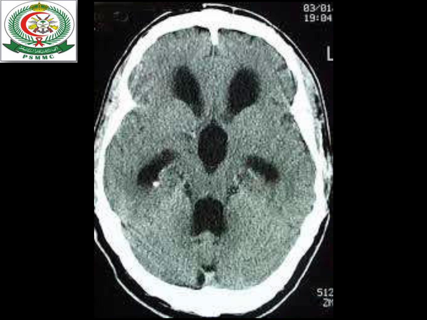
Hydrocephalus

Anatomy &
Pathophysiology


Types


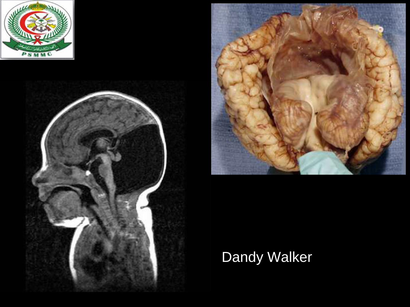
Dandy Walker

Clinical


other types
• Hydrocephalus ex vacuo
• Normal pressure hydrocephalus

Chiari Malformation

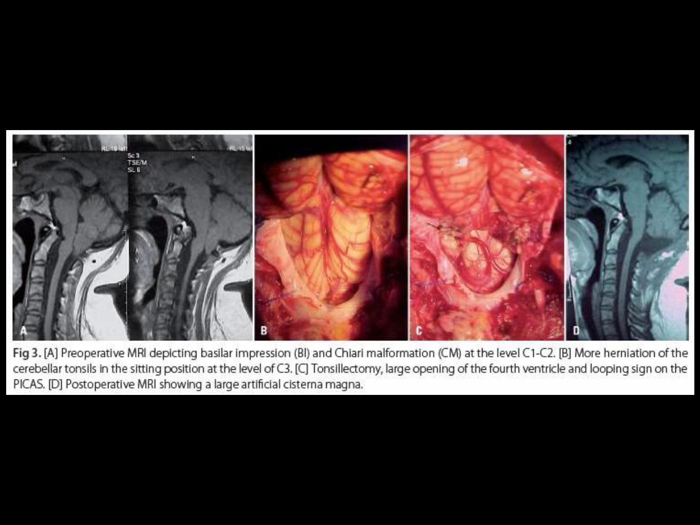
Chiari I
T“adult-onset Chiari” presenting by 4th decade
downward displacement of the cerebellar tonsils
through the foramen magnum


Chiari I Clinical
symptoms/ suboccipital headache
Neck pain
subjective weakness
numbness, loss of temperature sensation 40-60%
signs/ hyperactive lower extremity reflexes
“Cape”-like sensory loss
nystagmus (downbeat)
gait disturbance, upper extremity weaknes(30-50% )

Chiari I imaging
MRI is diagnostic
compression of brain stem
at FM
Hydrocephalus
Syringomyelia
Descent of cerebellar tonsils
through foramen magnum

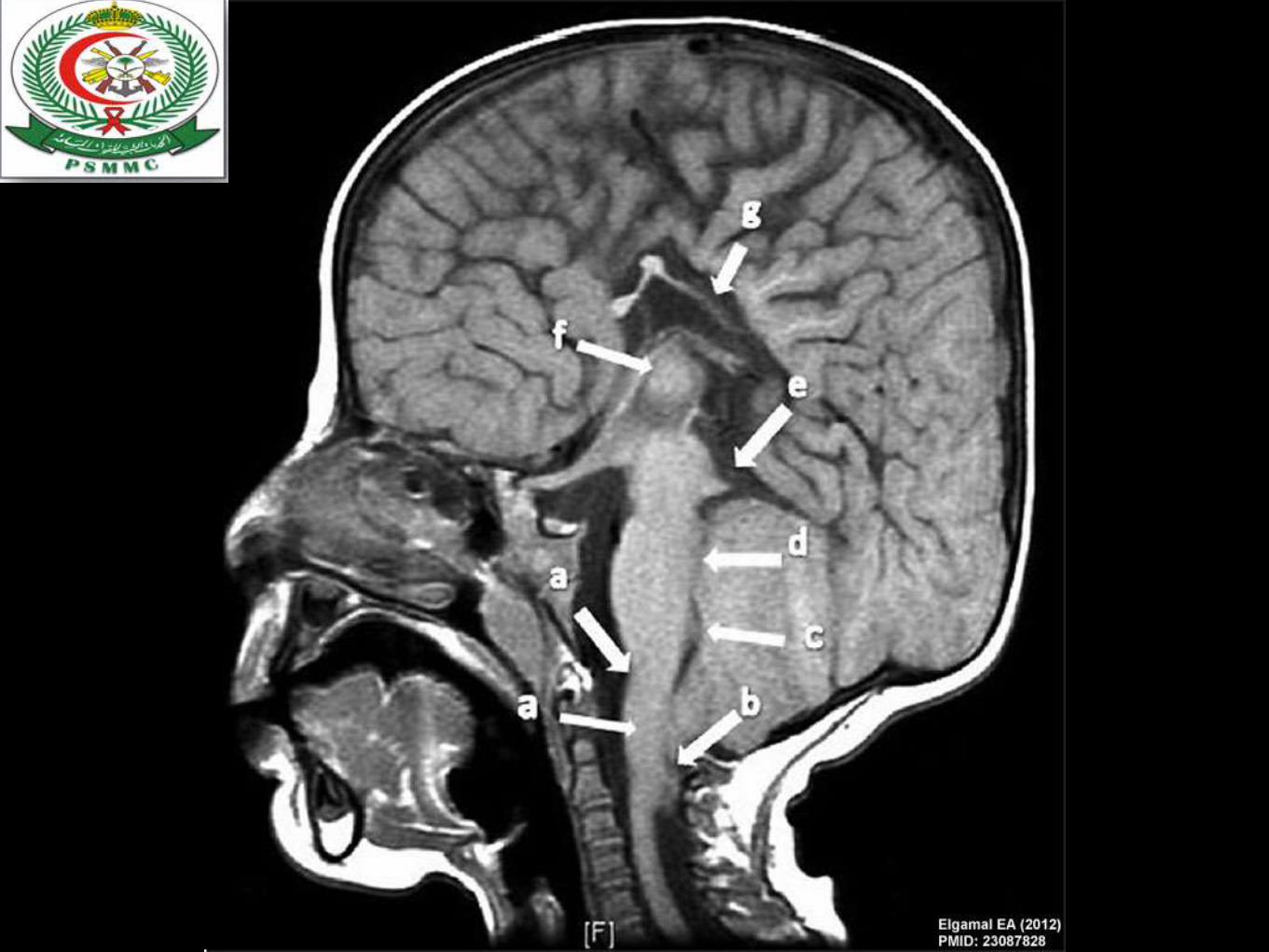
Chiari II
“Arnold-Chiari malformation.”Presents in childhood
Usually the younger
S&S/ secondary to brainstem and lower cranial
nerve dysfunction.
Findings (best seen on MRI):
Caudal displacement of posterior fossa structures,
including cervicomedullary junction, pons,
medulla, 4th ventricle, and cerebellar tonsils


Spinal Dysraphism

General
• general term for a family of congenital
malformations of the spine and spinal cord.
• also known as neural tube defects

Causes
Results from failure of the neural tube to close
spontaneously between the 3rd-4th week of in utero
development
Possible etiologic factors:
1-Radiation
2-Drugs
3-Malnutrition
4-Chemicals
5-Genetic determinants (mutations in folate-
responsive and folate-dependent pathways)
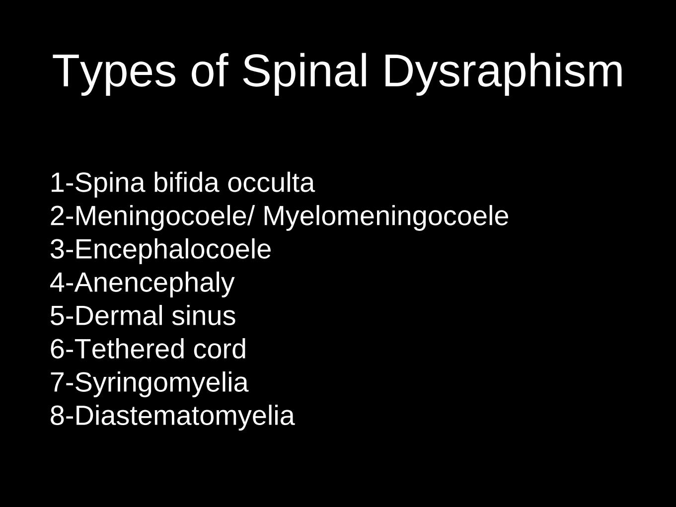
Types of Spinal Dysraphism
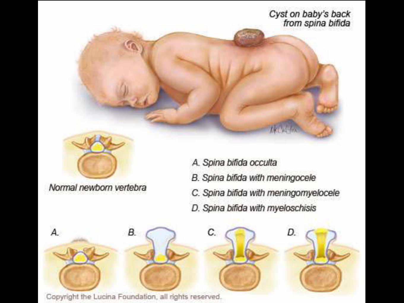
1-Spina bifida occulta
2-Meningocoele/ Myelomeningocoele
3-Encephalocoele
4-Anencephaly
5-Dermal sinus
6-Tethered cord
7-Syringomyelia
8-Diastematomyelia


Diagnostic Tools
Failure of closure of the neural tube allows excretion
(AFP, acetylcholinesterase) into amniotic fluid
Prenatal screening of maternal serum for AFP during 16-
18 week AOG
AF AFP obtained between 15-20 weeks’ gestation is most
specific
Rostral end of the NT closes on the 23rd day and the
caudal neuropore closes by the 27th day of development
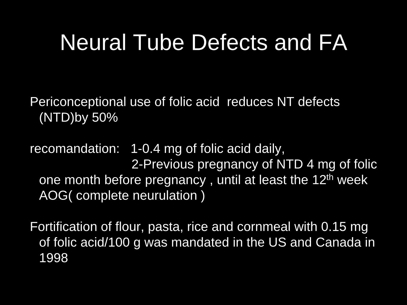
Neural Tube Defects and FA
Periconceptional use of folic acid reduces NT defects
(NTD)by 50%
recomandation: 1-0.4 mg of folic acid daily,
2-Previous pregnancy of NTD 4 mg of folic
one month before pregnancy , until at least the 12th week
AOG( complete neurulation )
Fortification of flour, pasta, rice and cornmeal with 0.15 mg
of folic acid/100 g was mandated in the US and Canada in
1998

Spina Bifida Occulta
Midline defect of the vertebral bodies without protrusion of
the SC or meninges
usually asymptomatic
In some, patches of hair, lipoma, discolouration of skin or
dermal sinus may be present
May be associated with syringomyelia, diastematomyelia, and
tethered cord
Recurrent meningitis of occult origin should prompt careful exam
for dermal sinus tract
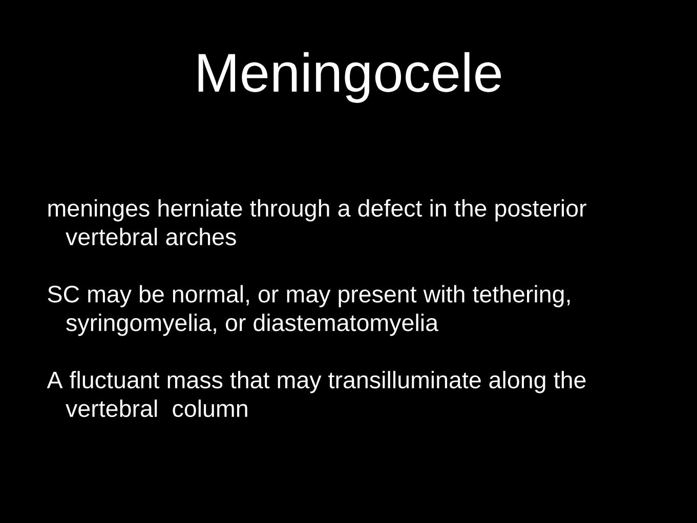
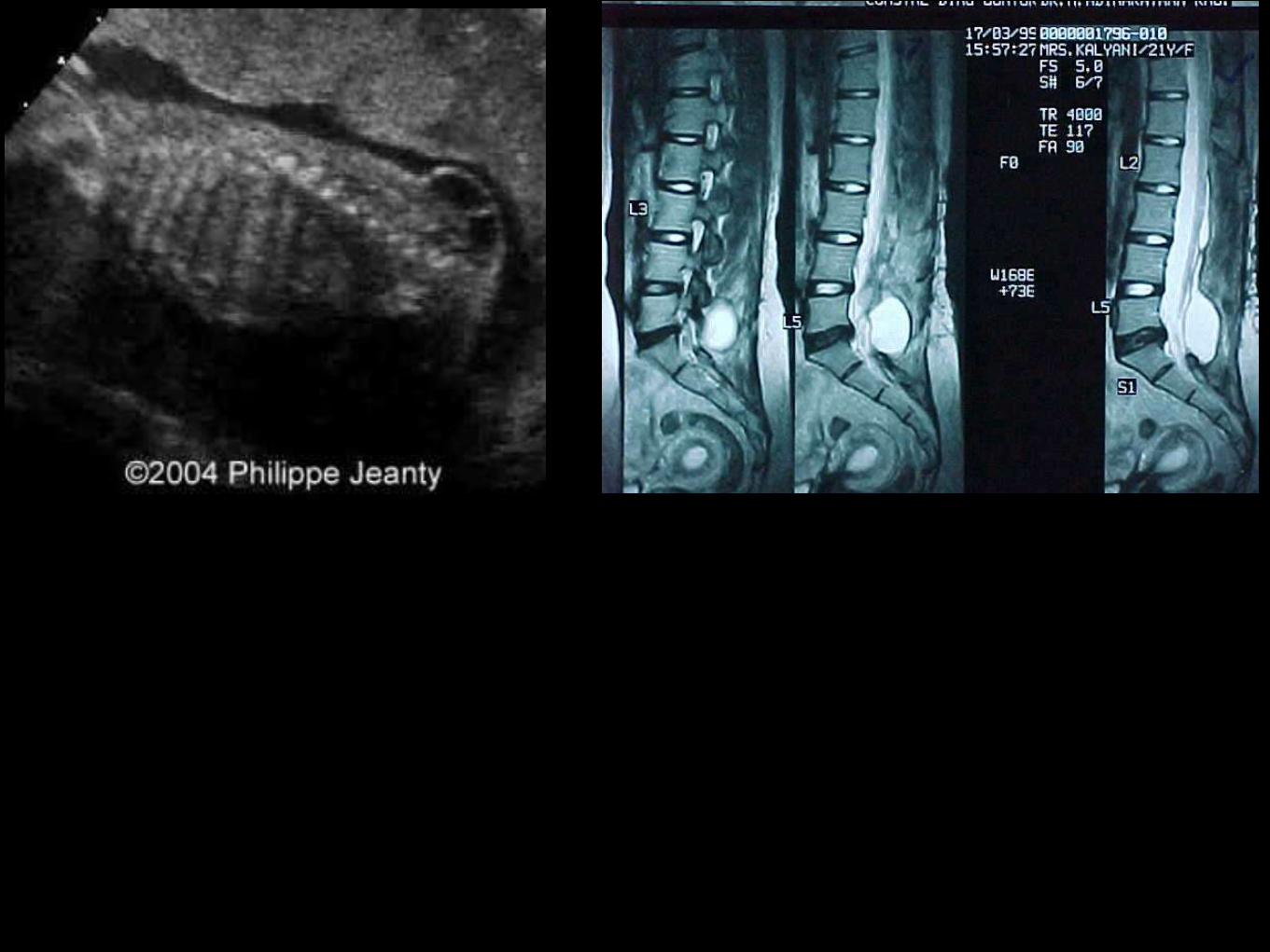
Meningocele
meninges herniate through a defect in the posterior
vertebral arches
SC may be normal, or may present with tethering,
syringomyelia, or diastematomyelia
A fluctuant mass that may transilluminate along the
vertebral column


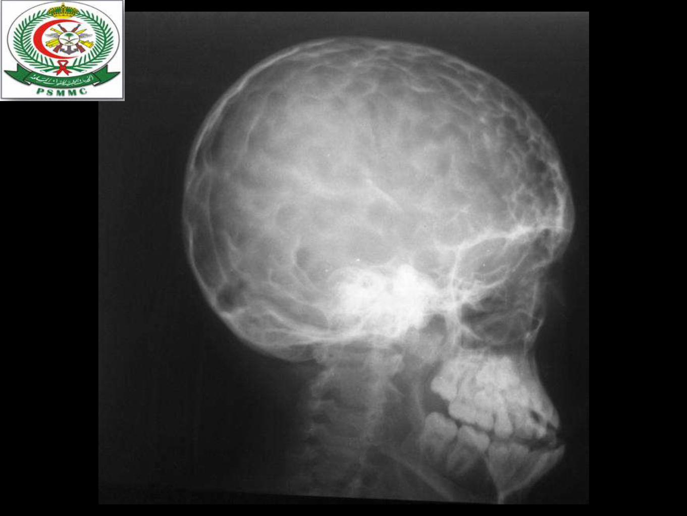
Myelomeningocoele
Most severe form, incidence of 1/4000 LB
Risk of recurrence after one affected child increases to
3-4% and increases to ~10% with 2 previous abnormal
pregnancies
Certain drugs that antagonize folic acid (TMP, AEDs:
CBZ, PHY, Pb, primidone) increase the risk of
myelomeningocoele
Valproic acid cause NT defects in ~1-2% of pregnancies

Myelomeningocoele
May be located anywhere along the neuraxis but
the LS region accounts for 75% of the cases
Extent and degree of the neuro deficit depend on
the location
CM: flaccid paralysis, absent DTRs, sensory
deficit below the affected level, postural abn of
the LE (clubfeet, subluxation of the hips),
constant urinary dribbling and a relaxed anal
sphincter

Myelomeningocoele
HCP in association with a type II Chiari defect develops
in at least 80% with myelomeningocoele
Infants with HCP and Chiari II develop symptoms of
hindbrain dysfunction: difficulty feeding, choking,
stridor, apnea, VC paralysis, pooling of secretions,
spasticity of UEs
Chiari crisis is due to downward herniation of the
medulla and cerebellar tonsils

Management
Requires a multidisciplinary approach: surgeon,
therapist, pediatrician
Surgery: repair and shunting; orthopaedic procedure,
urologic evaluation
GUT: regular catheterization to prevent UTI and reflux
leading to PN and hydronephrosis, urine cult, serum
elec, creatinine, renal scan, IV pyelogram, Utz
Rehab: functional ambulation (sacral or LS lesion)

Prognosis
MR- 10-15%
Most deaths occur before age 4 years
70% have normal intelligence, but learning
problems and seizure disorders are common
History of meningitis or ventriculitis adversely
affect the ultimate IQ

Craniosynostosis

Definition
• Craniosynostosis refers to premature closure of
cranial sutures, or joints between the bones of the
skull


Trauma
History
• indian 30 years male pedestrian
• hit by a car in front of PSMMC
• brought to trauma room
• not opening eyes ,localising to painful stimuli,and
producing sounds


Main points in head injury
stratification of head injury and when to do CT
3 categories

CATEGORY 1.
LOW RISK FOR INTRACRANIAL INJURY
Extremely low likelihood of
intracranial injury
even if a skull fracture is
present on SXR (incidence
of ICI 8.5 in 10,000 cases
with 95% confidence level
this category excludes
patients with a history of
loss of consciousness.


CATEGORY2.
MODERATE RISK FOR INTRACRANIAL
INJURY

CATIGORY 3
HIGH RISK FOR INTRACRANIAL
INJURY

Intracranial pressure
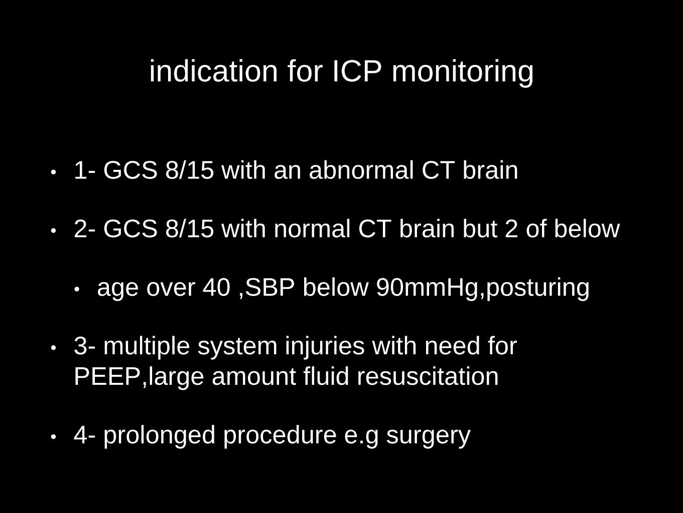
indication for ICP monitoring
• 1- GCS 8/15 with an abnormal CT brain
• 2- GCS 8/15 with normal CT brain but 2 of below
• age over 40 ,SBP below 90mmHg,posturing
• 3- multiple system injuries with need for
PEEP,large amount fluid resuscitation
• 4- prolonged procedure e.g surgery


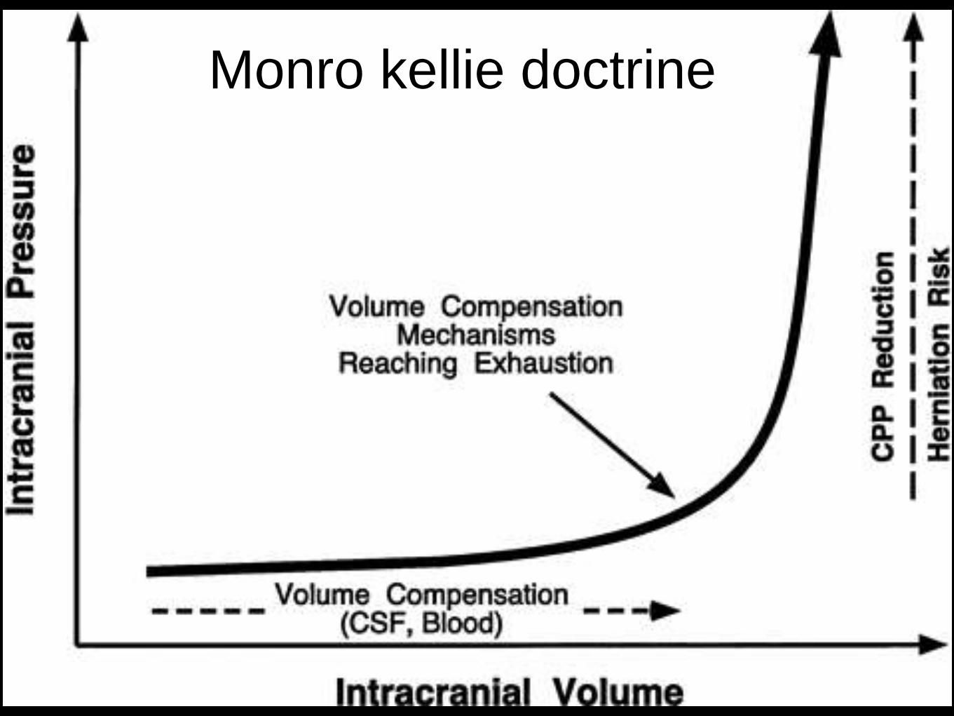
Monro kellie doctrine


Causes
1-Metabolic or collagen disease
2-Malignancy
3-Endo or exo-toxins
4-Ischaemia
5-Radiation * infection:leprosy
6-Trauma
a-Thermal
b-Chemical
c-Mechanical

Types of Injuries
primary injury
Results from same trauma that injures a bone or joint
Radial nerve is the most commonly injured. Of humeral
shaft fractures, 14 % is complicated by radial nerve
injuries
Displaced osseous fragments
Stretching
Manipulation
secondary injury
Results from involvement of nerve by infection, scar,
callous or vascular complications which may be
hematoma, AV fistula, Ischemia or aneurysm

Neuronal degeneration and regeneration
Any part of neuron detached from its nucleus
degenerates & is destroyed by phagocytosis.
Distal – Secondary / Wallerian Degeneration
Proximal - Primary / Traumatic / Retrograde
Degeneration
Time required for degeneration varies between sensory
and motor fibers and is also related to size &
myelination of fibers
Advancing Tinel sign and presence of motor march
phenomena are signs of regeneration


Diagnosis of Peripheral nerve injuries
History
Which nerve ?
What level ?
What is the cause ?
What degree of injury ?
Old or fresh injury ?

Diagnosis of Peripheral nerve injuries
Motor:
All muscles distal to the injury – paralyzed & atonic
Atrophy : 50 -70 % in 1st two months
Striations & motor end plate configurations retained for 12 – 18
months (critical limit of delay)
Sonsory :
Sensory loss follows a definite anatomical pattern, some overlap
from adjacent nerves may be present
Autonomous zone
Weber 2 point discrimination test
Tinel’s sign

Reflexes & Autonomic
Abolishes all reflexes transmitted by that nerve, either afferent or
efferent arc.
Complete & incomplete lesion. So , not a reliable guide to injury
severity.
Autonomic :
Loss of sweating
Loss of pilomotor response and
Vasomotor paralysis in autonomous zone

Others
Trophic ChangesEsp. hand and feet
Skin – thin, glistening,
breaks easily to form
ulcers that heal slowly
FingernailsRidged, distorted and
brittle
Osteoporosis (Reflex
sympathetic dystrophy)
BRACHIAL PLEXUS INJURIES
BRACHIAL PLEXUS INJURIES
etiology include:
1. penetrating trauma
2. traction (stretch injuries): more likely to affect the posterior and lateral cords
than the medial cord and median nerve
3. first rib fractures
4. compression by hematoma
Initial exam seeks to differentiate preganglionic injuries (proximal to dorsal root
ganglion) which cannot be repaired surgically, from postganglionic injuries.


preganglionic injury include:
1. Horner's syndrome: pre-ganglionic injury interrupts white rami
communicantes
2. paralysis of serratus anterior (long thoracic nerve): produces
winging of scapula
3. paralysis of rhomboids (dorsal scapular nerve)
4. early neuropathic pain suggests nerve root avulsion. MRI or
myelogram will show
pseudomeningoceles at the avulsed levels
5. EMG: requires ~ 3 weeks from injury for some findings. Look for:
A.denervation potentials in paraspinal muscles due to loss of neural
input. The posterior ramus of the spinal nerve originates just distal
to the dorsal root ganglion. Due to overlap, cannot localize to a
specific segment


preganglionic injury include: 2
B. normal sensory nerve action potential (SNAP):
preganglionic injuries leave the dorsal ganglion sensory cell
body and the distal axon intact, so that normal
SNAP can be recorded proximally even in an anesthetic
region
6-pseudomeningocele on myelography or MRI: suggests
nerve root avulsion (very proximal)

(Duchene)-Erb's palsy
Upper brachial plexus injury (C5 & 6, some authors include C7) e.g.
from forceful separation of humeral head from shoulder, commonly
due to difficult parturition or motorcycle accident (downward force on
shoulder can cause traumatic nerve root avulsion from the spinal
cord).
Paralysis of deltoid, biceps, rhomboids, brachioradialis, supra- &
infra-spinatus, and occasionally supinator. C7 involvement produces
weak wrist extension.
Motor: arm hangs at side internally rotated & extended at elbow and
flexed at the wrist ("Bellhop's tip position"). Hand motion is
unaffected.

Mechanism of injury


Klumpke's palsy
Injury to lower brachial plexus (C8 & Tl, some authors include C7),
from traction of abducted arm e.g. in catching onesulf during a fall from a
height, or by Pancoast tumor
Characteristic claw deformity (also seen with ulnar nerve injury) with
weakness and wasting of small hand muscles. Possible Horner's
syndrome if T1 involved.

Mechanism of injury

MEDIAN NERVE ENTRAPMENT
Above the elbow, the median nerve may rarely be
compressed by Struther's ligament
At the elbow and forearm, the median nerve may rarely
be trapped at any of three sites:
1) lacertus fibrosus (bicipital aponeurosis)
2) pronator teres
3) sublimis bridge

STRUTHER'S LIGAMENT
Distinct from struthers arcade which is a normal finding
The supra condylar process (SCP) is an anatomical variant
located 5-7 cm above medial epicondyle
present in 0.7-2.7% of population.
Struther's ligament bridges the SCP to the medial epicondyle.
The median nerve and brachial artery pass underneath, the ulnar
nerve may also.
Usually asymptomatic, occasionally may cause typical median
nerve syndrome.

PRONATOR (TERES} SYNDROME
From direct trauma or repeated pronation with tight hand-grip
Trapped between 2 heads of pronator teres.
Causes vague aching and easy fatiguing of forearm muscles with weak grip
and poorly localized paresthesias in index finger and thumb
no Nocturnal exacerbations Pain in palm distinguishes this from carpal tunnel
syndrome (CTS) since the median palmar cutaneous branch (PCB) exits
before the TCL and is spared in CTS
Treat with resting forearm.
Surgical decompression indicated for cases that progress while on rest or
when continued trauma is unavoidable.
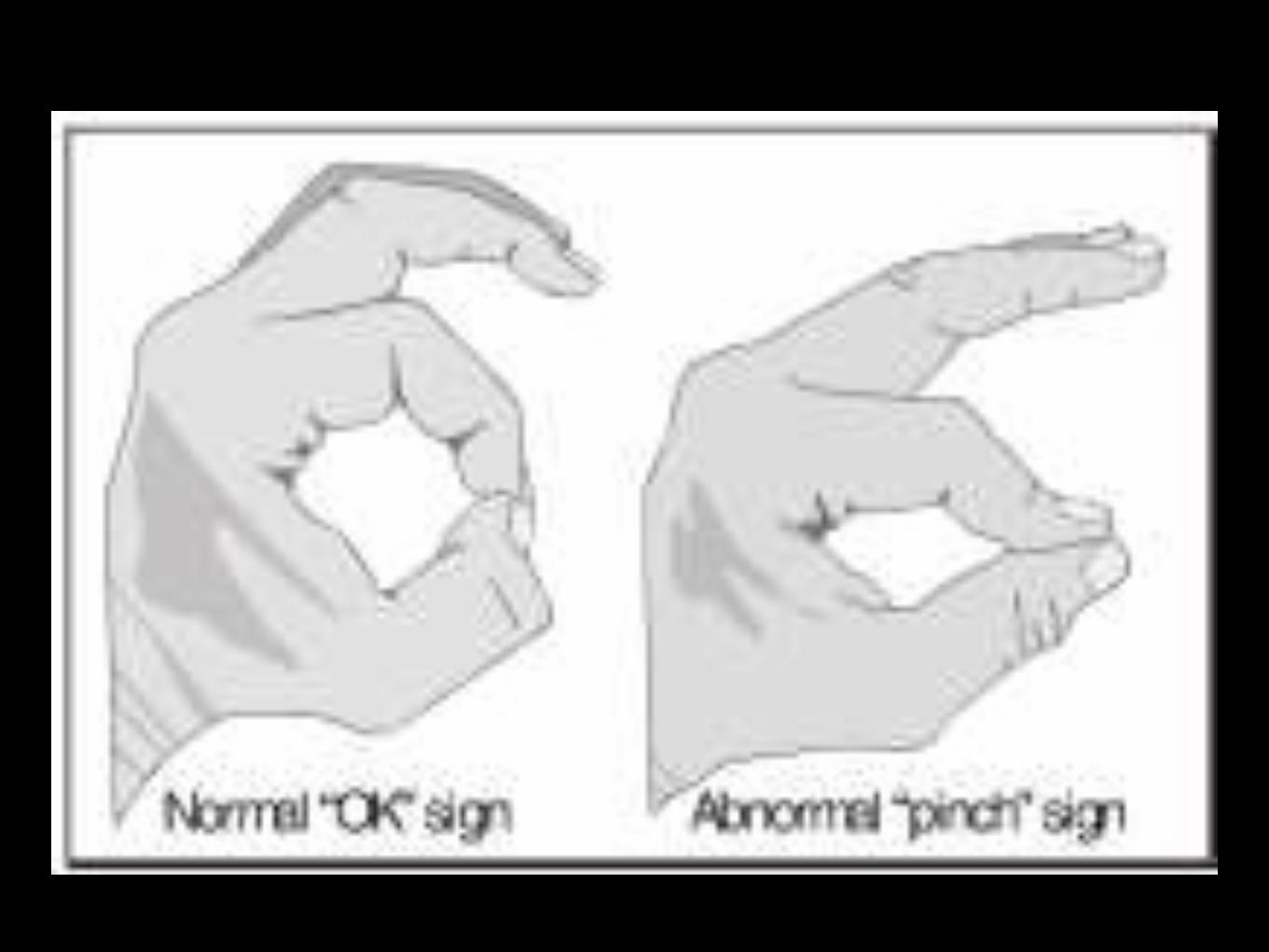
ANTERIOR INTEROSSEOUS NEUROPATHY
a purely motor branch of the median nerve that arises in the upper
forearm
produces no sensory loss and weakness of the 3 muscles supplied by
the nerve:
1. flexor digitorum profundus (FDP) I & II: flexion of distal phalanx of
digits 2 & 3
2. flexor pollicis longus (FPL): flexion of distal phalanx of thumb
3. pronator quadratus (in the distal forearm): difficult to isolate
clinically


CTS
Most common entrapment neuropathy
Median nerve compression by the transverse carpal lig
More women than men
Often bilat but almost without exception more prominent in
the dominant hand
Paresthesia usually in median nerve distribution
(thenar eminance spared as supplied by palmar cutaneous
sensory branch which comes off proximal to the carpal
tunnel
Advanced cases may have thenar muscle
weakness/wasting effecting thumb opposition and ab
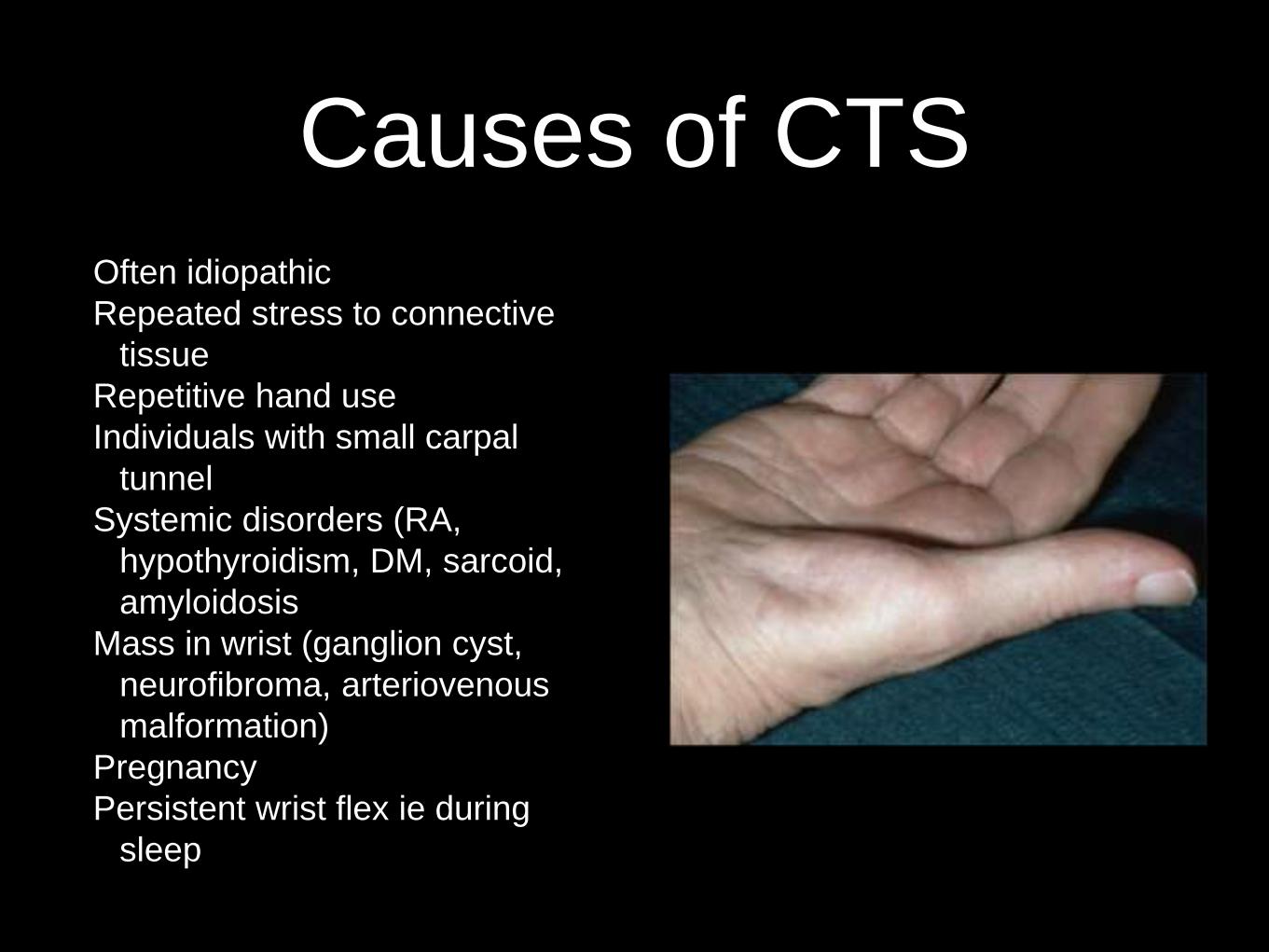
Causes of CTS
Often idiopathic
Repeated stress to connective
tissue
Repetitive hand use
Individuals with small carpal
tunnel
Systemic disorders (RA,
hypothyroidism, DM, sarcoid,
amyloidosis
Mass in wrist (ganglion cyst,
neurofibroma, arteriovenous
malformation)
Pregnancy
Persistent wrist flex ie during
sleep

Examination
Phalan’s good specific (75-93%)
and moderate sensitive (64-
75%) for CTS
Tinel’s similar spec & sens (tetro
et al, 1995 Bolland et al, 2008)
Carpal compression test more
spec less sensitive
NCS and EMG can help confirm
diagnosis and discount others
(however can be normal in 25%
of cases)

Differential diagnosis
1-C6-7 radiculopathy
2-Bracial plexopathy
3-Proximal median neuropathyThese can be identified by pain in the neck, reduced
reflexes, weakness outside median nerve distribution,
sensory loss in the thenar eminence

Treatment
Remove causative factorsSplints (night)NSAIDsInjection may be particularly helpful during pregnancy or other reversible condition i.e. Hypothyroidism
Surgical decompression

Ulnar Nerve entrapment
Second most common PNE in upper limb
Caused by compression of ulnar nerve in
the ulnar groove or cubital tunnel
Results from repeated trauma,OA
following #, ganglion/tumours/fibrous
tissue
Manifests as progressive loss of grip and
pinch strength and interosseus muscle
function Clumsiness
Wasting of thenar and hypothenar
eminence
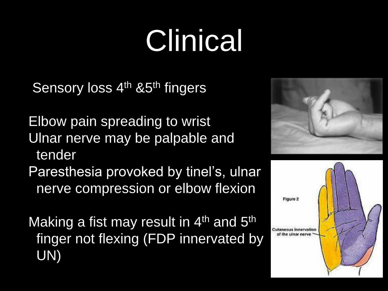
Clinical
Sensory loss 4th &5th fingers
Elbow pain spreading to wrist
Ulnar nerve may be palpable and
tender
Paresthesia provoked by tinel’s, ulnar
nerve compression or elbow flexion
Making a fist may result in 4th and 5th
finger not flexing (FDP innervated by
UN)

Several classic hand postures
may be present
1-Benediction posture
2-Wartenberg’s sign
3-Froment’s sign

Treatment
Conservative treatment
Avoid aggravating factors
Jt protection
Elbow splint
Surgical options
Transportation
Decompression cubital tunnel
Medial epicondylectomy
Many will recover spontaneously
but surgery very effective
90% of pt’s with mild symptoms
will recover with conservative Rx

Ulnar nerve compression at the wrist
Similar manifestation with weakness of
the hand intrinsics and thenar and
hypothenar eminences
Exacerbated by activities such as riding
bike or manual labour that repetitively
compresses ulnar side of the wrist and
Guyon’s canal
#, trauma, ganglion cysts, ulnar artery
thombus


Diagnosis and Treatment
May require MRI or CT
for diagnosis as well as
EMG and NCS
Conservative treatment
usually successful but
may require
decompression if mass
present
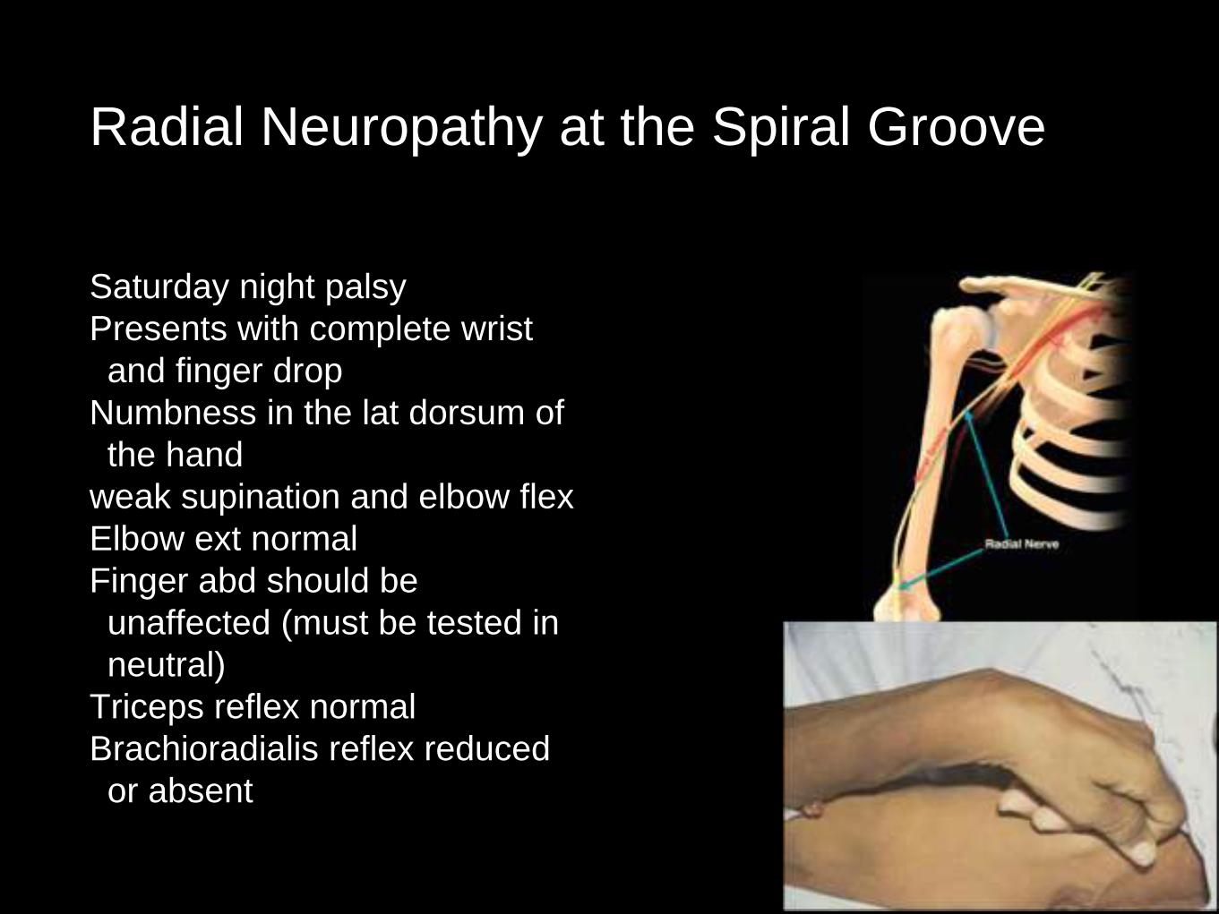
Radial Neuropathy at the Spiral Groove
Saturday night palsy
Presents with complete wrist
and finger drop
Numbness in the lat dorsum of
the hand
weak supination and elbow flex
Elbow ext normal
Finger abd should be
unaffected (must be tested in
neutral)
Triceps reflex normal
Brachioradialis reflex reduced
or absent

Radial nerve entrapment cause
Radial nerve lies in
juxtaposed to spiral
groove making it liable
to compression
Prolonged compression
leads to demylination
Can result from #
humerus, vasculitis or
stenuous muscle effort

Peroneal Neuropathy at the Fibular Head
Usually involves both deep and
superficial peroneal nerves
Therefore weakness in ankle df
and eversion
Sensory loss over dorsum of the
foot and lat calf
May be pain and Tinel’s over fib
neck
Ankle inversion spared as
innervated by Tib nerve.

Causes
Habitual leg crossing
Repetitive stretch from
squatting
Thin pt’s
Ganglions cyst
Associated to ankle
inversion injury including #
fib
Traction to nerve
Prolonged immobilisation
(especially sedated pt’s)

Treatment
local injected
AFO
Stretches to prevent contractures
Gait rehab
Proprioceptive work
Eliminate offending activities ie leg
crossing
Surgery rarely needed except
where extensive nerve damage
or mass present
