mediastinal germ cell tumor: a rare case report acute mast
TRANSCRIPT
Page 1/16
Acute Mast Cell Leukemia Preceded by MalignantMediastinal Germ Cell Tumor: A Rare Case Reportand Literature ReviewHuafang Wang
Zhejiang Provincial People's HospitalYuan Chen
Zhejiang Provincial People's HospitalHuijun Lin
Zhejiang Provincial People's HospitalJianping Lan
Zhejiang Provincial People's HospitalLai Jin ( [email protected] )
Zhejiang Provincial People's Hospital https://orcid.org/0000-0002-6203-6864
Case Report
Keywords: Case report, Mast cell leukemia; Germ cell tumor; Mediastinum; KIT; TP53
Posted Date: October 20th, 2021
DOI: https://doi.org/10.21203/rs.3.rs-968601/v1
License: This work is licensed under a Creative Commons Attribution 4.0 International License. Read Full License
Page 2/16
AbstractBackground: Mast cell leukemia (MCL) is a highly life-threatening and extremely rare subtype of systemicmastocytosis (SM). MCL, divided into de novo subtype and secondary to mastocytosis subtype, oftengenetically contains one or more somatic mutations, particularly the activating mutations of KIT. In thisstudy, an acute MCL case was reported, who had rare phenotype and genetic mutants with a history ofprimary malignant mediastinal germ cell tumor (GCT).
Case presentation: A male patient aged 30, who underwent two rounds of surgery and chemotherapy witha history of malignant mediastinal GCT, was admitted to our hospital due to persistent chest pain andsevere fatigue. The diagnosis of acute MCL was con�rmed by morphology analysis and chemicalstaining of marrow aspirate and biopsy, with the addition of C-�ndings including splenomegaly andcytopenia. The atypical MCs phenotypically expressed CD9, but no CD2 and CD25. Next gene sequencingof marrow aspirate identi�ed heterozygous mutations in TP53 P301Qfs*44, FLT3 R973X, SETBP1 N272D,and JAK3 I688F, whereas the mutations in KIT were not found. Although the initial therapy ofcorticosteroids and dasatinib-based regimen was effective, he died of acute respiratory distresssyndrome after the �rst cycle of chemotherapy with cladribine and cytarabine. The survival time of thepatient was 2.4 months after the initial presentation of MCL.
Conclusion: In this case, MCL preceded by malignant mediastinal GCT has similar clinical symptoms andmorphological manifestations but distinctly different genetic pro�les in contrast to primary MCL. Thecharacteristic morphology of MCL provides the most pivotal evidence that led our diagnosis in the rightdirection. A hypothesis was speculated that there is a common embryonal cancer stem cell betweenprimary malignant GCT and secondary de novo MCL, whereas the latter is gradually developed in thecontext of additional “driver mutations”.
IntroductionMastocytosis is a malignant disease characterized by the clonal expansion and in�ltration of mast cells(MCs) in the skin, marrow, and other organs. Due to its unique clinical and pathological features,mastocytosis has been excluded from the myeloproliferative neoplasm category and is regarded as adistinct disease category, according to the 2016 version WHO classi�cation of myeloid neoplasms [1].Mastocytosis is clinically subdivided into cutaneous mastocytosis, systemic mastocytosis (SM), andlocalized MC tumors, in which the �rst form usually appears in childhood with a favorable prognosis andthe latter two forms frequently develop in adulthood [2]. SM is a rare subtype characterized by multifocalin�ltration of high-grade MCs in the bone marrow and other various organs.
The diagnostic criteria of SM are categorized into major and minor criteria. The major criteria indicatemultifocal dense in�ltrates of MCs (≥15 MCs in aggregates) in BM biopsies and/or in otherextracutaneous organ(s). The minor criteria include a). > 25% of atypical MCs are detected on BM smearsor are spindle-shaped in MC in�ltrates detected on other organs. b). An activating point mutation at
Page 3/16
codon 816 of KIT in the marrow or another extra-cutaneous organ. c). MCs in the marrow, blood, oranother extra-cutaneous organ exhibit CD2 and/or CD25. d). Baseline serum tryptase level is >.20 ng/mL(in case of an unrelated myeloid neoplasm, item d is not valid as an SM criterion). SM is con�rmed withthe major criteria and at least 1 minor criteria, or more than 3 minor criteria [2, 3]. SM is classi�ed intoindolent SM, smoldering SM, SM with an associated hematologic (non-MC lineage) neoplasm, aggressiveSM (ASM), and mast cell leukemia (MCL) in the updated 2016 WHO classi�cation document [4, 5].
MCL is a highly life-threatening and extremely rare subtype of SM, accounting for less than 0.5% of SM[6]. It can be divided into de novo or secondary to earlier mastocytosis, and the ratio of the two subtypesis approximately 3:1 [7]. MCL is fatal because of its systemic nature and resistance to current therapeuticagents [7]. The diagnostic criteria for MCL are as follows: 1) the establishment of SM diagnosis; 2) theneoplastic in�ltration by atypical MCs in BM biopsy; 3) the presence of atypical MCs in marrow with orwithout other internal organs (more than 20% of BM nucleated cells) [5]. Traditionally, MCL includes analeukemic variant (most cases) when the percentage of atypical MCs < 10% of peripheral bloodmononuclear cells (PBMCs) and a classical/leukemic variant when the percentage ≥ 10% [8]. MCL canbe further subdivided into chronic versus acute MCL, and the latter follows a more aggressive course,with the presence of ≥ 1 C �ndings (including cytopenia, hepatomegaly, splenomegaly, andgastrointestinal or skeletal involvement) [3]. Neoplastic MCs usually express proto-oncogene KIT(CD117), tryptase, and CD25, with or without co-expression of CD2. Genetically, they often contain one ormore somatic mutations, represented by the activating mutations of KIT. In this study, a primary acuteMCL case of a young male patient was reported herein, who had rare phenotype and genetic mutantswith a history of primary malignant mediastinal germ cell tumor (GCT).
Case PresentationThis is a 30-year-old male patient, who had no family history of tumor and genetic disease, was �rstdiagnosed with a primary malignant mixed GCT encompassing seminoma and immature teratoma in themediastinum in 2010. At that time, he received the surgical tumorectomy and then was consolidated withradiation therapy concurrent with 4 cycles of cisplatin-based chemotherapy. At the end of 2014, however,he experienced the recurrence of GCT with a metastatic lesion located on the posterior basal segment ofthe lower lobe of the left lung, followed by undergoing pneumoresection and 6 cycles of consolidatedchemotherapy including vindesine, ifosfamide, and cis-platinum again. The pathological result revealed asingle immature teratoma. The patient afterward remained in a stable remission condition.
At the beginning of 2020, he was urgently admitted to our hospital with 1 day of persistent chest pain andsevere fatigue. Clinical symptoms showed low-grade fever, skin �ush, and splenomegaly. Complete bloodcell count showed anemia (hemoglobin 8.2 g/dL) and thrombocytopenia (38*109/L). Coagulation testspresented a higher D-dimer value (2,340 ug/L) and a slightly prolonged activated partial thromboplastintime (37.4s). The routine chemical analysis detected a high level of LDH (878 U/L) in peripheral blood.The results of computed tomography scan and transabdominal ultrasound revealed hepatosplenomegalyand seroperitoneum. PET-CT detected a diffuse increase in systemic bone metabolism,
Page 4/16
hepatosplenomegaly with slightly increased FDG metabolism, which was consistent with themanifestation of blood system diseases. There was no evidence pointing to the second recurrence ofGCT.
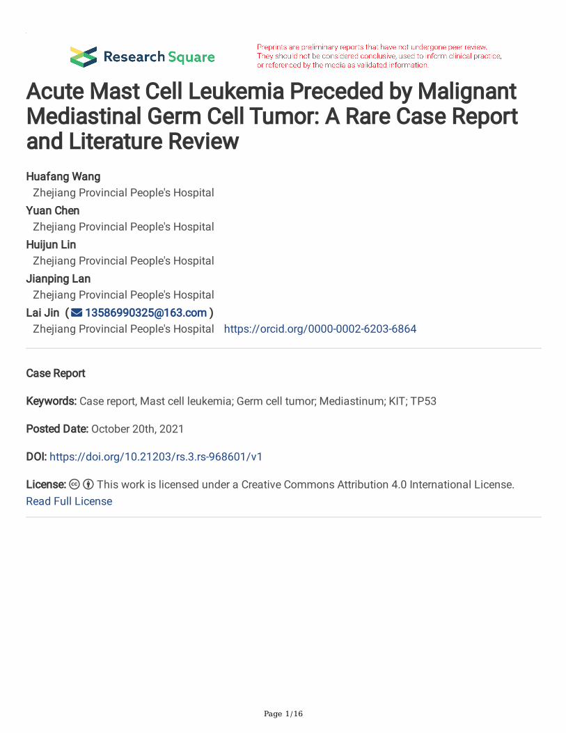
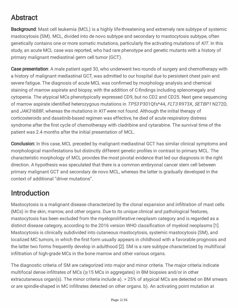
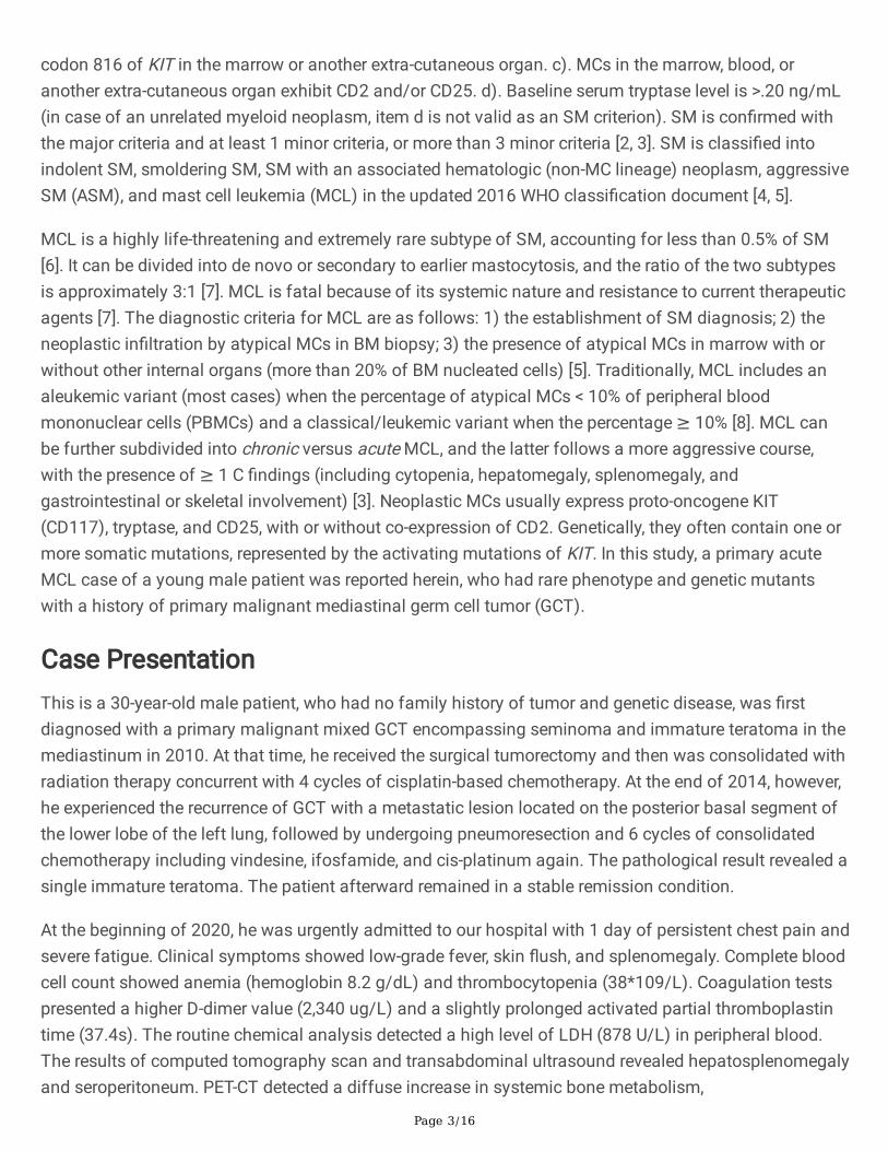
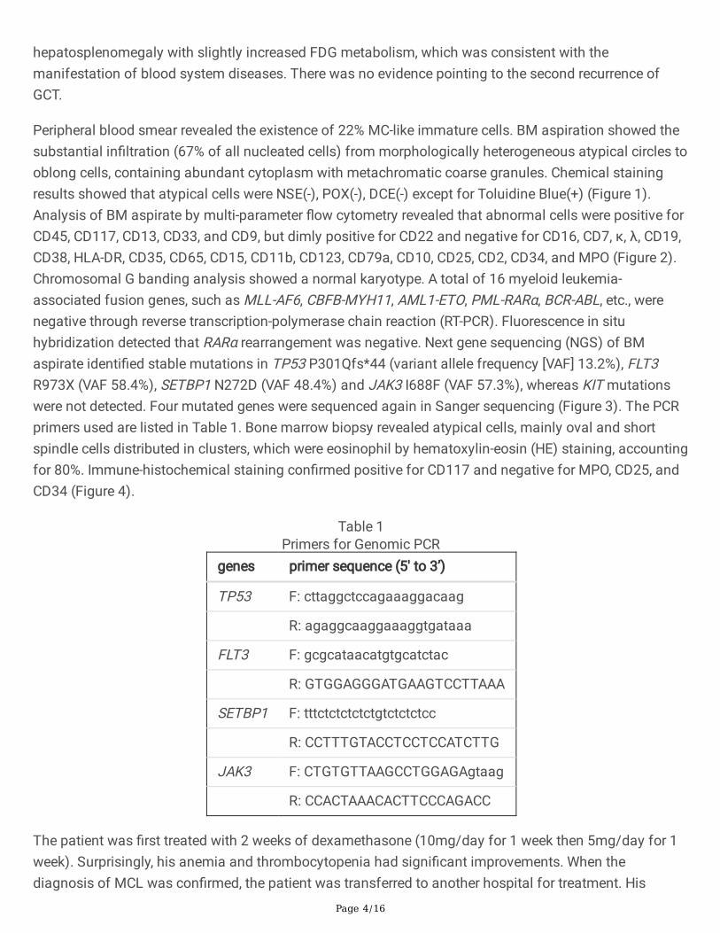
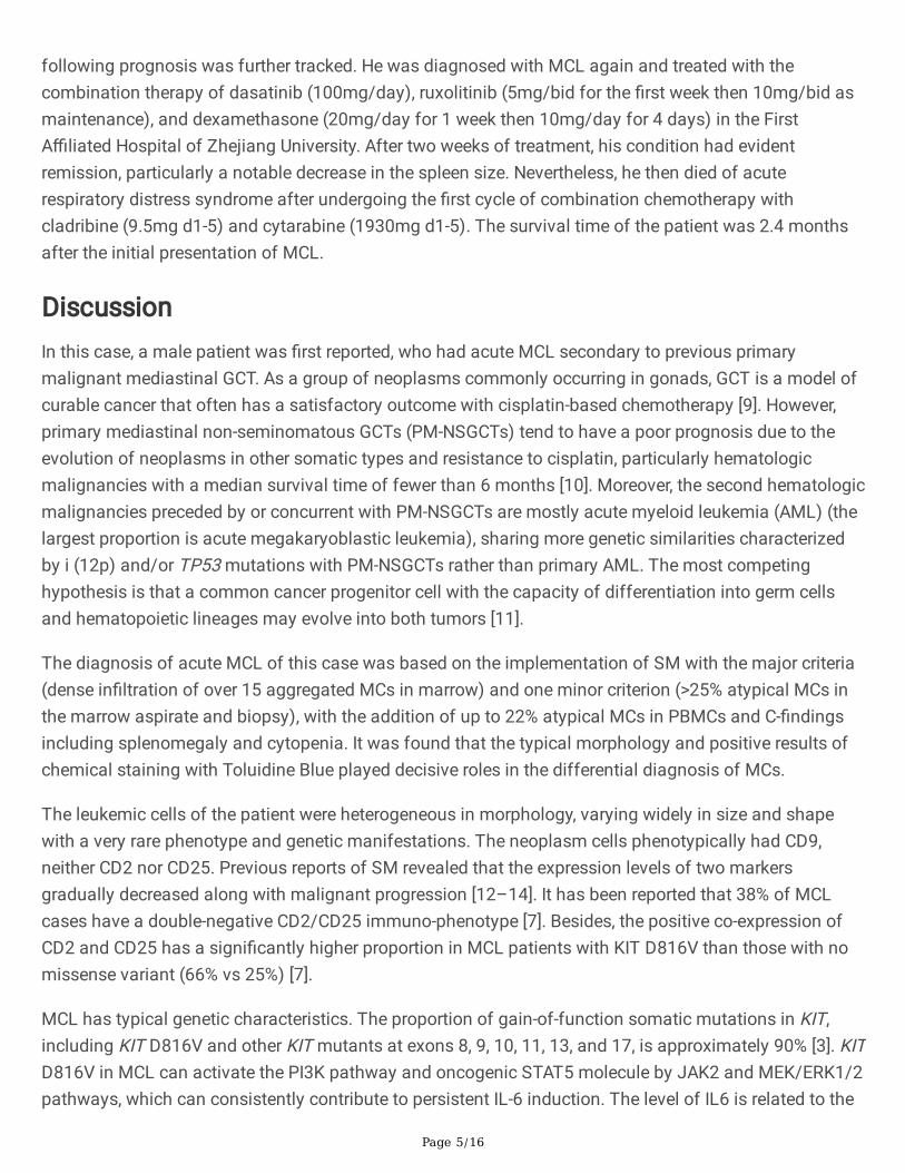
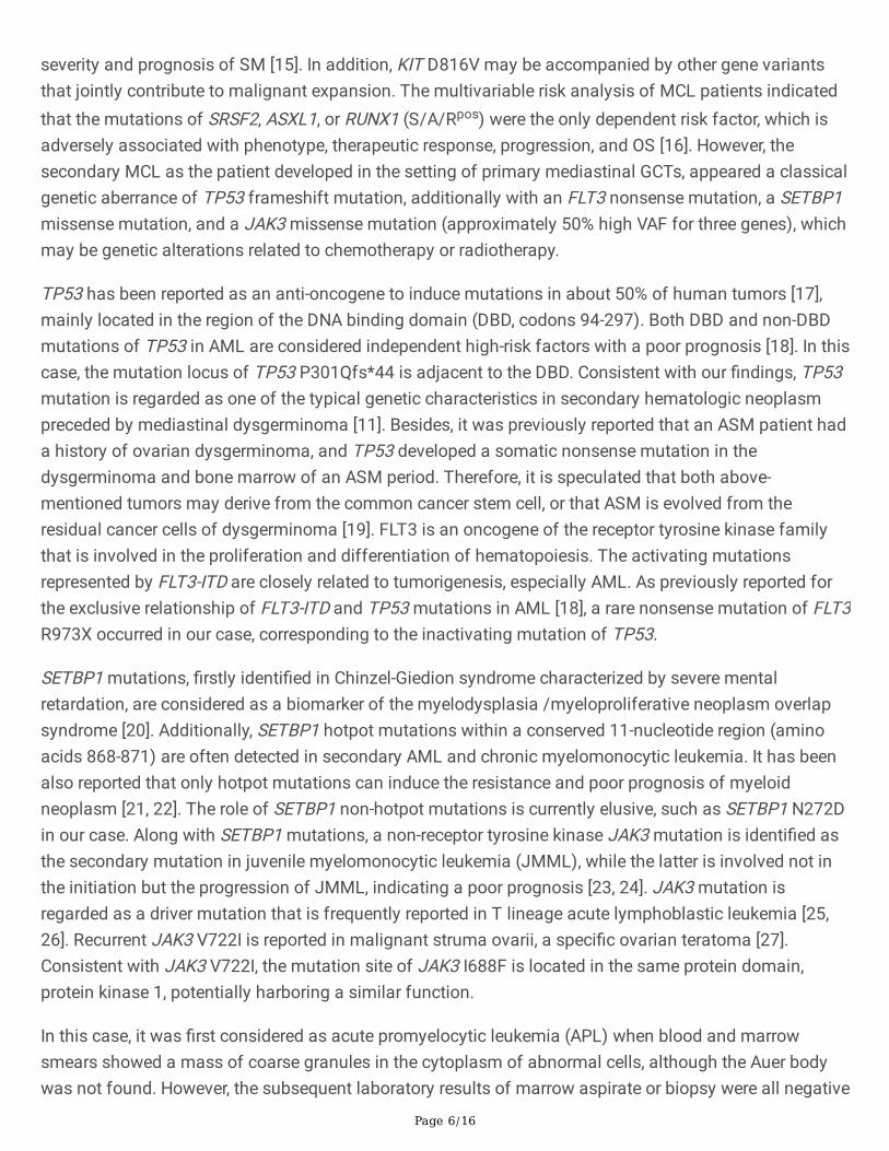
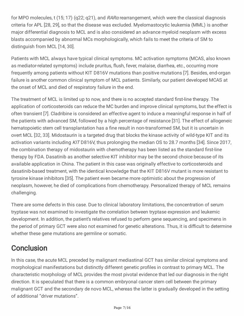
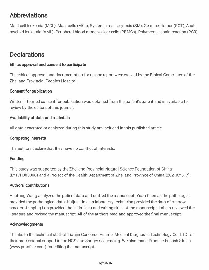
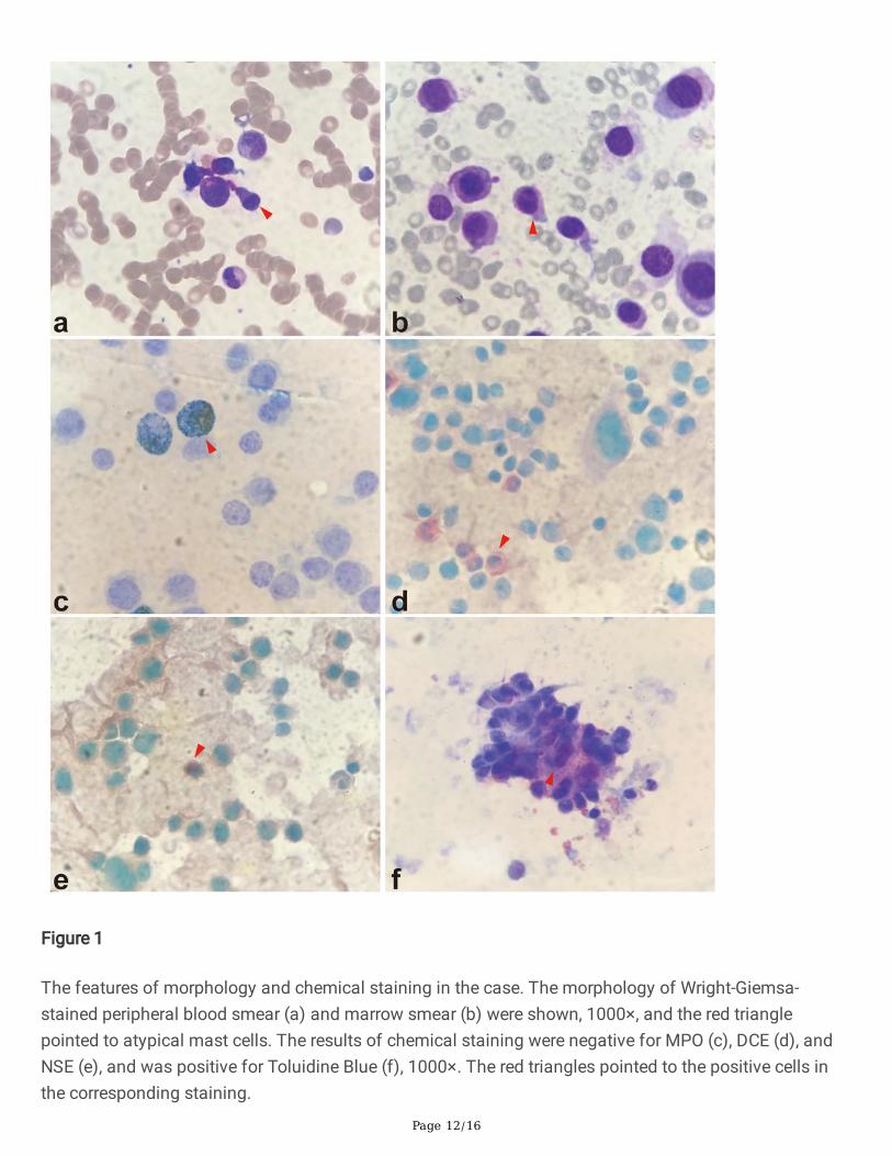
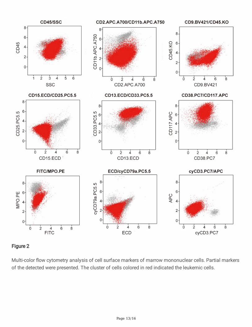
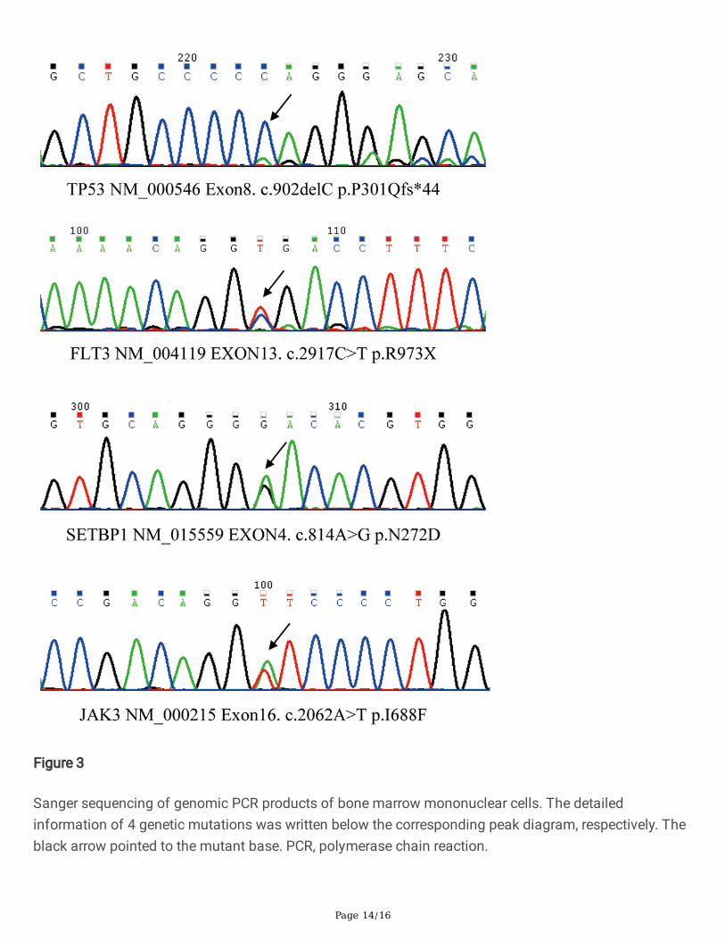
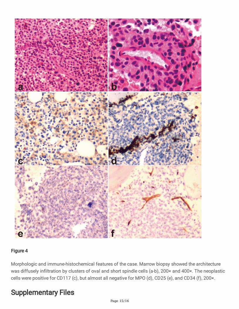
Peripheral blood smear revealed the existence of 22% MC-like immature cells. BM aspiration showed thesubstantial in�ltration (67% of all nucleated cells) from morphologically heterogeneous atypical circles tooblong cells, containing abundant cytoplasm with metachromatic coarse granules. Chemical stainingresults showed that atypical cells were NSE(-), POX(-), DCE(-) except for Toluidine Blue(+) (Figure 1).Analysis of BM aspirate by multi-parameter �ow cytometry revealed that abnormal cells were positive forCD45, CD117, CD13, CD33, and CD9, but dimly positive for CD22 and negative for CD16, CD7, κ, λ, CD19,CD38, HLA-DR, CD35, CD65, CD15, CD11b, CD123, CD79a, CD10, CD25, CD2, CD34, and MPO (Figure 2).Chromosomal G banding analysis showed a normal karyotype. A total of 16 myeloid leukemia-associated fusion genes, such as MLL-AF6, CBFB-MYH11, AML1-ETO, PML-RARα, BCR-ABL, etc., werenegative through reverse transcription-polymerase chain reaction (RT-PCR). Fluorescence in situhybridization detected that RARα rearrangement was negative. Next gene sequencing (NGS) of BMaspirate identi�ed stable mutations in TP53 P301Qfs*44 (variant allele frequency [VAF] 13.2%), FLT3R973X (VAF 58.4%), SETBP1 N272D (VAF 48.4%) and JAK3 I688F (VAF 57.3%), whereas KIT mutationswere not detected. Four mutated genes were sequenced again in Sanger sequencing (Figure 3). The PCRprimers used are listed in Table 1. Bone marrow biopsy revealed atypical cells, mainly oval and shortspindle cells distributed in clusters, which were eosinophil by hematoxylin-eosin (HE) staining, accountingfor 80%. Immune-histochemical staining con�rmed positive for CD117 and negative for MPO, CD25, andCD34 (Figure 4).
Table 1Primers for Genomic PCR
genes primer sequence (5' to 3’)
TP53 F: cttaggctccagaaaggacaag
R: agaggcaaggaaaggtgataaa
FLT3 F: gcgcataacatgtgcatctac
R: GTGGAGGGATGAAGTCCTTAAA
SETBP1 F: tttctctctctctgtctctctcc
R: CCTTTGTACCTCCTCCATCTTG
JAK3 F: CTGTGTTAAGCCTGGAGAgtaag
R: CCACTAAACACTTCCCAGACC
The patient was �rst treated with 2 weeks of dexamethasone (10mg/day for 1 week then 5mg/day for 1week). Surprisingly, his anemia and thrombocytopenia had signi�cant improvements. When thediagnosis of MCL was con�rmed, the patient was transferred to another hospital for treatment. His
Page 5/16
following prognosis was further tracked. He was diagnosed with MCL again and treated with thecombination therapy of dasatinib (100mg/day), ruxolitinib (5mg/bid for the �rst week then 10mg/bid asmaintenance), and dexamethasone (20mg/day for 1 week then 10mg/day for 4 days) in the FirstA�liated Hospital of Zhejiang University. After two weeks of treatment, his condition had evidentremission, particularly a notable decrease in the spleen size. Nevertheless, he then died of acuterespiratory distress syndrome after undergoing the �rst cycle of combination chemotherapy withcladribine (9.5mg d1-5) and cytarabine (1930mg d1-5). The survival time of the patient was 2.4 monthsafter the initial presentation of MCL.
DiscussionIn this case, a male patient was �rst reported, who had acute MCL secondary to previous primarymalignant mediastinal GCT. As a group of neoplasms commonly occurring in gonads, GCT is a model ofcurable cancer that often has a satisfactory outcome with cisplatin-based chemotherapy [9]. However,primary mediastinal non-seminomatous GCTs (PM-NSGCTs) tend to have a poor prognosis due to theevolution of neoplasms in other somatic types and resistance to cisplatin, particularly hematologicmalignancies with a median survival time of fewer than 6 months [10]. Moreover, the second hematologicmalignancies preceded by or concurrent with PM-NSGCTs are mostly acute myeloid leukemia (AML) (thelargest proportion is acute megakaryoblastic leukemia), sharing more genetic similarities characterizedby i (12p) and/or TP53 mutations with PM-NSGCTs rather than primary AML. The most competinghypothesis is that a common cancer progenitor cell with the capacity of differentiation into germ cellsand hematopoietic lineages may evolve into both tumors [11].
The diagnosis of acute MCL of this case was based on the implementation of SM with the major criteria(dense in�ltration of over 15 aggregated MCs in marrow) and one minor criterion (>25% atypical MCs inthe marrow aspirate and biopsy), with the addition of up to 22% atypical MCs in PBMCs and C-�ndingsincluding splenomegaly and cytopenia. It was found that the typical morphology and positive results ofchemical staining with Toluidine Blue played decisive roles in the differential diagnosis of MCs.
The leukemic cells of the patient were heterogeneous in morphology, varying widely in size and shapewith a very rare phenotype and genetic manifestations. The neoplasm cells phenotypically had CD9,neither CD2 nor CD25. Previous reports of SM revealed that the expression levels of two markersgradually decreased along with malignant progression [12–14]. It has been reported that 38% of MCLcases have a double-negative CD2/CD25 immuno-phenotype [7]. Besides, the positive co-expression ofCD2 and CD25 has a signi�cantly higher proportion in MCL patients with KIT D816V than those with nomissense variant (66% vs 25%) [7].
MCL has typical genetic characteristics. The proportion of gain-of-function somatic mutations in KIT,including KIT D816V and other KIT mutants at exons 8, 9, 10, 11, 13, and 17, is approximately 90% [3]. KITD816V in MCL can activate the PI3K pathway and oncogenic STAT5 molecule by JAK2 and MEK/ERK1/2pathways, which can consistently contribute to persistent IL-6 induction. The level of IL6 is related to the
Page 6/16
severity and prognosis of SM [15]. In addition, KIT D816V may be accompanied by other gene variantsthat jointly contribute to malignant expansion. The multivariable risk analysis of MCL patients indicatedthat the mutations of SRSF2, ASXL1, or RUNX1 (S/A/Rpos) were the only dependent risk factor, which isadversely associated with phenotype, therapeutic response, progression, and OS [16]. However, thesecondary MCL as the patient developed in the setting of primary mediastinal GCTs, appeared a classicalgenetic aberrance of TP53 frameshift mutation, additionally with an FLT3 nonsense mutation, a SETBP1missense mutation, and a JAK3 missense mutation (approximately 50% high VAF for three genes), whichmay be genetic alterations related to chemotherapy or radiotherapy.
TP53 has been reported as an anti-oncogene to induce mutations in about 50% of human tumors [17],mainly located in the region of the DNA binding domain (DBD, codons 94-297). Both DBD and non-DBDmutations of TP53 in AML are considered independent high-risk factors with a poor prognosis [18]. In thiscase, the mutation locus of TP53 P301Qfs*44 is adjacent to the DBD. Consistent with our �ndings, TP53mutation is regarded as one of the typical genetic characteristics in secondary hematologic neoplasmpreceded by mediastinal dysgerminoma [11]. Besides, it was previously reported that an ASM patient hada history of ovarian dysgerminoma, and TP53 developed a somatic nonsense mutation in thedysgerminoma and bone marrow of an ASM period. Therefore, it is speculated that both above-mentioned tumors may derive from the common cancer stem cell, or that ASM is evolved from theresidual cancer cells of dysgerminoma [19]. FLT3 is an oncogene of the receptor tyrosine kinase familythat is involved in the proliferation and differentiation of hematopoiesis. The activating mutationsrepresented by FLT3-ITD are closely related to tumorigenesis, especially AML. As previously reported forthe exclusive relationship of FLT3-ITD and TP53 mutations in AML [18], a rare nonsense mutation of FLT3R973X occurred in our case, corresponding to the inactivating mutation of TP53.
SETBP1 mutations, �rstly identi�ed in Chinzel-Giedion syndrome characterized by severe mentalretardation, are considered as a biomarker of the myelodysplasia /myeloproliferative neoplasm overlapsyndrome [20]. Additionally, SETBP1 hotpot mutations within a conserved 11-nucleotide region (aminoacids 868-871) are often detected in secondary AML and chronic myelomonocytic leukemia. It has beenalso reported that only hotpot mutations can induce the resistance and poor prognosis of myeloidneoplasm [21, 22]. The role of SETBP1 non-hotpot mutations is currently elusive, such as SETBP1 N272Din our case. Along with SETBP1 mutations, a non-receptor tyrosine kinase JAK3 mutation is identi�ed asthe secondary mutation in juvenile myelomonocytic leukemia (JMML), while the latter is involved not inthe initiation but the progression of JMML, indicating a poor prognosis [23, 24]. JAK3 mutation isregarded as a driver mutation that is frequently reported in T lineage acute lymphoblastic leukemia [25,26]. Recurrent JAK3 V722I is reported in malignant struma ovarii, a speci�c ovarian teratoma [27].Consistent with JAK3 V722I, the mutation site of JAK3 I688F is located in the same protein domain,protein kinase 1, potentially harboring a similar function.
In this case, it was �rst considered as acute promyelocytic leukemia (APL) when blood and marrowsmears showed a mass of coarse granules in the cytoplasm of abnormal cells, although the Auer bodywas not found. However, the subsequent laboratory results of marrow aspirate or biopsy were all negative
Page 7/16
for MPO molecules, t (15; 17) (q22; q21), and RARα rearrangement, which were the classical diagnosiscriteria for APL [28, 29], so that the disease was excluded. Myelomastocytic leukemia (MML) is anothermajor differential diagnosis to MCL and is also considered an advance myeloid neoplasm with excessblasts accompanied by abnormal MCs morphologically, which fails to meet the criteria of SM todistinguish from MCL [14, 30].
Patients with MCL always have typical clinical symptoms. MC activation symptoms (MCAS, also knownas mediator-related symptoms) include pruritus, �ush, fever, malaise, diarrhea, etc., occurring morefrequently among patients without KIT D816V mutations than positive mutations [7]. Besides, end-organfailure is another common clinical symptom of MCL patients. Similarly, our patient developed MCAS atthe onset of MCL and died of respiratory failure in the end.
The treatment of MCL is limited up to now, and there is no accepted standard �rst-line therapy. Theapplication of corticosteroids can reduce the MC burden and improve clinical symptoms, but the effect isoften transient [7]. Cladribine is considered an effective agent to induce a meaningful response in half ofthe patients with advanced SM, followed by a high percentage of resistance [31]. The effect of allogeneichematopoietic stem cell transplantation has a �ne result in non-transformed SM, but it is uncertain inovert MCL [32, 33]. Midostaurin is a targeted drug that blocks the kinase activity of wild-type KIT and itsactivation variants including KIT D816V, thus prolonging the median OS to 28.7 months [34]. Since 2017,the combination therapy of midostaurin with chemotherapy has been listed as the standard �rst-linetherapy by FDA. Dasatinib as another selective KIT inhibitor may be the second choice because of itsavailable application in China. The patient in this case was originally effective to corticosteroids anddasatinib-based treatment, with the identical knowledge that the KIT D816V mutant is more resistant totyrosine kinase inhibitors [35]. The patient even became more optimistic about the progression ofneoplasm, however, he died of complications from chemotherapy. Personalized therapy of MCL remainschallenging.
There are some defects in this case. Due to clinical laboratory limitations, the concentration of serumtryptase was not examined to investigate the correlation between tryptase expression and leukemicdevelopment. In addition, the patient’s relatives refused to perform gene sequencing, and specimens inthe period of primary GCT were also not examined for genetic alterations. Thus, it is di�cult to determinewhether these gene mutations are germline or somatic.
ConclusionIn this case, the acute MCL preceded by malignant mediastinal GCT has similar clinical symptoms andmorphological manifestations but distinctly different genetic pro�les in contrast to primary MCL. Thecharacteristic morphology of MCL provides the most pivotal evidence that led our diagnosis in the rightdirection. It is speculated that there is a common embryonal cancer stem cell between the primarymalignant GCT and the secondary de novo MCL, whereas the latter is gradually developed in the settingof additional “driver mutations”.
Page 8/16
AbbreviationsMast cell leukemia (MCL); Mast cells (MCs); Systemic mastocytosis (SM); Germ cell tumor (GCT); Acutemyeloid leukemia (AML); Peripheral blood mononuclear cells (PBMCs); Polymerase chain reaction (PCR).
DeclarationsEthics approval and consent to participate
The ethical approval and documentation for a case report were waived by the Ethical Committee of theZhejiang Provincial People’s Hospital.
Consent for publication
Written informed consent for publication was obtained from the patient’s parent and is available forreview by the editors of this journal.
Availability of data and materials
All data generated or analyzed during this study are included in this published article.
Competing interests
The authors declare that they have no conflict of interests.
Funding
This study was supported by the Zhejiang Provincial Natural Science Foundation of China(LY17H080008) and a Project of the Health Department of Zhejiang Province of China (2021KY517).
Authors’ contributions
Huafang Wang analyzed the patient data and drafted the manuscript. Yuan Chen as the pathologistprovided the pathological data. Huijun Lin as a laboratory technician provided the data of marrowsmears. Jianping Lan provided the initial idea and writing skills of the manuscript. Lai Jin reviewed theliterature and revised the manuscript. All of the authors read and approved the �nal manuscript.
Acknowledgments
Thanks to the technical staff of Tianjin Concorde Huamei Medical Diagnostic Technology Co., LTD fortheir professional support in the NGS and Sanger sequencing. We also thank Proo�ne English Studia(www.proo�ne.com) for editing the manuscript.
Page 9/16
References1. Arber DA, Orazi A, Hasserjian R, Thiele J, Borowitz MJ, Le Beau MM, Bloom�eld CD, Cazzola M,
Vardiman JW. The 2016 revision to the World Health Organization classi�cation of myeloidneoplasms and acute leukemia. Blood. 2016;127:2391–405.
2. Peter Valent CA, Metcalfe DD. Mastocytosis: 2016 updated WHO classi�cation and novel emergingtreatment concepts. Blood. 2017;129:1420–7.
3. Leguit R, Hebeda K, Kremer M, van der Walt J, Gianelli U, Tzankov A, Orazi A. The Spectrum ofAggressive Mastocytosis: A Workshop Report and Literature Review. Pathobiology. 2020;87:2–19.
4. Polyatskin IL, Artemyeva AS, Krivolapov YA. [Revised WHO classi�cation of tumors of hematopoieticand lymphoid tissues, 2017 (4th edition):lymphoid tumors]. Arkh Patol. 2019;81:59–65.
5. Pardanani A. Systemic mastocytosis in adults: 2019 update on diagnosis, risk strati�cation andmanagement. Am J Hematol. 2019;94:363–77.
�. Zheng Y, Nong L, Liang L, Wang W, Li T. De novo mast cell leukemia without CD25 expression andKIT mutations: a rare case report in a 13-year-old child. Diagn Pathol. 2018;13:14.
7. Georgin-Lavialle S, Lhermitte L, Dubreuil P, Chandesris MO, Hermine O, Damaj G. Mast cell leukemia.Blood. 2013;121:1285–95.
�. Preetesh Jain SW, Keyur P, Patel N, Sarwari J, Cortes. Hagop Kantarjian, Srdan Verstovsek: Mast cellleukemia (MCL): Clinico-pathologic and molecular features and survival outcome. Leuk Res.2017;59:105–9.
9. Cheng L, Albers P, Berney D, Feldman D, Daugaard G, Gilligan T, Looijenga L. Testicular cancer. Naturereviews Disease primers. 2018;4:29.
10. Hartmann J, Nichols C, Droz J, Horwich A, Gerl A, Fossa S, Beyer J, Pont J, Fizazi K, Einhorn L, et al.Hematologic disorders associated with primary mediastinal nonseminomatous germ cell tumors. JNatl Cancer Inst. 2000;92:54–61.
11. Taylor J, Donoghue M, Ho C, Petrova-Drus K, Al-Ahmadie H, Funt S, Zhang Y, Aypar U, Rao P, ChavanS, et al. Germ cell tumors and associated hematologic malignancies evolve from a common sharedprecursor. J Clin Investig. 2020;130:6668–76.
12. Escribano L, Díaz-Agustín B, Bellas C, Navalón R, Nuñez R, Sperr WR, Schernthaner GH, Valent P,Orfao A. Utility of �ow cytometric analysis of mast cells in the diagnosis and classi�cation of adultmastocytosis. Leuk Res. 2001;25:563–70.
13. Escribano L, Orfao A, Díaz-Agustin B, Villarrubia J, Cerveró C, López A, Marcos MA, Bellas C,Fernández-Cañadas S, Cuevas M, et al. Indolent systemic mast cell disease in adults:immunophenotypic characterization of bone marrow mast cells and its diagnostic implications.Blood. 1998;91:2731–6.
14. Valent P, Sotlar K, Sperr WR, Escribano L, Yavuz S, Reiter A, George TI, Kluin-Nelemans HC, Hermine O,Butter�eld JH, et al. Re�ned diagnostic criteria and classi�cation of mast cell leukemia (MCL) andmyelomastocytic leukemia (MML): a consensus proposal. Ann Oncol. 2014;25:1691–700.
Page 10/16
15. Araceli Tobío GB, Denise A. Morris,1 D-K, Kim MP, O’Connell HD, Komarow MC, Carter, Daniel Smrz,,Olivera DDMaA: Oncogenic D816V-KIT signaling in mast cells causes persistent IL-6 production.Haematologica 2020, 105:124-135.
1�. Jawhar M, Schwaab J, Meggendorfer M, Naumann N, Horny HP, Sotlar K, Haferlach T, Schmitt K,Fabarius A, Valent P, et al. The clinical and molecular diversity of mast cell leukemia with or withoutassociated hematologic neoplasm. Haematologica. 2017;102:1035–43.
17. Muller P, Vousden K. Mutant p53 in cancer: new functions and therapeutic opportunities. Cancer cell.2014;25:304–17.
1�. Terada K, Yamaguchi H, Ueki T, Usuki K, Kobayashi Y, Tajika K, Gomi S, Kurosawa S, Miyadera K,Tokura T, et al. Full-length mutation search of the TP53 gene in acute myeloid leukemia hasincreased signi�cance as a prognostic factor. Annals of hematology. 2018;97:51–61.
19. Tsutsumi M, Miura H, Inagaki H, Shinkai Y, Kato A, Kato T, Hamada-Tsutsumi S, Tanaka M, Kudo K,Yoshikawa T, Kurahashi H. An aggressive systemic mastocytosis preceded by ovariandysgerminoma. BMC Cancer. 2020;20:1162.
20. Linder K, Iragavarapu C, Liu D. SETBP1 mutations as a biomarker for myelodysplasia/myeloproliferative neoplasm overlap syndrome. Biomark Res. 2017;5:33.
21. Winkelmann N, Schäfer V, Rinke J, Kaiser A, Ernst P, Scholl S, Hochhaus A, Ernst T. Only SETBP1hotspot mutations are associated with refractory disease in myeloid malignancies. J Cancer Res ClinOncol. 2017;143:2511–9.
22. Makishima H. Somatic SETBP1 mutations in myeloid neoplasms. Int J Hematol. 2017;105:732–42.
23. Sakaguchi H, Okuno Y, Muramatsu H, Yoshida K, Shiraishi Y, Takahashi M, Kon A, Sanada M, ChibaK, Tanaka H, et al. Exome sequencing identi�es secondary mutations of SETBP1 and JAK3 injuvenile myelomonocytic leukemia. Nat Genet. 2013;45:937–41.
24. Wakamatsu M, Okuno Y, Murakami N, Miwata S, Kitazawa H, Narita K, Kataoka S, Ichikawa D,Hamada M, Taniguchi R, et al. Detection of subclonal SETBP1 and JAK3 mutations in juvenilemyelomonocytic leukemia using droplet digital PCR. Leukemia. 2021;35:259–63.
25. Liu Y, Easton J, Shao Y, Maciaszek J, Wang Z, Wilkinson MR, McCastlain K, Edmonson M, PoundsSB, Shi L, et al. The genomic landscape of pediatric and young adult T-lineage acute lymphoblasticleukemia. Nat Genet. 2017;49:1211–8.
2�. de Bock CE, Cools J. JAK3 mutations and HOXA9 expression are important cooperating events in T-cell acute lymphoblastic leukemia. Mol Cell Oncol. 2018;5:e1458014.
27. Poli R, Scatolini M, Grosso E, Maletta F, Gallo M, Liscia D, Nelva A, Cesario F, Forte G, Metovic J, et al.Malignant struma ovarii: next-generation sequencing of six cases revealed Nras, Braf, and Jak3mutations. Endocrine. 2021;71:216–24.
2�. Wang ZY, Chen Z. Acute promyelocytic leukemia: from highly fatal to highly curable. Blood.2008;111:2505–15.
29. Tran VT, Phan TT, Mac HP, Tran TT, Ho TT, Pho SP, Nguyen VN, Vo TM, Nguyen HT, Le TT, et al. Thediagnostic power of CD117, CD13, CD56, CD64, and MPO in rapid screening acute promyelocytic
Page 11/16
leukemia. BMC Res Notes. 2020;13:394.
30. Arredondo AR, Gotlib J, Shier L, Medeiros B, Wong K, Cherry A, Corless C, Arber DA, Valent P, GeorgeTI. Myelomastocytic leukemia versus mast cell leukemia versus systemic mastocytosis associatedwith acute myeloid leukemia: a diagnostic challenge. Am J Hematol. 2010;85:600–6.
31. Barete S, Lortholary O, Damaj G, Hirsch I, Chandesris MO, Elie C, Hamidou M, Durieu I, Suarez F,Grosbois B, et al. Long-term e�cacy and safety of cladribine (2-CdA) in adult patients withmastocytosis. Blood. 2015;126:1009–16. quiz 1050.
32. Ustun C, Reiter A, Scott BL, Nakamura R, Damaj G, Kreil S, Shanley R, Hogan WJ, Perales MA, Shore T,et al. Hematopoietic stem-cell transplantation for advanced systemic mastocytosis. J Clin Oncol.2014;32:3264–74.
33. Ustun C, Gotlib J, Popat U, Artz A, Litzow M, Reiter A, Nakamura R, Kluin-Nelemans HC, Verstovsek S,Gajewski J, et al. Consensus Opinion on Allogeneic Hematopoietic Cell Transplantation in AdvancedSystemic Mastocytosis. Biol Blood Marrow Transplant. 2016;22:1348–56.
34. Gotlib J, Kluin-Nelemans HC, George TI, Akin C, Sotlar K, Hermine O, Awan FT, Hexner E, Mauro MJ,Sternberg DW, et al. E�cacy and Safety of Midostaurin in Advanced Systemic Mastocytosis. N EnglJ Med. 2016;374:2530–41.
35. Zermati Y, De Sepulveda P, Féger F, Létard S, Kersual J, Castéran N, Gorochov G, Dy M, RibadeauDumas A, Dorgham K, et al. Effect of tyrosine kinase inhibitor STI571 on the kinase activity of wild-type and various mutated c-kit receptors found in mast cell neoplasms. Oncogene. 2003;22:660–4.
Figures
Page 12/16
Figure 1
The features of morphology and chemical staining in the case. The morphology of Wright-Giemsa-stained peripheral blood smear (a) and marrow smear (b) were shown, 1000×, and the red trianglepointed to atypical mast cells. The results of chemical staining were negative for MPO (c), DCE (d), andNSE (e), and was positive for Toluidine Blue (f), 1000×. The red triangles pointed to the positive cells inthe corresponding staining.
Page 13/16
Figure 2
Multi-color �ow cytometry analysis of cell surface markers of marrow mononuclear cells. Partial markersof the detected were presented. The cluster of cells colored in red indicated the leukemic cells.
Page 14/16
Figure 3
Sanger sequencing of genomic PCR products of bone marrow mononuclear cells. The detailedinformation of 4 genetic mutations was written below the corresponding peak diagram, respectively. Theblack arrow pointed to the mutant base. PCR, polymerase chain reaction.
Page 15/16
Figure 4
Morphologic and immune-histochemical features of the case. Marrow biopsy showed the architecturewas diffusely in�ltration by clusters of oval and short spindle cells (a-b), 200× and 400×. The neoplasticcells were positive for CD117 (c), but almost all negative for MPO (d), CD25 (e), and CD34 (f), 200×.
Supplementary Files

Page 16/16
This is a list of supplementary �les associated with this preprint. Click to download.
CAREchecklist.docx