mechanistic study of proton transfer and hysteresis in catalytic antibody 16e7 by site-directed...
TRANSCRIPT
Bioorganic & Medicinal Chemistry 13 (2005) 1021–1029
Mechanistic study of proton transfer and hysteresisin catalytic antibody 16E7 by site-directed
mutagenesis and homology modeling
Lei Zheng,a Roman Manetsch,b,� Wolf-Dietrich Woggon,b
Ulrich Baumanna and Jean-Louis Reymonda,*
aDepartment of Chemistry and Biochemistry, University of Berne, Freiestrasse 3, CH-3012 Berne, SwitzerlandbDepartment of Chemistry, University of Basel, St. Johanns-Ring 19, CH-4056 Basel, Switzerland
Received 11 October 2004; revised 19 November 2004; accepted 22 November 2004
Available online 19 December 2004
Abstract—Antibody 16E7 catalyzes the carbon protonation of enol ether 2 to hemiacetal 3, and the carbon deprotonation of benz-isoxazole 7 to phenol 8. This antibody shows an extreme case of hysteresis, requiring several hours to reach full activity. Antibody16E7 was expressed as recombinant chimeric Fab in Escherichia coli. A model for the three-dimensional structure was produced byhomology modeling and used for a docking procedure to obtain models for antibody–ligand complexes. Site-direct mutagenesis ofGluL39, identified as a possible catalytic residue by the model, to either glutamine or alanine abolished catalysis, showing that boththe protonation reaction of enol ether 2 and the deprotonation of benzisoxazole 7 are promoted by the same residue. The modelfurthermore suggested that substrate access to the catalytic site might be hindered by a flexible HCDR3 loop held in closed positionby a hydrogen bond between SerH99 and GluL39, which could explain the observed hysteresis effect. In agreement with this model,mutagenesis of SerH99 to alanine, or deletion of this residue, was found to reduce hysteresis by approximately 50%.� 2004 Elsevier Ltd. All rights reserved.
1. Introduction
Catalytic antibodies can be obtained by immunizationagainst small molecule haptens that are either transitionstate analogs or reactive probes corresponding to agiven reaction.1 Most catalytic antibodies acceleratebase-promoted hydrolytic processes or pericyclic reac-tions.2 Catalytic antibodies for carbon protonationand deprotonation reactions are of particular interestdue to the fundamental nature of this step, which isthe key to catalysis in many natural enzymes.3 We re-cently reported the preparation of catalytic antibody16E7, obtained by immunization against the guanidi-nium hapten 1.4 This antibody catalyzes proton transferto and from carbon in two separate reactions, which arethe acid-promoted protonation of enol ethers 2 to form
0968-0896/$ - see front matter � 2004 Elsevier Ltd. All rights reserved.
doi:10.1016/j.bmc.2004.11.041
Keywords: Catalytic antibody; Proton transfer; Homology modeling;
Site-directed mutagenesis.* Corresponding author. Tel.: +41 31 631 4325; fax: +41 31 631
8057; e-mail: [email protected]�Present address: The Scripps Research Institute, 10550 North Torrey
Pines Road, BCC-315, La Jolla, CA 92037, USA.
hemiacetal 3, and the base-promoted deprotonation ofbenzisoxazole 7 to form cyanophenol 8 (Fig. 1). Bothtypes of reactions have been previously observed indi-vidually in different catalytic antibodies.5–7 However,no catalytic antibody has been reported previously tocatalyze both reaction types within the same bindingpocket, which renders the molecular reaction mecha-nism of antibody 16E7 particularly interesting. In addi-tion, catalytic antibody 16E7 exhibits an unusual case ofhysteresis for both its protonation and its deprotonationreactions, whereby catalysis requires several hours toreach full activity in the presence of substrate. Thisretardation effect is reversible, suggesting an underlyingprotein dynamics effect.
Herein we report a site-directed mutagenesis study ofcatalytic antibody 16E7 guided by homology modeling.A three-dimensional model of catalytic antibody 16E7was produced and used in a docking procedure to gen-erate a model of the antibody complexes with hapten 1and substrates 2 and 7. Residue GluL39 was identifiedas the key catalytic residue responsible for both the pro-tonation and the deprotonation activity of antibody

MeO
NH
NH
NH
OH
O
CF3COO
1
MeO HO
O OR
Ab 16E7
2
OMeOH
MeO
OOH Me
HO
H
3 (R = H)4 (R = Me)
MeO
OHOH
OMeab
a
5 6
b
O2N
ON
7
Ab 16E7O2N
8
CN
O
H
H
Ab-COO
Ab 16E7
Figure 1. Carbon protonation and deprotonation reaction catalyzed
by antibody 16E7 (anti-1).
1022 L. Zheng et al. / Bioorg. Med. Chem. 13 (2005) 1021–1029
16E7. Furthermore, the modeling study provided amechanistic hypothesis linking the hysteresis effect to aslow breathing motion of the HCDR3 loop, which wastested by mutagenesis.
2. Materials and methods
2.1. Cloning of antibody 16E7 Fab gene fragment
Total RNA was isolated from the hybridoma cell line16E7 with RNeasy kit (Qiagen) and its cDNA librarywas constructed with the Omniscript kit (Qiagen). TheFd and kappa chain gene fragments of 16E7 were ampli-fied by PCR with the degenerated primer set as de-scribed.8 Amplified PCR products were digested withXhoI-SpeI for the Fd gene fragment and SacI-XbaIfor the kappa chain gene fragment, respectively, andligated into phage display vector pcomb3H8 to givepcomb3H-16E7. The resulting clone was checked forits affinity to the hapten-BSA conjugate with phage dis-play panning methods.8 All PCRs were carried out withVent proofreading polymerase (New England Biolab).
2.2. Construction of chimeric Fab fragment
The VL and VH genes of Ab 16E7 were amplified usingpcomb3H-16E7 as the template with the followingprimers: VL sense, CCAGATGTGAGCTCGTGACC-
CAGACTCCA (the SacI restriction site is underlined);VL anti-sense, GCAGCATCAGCCCGTTTTATTTC-AAGCTTGG (HindIII); VH sense, AGGTCCAGC-TGCTCGAGTCTGG (XhoI); VH anti-sense,GAGACGGTGACCAGAGTCCCTTGG (BstEII).The amplified fragments were cloned into appropriatesites in the vector p4xH-M139 to give a plasmid in whichthe VL and VH segments of the antibody 16E7 are fusedto human Cj and gamma1 CH1 regions. The chimericFab 16E7 gene fragment was subcloned intopBAD-43C9 vector10 by PCR with VL sense and CH1anti-sense primer, CACCGCCGGTCGACTCAGTGG-TGGTGGTGGTGGTGTGTGTGAGTTTTGTCAC(SalI), 6XHistidine tag was added at the C-terminus ofchimeric Fd. The resulting expression plasmid pBAD-16E7his allows one to express a 6X Histidine containingchimeric Fab 16E7.
2.3. Production, purification, and characterization ofchimeric Fab fragments
The Escherichia coli strain TOP10 (Invitrogen) wastransformed with pBAD-16E7his and its mutagenizedderivatives for expression of the wild type and mutantantibodies. 1/40 volume of preculture was inoculatedinto LB medium containing 100 lg/mL ampicillin at20 �C and overexpression was performed after inductionwith 0.1% LL(+)-arabinose at OD600 of 1.5. 1/10 culturevolume of Terrific Broth was added to implementnutrition for overexpression. After 14 h induction, thecrude periplasmic lysate was prepared by sonication.The chimeric Fab 16E7 fragment was purified by Ni2+
NTA affinity chromatography (Qiagen). Concentrationsof purified antibodies were determined by measuringA280 and A260.
11 The purity of the sample was con-firmed by SDS-PAGE. The same procedure was appliedfor the production and purification of mutants (seebelow). WT and mutant chimeric 16E7-Fab fragmentswere obtained in similar purified yields of 0.6–1.0 mg/Lculture.
2.4. Site-directed mutagenesis
Site-directed mutagenesis was accomplished by a Quik-ChangeTM site-directed mutagenesis protocol with minormodification and including silent restriction site. AllPCRs were performed with the High Fidelity System(Roche). Mutant codons were generally chosen to ex-ploit the most frequently used codon for a particularamino acid among highly expressed genes in E. coli. Oli-gonucleotides were custom-synthesized and purified byMicroSynth (Balgach, Switzerland) as follows:
DH50A_for GGAGcTATTTACCCTGGAtccGGGAA-TACTTACTAC (BamHI)DH50A_rev CCCggaTCCAGGGTAAATAgCTCCAA-TCCACTCAAGTCCEL39Q_for CCTATTTAcaATGGTAtCTGCAGAAA-CCAGGCC (KpnI deletion)EL39Q_rev GCAGaTACCATtgTAAATAGGTGTTTC-CATTACEL39A_for CCTATTTAGcATGGTAtCTGCAGAAA-CCAGGCC (KpnI deletion)

L. Zheng et al. / Bioorg. Med. Chem. 13 (2005) 1021–1029 1023
EL39A_rev GCAGaTACCATgCTAAATAGGTGTT-TCCATTACSH99A_for GGTACTACGGTgcTGGCGCTGTCTCC-TGGGGCSH99A_rev CAGCGCCAgcACCGTAGTACCCgCgg-GCACAGAAATAGAC (SacII)SH99del_forGGTACTACGGTGGCGCTGTCTCCTGGGGCGSH99del_rev GAGACAGCGCCACCGTAGTACCCg-CggGCACAGAAATAG (SacII)
For all primers, mutagenized positions are denoted inlowcase. The restriction sites are underlined.
2.5. Kinetic measurements
A solution of antibody in BisTris buffer, pH 6.24 at37 �C was mixed with a substrate solution (acetoni-trile/buffer = 1:1) providing a final solution containing29 mM BisTris, 104 mM NaCl, 10% acetonitrile. Thefinal antibody concentration was 250–540 lg/mL forthe hydrolysis of the enol ether and 24–90 lg/mL forthe Kemp elimination. Product formation was followedby RP18-HPLC on a Bischoff LiChrospher 100(4.6 · 125 mm) column using isocratic elution at 1 mL/min with a premixed acetonitrile/water solution withthe desired proportion. The 16E7-catalyzed eliminationwas followed by measuring A380 using a Molecular De-vices microtiter plate reader. Measurements were takenat intervals of 60, 90, or 120 s. With all substrates, theantibody-catalyzed reactions were carried out in parallelwith an equivalent reaction containing excess hapten 1(20 lM) for inhibition. All reactions of WT and mutantFab-16E7 in the presence of hapten 1 gave backgroundlevel reaction rates, indicating that the observed activi-ties originated from the antigen combining site of theantibodies.
2.6. Data treatment
For the hydrolysis of the enol ether, the values for theinitial velocity vi and for the final velocity vss were graph-ically estimated from the slopes of the progress curve.From following equation:
T ¼ ðvss � viÞvss
� s, ð1Þ
the value T of the intercept on the time axis was used todetermine the apparent rate constant s for the transitionbetween the two velocities vi and vss.
4,12 For the Kempelimination the data were analyzed with Kaleidagraph(Abelbeck Software). The three parameters vi, vss, ands were obtained from the following equation:4,12
½P � ¼ vss � t � ðvss � viÞ � ð1� e�t=sÞ � s: ð2Þ
2.7. Michaelis-Menten kinetics
The hydrolysis of the enol ethers was initiated by addingthe substrate to the antibody in a capped microtiterplate. The microtiter plate was kept in a closed plasticbox with a wet paper towel inside for humidity satura-
tion at 37 �C. Substrate and product concentrationswere measured by RP-C18-HPLC after 12, 20, and28 h incubation time, allowing to determine the initialvelocities vi and the final velocities vss for the differentsubstrate concentrations used in the measurements.After correction of these rates for the uncatalyzed reac-tion rates in BisTris buffer, the net rates vi and vss wereobtained. These rates were used to derive the Michaelis-Menten constants KM and the maximum velocity Vmax
from the Lineweaver–Burk plot of 1/V versus 1/[S].The catalytic constant kcat was obtained by dividingVmax by the antibody concentration. For the Kempelimination the obtained velocities vi and vss fromEq. (2) were used to determine the value KM/Vmax fromthe Lineweaver–Burk plot of 1/V versus 1/[S].
3. Results
3.1. Hysteresis character of chimeric Fab 16E7
Antibody 16E7 was expressed as chimeric Fab in E. coliafter joining the variable heavy and light chains regionswith the human kappa chain constant region and heavychain IgG1 CH1 region, respectively. This chimeric anti-body catalyzes both the protonation of enol ether 2 andthe deprotonation of benzisoxazole 7 with activityparameters comparable to those of the hybridoma de-rived IgG antibody, including the hysteresis phenome-non characterized by the retardation time s, the initialvelocity vi and the steady-state velocity vss (Table 1).In the case of enol ether 2 the transition between the ini-tial phase of catalysis (vi) and the steady-state phase ofcatalysis (mss) is accompanied by an increase in substratebinding (threefold lower KM), while the catalytic rateconstant kcat is unaffected (Table 2). Only the specificityconstant kcat/KM was determined for benzisoxazole 7since no substrate saturation occurred within the con-centration range accessible with this substrate, and wasfound to increase by two-fold between the initial phaseand the steady-state phase.
3.2. Homology modeling and docking
The 3-D structure of the antibody 16E7 Fv domain wasconstructed using the online antibody structure model-ing service WAM (Web Antibody Modeling, http://antibody.bath.ac.uk/).13 The whole molecule energyminimization procedure resulted in 200 antibody struc-tures for antibody 16E7, which were ordered withascending minimization energy. The obtained antibodymodel was then used in a docking procedure for the dif-ferent ligands using the docking program AUTODOCKAUTODOCK
3.053.05.14 All docking attempts with the tight binding hap-ten 1 or the substrates 2 and 7 failed starting with thelowest energy model 16E7-E1. However, the dockingprocedure was successful when starting from the secondlowest energy model 16E7-E2.
In the resulting model of the antibody–hapten complex,ligand 1 appears to be buried in a very deep pocketlocated at the center of Fv domain. The hapten is indirect contact with two carboxylate residues, GluL39
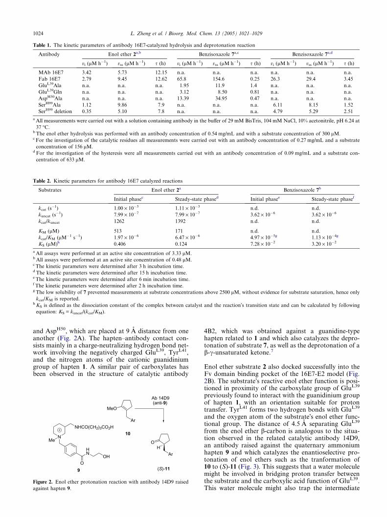
Table 1. The kinetic parameters of antibody 16E7-catalyzed hydrolysis and deprotonation reaction
Antibody Enol ether 2a,b Benzisoxazole 7a,c Benzisoxazole 7a,d
vi (lM h�1) vss (lM h�1) s (h) vi (lM h�1) vss (lM h�1) s (h) vi (lM h�1) vss (lM h�1) s (h)
MAb 16E7 3.42 5.73 12.15 n.a. n.a. n.a. n.a. n.a. n.a.
Fab 16E7 2.79 9.45 12.62 65.8 154.6 0.25 26.3 29.4 3.45
GluL39Ala n.a. n.a. n.a. 1.95 11.9 1.4 n.a. n.a. n.a.
GluL39Gln n.a. n.a. n.a. 3.12 8.50 0.81 n.a. n.a. n.a.
AspH50Ala n.a. n.a. n.a. 13.39 34.95 0.47 n.a. n.a. n.a.
SerH99Ala 1.12 9.86 7.9 n.a. n.a. n.a. 6.11 8.15 1.52
SerH99 deletion 0.35 5.10 7.8 n.a. n.a. n.a. 4.79 5.29 2.51
a All measurements were carried out with a solution containing antibody in the buffer of 29 mM BisTris, 104 mM NaCl, 10% acetonitrile, pH 6.24 at
37 �C.b The enol ether hydrolysis was performed with an antibody concentration of 0.54 mg/mL and with a substrate concentration of 300 lM.c For the investigation of the catalytic residues all measurements were carried out with an antibody concentration of 0.27 mg/mL and a substrate
concentration of 156 lM.d For the investigation of the hysteresis were all measurements carried out with an antibody concentration of 0.09 mg/mL and a substrate con-
centration of 633 lM.
Table 2. Kinetic parameters for antibody 16E7 catalyzed reactions
Substrates Enol ether 2a Benzisoxazole 7b
Initial phasec Steady-state phased Initial phasee Steady-state phasef
kcat (s�1) 1.00 · 10�3 1.11 · 10�3 n.d. n.d.
kuncat (s�1) 7.99 · 10�7 7.99 · 10�7 3.62 · 10�6 3.62 · 10�6
kcat/kuncat 1262 1392 n.d. n.d.
KM (lM) 513 171 n.d. n.d.
kcat/KM (lM�1 s�1) 1.97 · 10�6 6.47 · 10�6 4.97 · 10�5g 1.13 · 10�4g
KS (lM)h 0.406 0.124 7.28 · 10�2 3.20 · 10�2
a All assays were performed at an active site concentration of 3.33 lM.bAll assays were performed at an active site concentration of 0.48 lM.c The kinetic parameters were determined after 3 h incubation time.d The kinetic parameters were determined after 15 h incubation time.e The kinetic parameters were determined after 6 min incubation time.f The kinetic parameters were determined after 2 h incubation time.g The low solubility of 7 prevented measurements at substrate concentrations above 2500 lM, without evidence for substrate saturation, hence only
kcat/KM is reported.h KS is defined as the dissociation constant of the complex between catalyst and the reaction�s transition state and can be calculated by following
equation: KS = kuncat/(kcat/KM).
1024 L. Zheng et al. / Bioorg. Med. Chem. 13 (2005) 1021–1029
and AspH50, which are placed at 9 A distance from oneanother (Fig. 2A). The hapten–antibody contact con-sists mainly in a charge-neutralizing hydrogen bond net-work involving the negatively charged GluL39, TyrL41,and the nitrogen atoms of the cationic guanidiniumgroup of hapten 1. A similar pair of carboxylates hasbeen observed in the structure of catalytic antibody
MeO
10
OH
Ab 14D9 (anti-9)
(S)- 11O
HN
OH
NMe
Ar
ArNHCO(CH2)3CO2H
9
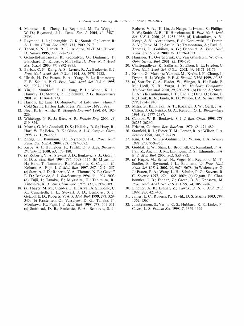
Figure 2. Enol ether protonation reaction with antibody 14D9 raised
against hapten 9.
4B2, which was obtained against a guanidine-typehapten related to 1 and which also catalyzes the depro-tonation of substrate 7, as well as the deprotonation of ab-c-unsaturated ketone.7
Enol ether substrate 2 also docked successfully into theFv domain binding pocket of the 16E7-E2 model (Fig.2B). The substrate�s reactive enol ether function is posi-tioned in proximity of the carboxylate group of GluL39
previously found to interact with the guanidinium groupof hapten 1, with an orientation suitable for protontransfer. TyrL41 forms two hydrogen bonds with GluL39
and the oxygen atom of the substrate�s enol ether func-tional group. The distance of 4.5 A separating GluL39
from the enol ether b-carbon is analogous to the situa-tion observed in the related catalytic antibody 14D9,an antibody raised against the quaternary ammoniumhapten 9 and which catalyzes the enantioselective pro-tonation of enol ethers such as the tranformation of10 to (S)-11 (Fig. 3). This suggests that a water moleculemight be involved in bridging proton transfer betweenthe substrate and the carboxylic acid function of GluL39.This water molecule might also trap the intermediate

Figure 3. Views of the 16E7 Fv model combining site. The residues in the binding pocket are drawn in gray stick and the ligands are shown in green.
(A) Complex with docked hapten 1. (B) Complex with enol ether substrate 2. (C) Complex with docked benzisoxazole substrate 7. (D) HCDR3
flexibility in models 16E7 E1-E7. E1: green; E2: cyan; E3: olive; E4: orange; E5: lime; E6: magenta; E7: salmon. The GluL39 and SerH99 of model E1,
E2 are shown in green and gray sticks, respectively.
L. Zheng et al. / Bioorg. Med. Chem. 13 (2005) 1021–1029 1025
oxocarbonium cation 5 to form the acyclic hemiacetal 6as the primary reaction product (Fig. 1). AspH50 forms ahydrogen bond with the phenolic OH-group of substrate2, holding this group away from the enol ether function.This orientation might explain the fact that enol ether 2does not undergo an intramolecular cyclization to thecorresponding acetal 4 under antibody catalysis, butrather undergoes hydrolysis to 3 as observed in thebackground reaction in water.
Benzisoxazole 7 also docked into 16E7-E2 model to pro-duce a stable complex (Fig. 2C). In this case, the criticalacidic residue GluL39 forms two hydrogen bonds, onewith TyrL41, and another with the nitrogen atom inthe substrate�s benzisoxazole ring. The substrate�sdeprotonation site is located at a distance of 3 A fromthe carboxylate of GluL39 in an arrangement suitablefor direct proton transfer.
3.3. Mutagenesis of catalytic residues
Mutagenesis experiments suggested by the modeling anddocking study were carried out to probe the reactionmechanism of antibody 16E7 (Table 1). The role ofGluL39, which appeared as the best candidate for a cat-alytic residue, was investigated first. Mutation of GluL39
to either glutamine or alanine abolished catalytic activ-ity in both, the protonation reaction with enol ether 2and the deprotonation reaction with benzisoxazole 7,establishing that this residue is critical for catalysis inantibody 16E7. Residue AspH50 was mutated to alanineto remove its hydrogen-bonding interaction with thephenolic hydroxyl group of enol ether 2 as observed inthe docked model (Fig. 2A), with the hope that theAspH50Ala mutant might promote an intramolecularcyclization of enol ether 2 to form the cyclic acetal 4.Unfortunately, the AspH50Ala mutant was completelyinactive for the protonation reaction. Nevertheless, thismutant retained 25% of the WT activity for the deproto-nation reaction with benzisoxazole 7. The selective lossof catalysis for the enol ether reaction suggests that thisreaction is more sensitive to perturbations than theKemp�s elimination reaction of benzisoxazole 7, inagreement with the fact that the later reaction is verysensitive to non-specific catalysis effects.16 The loss ofactivity in the case of enol ether 2 might be due to per-turbed substrate binding.
3.4. Analysis of loop movements
The fact that docking hapten 1 or the substrates ontothe lowest energy 16E7-E1 model did not produce a

Figure 4. Comparsion of 16E7 models molecular surface. Top: the
16E7-E1 model, bottom: the 16E7-E2 model.
Table 3. The conformation characters of HCDR3 with increasing
energy
Energy minimization
conformation
DG
(kcal/mol)
Loop flexibility
(deg)
E1 0 0
E2 1.30 67.64
E3 2.07 33.47
E4 4.22 54.21
E5 4.48 47.68
E6 4.65 42.03
E7 4.82 23.15
E1–E7 are the generated conformations during whole molecular min-
imization and ordered by increasing energies. DG is the energy differ-
ence between every conformation and the minimized conformation.
Loop angles were measured with the program Swisspdbviewer. The Caatom of GlycineH98 in the HCDR3 conformation of E1 was chosen as
reference point and defined as an angle of 0�. Angles are defined for the
motion of the Ca atom of GlyH98 in relation to the Ca atom of ArgH95
as fixed point.
0
0.1
0.2
0.3
0.4
0.5
0.6
0.7
0.8
0 10 20 30 40 50Model number
Mov
emen
t (nm
)
-0.4
-0.3
-0.2
-0.1
0
0.1
0.2
0.3
0.4
0 10 20 30 40 50Model number
Mov
emen
t (nm
)
Figure 5. Scalar representation of CDR3 loops flexibility of 16E7 Fv
models E1–E50. Two atoms on the tip of the CDR3 loops of heavy
and light chain were used to calculate the movement of their loops.
Top: Ca atom of the GlyH98, bottom: Ca atom of ValL99. Their
positions in the E1 model are set to zero and compared to the
corresponding atoms in the conformations of the other models.
1026 L. Zheng et al. / Bioorg. Med. Chem. 13 (2005) 1021–1029
complex suggested that the ligands could not enter thebinding pocket in this model. Indeed AUTODOCKAUTODOCK 3.053.05
employs a molecule �walking� procedure on the surfaceof the molecule to search the potential binding pocket.The structural difference between the 16E7-E1 modeland the 16E7-E2 model used to produce dockedcomplexes was identified as a large conformationalchange in the HCDR3 loop. In the 16E7-E1 model, thisloop closes the binding pocket unusually deeply, pre-venting ligand access. In the 16E7-E2 model, by con-trast, the loop is placed such as to leave the pocketopen (Fig. 4).
The five next lower energy models for 16E7 were exam-ined. These were found to represent intermediate statesof a loop movement between the 16E7-E1 and the16E7-E2 conformation (Fig. 2D). The HCDR3loop movement involved seven contiguous residues(TyrH96-GlyH97-GlyH98-SerH99-AlaH100-ValH101-SerH102)and featured a rotation spanning 67� as measured forthe Ca atom of GlyH98 in relation to the Ca atom ofArgH95 as fixed point. The calculated energy differencebetween E1 and the least stable of these seven confor-mers amounted to 4.8 kcal/mol, suggesting that the looprearrangement required a significant activation energy(Table 3). Further examination of the 50 lowest energyconformers showed that the HCDR3 loop was moreflexible compared to its LCDR3 counterpart (Fig. 5).
3.5. Mutagenesis of Serine H99
The movement of the HCDR3 observed between the16E7-E1 model and the 16E7-E2 model might explainthe hysteresis behavior of antibody 16E7. Indeed, thesubstrates might meet a similar entry problem in the realantibody to that observed in the docking procedure withthe 16E7-E1 model. Thus, the 16E7-E1 model mightrepresent a conformation existing in the catalytic anti-body during the initial kinetic phase (vi), while the16E7-E2 model might represent the antibody conforma-tion during the activated state of the kinetic phase (vss).

L. Zheng et al. / Bioorg. Med. Chem. 13 (2005) 1021–1029 1027
Since SerH99 played a crucial role in locking the closedconformation of the 16E7-E1 model by hydrogen-bond-ing interactions with GluL39, we reasoned that mutatingthis residue to an alanine might destabilize the inactivestate of antibody 16E7 and therefore reduce the activa-tion time s for reaching full activity. Substituting alaninefor serine at position H99 produced a catalytically activemutant for both the protonation of enol ether 2 and thedeprotonation of benzisoxazole 7, showing approxi-mately 25% of WT activity in both cases. More impor-tantly, the hysteresis time s in the SerH99Ala mutantwas reduced to 44% of WT for the reaction of benzisox-azole 7, and to 62% of WT for the protonation of enolether 2 (Table 1, Fig. 6). Similar reductions in hysteresiswere observed when SerH99 was deleted. These effects areconsistent with the mechanistic hypothesis assigning thehysteresis behavior to a movement of the HCDR3 loopbetween a more stable, catalytically less active con-former (model 16E7-E1) and a slightly less stable, cata-
0
50
100
150
200
0 5 10 15 20 25time / (h)
[pro
duct
] / (µ
M)
0
40
80
120
160
0 4 8 12 16 20 24time / (h)
[pro
duct
] / (µ
M)
(B)
(A)
Figure 6. Hysteresis effect in catalytic antibody 16E7. (A) Hydrolysis
of the enol ether 2. Hysteresis observed for WT Fab-16E7 (h) and for
the mutated Fab-16E7(SerH99Ala) (j). The values for the initial
velocity mi and final velocity mss are fitted according to the equation
described in the literature.4,12 Assays were performed at an initial
substrate concentration of 300 lM and an antibody concentration of
0.54 mg/mL (29 mM BisTris, 104 mM NaCl, 10% acetonitrile,
pH 6.24, 37 �C). (B) Observed hysteresis of the chimeric WT Fab
16E7-catalyzed (h) and of the Fab-16E7 (SerH99Ala)-catalyzed (j)
elimination of the Kemp substrate 7. Assays were performed at an
initial substrate concentration of 475 lM and an antibody concentra-
tion of 0.09 mg/mL (29 mM BisTris, 104 mM NaCl, 10% acetonitrile,
pH 6.24, 37 �C).
lytically more active conformer (model 16E7-E2), takingplace in a slow transition requiring a significant activa-tion energy.
4. Discussion
4.1. Homology modeling and docking
Homology modeling has been used broadly for antibod-ies17 and the validity of the models produced has beenconfirmed in several cases by crystallographic studies.18
Computational simulations have also been used exten-sively to analyze protein dynamics,19 and the resultsconfirmed by fluorescence,20 CD spectroscopy andNMR studies.21 While the overall structure of antibod-ies is largely conserved, modeling concentrates on pre-dicting the conformation of the CDR in the active site.In this study we used the online antibody structure mod-eling service WAM (Web Antibody Modeling, http://antibody.bath.ac.uk/),13 which uses a combination of se-quence comparison and conformation searches for pre-dicting the three-dimensional structure from sequencedata. In order to probe the reliability of this service wesubmitted the sequence of catalytic antibody 14D9 fromour group prior to its publication.15 In this case we ob-tained a modeled structure, which was very close to theactual structure as observed in the crystal structure,showing that the model is capable of accurate predic-tions for antibodies raised against small molecule hap-tens. Docking of ligands into the 16E7 model wasaccomplished using the docking program AUTODOCKAUTODOCK
3.0514 This program has been described to be generallysuitable for elucidating interactions between antibodiesand ligands.22 In the present study modeling and dock-ing were used as a guide for selecting mutagenesis exper-iments to probe the reaction mechanism and hysteresiseffect.
4.2. Catalytic mechanism of antibody 16E7
The mechanism of proton transfer to and from carbonin enzyme catalysis is of particular interest since this stepis generally kinetically slow and may be rate limiting in acatalytic cycle. The data above for antibody 16E7 clearlypoint to GluL39 as the key catalytic residue responsiblefor both the protonation catalysis with enol ether 2and the deprotonation catalysis with benzisoxazole 7.The direct carbon deprotonation reaction of substrate7 by GluL39 proposed here for antibody 16E7 is similarto the general base mechanism proposed for catalyticantibody 2B4 on the basis of its crystal structure.7 Onthe other hand, we observed a distance of 4.5 A betweenthe catalytic carboxyl group of GluL39 and the b-carbonof enol ether 2 in the docked model of 16E7. This situ-ation is analogous to that observed in the crystal struc-ture of catalytic antibody 14D9, which also catalyzes anenol ether protonation reaction, suggesting a similarmechanism with an intervening water molecule.15 Thedata with antibody 16E7 thus bridges both situationsand suggests a common mechanism for proton transferto and from carbon in catalytic antibodies. The situationis also similar in several enzymes where glutamate

1028 L. Zheng et al. / Bioorg. Med. Chem. 13 (2005) 1021–1029
residues have been found to catalyze protonation ordeprotonation reactions.7,23
The hydrogen-bonding interaction between the phenolichydroxyl group of substrate 2 and AspH50 observed inthe docked model of antibody 16E7 with the enol ether,which places this phenolic hydroxyl group away fromthe enol ether, provides a structural model to explainthe exclusive trapping of the intermediate carbocationby water, which takes place in the antibody catalysisdespite of the desolvation occurring upon substratebinding. Desolvation should indeed rather favor anintramolecular cyclization pathway to acetal 4, asobserved upon treatment of enol ether 2 with strongacid under non-aqueous conditions.4 The bindingconformation of substrate 2 in the docked model isalso in agreement with the observation that antibody16E7 is not inhibited by product 3 and its cyclic acetalanalog 4, an observation which alone suggests that thebinding pocket is not suited for forming the cyclizedproduct.
4.3. Hysteresis effect
Hysteresis effects in catalysis have been observed andcharacterized in several enzymes and linked to proteinmotions such as loop movements and quaternary con-formational rearrangements. These phenomena are re-ferred to as �protein breathing� when their time scale ison the order of several hours.24,25 Antibodies have alsobeen shown to display protein dynamics effects rangingfrom adjustment of a single amino acid side chain26
and loop rearrangements27 to entire domain move-ments.28 By contrast, catalytic antibodies have been gen-erally regarded as possessing a relatively rigid bindingpocket, and numerous crystal structures of catalyticantibodies show no significant differences between apo-and hapten-bound structures.29 A study by Tawfik andco-workers suggested that a protein conformationalchange might be responsible for a relatively short hyster-esis effect, observed as a delay of a few tens of seconds,in the activation of a catalytic antibody upon substratebinding.30 The protein dynamical aspects of antibodybinding have been recently demonstrated by the sameauthors by the crystallographic observation of two isola-ble distinct conformations of a single antibody bindingto two different haptens.31
The homology modeling suggests that the unusual hys-teresis in antibody 16E7 catalysis, which is observedwith both substrates, originates in a slow conforma-tional movement of the HCDR3 loop switching froma closed conformation blocking substrate access to thecatalytic site (16E7-E1 model) to a catalytically activeopen conformation (16E7-E2 model). This mechanismis supported by the 38–56% reduction in hysteresis timeobserved in the SerH99Ala and the SerH99 deletionmutants (Table 1). This effect can be interpreted in termsof residue SerH99 locking the closed conformation byhydrogen bonding to GluL39 as suggested by themodel. Interestingly models E3 to E7 displaying inter-mediate states of the loop movement have a significantlyhigher calculated energy relative to E1 and E2, which is
consistent with the slow hysteresis if attributed to themodeled conformational rearrangement between E1and E2. However, the energy barrier of 4.8 kcal/molgiven by the model is not sufficient to quantitativelyexplain the very long activation times observed. Themodeled conformational rearrangement is also consis-tent with the threefold tighter binding of enol ether 2in the steady state (open conformer E2) versus initialphase (closed conformed E1). Although the KM valuesare not accessible, the twofold higher activity with benz-isoxazole 7 in the steady state versus initial phase mightalso be caused by tighter substrate binding.
Induced-fit upon ligand binding and pre-equilibriumhave been considered as driving forces for conforma-tional changes in several hysteresis enzymes and anti-bodies.27,30,32 In the present case of antibody 16E7, theclosed, catalytically inactive E1 conformation could befavored in the absence of substrate. The open E2 confor-mation would represent the tighter binding conforma-tion, and would exist mainly with bound ligands. Thesubstrate–antibody interaction in this model may be de-scribed either as induced-fit or as pre-equilibriumdepending on whether the conformational rearrange-ment occurs with or without bound substrate. In anyevent this two-state model is consistent with the fact thatthe constant s decreases upon increasing substrate con-centration, and with the fact that the inactive state isregenerated upon repurification of the antibody by pro-tein G affinity chromatography.
5. Conclusion
The mechanistic model proposed on the basis of site-directed mutagenesis and molecular modeling providesa consistent picture of the mechanism of proton transferto and from carbon in catalytic antibody 16E7. Muta-genesis of the key residues GluL39, AspH50, andSerH99 shows that these residues control the catalysisand hysteresis phenomenon of the antibody. Protontransfer depends on GluL39 and proceeds analogouslyto proton transfer steps observed in related systems,which supports a common mechanism for proton trans-fer in catalytic antibodies. The unusual hysteresis ofantibody 16E7 might be explained by a slow conforma-tional movement of the HCDR3 loop.
Acknowledgements
This work was supported by the Swiss National ScienceFoundation, the University of Basel, and the Universityof Berne.
References and notes
1. Schultz, P. G.; Yin, J.; Lerner, R. A. Angew. Chem., Int.Ed. 2002, 41, 4427–4437.
2. Stevenson, J. D.; Thomas, N. R. Nat. Prod. Rep. 2000, 17,535–537.
3. Kemp, D. S. Nature 1995, 373, 196–197.
L. Zheng et al. / Bioorg. Med. Chem. 13 (2005) 1021–1029 1029
4. Manetsch, R.; Zheng, L.; Reymond, M. T.; Woggon,W.-D.; Reymond, J.-L. Chem. Eur. J. 2004, 10, 2487–2506.
5. Reymond, J.-L.; Jahanghiri, G. K.; Stoudt, C.; Lerner, R.A. J. Am. Chem. Soc. 1993, 115, 3909–3917.
6. Thorn, S. N.; Daniels, R. G.; Auditor, M.-T. M.; Hilvert,D. Nature 1995, 373, 228–230.
7. Golinelli-Pimpaneau, B.; Goncalves, O.; Dintinger, T.;Blanchard, D.; Knossow, M.; Tellier, C. Proc. Natl. Acad.Sci. U.S.A. 2000, 97, 9892–9895.
8. Barbas, C. F.; Kang, A. S.; Lerner, R. A.; Benkovic, S. J.Proc. Natl. Acad. Sci. U.S.A. 1991, 88, 7978–7982.
9. Ulrich, H. D.; Patten, P. A.; Yang, P. L.; Romesberg,F. E.; Schultz, P. G. Proc. Natl. Acad. Sci. U.S.A. 1995,92, 11907–11911.
10. Yin, J.; Mundorff, E. C.; Yang, P. L.; Wendt, K. U.;Hanway, D.; Stevens, R. C.; Schultz, P. G. Biochemistry2001, 40, 10764–10773.
11. Harlow, E.; Lane, D. Antibodies: A Laboratory Manual;Cold Spring Harbor Lab. Press: Plainview, NY, 1988.
12. Neet, K. E.; Ainslie, R. Methods Enzymol. 1980, 64, 192–226.
13. Whitelegg, N. R. J.; Rees, A. R. Protein Eng. 2000, 13,819–824.
14. Morris, G. M.; Goodsell, D. S.; Halliday, R. S.; Huey, R.;Hart, W. E.; Belew, R. K.; Olson, A. J. J. Comput. Chem.1998, 19, 1639–1662.
15. Zheng, L.; Baumann, U.; Reymond, J.-L. Proc. Natl.Acad. Sci. U.S.A. 2004, 101, 3387–3392.
16. Kirby, A. J.; Hollfelder, F.; Tawfik, D. S. Appl. Biochem.Biotechnol. 2000, 83, 173–180.
17. (a) Roberts, V. A.; Stewart, J. D.; Benkovic, S. J.; Getzoff,E. D. J. Mol. Biol. 1994, 235, 1098–1116; (b) Miyashita,H.; Hara, T.; Tanimura, R.; Fukuyama, S.; Cagnon, C.;Kohara, A.; Fujii, I. J. Mol. Biol. 1997, 267, 1247–1257;(c) Stewart, J. D.; Roberts, V. A.; Thomas, N. R.; Getzoff,E. D.; Benkovic, S. J. Biochemistry 1994, 33, 1994–2003;(d) Fujii, I.; Tanaka, F.; Miyashita, H.; Tanimura, R.;Kinoshita, K. J. Am. Chem. Soc. 1995, 117, 6199–6209.
18. (a) Thayer, M. M.; Olender, E. H.; Arvai, A. S.; Koike, C.K.; Canestrelli, I. L.; Stewart, J. D.; Benkovic, S. J.;Getzoff, E. D.; Roberts, V. A. J. Mol. Biol. 1999, 291, 329–345; (b) Kristensen, O.; Vassylyev, D. G.; Tanaka, F.;Morikawa, K.; Fujii, I. J. Mol. Biol. 1998, 281, 501–511;(c) Smithrud, D. B.; Benkovic, P. A.; Benkovic, S. J.;
Roberts, V. A., III; Liu, J.; Neagu, I.; Iwama, S.; Phillips,B. W.; Smith, A. B., III; Hirschmann, R. Proc. Natl. Acad.Sci. U.S.A. 2000, 97, 1953–1958; (d) Kolesnikov, A. V.;Kozyr, A. V.; Alexandrova, E. S.; Koralewski, F.; Demin,A. V.; Titov, M. I.; Avalle, B.; Tramontano, A.; Paul, S.;Thomas, D.; Gabibov, A. G.; Friboulet, A. Proc. Natl.Acad. Sci. U.S.A. 2000, 97, 13526–13531.
19. Hansson, T.; Oostenbrink, C.; Van Gunsteren, W. Curr.Opin. Struct. Biol. 2002, 12, 190–196.
20. Chattopadhyay, K.; Saffarian, S.; Elson, E. L.; Frieden, C.Proc. Natl. Acad. Sci. U.S.A. 2002, 99, 14171–14176.
21. Kroon, G.; Martinez-Yamout, M.; Krebs, J. F.; Chung, J.;Dyson, H. J.; Wright, P. E. J. Biomol. NMR 1999, 15, 83.
22. (a) Sotriffer, C. A.; Flader, W.; Winger, R. H.; Rode, B.M.; Liedl, K. R.; Varga, J. M. Methods: CompanionMethods Enzymol. 2000, 20, 280–291; (b) Heine, A.; Stura,E. A.; Yli-Kauhaluoma, J. T.; Gao, C.; Deng, Q.; Beno, B.R.; Houk, K. N.; Janda, K. D.; Wilson, I. A. Science 1998,279, 1934–1940.
23. Mitra, B.; Kallarakal, A. T.; Kozarich, J. W.; Gerlt, J. A.;Clifton, J. G.; Petsko, G. A.; Kenyon, G. L. Biochemistry1995, 34, 2777–2787.
24. Cannon, W. R.; Benkovic, S. J. J. Biol. Chem. 1998, 273,26257–26260.
25. Frieden, C. Annu. Rev. Biochem. 1979, 48, 471–489.26. Stanfield, R. L.; Fieser, T. M.; Lerner, R. A.; Wilson, I. A.
Science 1990, 248, 712–719.27. Rini, J. M.; Schulze-Gahmen, U.; Wilson, I. A. Science
1992, 255, 959–965.28. Guddat, L. W.; Shan, L.; Broomell, C.; Ramsland, P. A.;
Fan, Z.; Anchin, J. M.; Linthicum, D. S.; Edmundson, A.B. J. Mol. Biol. 2000, 302, 853–872.
29. (a) Hugot, M.; Bensel, N.; Vogel, M.; Reymond, M. T.;Stadler, B.; Reymond, J.-L.; Baumann, U. Proc. Natl.Acad. Sci. U.S.A. 2002, 99, 9674–9678; (b) Wedemayer, G.J.; Patten, P. A.; Wang, L. H.; Schultz, P. G.; Stevens, R.C. Science 1997, 276, 1665–1669; (c) Gigant, B.; Char-bonnier, J. B.; Eshhar, Z.; Green, B. S.; Knossow, M.Proc. Natl. Acad. Sci. U.S.A. 1999, 94, 7857–7861.
30. Lindner, A. B.; Eshhar, Z.; Tawfik, D. S. J. Mol. Biol.1999, 285, 421–430.
31. James, L. C.; Roversi, P.; Tawfik, D. S. Science 2003, 299,1362–1367.
32. Jaaskelainen, S.; Verma, C. S.; Hubbard, R. E.; Linko, P.;Caves, L. S. Protein Sci. 1998, 7, 1359–1367.