mechanism of polycomb group gene silencing
TRANSCRIPT

The Drosophila trithorax-group (trxG) and Polycomb-group (PcG) proteins function in an antagonistic mannerto maintain the transcriptionally active and silence statesof target genes, respectively. Although they regulate nu-merous genes, mutant alleles of most trxG and PcG geneswere first identified on the basis of homeotic phenotypesresulting from misexpression of Hox genes of the Anten-napedia and bithorax gene complexes. Drosophila Hoxgenes, which encode transcription factors that regulatenumerous downstream genes, must be continuously ex-pressed in appropriate patterns throughout embryonic andlarval development in order to assign segmental identitiesto cells along the anterior–posterior body axis. The ex-pression patterns of the Hox genes are initially estab-lished in early embryos by activators and repressors en-coded by gap and pair rule genes, but soon after Hox geneexpression is initiated, these activators and repressors de-cay. It is during this window of time that trxG and PcGproteins somehow recognize the transcriptionally activeor repressed states of Hox genes and become responsiblefor maintaining their expression states in cell lineagesthroughout embryonic and larval development. Thus,trxG and PcG proteins serve as molecular memory sys-tems central to the process of cellular determination(Francis and Kingston 2001; Simon and Tamkun 2002).
Here we discuss our recent progress in understandingthe mechanisms of PcG silencing, but, because PcG pro-teins function antagonistically to the trxG, we will firstbriefly describe the trxG and the mechanisms by whichthey help maintain transcriptional activity. The trxG com-prises approximately 20 genes. Several encode compo-nents of the 2-MD Brahma (BRM) complex, which is amember of the SWI/SNF family of nucleosome remodel-ing complexes (Papoulas et al. 1998; Kal et al. 2000), andothers encode proteins that are members of theSWI2/SNF2 family of ATPases, but are physically inde-pendent of the BRM complex (Daubresse et al. 1999;Ruhf et al. 2001). Two members of the trxG, Trithorax(Trx) and Abnormal small or homeotic discs-1 (ASH-1),contain SET domains [Su(var)3-9, Enhancer of zeste,Trx], conserved domains present in numerous chromatinproteins that possess histone lysine methyltransferase(HMTase) activity (Jenuwein et al. 1998; Rea et al.2000). Both Trx and ASH-1 methylate histone H3 at ly-sine 4 (H3-K4) (Beisel et al. 2002; Byrd and Shearn 2003;Smith et al. 2004), a modification generally associatedwith gene activation (Bernstein et al. 2002). Trx coexists
in the 1-MD TAC1 complex with dCBP, a histone acetyl-transferase, and dSbf1 (Petruk et al. 2001).
Originally identified as regulators of Drosophila Hoxgenes, PcG homologs have since been identified across awide phylogenetic spectrum, including Caenorhabditis el-egans, Arabidopsis thalania, and mammals. A list ofDrosophila PcG proteins and their mammalian homologsare provided in Table 1. Thus, PcG proteins appear to bean evolutionarily conserved gene-silencing system that hasbeen adapted for the regulation of different genes and de-velopmental purposes. The Drosophila PcG comprises ap-proximately 15 genes, many of which encode componentsof multiprotein complexes. The Polycomb repressive com-plex 1 (PRC1) contains the PcG proteins Polycomb (Pc),
Mechanism of Polycomb Group Gene Silencing
Y. ZHANG,* R. CAO,* L. WANG,† AND R.S. JONES†
*Department of Biochemistry and Biophysics, Lineberger Comprehensive Cancer Center,University of North Carolina at Chapel Hill, Chapel Hill, North Carolina 27599-7295;
†Department of Biological Sciences, Southern Methodist University, Dallas, Texas 75275
Cold Spring Harbor Symposia on Quantitative Biology, Volume LXIX. © 2004 Cold Spring Harbor Laboratory Press 0-87969-729-6/04. 309
Table 1. A List of Known PcG Proteins in Drosophila andMammalians
Drosophila Human Mouse proteins proteins proteins
Sequence-specific DNA-binding proteinsPho YY1 Yy1Phol YY1 Yy1
Esc-E(z) complexEsc EED EedE(z) EZH1 Ezh1/Enx2
EZH2 Ezh2/Enx1Su(z)12 SUZ12 Suz12
PRC1 complexPc HPC1/CBX2 M33/Cbx2
HPC2/CBX4 Mpc2/Cbx4HPC3/CBX8
Ph HPH1/EDR1 Mph1/Rae28/Rae28HPH2/EDR2 Mph2/Edr2HPH3/EDR3
dRing/Sce RING1/RNF1/RING1A Ring1/Ring1aRING1B/RNF2 Ring1b/Rnf2
Psc BMI1 Bmi1ZFP144/RNF110 Mel18/Zfp144/Rnf110ZNF134 Znf134/Mblr
Undefined functionAsx ASXL1
ASXL2CrmMxcScm SCML1 Scmh1
SCML2 Scmh2Pcl hMTF2 MTF2
PHF1Sxc
37_Symp69_Zhang_p.309_318 4/21/05 10:10 AM Page 309

polyhomeotic (Ph), Posterior sex combs (Psc), dRing1(also known as Sex combs extra, Sce; Fritsch et al. 2003),in addition to Zeste (which has been also classified as atrxG protein), dSbf1, HSC4, and five general transcriptionfactors (dTAFIIs 250, 110, 85, 62, and 42) (Saurin et al.2001). A second complex, referred to as Esc-E(z), containsthe PcG proteins Extra sex combs (Esc), Enhancer of zeste[E(z)], and Suppressor 12 of zeste [Su(z)12], in addition tothe histone-binding protein NURF-55. The histonedeacetylase HDAC1 (Rpd3) has been identified in someforms of the complex (Tie et al. 2001; Czermin et al.2002), but is absent from others (Müller et al. 2002). Thehuman counterparts of both complexes have been purifiedand the core components are found to be conserved (Caoet al. 2002; Levine et al. 2002).
To fully understand the molecular mechanism of PcG-mediated gene silencing, several major questions must beaddressed. (1) How is the repressed state of target genesinitially recognized? (2) What are the mechanisms bywhich PcG proteins repress transcription? (3) How is thesilenced state faithfully transmitted through many cyclesof cell division? Here, we describe our recent studiesaimed at addressing the latter two questions. In particular,we will discuss the role of sequence-specific DNA-bind-ing PcG proteins Pleiohomeotic (Pho) and Pho-like (Phol)and H3-K27 methylation by ESC-E(Z)/EED-EZH2 com-plexes in maintenance of transcriptional silencing. Wewill also examine the roles of both catalytic and noncat-alytic subunits of this HMTase complex and how theycontribute to H3-K27 methylation. In addition, we willdiscuss the mechanisms by which PcG proteins may re-press transcription, including the contribution of a novelenzymatic activity associated with the PRC1 complex.
MATERIALS AND METHODS
All materials used and methods described in the stud-ies presented here have been previously described as in-dicated throughout the text.
RESULTS
Purification and Characterization of the EED-EZH2HMTase Complex
Histone tails are rich in covalent modifications that in-clude acetylation, methylation, ubiquitination, and phos-phorylation (van Holde 1988). While acetylation on ly-sine residues generally correlates with gene activation,methylation on lysine residues results in either gene acti-vation or repression depending on the particular lysineresidues that are methylated (Zhang and Reinberg 2001;Lachner et al. 2003). In an attempt to understand the func-tion of histone methylation, we have been using a sys-tematic biochemical approach to purify and characterizehistone methyltransferases from HeLa cells (Fang et al.2003). Of the six HMTases that we have characterized sofar, the EED-EZH2/ESC-E(Z) complex is of particularinterest because of its roles in diverse biological pro-cesses including PcG silencing, X-inactivation, germ-linedevelopment, stem cell pluripotency, and cancer (Caoand Zhang 2004a).
By following a nucleosomal histone H3 methyltrans-ferase activity, we had previously purified a protein com-plex of about 500 kD from HeLa cells (Cao et al. 2002).The complex is composed of five subunits includingEZH2, SUZ12, AEBP2, EED, and RbAp48 (Fig. 1a). Asimilar protein complex was also purified independentlyby several other groups (Czermin et al. 2002; Kuzmichevet al. 2002; Müller et al. 2002). RbAp48 is a WD40-re-peat protein initially identified as a Rb-binding protein(Qian et al. 1993). Subsequent studies revealed the pres-ence of this protein in many protein complexes involvedin histone modification and nucleosome remodeling, con-sistent with the notion that this protein is a histone-bind-ing protein (Verreault et al. 1998). AEBP2 is a zinc fin-ger transcriptional repressor that may contribute totargeting of the complex to specific genes (He et al.1999). EZH2, EED, and SUZ12 are PcG proteins (Table1). Since, with the exception of AEBP2, the composition
310 ZHANG ET AL.
Figure 1. Purification and characterization of the EED-EZH2 histone methyltransferase complex. (a) Coomassie-stained polyacryl-amide-SDS gel containing the purified EED-EZH2 complex. The identity of the proteins in the complex is indicated. The largestprominent protein is a contaminant. The protein size markers are indicated. (b) The EED-EZH2 HMTase complex prefers oligonu-cleosomal histone substrate. Equal amounts of the enzyme complex were used to methylate equal amounts of histone H3 alone, in oc-tamer, mono-, and oligonucleosome forms (bottom panel). The top panel is an autoradiography of the bottom panel. Quantification ofthe autoradiography is presented in the middle panel. (c) EED-EZH2 complex methylates H3 at lysine 27. Equal amounts of wild-type and mutant histone H3 (bottom panel) were methylated by EED-EZH2 complex (top panel) and SUV39H1 (middle panel), re-spectively. The lysines that were mutated are indicated on top of the panel. (Adapted, with permission, from Cao et al. 2002[©AAAS].)
37_Symp69_Zhang_p.309_318 4/21/05 10:10 AM Page 310

MECHANISM OF PcG SILENCING 311
al. 1996). As a result, E(z)61 produces multiple homeoticphenotypes because of derepression of Hox genes (Jonesand Gelbart 1990). Therefore, if E(Z) is responsible forH3-K27 methylation in vivo, we expect partial or com-plete loss of H3-K27 methylation when E(Z)61 mutantsare shifted from 18ºC to 29ºC. Results shown in Figure 2aconfirm this prediction and demonstrate that H3-K27methylation is dramatically decreased in the E(z)61 em-bryos at 29ºC (middle panel). However, these conditionsdo not affect H3-K9 methylation (top panel). Therefore,we conclude that functional E(Z) protein is required forH3-K27 methylation in vivo.
Previous studies have demonstrated that transcriptionalsilencing of the Ubx gene requires both the ESC-E(Z) andthe PRC1 complexes, in addition to a cis-acting Poly-comb response element (PRE), to which the two com-plexes bind. To understand the functional relationship be-tween E(Z)-mediated H3-K27 methylation and Hox genesilencing, we analyzed E(Z) binding, H3-K27 methyla-tion, and recruitment of PC, a core component of thePRC1 complex (Francis et al. 2001), to the major UbxPRE (PRED) by chromatin immunoprecipitation (ChIP)(Fig. 2b). Analysis of S2 tissue culture cells revealed aprecise colocalization of E(Z), H3-K27 methylation, andPC binding to the PRED region (Cao et al. 2002). Impor-tantly, disruption of the ESC-E(Z) complex by RNAi re-sulted in greatly reduced E(Z) binding, H3-K27 methyla-tion, and concomitant loss of PC binding to the PRE (Caoet al. 2002), suggesting that ESC-E(Z)-mediated H3-K27methylation contributes to PRE binding by PC. We alsoperformed similar experiments using dissected wingimaginal discs from homozygous E(z)61 larvae, whichhad been either reared continuously at 18ºC or shiftedfrom 18ºC to 29ºC ~48 hours prior to dissection. Resultsshown in Figure 2c (left panels) demonstrate that at per-missive temperatures, as in S2 cells, E(Z)61 binding, H3-K27 methylation, and PC binding colocalize at the PRED
region. At restrictive temperatures, however, loss ofE(Z)61 binding is concomitant with loss of H3-K27methylation and PC binding (Fig. 2c, right panels). Incontrast, similar changes in H3-K9 methylation were notobserved under the same conditions (Fig. 2c). Similar in-activation of an E(z) temperature-sensitive allele duringlarval development has been shown to result in signifi-cant derepression of Ubx in wing discs (LaJeunesse andShearn 1996). Collectively, these data suggest that H3-K27 methylation plays an important role in the mainte-nance of Ubx gene silencing.
PC Chromodomain Recognizes Methyl-K27 of H3
The “histone code” hypothesis predicts that single orcombinational histone modifications may serve as molec-ular marks that can be recognized by specific proteinmodules or domains that in turn direct the functional con-sequence of the modification (Strahl and Allis 2000;Turner 2000). Consistent with this hypothesis, the chro-modomain of the heterochromatin protein HP1 has beendemonstrated to specifically bind to H3 tails that aremethylated at K9 by the HMTase SUV39H1 (Bannister etal. 2001; Lachner et al. 2001). Several lines of evidence
of this complex is conserved in the Drosophila ESC-E(Z)complex (Ng et al. 2000; Czermin et al. 2002; Kuzmichevet al. 2002; Müller et al. 2002), we refer to it as the EED-EZH2 complex. The facts that most subunits of the com-plex belong to the PcG proteins and that EZH2 contains aSET domain suggest a potential link between the intrinsicHMTase activity of the complex and PcG silencing.
To understand the relationship between PcG silencingand the HMTase activity, we characterized the enzymaticactivity further by determining its substrate specificityand the lysine residue on H3 that the complex methylates.Toward this end, equivalent amounts of isolated histoneH3, histone H3 assembled with other core histones, andmono- or oligonucleosomes were subjected to methyla-tion by equal amounts of the enzyme complex. Resultsshown in Figure 1b indicate that the enzyme complex hasa strong preference for H3 in oligonucleosome form. Toidentify the lysine residue that the complex methylates,we generated H3 mutants in which each of the five po-tential methylation sites (K4, K9, K27, K36, and K79)was individually mutated. The effect of the mutations onthe ability of H3 to serve as substrates for the enzymecomplex was evaluated. As a control, the ability of theseH3 mutants to serve as substrates for the H3-K9 methyl-transferase SUV39H1 was also analyzed. Results shownin Figure 1c (top panel) indicate that mutation on K27completely abolished the ability of H3 to serve as a sub-strate, whereas mutations on other sites had little effect.As expected, only mutation of K9 affected theSUV39H1-mediated H3 methylation (Fig. 1c, middlepanel). These results strongly suggest that H3-K27 is thetarget site of methylation for the complex. To further ver-ify the result, oligonucleosomes were subjected to methylation. After purification, the methylated H3 wassubjected to microsequencing followed by liquid scintil-lation counting. This again revealed that K27 is the targetsite (Cao et al. 2002). Therefore, we conclude that theEED-EZH2 complex prefers oligonucleosomal substratesand methylates H3-K27.
H3-K27 Methylation Is Required for PRE Bindingby PC and Ubx Gene Silencing
To study the function of H3-K27 methylation in vivo,we generated a polyclonal antibody that recognizesmethylated, but not nonmethylated, H3-K27 (Cao et al.2002). Using this antibody, we evaluated whether theDrosophila ESC-E(Z) complex is responsible for H3-K27 methylation in vivo. Previous studies have identifiedan E(z) temperature-sensitive allele, E(z)61, which con-tains a Cys-to-Tyr substitution (C603Y) in the cysteine-rich region immediately preceding the SET domain (Car-rington and Jones 1996). At 18ºC (permissivetemperature), the protein functions normally and E(z)61
homozygotes exhibit no detectable mutant phenotype andmaintain wild-type expression patterns of Hox genes,such as Ubx (Jones and Gelbart 1990; Carrington andJones 1996). However, at 29ºC (restrictive temperature),E(Z)61protein fails to bind to chromatin leading to dis-ruption of chromosome binding by Polycomb (PC) andother PRC1 components (Rastelli et al. 1993; Platero et
37_Symp69_Zhang_p.309_318 4/21/05 10:10 AM Page 311

suggest that the chromodomain of PC may recognize H3tails methylated at K27, analogous to that of the HP1binding to H3 tails methylated on K9. First, the chro-modomain of PC is both necessary and sufficient for tar-geting PC, as well as other components of the PRC1 com-plex, to specific chromosomal locations in vivo(Messmer et al. 1992; Platero et al. 1995). Second, loss ofE(Z) function abolishes H3-K27 methylation as well asPC binding to the Ubx PRE (Fig. 2c). Third, all the aminoacids in HP1 chromodomain that are involved in methyl-lysine binding are conserved in the PC chromodomain.These lines of evidence prompted us to test theDrosophila PC protein, generated using the rabbit reticu-locyte transcription/translation system, for its ability tobind to biotinylated H3 peptides with or without K27methylation. Results shown in Figure 3a (top panel) indi-cated that methylation on K27 facilitates binding of PC tothe H3 peptide. This binding is mediated through thechromodomain as mutations in two of the highly con-served amino acids within the chromodomain (W47A,W50A) abolished preferential binding of PC to themethylated peptide (Fig. 3a, middle panel). Binding ofPC to the peptides is specific because the chromodomain-containing protein HP1 failed to bind to the same peptidesunder the same conditions (Fig. 3a, bottom panel).
The above in vitro binding results were recently con-firmed by structural studies in which the PC chromo-domain in complex with an H3 peptide trimethylated onK27 was crystallized and the structure solved (Fischle etal. 2003; Min et al. 2003). The study revealed a conservedmode of methyl-lysine binding and provided structural
basis for specific recognition of PC chromodomain to his-tone H3 methylated on K27, but not K9. As shown in Fig-ure 3b, the Drosophila PC chromodomain consists ofthree β strands (β1–β3) and a carboxy-terminal helix(αA). The histone H3 peptide is bound in a cleft formedbetween the PC amino terminal to β1 and the loop con-necting β3 and αA. Although the overall structures of PCand HP1 chromodomains are very similar (Jacobs andKhorasanizadeh 2002; Nielsen et al. 2002), differencesbetween the two chromodomains are noticeable. For ex-ample, while the methyl-lysine-binding pocket of HP1 in-teracts with methyl-K9 via hydrophobic interaction, thecorresponding aromatic residues on PC interact withmethyl-K27 through cation–π interactions. In addition,unique interactions between Leu 20, Thr 22 of histoneH3, and Arg 67 of PC were noticed. However, these in-teractions cannot account for the binding specificity ofPC chromodomain to methyl-K27, but not methyl-K9,because only the main-chain atoms of histone H3 are in-volved in the interaction (Min et al. 2003).
A careful examination of the cocrystal structure identi-fied a potential chromodomain dimer that can account forthe binding specificity of PC chromodomain to methyl-K27. As depicted in Figure 3c, the chromodomain dimerinteracts via intermolecular hydrogen bonds between themain-chain atoms of Leu 64 and Arg 66, which appear tobe specific to the PC family of proteins. An additional hy-drogen bond can also form between Arg 66 and Val 61.The chromodomain dimer juxtaposes the two H3-bindingclefts in an antiparallel fashion and results in histone–his-tone interactions involving Leu 20, Thr 22, and Ala 24
312 ZHANG ET AL.
Figure 2. Loss of E(Z) function abolishes H3-K27 methylation, PC binding, and Ubx gene silencing. (a) H3-K27 methylation de-pends on functional E(Z) protein. Equivalent amounts (bottom panel) of histones purified from wild-type (lane 2) and mutant E(z)61
(lanes 3 and 4) Drosophila embryos were probed with H3-2mK9- (top panel) or H3-2mK27- (middle panel) specific antibodies. (b)Schematic representation of the Ubx promoter and bxd PRE regions. The regions amplified by PCR in these and subsequent ChIP as-says, p1–p4 and b1–b9, are depicted as horizontal lines below. (Adapted from Wang et al. 2004.) (c) ChIP assays demonstrate colo-calization of E(Z) binding and H3-K27 methylation in E(z)61 wing imaginal discs at 18ºC (left panel) and loss of binding in wing discsat 29ºC (right panel). Antibodies used in each assay are indicated on the left. Genomic DNA from pooled collection of wing imagi-nal discs was PCR amplified as controls for efficiencies of PCR primers. Numbers below the panels indicate the PCR primers used ineach ChIP assay. Lanes 1–9 corresponding to the regions are as indicated in b; lane 10 is a PCR product of RpII140 promoter, whichserved as a negative control. (Adapted, with permission, from Cao et al. 2002 [©AAAS].)
37_Symp69_Zhang_p.309_318 4/21/05 10:10 AM Page 312

(Fig. 3d). This recognition mode can effectively excludethe binding of a histone H3 peptide encompassing methylated Lys 9, as the residues corresponding to Leu 20,Thr 22, and Ala 24 of H3 would be Arg 2, Lys 4, and Thr5, respectively. Therefore, the key determinants that con-fer specific recognition of methyl-K27 by PC chromod-omain are both the histone H3 sequence (Leu 20, Thr 22,and Ala 24) and the dimerization of the PC chromodomain.
H3-K27 Methylation Contributes to PC/PRC1Recruitment
As described above, loss of E(Z) results in rapid loss ofH3-K27 methylation and PC binding to the PRED region.Previous studies also suggest that the E(Z) complex cantransiently interact with components of the PRC1 com-plex (Poux et al. 2001). Therefore, results from the abovestudy cannot distinguish between the contribution of H3-K27 methylation and the physical interaction between theESC-E(Z) complex and the PRC1 components in PC re-cruitment. However, ChIP analysis indicate that E(Z),PC, and trimethyl H3-K27 are also present near the Ubxpromoter in wing imaginal discs (Fig. 4a). Following in-activation of E(Z)61 and loss of the HMTase complex,H3-K27 methylation is maintained near the Ubx pro-moter for ~24 hours. PC also remains near the Ubx pro-
moter in the absence of E(Z), but is finally lost when H3-K27 methylation is no longer detectable (Fig. 4b). Thus,PC binding correlates with H3-K27 methylation, but notwith the physical presence of E(Z)-containing complex,consistent with H3-K27 methylation serving as a tag thatis primarily responsible for recruiting PC-containingcomplexes.
MECHANISM OF PcG SILENCING 313
Figure 3. H3-K27 methylation facilitates binding of PC to H3 through its chromodomain. (a) Autoradiographs of peptide pulldownexperiments. 35S-labeled PC, PC mutant (W47A, W50A), and HP1-α were incubated with biotinylated H3 peptides (aa 19–35), whichwere either methylated or unmethylated at K27, in the presence of streptavidin-conjugated Sepharose beads. After extensive wash-ing, the beads were boiled with SDS loading buffer and resolved in SDS-polyacrylamide gels. In: 10% of the total input used for thepulldown assays; B: bound; Ft: flowthrough. (b) Overall structure of the Drosophila PC chromodomain (aa 23–77) in complex witha histone H3 peptide (aa 19–33) trimethylated on K27. The choromodomain is shown in a ribbon diagram (brown), and the H3 pep-tide is shown as a ball-and-stick model (red, oxygen; blue, nitrogen; and yellow, carbon). (c) The PC chromodomain dimer. The PCchromodomain are shown in a ribbon representation (brown and cyan). Key residues involved in dimerization are shown in a bondmodel. Hydrogen bonds involving these residues are indicated with broken lines. (d) The PC chromodomain dimer juxtaposes the twobinding sites of methyl-K27 of H3. The PC chromodomain dimer is shown as surface representation (red, negatively charged area;blue, positively charged area; white, neutral). Two bound H3-3mK27 peptides are shown in a ball-and-stick model. (Courtesy of Dr.Rui-Ming Xu.)
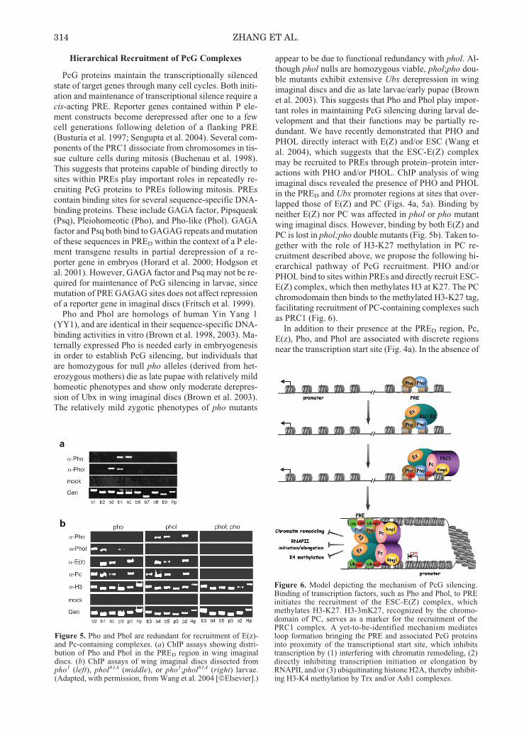
Figure 4. Pc binding at the Ubx promoter and PRED regions isdependent on H3-K27 methylation. (a) ChIP assays showingdistribution of PcG proteins and H3-3mK27 in the Ubx pro-moter region in wing imaginal discs. Wing imaginal discs weredissected from E(z)61 larvae reared continuously at 18ºC. (b)ChIP assays of wing imaginal discs from E(z)61 larvae shiftedfrom 18ºC to 29ºC (left) 24 hr or (right) 48 hr prior to dissection.α-H3, anti-histone H3 was used as a positive control in the ChIPassays shown in this figure and in Fig. 5. (Adapted, with per-mission, from Wang et al. 2004 [©Elsevier].)
37_Symp69_Zhang_p.309_318 4/21/05 10:10 AM Page 313
Hierarchical Recruitment of PcG Complexes
PcG proteins maintain the transcriptionally silencedstate of target genes through many cell cycles. Both initi-ation and maintenance of transcriptional silence require acis-acting PRE. Reporter genes contained within P ele-ment constructs become derepressed after one to a fewcell generations following deletion of a flanking PRE(Busturia et al. 1997; Sengupta et al. 2004). Several com-ponents of the PRC1 dissociate from chromosomes in tis-sue culture cells during mitosis (Buchenau et al. 1998).This suggests that proteins capable of binding directly tosites within PREs play important roles in repeatedly re-cruiting PcG proteins to PREs following mitosis. PREscontain binding sites for several sequence-specific DNA-binding proteins. These include GAGA factor, Pipsqueak(Psq), Pleiohomeotic (Pho), and Pho-like (Phol). GAGAfactor and Psq both bind to GAGAG repeats and mutationof these sequences in PRED within the context of a P ele-ment transgene results in partial derepression of a re-porter gene in embryos (Horard et al. 2000; Hodgson etal. 2001). However, GAGA factor and Psq may not be re-quired for maintenance of PcG silencing in larvae, sincemutation of PRE GAGAG sites does not affect repressionof a reporter gene in imaginal discs (Fritsch et al. 1999).
Pho and Phol are homologs of human Yin Yang 1(YY1), and are identical in their sequence-specific DNA-binding activities in vitro (Brown et al. 1998, 2003). Ma-ternally expressed Pho is needed early in embryogenesisin order to establish PcG silencing, but individuals thatare homozygous for null pho alleles (derived from het-erozygous mothers) die as late pupae with relatively mildhomeotic phenotypes and show only moderate derepres-sion of Ubx in wing imaginal discs (Brown et al. 2003).The relatively mild zygotic phenotypes of pho mutants
appear to be due to functional redundancy with phol. Al-though phol nulls are homozygous viable, phol;pho dou-ble mutants exhibit extensive Ubx derepression in wingimaginal discs and die as late larvae/early pupae (Brownet al. 2003). This suggests that Pho and Phol play impor-tant roles in maintaining PcG silencing during larval de-velopment and that their functions may be partially re-dundant. We have recently demonstrated that PHO andPHOL directly interact with E(Z) and/or ESC (Wang etal. 2004), which suggests that the ESC-E(Z) complexmay be recruited to PREs through protein–protein inter-actions with PHO and/or PHOL. ChIP analysis of wingimaginal discs revealed the presence of PHO and PHOLin the PRED and Ubx promoter regions at sites that over-lapped those of E(Z) and PC (Figs. 4a, 5a). Binding byneither E(Z) nor PC was affected in phol or pho mutantwing imaginal discs. However, binding by both E(Z) andPC is lost in phol;pho double mutants (Fig. 5b). Taken to-gether with the role of H3-K27 methylation in PC re-cruitment described above, we propose the following hi-erarchical pathway of PcG recruitment. PHO and/orPHOL bind to sites within PREs and directly recruit ESC-E(Z) complex, which then methylates H3 at K27. The PCchromodomain then binds to the methylated H3-K27 tag,facilitating recruitment of PC-containing complexes suchas PRC1 (Fig. 6).
In addition to their presence at the PRED region, Pc,E(z), Pho, and Phol are associated with discrete regionsnear the transcription start site (Fig. 4a). In the absence of
314 ZHANG ET AL.
Figure 5. Pho and Phol are redundant for recruitment of E(z)-and Pc-containing complexes. (a) ChIP assays showing distri-bution of Pho and Phol in the PRED region in wing imaginaldiscs. (b) ChIP assays of wing imaginal discs dissected frompho1 (left), phol81A (middle), or pho1;phol81A (right) larvae.(Adapted, with permission, from Wang et al. 2004 [©Elsevier].)
Figure 6. Model depicting the mechanism of PcG silencing.Binding of transcription factors, such as Pho and Phol, to PREinitiates the recruitment of the ESC-E(Z) complex, whichmethylates H3-K27. H3-3mK27, recognized by the chromo-domain of PC, serves as a marker for the recruitment of thePRC1 complex. A yet-to-be-identified mechanism mediatesloop formation bringing the PRE and associated PcG proteinsinto proximity of the transcriptional start site, which inhibitstranscription by (1) interfering with chromatin remodeling, (2)directly inhibiting transcription initiation or elongation byRNAPII, and/or (3) ubiquitinating histone H2A, thereby inhibit-ing H3-K4 methylation by Trx and/or Ash1 complexes.
37_Symp69_Zhang_p.309_318 4/21/05 10:10 AM Page 314

E(z), Pho and Phol remain at the PRE, but are no longerdetected near the Ubx promoter (Fig. 4b). This is consis-tent with a model in which PcG proteins assemble at thePRE followed by the formation of a loop that brings theminto contact with the promoter (Fig. 6). Assuming thismodel is correct, it is not clear what may mediate loopformation. PRC1 has been shown to be able to recruitchromatin templates in trans (Lavigne et al. 2004). Alter-natively, the sequence-specific DNA-binding proteinZeste has been shown to be a component of PRC1 (Saurinet al. 2001), raising the possibility that Zeste may medi-ate loop formation.
Evolutionary Conservation of PcG Gene Silencing
As listed in Table 1, PcG proteins have been struc-turally and functionally conserved during evolution. Inaddition, the core components of the ESC-E(Z)/EED-EZH2 and the PRC1 complexes are conserved fromDrosophila to human (Francis et al. 2001; Cao et al.2002; Levine et al. 2002; Muller et al. 2002). One of theconserved functions of PcG proteins is their involvementin Hox gene silencing. For example, PcG mutations inDrosophila or mice result in homeotic transformation be-cause of derepression of Hox genes (Kmita and Duboule2003). Data presented above illustrate the importance ofESC-E(Z)-mediated H3-K27 methylation in Ubx gene si-lencing. To examine whether the function of H3-K27methylation is conserved in mammalian cells, we recon-stituted the human EED-EZH2 complex and demon-strated that the HMTase activity requires a minimum ofthree components, including EZH2, EED, and SUZ12.Addition of RbAp48 and AEBP2 stimulated the enzy-matic activity (Cao and Zhang 2004b).
To evaluate the role of SUZ12 in H3-K27 methylationin vivo, we generated a stable SUZ12 knockdown cellline that expresses ~25% of the normal levels of SUZ12protein and ~35% of the normal levels of SUZ12 mRNA(Fig. 7a). Compared with the control empty vector knock-down cells, SUZ12-targeted knockdown resulted in a sig-
nificant decrease on the trimethyl-K27 level but had littleeffect on the trimethyl-K9 level (Fig. 7b, third and fourthpanels). Interestingly, an increase in monomethyl-K27and a moderate decrease in dimethyl-K27 were also ob-served (Fig. 7b, top two panels). The fact that SUZ12knockdown does not affect EZH2 level (Fig. 7a) in com-bination with the requirement of SUZ12 for H3-K27methyltransferase activity in vitro (Fig. 7b) allows us toconclude that SUZ12 directly contributes to H3-K27methylation in vivo.
Previous studies in Drosophila have established a crit-ical role for Su(z)12 in Hox gene silencing (Birve et al.2001). The fact that SUZ12 is required for H3-K27methylation in combination with the fact that H3-K27methylation is critical in Hox gene silencing (Cao et al.2002; Muller et al. 2002) predict that SUZ12 knockdownwill result in derepression of at least some Hox genes.Analysis of HoxC6, HoxC8, and HoxA9 in the knock-down cells and the parallel control cells revealed dere-pression of HoxC8 and HoxA9 genes in the knockdowncells (Fig. 7c). These data support the notion that, likemost other PcG proteins, the function of SUZ12/Su(z)12in Hox gene silencing is conserved from human toDrosophila.
CONCLUSIONS AND FUTURE DIRECTIONS
As a result of these and other studies, we can now be-gin to assign molecular/biochemical activities to morethan half of the known PcG proteins. We propose thatPcG proteins may be placed in either of two categories:Recruiters or Effectors. Proteins such as Pho, Phol, ortheir mammalian homolog YY1 and components of theEsc-E(z)/EED-EZH2 complex primarily function as Re-cruiters. The sequence-specific DNA-binding Pho andPhol bind to sites within PREs and directly recruit ESC-E(Z) complexes, which in turn methylates H3 at K27 inthe immediate vicinity of the PRE. The PC chromo-domain binds to the methylated H3-K27 tag, facilitatingrecruitment of PRC1, or related complexes (Fig. 6). Thus,
MECHANISM OF PcG SILENCING 315
Figure 7. SUZ12 knockdown affects H3-K27methylation and Hox gene expression. (a) West-ern blot (left panel) and quantitative RT-PCR(right panel) analysis of a SUZ12 stable knock-down cell line and a parallel mock knockdowncell line. Tubulin serves as a loading control forWestern blotting. GAPDH serves as control fornormalization in the quantitative RT-PCR. (b)Western blot analysis of histones extracted fromcontrol and knockdown HeLa cells with antibod-ies specific for mono-, di-, or trimethylated K27and trimethylated K9. Equal loading of histoneH3 was verified by Coomassie staining of a par-allel gel (bottom panel). (c) Quantitative RT-PCR analysis of HoxC6, HoxC8, and HoxA9 ex-pression in SUZ12 knockdown and mockknockdown cells. GAPDH was used as a controlfor normalization. Quantification is an averageof two independent experiments with error bars.(Adapted from Cao and Zhang 2004b.)
37_Symp69_Zhang_p.309_318 4/21/05 10:10 AM Page 315
the primary function of H3-K27 methylation in PcG si-lencing appears to be recruitment of PC-containing com-plexes. In vitro studies suggest that PRC1 may be classi-fied as an Effector of transcriptional repression, whichmay inhibit transcription by any of several possiblemechanisms. For example, PRC1 inhibits nucleosome re-modeling by SWI/SNF complexes (Shao et al. 1999;Francis et al. 2001). Therefore, it may antagonize the nu-cleosome remodeling activity of the trxG BRM complex,thus interfering with activator binding or assembly of thepreinitiation complex. In addition, PRC1 has been shownto be able to block transcription of chromatin or nakedDNA templates by RNA polymerase II or T7 RNA poly-merase (King et al. 2002). PRC1 does not appear to blockactivator binding in these assays, but instead seems to actupon the template to interfere with transcription initiationor elongation. These observations are consistent with invivo studies in which RNA polymerase II and basal tran-scription factors were shown to be present at promotersunder conditions of PcG repression (Dellino et al. 2004)and the presence of Pc- and E(z)-containing complexes ata discrete site just downstream of a silenced endogenoustarget gene, Ubx, in wing imaginal discs (Wang et al.2004). In addition, our recent studies indicate that aPRC1-like complex possesses H2A ubiquitin ligase ac-tivity. Human Ring 2, a homolog of dRing/Sce, was iden-tified as the catalytic subunit (data not shown). Althoughthe mechanism by which this activity affects transcriptionhas not been determined, it nevertheless suggests thatPRC1 may interfere with transcription by multiple mech-anisms.
Among the questions to be addressed in the near futureis whether PRE-promoter loops actually form, what is themechanistic basis for loop formation, and how are PcGcomplexes targeted to a site just downstream of the tran-scription start site. Once positioned downstream of thetranscription start site, what is the mechanism by whichtranscription is prevented? Does PRC1 directly act uponthe DNA template to prevent duplex melting, or might itinterfere with some step in initiation such as RNA poly-merase II CTD phosphorylation? What is the effect ofH2A ubiquitination by dRing/hRing2? It is also impor-tant to point out that of the 15 genetically identified PcGgenes, the products of only 9 have been identified eitheras sequence-specific DNA-binding proteins (Pho andPhol) or components of the PRC1 (Pc, Ph, Psc, dRing) orEsc-E(z) (Esc, E(z), Su(z)12) complexes. The remainingPcG proteins also play important roles in transcriptionalsilencing, but their activities are yet to be defined. In ad-dition, other proteins, which may have pleiotropic func-tions and therefore are not easily classifiable as membersof the PcG on the basis of genetic studies, also contributeto PcG silencing. Full understanding of this epigeneticgene regulation system will require an understanding ofthese other players in addition to those that have receivedthe bulk of our attention to date.
ACKNOWLEDGMENTS
This work was supported by NIH grants GM068804(Y.Z.) and GM46567 (R.S.J.).
REFERENCES
Bannister A.J., Zegerman P., Partridge J.F., Miska E.A., ThomasJ.O., Allshire R.C., and Kouzarides T. 2001. Selective recog-nition of methylated lysine 9 on histone H3 by the HP1chromo domain. Nature 410: 120.
Beisel C., Imhof A., Greene J., Kremmer E., and Sauer F. 2002.Histone methylation by the Drosophila epigenetic transcrip-tional regulator Ash1. Nature 419: 857.
Bernstein B.E., Humphrey E.L., Erlich R.L., Schneider R.,Bouman P., Liu J.S., Kouzarides T., and Schreiber S.L. 2002.Methylation of histone H3 Lys 4 in coding regions of activegenes. Proc. Natl. Acad. Sci. 99: 8695.
Birve A., Sengupta A.K., Beuchle D., Larsson J., Kennison J.A.,Rasmuson-Lestander A., and Muller J. 2001. Su(z)12, a novelDrosophila Polycomb group gene that is conserved in verte-brates and plants. Development 128: 3371.
Brown J.L., Fritsch C., Mueller J., and Kassis J.A. 2003. TheDrosophila pho-like gene encodes a YY1-related DNA bind-ing protein that is redundant with pleiohomeotic in homeoticgene silencing. Development 130: 285.
Brown J.L., Mucci D., Whiteley M., Dirksen M.L., and KassisJ.A. 1998. The Drosophila Polycomb group gene pleio-homeotic encodes a DNA binding protein with homology tothe transcription factor YY1. Mol. Cell 1: 1057.
Buchenau P., Hodgson J., Strutt H., and Arndt-Jovin D.J. 1998.The distribution of polycomb-group proteins during cell divi-sion and development in Drosophila embryos: Impact onmodels for silencing. J. Cell Biol. 141: 469.
Busturia A., Wightman C.D., and Sakonju S. 1997. A silencer isrequired for maintenance of transcriptional repressionthroughout Drosophila development. Development 124:4343.
Byrd K.N. and Shearn A. 2003. ASH1, a Drosophila trithoraxgroup protein, is required for methylation of lysine 4 residueson histone H3. Proc. Natl. Acad. Sci. 100: 11535.
Cao R. and Zhang Y. 2004a. The functions of E(Z)/EZH2-medi-ated methylation of lysine 27 in histone H3. Curr. Opin.Genet. Dev. 14: 155.
_______. 2004b. SUZ12 is required for both the histone methyl-transferase activity and the silencing function of the EED-EZH2 complex. Mol. Cell 15: 57.
Cao R., Wang L., Wang H., Xia L., Erdjument-Bromage H.,Tempst P., Jones R.S., and Zhang Y. 2002. Role of histone H3lysine 27 methylation in Polycomb-group silencing. Science298: 1039.
Carrington E.A. and Jones R.S. 1996. The Drosophila Enhancerof zeste gene encodes a chromosomal protein: Examination ofwild-type and mutant protein distribution. Development 122:4073.
Czermin B., Melfi R., McCabe D., Seitz V., Imhof A., and Pir-rotta V. 2002. Drosophila Enhancer of Zeste/ESC complexeshave a histone H3 methyltransferase activity that marks chro-mosomal Polycomb sites. Cell 111: 185.
Daubresse G., Deuring R., Moore L., Papoulas O., Zakrajsek I.,Waldrip W.R., Scott M.P., Kennison J.A., and Tamkun J.W.1999. The Drosophila kismet gene is related to chromatin-re-modeling factors and is required for both segmentation andsegment identity. Development 126: 1175.
Dellino G.I., Schwartz Y.B., Farkas G., McCabe D., Elgin S.C.,and Pirrotta V. 2004. Polycomb silencing blocks transcriptioninitiation. Mol. Cell 13: 887.
Fang J., Wang H., and Zhang Y. 2003. Purification of histonemethyltransferases from HeLa cells. Methods Enzymol. 377:213.
Fischle W., Wang Y., Jacobs S.A., Kim Y., Allis C.D., and Kho-rasanizadeh S. 2003. Molecular basis for the discrimination ofrepressive methyl-lysine marks in histone H3 by Polycomband HP1 chromodomains. Genes Dev. 17: 1870.
Francis N.J. and Kingston R.E. 2001. Mechanisms of transcrip-tional memory. Nat. Rev. Mol. Cell Biol. 2: 409.
Francis N.J., Saurin A.J., Shao Z., and Kingston R.E. 2001. Re-constitution of a functional core polycomb repressive com-plex. Mol. Cell 8: 545.
Fritsch C., Beuchle D., and Muller J. 2003. Molecular and ge-
316 ZHANG ET AL.
37_Symp69_Zhang_p.309_318 4/21/05 10:10 AM Page 316

netic analysis of the Polycomb group gene Sex combs ex-tra/Ring in Drosophila. Mech. Dev. 120: 949.
Fritsch C., Brown J.L., Kassis J.A., and Muller J. 1999. TheDNA-binding polycomb group protein pleiohomeotic medi-ates silencing of a Drosophila homeotic gene. Development126: 3905.
He G.P., Kim S., and Ro H.S. 1999. Cloning and characteriza-tion of a novel zinc finger transcriptional repressor. A directrole of the zinc finger motif in repression. J. Biol. Chem. 274:14678.
Hodgson J.W., Argiropoulos B., and Brock H.W. 2001. Site-specific recognition of a 70-base-pair element containingd(GA)(n) repeats mediates bithoraxoid polycomb group re-sponse element-dependent silencing. Mol. Cell. Biol. 21:4528.
Horard B., Tatout C., Poux S., and Pirrotta V. 2000. Structure ofa polycomb response element and in vitro binding of poly-comb group complexes containing GAGA factor. Mol. Cell.Biol. 20: 3187.
Jacobs S.A. and Khorasanizadeh S. 2002. Structure of HP1 chro-modomain bound to a lysine 9-methylated histone H3 tail.Science 295: 2080.
Jenuwein T., Laible G., Dorn R., and Reuter G. 1998. SET do-main proteins modulate chromatin domains in eu- and het-erochromatin. Cell. Mol. Life Sci. 54: 80.
Jones R.S. and Gelbart W.M. 1990. Genetic analysis of the en-hancer of zeste locus and its role in gene regulation inDrosophila melanogaster. Genetics 126: 185.
Kal A.J., Mahmoudi T., Zak N.B., and Verrijzer C.P. 2000. TheDrosophila brahma complex is an essential coactivator for thetrithorax group protein zeste. Genes Dev. 14: 1058.
King I.F., Francis N.J., and Kingston R.E. 2002. Native and re-combinant polycomb group complexes establish a selectiveblock to template accessibility to repress transcription invitro. Mol. Cell. Biol. 22: 7919.
Kmita M. and Duboule D. 2003. Organizing axes in time andspace; 25 years of colinear tinkering. Science 301: 331.
Kuzmichev A., Nishioka K., Erdjument-Bromage H., Tempst P.,and Reinberg D. 2002. Histone methyltransferase activity as-sociated with a human multiprotein complex containing theEnhancer of Zeste protein. Genes Dev. 16: 2893.
Lachner M., O’Sullivan R.J., and Jenuwein T. 2003. An epige-netic road map for histone lysine methylation. J. Cell Sci. 116:2117.
Lachner M., O’Carroll D., Rea S., Mechtler K., and Jenuwein T.2001. Methylation of histone H3 lysine 9 creates a bindingsite for HP1 proteins. Nature 410: 116.
LaJeunesse D. and Shearn A. 1996. E(z): A polycomb groupgene or a trithorax group gene? Development 122: 2189.
Lavigne M., Francis N.J., King I.F., and Kingston R.E. 2004.Propagation of silencing; recruitment and repression of naivechromatin in trans by polycomb repressed chromatin. Mol.Cell 13: 415.
Levine S.S., Weiss A., Erdjument-Bromage H., Shao Z., TempstP., and Kingston R.E. 2002. The core of the polycomb re-pressive complex is compositionally and functionally con-served in flies and humans. Mol. Cell. Biol. 22: 6070.
Messmer S., Franke A., and Paro R. 1992. Analysis of the func-tional role of the Polycomb chromo domain in Drosophilamelanogaster. Genes Dev. 6: 1241.
Min J., Zhang Y., and Xu R.M. 2003. Structural basis for spe-cific binding of Polycomb chromodomain to histone H3methylated at Lys 27. Genes Dev. 17: 1823.
Müller J., Hart C.M., Francis N.J., Vargas M.L., Sengupta A.,Wild B., Miller E.L., O’Connor M.B., Kingston R.E., and Si-mon J.A. 2002. Histone methyltransferase activity of aDrosophila Polycomb group repressor complex. Cell 111:197.
Ng J., Hart C.M., Morgan K., and Simon J.A. 2000. ADrosophila ESC-E(Z) protein complex is distinct from otherpolycomb group complexes and contains covalently modifiedESC. Mol. Cell. Biol. 20: 3069.
Nielsen P.R., Nietlispach D., Mott H.R., Callaghan J., Bannister
A., Kouzarides T., Murzin A.G., Murzina N.V., and LaueE.D. 2002. Structure of the HP1 chromodomain bound to his-tone H3 methylated at lysine 9. Nature 416: 103.
Papoulas O., Beek S.J., Moseley S.L., McCallum C.M., SarteM., Shearn A., and Tamkun J.W. 1998. The Drosophilatrithorax group proteins BRM, ASH1 and ASH2 are subunitsof distinct protein complexes. Development 125: 3955.
Petruk S., Sedkov Y., Smith S., Tillib S., Kraevski V., NakamuraT., Canaani E., Croce C.M., and Mazo A. 2001. Trithorax anddCBP acting in a complex to maintain expression of ahomeotic gene. Science 294: 1331.
Platero J.S., Hartnett T., and Eissenberg J.C. 1995. Functionalanalysis of the chromo domain of HP1. EMBO J. 14: 3977.
Platero J.S., Sharp E.J., Adler P.N., and Eissenberg J.C. 1996. Invivo assay for protein-protein interactions using Drosophilachromosomes. Chromosoma 104: 393.
Poux S., Melfi R., and Pirrotta V. 2001. Establishment of Poly-comb silencing requires a transient interaction between PCand ESC. Genes Dev. 15: 2509.
Qian Y.-W., Wang Y.-C.J., Hollingsworth R.E.J., Jones D., LingN., and Lee E.Y.-H.P. 1993. A retinoblastoma-binding pro-tein related to a negative regulator of Ras in yeast. Nature364: 648.
Rastelli L., Chan C.S., and Pirrotta V. 1993. Related chromo-some binding sites for zeste, suppressors of zeste and Poly-comb group proteins in Drosophila and their dependence onEnhancer of zeste function. EMBO J. 12: 1513.
Rea S., Eisenhaber F., O’Carroll D., Strahl B.D., Sun Z.W.,Schmid M., Opravil S., Mechtler K., Ponting C.P., Allis C.D.,and Jenuwein T. 2000. Regulation of chromatin structure bysite-specific histone H3 methyltransferases. Nature 406: 593.
Ruhf M.L., Braun A., Papoulas O., Tamkun J.W., Randsholt N.,and Meister M. 2001. The domino gene of Drosophila en-codes novel members of the SWI2/SNF2 family of DNA-de-pendent ATPases, which contribute to the silencing ofhomeotic genes. Development 128: 1429.
Saurin A.J., Shao Z., Erdjument-Bromage H., Tempst P., andKingston R.E. 2001. A Drosophila Polycomb group complexincludes Zeste and dTAFII proteins. Nature 412: 655.
Sengupta A.K., Kuhrs A., and Muller J. 2004. General tran-scriptional silencing by a Polycomb response element inDrosophila. Development 131: 1959.
Shao Z., Raible F., Mollaaghababa R., Guyon J.R., Wu C.T.,Bender W., and Kingston R.E. 1999. Stabilization of chro-matin structure by PRC1, a Polycomb complex. Cell 98: 37.
Simon J.A. and Tamkun J.W. 2002. Programming off and onstates in chromatin: Mechanisms of Polycomb and trithoraxgroup complexes. Curr. Opin. Genet. Dev. 12: 210.
Smith S.T., Petruk S., Sedkov Y., Cho E., Tillib S., Canaani E.,and Mazo A. 2004. Modulation of heat shock gene expressionby the TAC1 chromatin-modifying complex. Nat. Cell Biol.6: 162.
Strahl B.D. and Allis C.D. 2000. The language of covalent his-tone modifications. Nature 403: 41.
Tie F., Furuyama T., Prasad-Sinha J., Jane E., and Harte P.J.2001. The Drosophila Polycomb Group proteins ESC andE(Z) are present in a complex containing the histone-bindingprotein p55 and the histone deacetylase RPD3. Development128: 275.
Turner B.M. 2000. Histone acetylation and an epigenetic code.Bioessays 22: 836.
van Holde K.E. 1988. Histone modifications. Springer, NewYork.
Verreault A., Kaufman P.D., Kobayashi R., and Stillman B.1998. Nucleosomal DNA regulates the core-histone-bindingsubunit of the human Hat1 acetyltransferase. Curr. Biol. 8:96.
Wang L., Brown J.L., Cao R., Zhang Y., Kassis J.A., and JonesR.S. 2004. Hierarchical recruitment of polycomb group si-lencing complexes. Mol. Cell 14: 637.
Zhang Y. and Reinberg D. 2001. Transcription regulation by histone methylation: Interplay between different covalentmodifications of the core histone tails. Genes Dev. 15: 2343.
MECHANISM OF PcG SILENCING 317
37_Symp69_Zhang_p.309_318 4/21/05 10:10 AM Page 317

37_Symp69_Zhang_p.309_318 4/21/05 10:10 AM Page 318