mathematical modelling of radiotherapy strategies for ...rmhkjsv/papers/2006jtbiol maths...
TRANSCRIPT

ARTICLE IN PRESS
0022-5193/$ - se
doi:10.1016/j.jtb
�Correspond��Also to be
fax: +448701 3
E-mail addr
anderson@mat
dundee.ac.uk (M
j.s.vaidya@dun
Journal of Theoretical Biology 241 (2006) 158–171
www.elsevier.com/locate/yjtbi
Mathematical modelling of radiotherapy strategies forearly breast cancer
Heiko Enderlinga,�, Alexander R.A. Andersona, Mark A.J. Chaplaina,Alastair J. Munrob, Jayant S. Vaidyab,��
aDivision of Mathematics, University of Dundee, Dundee DD1 4HN, Scotland, UKbDepartment of Surgery and Molecular Oncology, Ninewells Hospital and Medical School, University of Dundee, Dundee DD1 9SY, Scotland, UK
Received 18 May 2005; received in revised form 11 November 2005; accepted 11 November 2005
Available online 28 December 2005
Abstract
Targeted intraoperative radiotherapy (Targit) is a new concept of partial breast irradiation where single fraction radiotherapy is
delivered directly to the tumour bed. Apart from logistic advantages, this strategy minimizes the risk of missing the tumour bed and
avoids delay between surgery and radiotherapy. It is presently being compared with the standard fractionated external beam
radiotherapy (EBRT) in randomized trials.
In this paper we present a mathematical model for the growth and invasion of a solid tumour into a domain of tissue (in this case
breast tissue), and then a model for surgery and radiation treatment of this tumour. We use the established linear-quadratic (LQ) model
to compute the survival probabilities for both tumour cells and irradiated breast tissue and then simulate the effects of conventional
EBRT and Targit.
True local recurrence of the tumour could arise either from stray tumour cells, or the tumour bed that harbours morphologically
normal cells having a predisposition to genetic changes, such as a loss of heterozygosity (LOH) in genes that are crucial for
tumourigenesis, e.g. tumour suppressor genes (TSGs). Our mathematical model predicts that the single high dose of radiotherapy
delivered by Targit would result in eliminating all these sources of recurrence, whereas the fractionated EBRT would eliminate stray
tumour cells, but allow (by virtue of its very schedule) the cells with LOH in TSGs or cell-cycle checkpoint genes to pass on low-dose
radiation-induced DNA damage and consequently mutations that may favour the development of a new tumour.
The mathematical model presented here is an initial attempt to model a biologically complex phenomenon that has until now received
little attention in the literature and provides a ‘proof of principle’ that it is possible to produce clinically testable hypotheses on the effects
of different approaches of radiotherapy for breast cancer.
r 2005 Elsevier Ltd. All rights reserved.
Keywords: Mathematical modelling; Tumour invasion; Breast cancer; Radiotherapy; Targit; Intraoperative radiotherapy; Local recurrence
1. Introduction
In recent years the treatment of breast cancer has shiftedaway from the very radical to more conservative surgicaloperations followed by radiotherapy (Vaidya et al., 2001).
e front matter r 2005 Elsevier Ltd. All rights reserved.
i.2005.11.015
ing author. Tel.: +441382 344470; fax: +44 1382 345516.
corresponded to Tel.: +44 1382 496434;
07403.
esses: [email protected] (H. Enderling),
hs.dundee.ac.uk (A.R.A. Anderson), chaplain@maths.
.A.J. Chaplain), [email protected] (A.J. Munro),
dee.ac.uk (J.S. Vaidya).
Conventional external beam radiotherapy (EBRT) ofabout 50–55Gy (1Gy ¼ 1Gray ¼ 1 Joule kg�1) is givenin fractions of 2Gy over a 4–6 week period (Reitsamer etal., 2002). This external delivery may miss the target andestimates of such a ‘geographical miss’ range from 24% to88% of cases (Vaidya et al., 2005). This could wellcontribute towards the persistent rate of local recurrenceof about a third of the rate without radiotherapy(EBCTCG, 2000; Reitsamer et al., 2002).Over the last few years studies have begun that focus on
how to obtain local tumour control with radiotherapydirected at the tumour bed (Veronesi et al., 2001; Vaidya

ARTICLE IN PRESSH. Enderling et al. / Journal of Theoretical Biology 241 (2006) 158–171 159
et al., 1999; Arthur, 2003). One reason for concentrating onthe tissue adjacent to the tumour is that over 90% of earlylocal recurrence occurs here (Vaidya et al., 1996). Thisregion is defined by a ‘shell’ of tissue which may containactual or potential tumour cells within a 10mm margin(Ebert and Carruthers, 2003). The potential tumour cellsare morphologically normal cells that may harbour geneticmutations similar to the alterations found in the tumour(Lakhani et al., 1999; Forsti et al., 2001; Li et al., 2002;Steinarsdottir et al., 2004).
One genetic mutation that is common in the carcinogen-esis cascade is the so-called loss of heterozygosity (LOH)(Dairkee, 1998; Tomlinson, 2001) in tumour suppressorgenes (TSGs). These genes (i.e. their mutations) areimportant for the initiation or early progression ofbreast cancer development (Deng et al., 1996). The ‘fieldeffect’ of the mutations in tissue adjacent to the tumour bedhas been known for some time (Deng et al., 1996), but itsaetiology is uncertain. It could be the result of clonalexpansion (Tomlinson, 2001; Larson et al., 2002) of only afew stem cells that mutated before puberty (Lakhani et al.,1999) and thus spread to whole segments of the ducto-lobular tree. In an elegant study of patients treated withbreast conservation surgery, LOH occurring in TSGs intissues adjacent to the primary tumour was shown toincrease the probability of recurrence 4–5-fold (Li et al.,2002).
Normal DNA-repair mechanisms at different checkpoints during the cell cycle are able to detect and to repairmost low-dose radiation-induced damage (Oldham, 2001)in a relatively short time (approximately 70.2min, Brenneret al., 1998). DNA double-strand breaks, as induced byionizing radiation, are repaired either by homologousrecombination or non-homologous end-joining mechan-isms (Sancar et al., 2004). The therapeutic index ofradiotherapy relies on the fact that cancer cells lack suchefficient DNA-repair mechanisms and therefore geneticdamage is carried on. If the resulting mutations limit thecells’ ability to survive, which is the case in about 1–2%of all radiaton-induced double-strand breaks (Turessonet al., 2003), a proportion dies with every small dose ofradiation.
The traditional aim of radiotherapy has been to achievea therapeutic index by inducing tumour cell death whilelimiting the damage to nearby healthy cells. Most radio-biological reaction-rate models lead to linear-quadratic(LQ) relations (Sachs et al., 1997). The biologicallyeffective dose (BED) can be obtained from this relation, i.e.
BED ¼ nðad þ bd2Þ, (1)
where d is the physical dose delivered per fraction, n isthe total number of fractions, a is the coefficient ofsingle-hit double-strand breaks, and b is the coefficientof the combination of two sub-lethal single-strand breaksto form a lethal double-strand break. With theknown BEDs for healthy cells and cancer cells onecan calculate the surviving probability S (Guerrero and
Allen Li, 2003) as
S ¼ exp �nda 1þd
ða=bÞ
� �� �, (2a)
which can then be used to predict the outcome of atreatment.Although individual cell repair is not considered in the
LQ-model, we use experimental results to estimate theproportion of cells that survive low-dose radiation damageafter repair. From Eq. (2a) we obtain the proportion ofcells that are not damaged by radiotherapy. However, 98%of the damage is likely to be repaired within the first fewhours after radiation (Brenner et al., 1998; Guerrero andAllen Li, 2003) and hence the cells survive low-dosetreatment. Turesson et al. (2003) have stated that eachradiation dose of 1Gy causes about 20–25 double-strandbreaks per cell. If a dose of more than 5Gy is delivered(and hence more than 100 double-strand breaks occur), itcan be assumed that even after repairing of 98% of damageat least one crucial double-strand break is still leftunrepaired which will force the cell to die. Therefore, weintroduce the observed cell repair probability of 98% fordoses dp5Gy and the new surviving portion S* that willbe applied to healthy cells arises from the proportion ofcells that survive sub-lethal radiation damage (i.e. S) plusthe proportion of the cells that were damaged and wererepaired (i.e. (1–S)� 0.98). Hence, we have
S� ¼S; if d45;
S þ ½ð1� SÞ � 0:98�; if dp5:
((2b)
2. Targeted intraoperative radiotherapy (Targit) for early
breast cancer
Intraoperative radiotherapy (IORT) delivers a largesingle dose to the tumour bed in the operating theatrewhile the patient is still anaesthetized. The applicator isplaced in the tumour bed formed after wide local excisionof the primary tumour (Vaidya et al., 2002). Since theapplicator is placed directly in the tumour bed, a‘geographical miss’ is avoided (Fig. 1). The tumour bedsurrounds the radiation source, so that the breast tissuethat was previously close to the tumour is now close to thespherical applicator and therefore receives the highest doseof radiotherapy (Vaidya et al., 2001).An electron-beam driven X-ray source is used for Targit.
This point source emits low energy (50 kV) X-raysisotropically and gives a uniform dose rate at the applicatorsurface. There is rapid attenuation of the dose within thetissues that absorb the radiation energy and thus there isonly a small high-dose region and distant normal tissuesare spared (Vaidya et al., 2001). Fig. 2 shows theexperimentally measured loss of energy of soft X-rays overshort distances (Vaidya et al., 2001) as well as anapproximated function f ðxÞ ¼ 5e1�x1:5
of this exponential

ARTICLE IN PRESS
Fig. 1. Diagrams showing the Targit system. Left: schematic diagram of the Intrabeam system. X-rays are generated when electrons that are accelerated
along the drift tube hit the tip. These X-rays are modulated by the spherical applicator delivering a uniform dose of radiation at the surface of the
applicator sphere. Right: The applicator being placed in the tumour bed, immediately after the excision of the tumour. From Vaidya et al. (2001).
Fig. 2. Targit observed radiation dose over distance (Vaidya et al., 2001)
and its approximation with a function f ðxÞ with exponential decay. The
rapid attenuation reduces the treatment area to tissue close to the tumour
bed.
Fig. 3. Plot of the biologically effective dose (BED) in Targit against
distance for both tissue and tumour cells. The biological ratios a/b are
5Gy for tumour and 3Gy for healthy breast tissue, respectively.
H. Enderling et al. / Journal of Theoretical Biology 241 (2006) 158–171160
decay. The dose is 5Gy at 1 cm distance from theapplicator surface. The highest radiation dose of 20Gy islocated close to the source, whereas at a distance of 2.7 cmfrom the source only a dose of 1Gy remains.
The approximated dose can be used to compute the BEDand survival fractions for any area in the tissue. Guerreroand Allen Li (2003) have stated that the a=b ratio for breasttumour varies from 13 to 5Gy, with a widely observedmean of 10Gy. Throughout this paper we have assumedthe biological ratio to be ða=bÞtumour ¼ 10Gy. The ratio forbreast tissue is widely assumed to be (a=bÞtissue ¼ 3Gy(Dale, 1996; Vaidya et al., 2001; Guerrero and Allen Li,2003). If the ratio ða=bÞtumour turns out to be lower forsingle dose radiotherapy (as indicated in Vaidya et al.,2001; Yarnold et al., 2005), from Eq. 2(a) and 2(b) weobtain a much smaller survival probability and thus aneven better therapeutic index. For Targit, by simplyapplying Eq. (1) we obtain the BED for both tumour andbreast tissues, which are plotted against the distance fromthe radiation source in Fig. 3. Close to the applicator, theeffective dose is higher for healthy tissue than for tumourcells, and in distant sites the radiation has almost the same
low effect on either tissue. The high BED for healthy tissueis reduced by repair mechanisms.By applying atumour ¼ 0:3Gy�1 and atissue ¼ 0:15Gy�1
for breast cancer (Guerrero and Allen Li, 2003) and breasttissue respectively (Booth, 2002) the survival rates S and S*in Targit are calculated using Eq. (2a), (2b) and are asshown in Table 1 and plotted against distance from theradiation source in Fig. 4.Targit is currently being compared in an international
randomized trial (Vaidya et al., 1999; Vaidya et al., 2004)with the conventional 4–6 week course of postoperativeradiotherapy. It is felt that Targit can virtually eliminatethe risk of a ‘geographical miss’ of the tumour bed whilesparing distant normal tissues (e.g. skin) and therefore canpotentially better the cosmetic outcome. It avoids delaysbefore radiotherapy treatment after surgery which couldincrease the risk of local recurrence (Wyatt et al., 2003),and has several logistic and financial advantages. Thus, ifTargit is found as effective as conventional radiotherapy, itcould become the new standard. Our mathematical modeldescribes the effects of this new radiotherapy technique aswell as that of the conventional radiotherapy. We provide

ARTICLE IN PRESS
Table 1
Table shows the BEDs (in Gy) for tumour and breast tissue and the corresponding survival fractions Stumour for tumour cells and Stissue and S*tissue for
breast tissue cells
Distance (cm) Dose (Gy) BEDtumour Stumour BEDtissue Stissue S*tissue
0.1 15 38 1.3� 10�5 90 1.4� 10�6 1.4� 10�6
0.2 12.5 28 2.2� 10�4 64 6.2� 10�5 6.2� 10�5
0.5 8.75 16 7.3� 10�3 34 5.9� 10�3 5.9� 10�3
1.0 5.0 8 0.1 13 0.14 0.983
1.3 4.0 5.6 0.19 9.3 0.25 0.985
2.0 2.0 2.4 0.48 3.3 0.6 0.992
2.7 1.0 1 0.7 1.3 0.82 0.996
Fig. 4. Plot of survival probabilities for tumour and tissue cells in Targit
as a function of distance as calculated in our model using equations (2a)
and (2b).
H. Enderling et al. / Journal of Theoretical Biology 241 (2006) 158–171 161
an elegant demonstration of how mathematical modellingcould help compare and contrast current treatments withnew treatment strategies.
3. A mathematical model for solid tumour growth and
invasion
The mathematical model is presented in three differentphases of tumour development and treatment. In the firstphase, we model a solid tumour invading the host breasttissue (cf. Anderson et al., 2000). After the tumour hasreached a specific size we simulate the wide local excision ofthe tumour and then we introduce radiation therapy in thesecond phase of our modelling. In the final phase wesimulate the development (or not) of local recurrence.
In the invasion model of Anderson et al. (2000), laterdeveloped and extended by Anderson (2005), the threevariables considered in this first phase are tumour cells,extracellular matrix (ECM) and matrix-degrading enzymes(MDE). These variables are denoted as n, f and m,respectively. The random motion of tumour cells ismodelled by simple diffusion (random motility). Thedirected movement of tumour cells up the gradients of
fixed or bound chemicals is denoted by haptotaxis. Thetumour cells are known to produce enzymes (modelled byproduction and decay) that diffuse throughout the sur-rounding matrix and degrade the tissue upon contact,which we model by a degradation term. In our model, weadd a term to simulate the effect of proliferation of tumourcells. For simplicity we neglect cell death and only considerproliferation to be modulated by available space.These assumptions lead to the following system of
equations describing the interactions of the tumour cells,ECM and MDEs as detailed in the previous paragraph
qn
qt¼ mnð1� n=n0 � f =f 0Þ
proliferation
þ Dnr2n
radom motility
� wr � ðnrf Þhaptotaxis
,
qf
qt¼ �kmf
degradation
,
qm
qt¼ Dmr
2m
diffusion
þ znð1�m=m0Þ
production
� omdecay
. ð3Þ
We assume that the tumour grows inside the breast withoutreaching the outer boundary—the skin. Hence we neglectthe breast geometry and assume that the tumour grows in aradially symmetric manner inside a uniformly shapeddomain of breast tissue. Therefore our system is consideredto hold in a one-dimensional spatial domain O (a region oftissue) with appropriate initial conditions for each variable.This domain may be seen as an approximation to a radiallysymmetric tumour geometry within the breast. We assumethat tumour cells, and consequently the MDEs, remainwithin the domain of tissue under consideration andtherefore no-flux boundary conditions are imposed ondO, the boundary of O.In order to solve the system numerically, we first of all
non-dimensionalize the equations in the standard way. Werescale distance with an appropriate length scale L (i.e. thesize of the breast ¼ 10 cm), time with t ¼ 1 year, tumourcell density with n0, ECM density with f0 and MDEconcentration with m0 (where n0, f0, m0 are appropriatereference density and concentration variables). Therefore,setting
~n ¼n
n0; ~f ¼
f
f 0
; ~m ¼m
m0; ~x ¼
x
L; ~t ¼
t
t,

ARTICLE IN PRESSH. Enderling et al. / Journal of Theoretical Biology 241 (2006) 158–171162
in (3) and dropping the tildes for notational convenience,we obtain the scaled system of equations:
qn
qt¼ lnð1� n� f Þ
proliferation
þ dnr2n
radom motility
� gr � ðnrf Þhaptotaxis
,
qf
qt¼ �Zmf
degradation
,
qm
qt¼ dmr
2m
diffusion
þ anð1�mÞproduction
� bmdecay
, ð4Þ
where l ¼ tm, dn ¼ tDn=L2, g ¼ twf 0=L2, Z ¼ tkm0,dm ¼ tDm=L2, a ¼ tzn0=m0, b ¼ to. We took a value forthe cell random motility parameter Dn of Dn�10
�9 cm2 s�1
(cf. Bray, 1992). It is known that the MDEs secreted bycancer cells are quickly bound to the matrix and localizednear the invading edge, i.e. the MDEs have a diffusion ratecomparable to the cancer cell random motility. Hence, wetook Dm�10
�9 cm2 s�1. The parameter f0 was taken to bef0�10
�11M estimated in line with Anderson et al. (2000).However, in contrast to Anderson et al. (2000) whomodelled generic invasive tumour growth, we took thehaptotactic parameter w to be much smaller in order toreflect early stage breast tumour invasive behaviour whichis characterized by more compact, localized spread.Estimates for the kinetic parameters k, z, o were notavailable since these are very difficult to obtain experimen-tally. The proliferation parameter m was chosen to reflectbreast cancer specific tumour growth with a mean tumourcell doubling time of 100 days (Guiot et al., 2003).
With the preceding estimates, in all the followingsimulations, the scaled parameter values used in Eq. (4)were as follows:
dn ¼ 0:0001; dm ¼ 0:0005; g ¼ 0:00005,
l ¼ 0:75; Z ¼ 10; a ¼ 0:1 and b ¼ 0.
We now solve the above system of Eq. (4) numerically inorder to simulate (i) breast cancer growth pre-treatment;(ii) breast cancer surgery, and finally (iii) postoperativetreatment with radiotherapy (conventional and Targit).
Early breast cancer is defined by a solid mass of cancercells which has a higher density than the surrounding tissue.We will interpret the term density in a slightly different wayin this paper—it means the probability of tumour cellsbeing present in a particular sample of tissue—so a lowdensity here means a low probability of finding a tumourcell within the tissue and not ‘the degree of compactness ofa substance’. First of all we examine the effect of varyingthe proliferation rate on invasion.
The difference in the tumour density due to differentproliferation rates is shown in Figs. 5a–c. Withoutproliferation (l ¼ 0) diffusion is the dominant process fortumour cells. As the proliferation rate increases the tumourmass achieves a higher compactness and the borderbetween tumour and healthy tissue becomes clearer. Witha proliferation rate of l ¼ 0:75 we can model a dense
tumour lump that has a sharp boundary within surround-ing breast tissue.
4. Modelling of surgery and radiotherapy
With increasing awareness about breast cancer and thewidespread use of screening using mammography, moreand more newly detected breast cancers are small in size.Indeed about 70% (Fritz et al., 2005) of all cases are of theearly stages I and II. The usual treatment for a tumour ofthis size is breast conserving surgery and adjuvant radio-therapy.Let us assume that the tumour develops as described in
our numerical computations of the model (4) in theprevious section and reaches a size of 2 cm. At this stage,we simulate breast conserving surgery at the beginning ofthe second phase in our model. In breast conservingsurgery a solid tumour lump is removed along with a rim ofthe surrounding normal breast tissue. In our model, wesimulate surgery by removing all areas of high tumour celldensity. In our simulation these areas are defined by theconcentration dominance of tumour cells over tissue cells.Therefore, the tumour cell concentration, the tissueconcentration and the enzyme concentration in this areabecome zero after surgical removal of the lump.To model conventional radiotherapy we apply the simple
LQ model introduced in Eq. (1). This model has beenmodified by Dale (1985, 1989) and recently by Herskind etal. (2005) to include dynamic processes between twoconsecutive fractions, e.g. tumour repopulation. We, incontrast, consider every fraction of irradiation separately,and apply the model of solid tumour growth and invasion(4) between two fractions to enable tumour and tissuedynamics to influence the outcome during the course ofconventional fractionated radiotherapy. Furthermore, tokeep the model simple, we have not included hypoxia in theLQ-relation. This is because we are modelling the effect ofradiotherapy on tissues left behind after surgery, and thesetissues are unlikely to be hypoxic.The dose delivered in conventional treatment is assumed
to be uniform throughout the domain. Targit, in contrast,is characterized by a rapid dose fall off over distance. Atabout 3 cm from the applicator surface both tumour andhealthy cells survive with a probability of 100%. Thesurvival rates for cells closer to the radiation source arecalculated using Eqs. (2a) and (2b) and the dose given inFig. 2. The distance-dependent surviving probabilities areshown in Fig. 4. During the relatively short time whensurgery and actual delivery of radiotherapy is performed,we can ignore all temporal processes described in the firstphase of our model as the time-scales for surgery, Targitand each fraction of conventional treatment are in therange of minutes whereas potential breast cancer doublingtime is in the range of months (e.g. a median of 100 days cf.Peer et al., 1993; Kopans et al., 2003; Michaelson et al.,2003).

ARTICLE IN PRESS
Fig. 5. Spatio-temporal evolution of growing and invading breast tumours with different proliferation rates l ¼ 0 (a), l ¼ 0:3 (b) and l ¼ 0:75 (c). Plots
are shown at times t ¼ 0, 10 and 20 in left, middle and right columns, respectively. Initially the tumour is located on the left-hand side of the domain (red),
with tissue (green) elsewhere. The tumour releases matrix degrading enzymes (solid blue line) which degrade the tissue upon contact and thus create space
for the tumour to grow into over time. With a higher proliferation rate l the cancer cells proliferate more rapidly and form a solid dense tumour mass.
Other parameter values as per text.
H. Enderling et al. / Journal of Theoretical Biology 241 (2006) 158–171 163
In the third phase of our framework, we apply the samemathematical model used in the first phase to simulatepotential local recurrence. If the adjacent tissue stillharbours tumour cells after surgery and radiotherapy,these cells may form a new tumour that invades the domainfurther. In the initial simulations we model the tworadiotherapy strategies that include the effects of straytumour cells in the tissue adjacent to the tumour bed.
In the final simulations, we model the presence of cellswith LOH in TSGs in the tissues immediately surroundingthe tumour and examine how this would change the clinicaloutcome with each of the two radiotherapy strategies. Atthis stage we have not modelled the actual emergence ofrecurrence from LOH because these cells are not yettransformed and modelling this transformation would needan individual-based cellular model rather than the con-tinuum cell-population model that we have described. Wehave instead included this cell population as being a
potential source of local ‘recurrence’ and assessed if aparticular radiotherapy strategy can eliminate it.
5. Results
In the first phase of the mathematical model for solidtumour growth and invasion we simulate biologicallyobserved breast tumour growth before diagnosis. In thisspecific situation, a solid and very dense tumour mass isproduced and this degrades the surrounding tissue. Only avery small fraction of tumour cells escape into the tissue.The system of partial differential equations (4) was solvednumerically using an explicit finite difference approxima-tion scheme for the parameter values
dn ¼ 0:0001; dm ¼ 0:0005; g ¼ 0:00005,
l ¼ 0:75; Z ¼ 10; a ¼ 0:1 and b ¼ 0.

ARTICLE IN PRESS
Fig. 6. Plots showing the simulation of surgery for early breast cancer. Tumour cell density is red, healthy tissue density is green and MDE concentration
is the blue line. When the dense tumour diameter (dominance of tumour cells over tissue cells) reaches 2 cm (left), surgery is simulated and all cells and
enzymes in the area of high tumour tissue density are removed (right). The presence of some tumour cells in the tissue after surgery (the small brown
triangle at the bottom left of the green area near the x-axis) emphasizes the need of radiotherapy following breast conserving surgery.
H. Enderling et al. / Journal of Theoretical Biology 241 (2006) 158–171164
The computational results were obtained using a Javaimplementation of the numerical scheme and all figuresshown were generated in Java. [Simulation videos areavailable at URL http://www.maths.dundee.ac.uk/�heikman/targit; http://www.dundee.ac.uk/surgery/targit/model].
With the above parameter values, the undiagnosed anduntreated tumour grows exactly as that shown in Fig. 5c.The distribution at t ¼ 0 shows the initial conditions for ourmodel with a cluster of tumour cells being located at the left-hand side of the domain (red), the presence of MDEs withinthis area (the solid blue line), and tissue cells (green)everywhere else. As time evolves the cluster of tumour cellsgrows and realeases MDEs, which degrade the ECM tomake space for the tumour to invade the domain further.We found that the chosen values of the parameters fortumour cell random motility and haptotaxis, dn and g,respectively, simulate the very localized lump characteristicof early stage breast cancer. This is in contrast to the originalsimulations for more aggressive invading tumour cells with ahigher random motility and haptotactic potential proposedby Anderson et al. (2000), where the initial cluster of tumourcells may be able to break into two separate clusters.
In the second phase of the model framework we modelbreast conserving surgery. Fig. 6 shows the effect ofsurgery. In our simulations we apply surgery as soon as thedense tumour reaches a 2 cm diameter, and all tissue on theleft side of this boundary i.e., the tumour and some normaltissue is removed. The denser tumour is defined by a higherconcentration of tumour cells than tissue cells. The tumourand tissue concentration, as well as the enzyme concentra-tion in this area, is set to zero. In our simulations thetumour reaches a detectable size after about four and a halfyears. Guiot et al. (2003) state that with a mean tumour celldoubling time of 100 days and 30 cell doublings necessaryto reach a ‘diagnostic’ stage the tumour had a developingtime of roughly 8 years. In our simulations the tumour is
‘detected’ in about half of this time, which is in line withour initial conditions where the tumour is already at halfthe detectable size.The presence of a few tumour cells (the small brown
triangle at the bottom left of the green area near the x-axisin Fig. 6) within the healthy tissue after surgery now needto be treated with radiotherapy. We assumed that 99% ofclonogenic cells are removed by surgery (Wyatt et al.,2003). Without radiotherapy, the tumour may recur even ifonly a few clonogenic cells are left in the breast tissue. If weassume that a small number of tumour cells remain (as seenin Fig. 6) and use this as the new initial conditions in ourmodel (4) for the next phase, then as can be seen in Fig. 7the tumour recurs. The computational simulations indicatea clinically detectable recurrence on a time-scale of 2–5years which is in line with clinical data (Demicheli et al.,1999; Fisher et al., 2002). Now, we model the effect ofdifferent radiotherapy strategies on our tumour growth-and-surgery model.
6. Simulation of the effects of conventional fractionated
radiotherapy
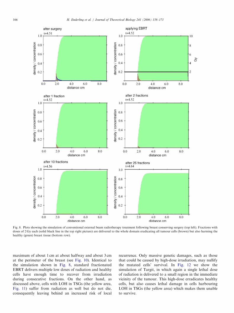
Fig. 6 shows the initial conditions used to model the effectof conventional EBRT. The dosage in EBRT is assumed tobe uniform all over the domain (Vaidya et al., 2001). Thehealthy breast tissue gets the same dose as the tumour bed,but during the interval between fractions, normal cells areable to repair the radiation-induced DNA damage asdiscussed above. The model of phase I is applied betweenthe fractions over the 5 week treatment period to considerthe effect of regrowth and repopulation during the course ofradiotherapy. Fig. 8 shows the simulation of EBRTtreatment after breast conserving surgery with 25 fractionseach of 2Gy over a period of 5 weeks. The condition aftersurgery and the delivery of the first fraction (solid black line)is shown in the top row, and the results after first, second,

ARTICLE IN PRESS
Fig. 7. Plots showing the spatio-temporal evolution of the recurrence of a breast tumour (red) when there are a few tumour cells left behind in the tissue
(green) after the surgery as shown in Fig. 6. The matrix degrading enzyme concentration is plotted as the blue line. The plot at time t ¼ 5 (left) shows the
tumour density about 6 months after surgery (surgery was at time t ¼ 4:51, see Fig. 6), and the plot at time t ¼ 10 (right) represents about 5 years after
surgery, by which time the tumour has already grown to a detectable size. Clinically, recurrence is typically detected between 2 and 4 years after surgery
which is in line with our simulations results.
H. Enderling et al. / Journal of Theoretical Biology 241 (2006) 158–171 165
10th and the final 25th fraction is shown in the rows below.All tumour cells are eliminated, but over time (fractions10–25) the healthy tissue throughout the domain also visiblysuffers from irradiation. This damage to normal tissue isrelatively small and distributed all over the breast.
7. Simulation of the effects of single fraction irradiation with
Targit
Targit is given over a period of about 25min (Vaidyaet al., 2001). We apply the survival probability fractionsSfn; f g as shown in table 1 to the tumour and tissue densities.The results in Fig. 9 show a simulation that uses theparameters discussed earlier for 25min of radiation with a50kV soft X-ray beam which affects normal tissue andtumour cells as seen in Fig. 4. The two images show theinitial distribution and coverage of radiation (Fig. 9, left plot)and the tumour, tissue and MDE densities after irradiation(Fig. 9, right plot). As with EBRT, all tumour cells geteradicated. Additionally, with Targit the normal breast tissueclose to the radiation source is also gradually destroyed.
Our simulations show that both treatment strategieseliminate stray tumour cells that are possibly left behind inthe tumour bed after breast conserving surgery. So itappears that local recurrence due to tumour cells in theadjacent tissue would be completely eliminated by both thetreatment strategies. However, we know that conventionalradiotherapy only eliminates 2/3rds of recurrences(EBCTCG, 2000). Thus let us assume that some of thelocal recurrence arises from genetically unstable cells in thevicinity of the tumour.
8. Irradiation of cells harbouring LOH in tumour suppressor
genes
LOH in TSGs is found at relatively high frequency inbreast cancers (Berardo et al., 1998; Utada et al., 2000).
Lacroix et al. (2004) argue that the origin of breast tumourscould be breast cancer stem cells which have a long life anda large replicating potential. As discussed previously,recurrence of the tumour may occur due to geneticallymutated cells in the adjacent tissue which are clones fromthe same stem cell as the primary tumour. It is plausiblethat the genetic mutations in TSGs in morphologicallyhealthy cells in the tissue adjacent to the primary tumourincrease their susceptibility to further mutations andeventually give rise to a new tumour. This hypothesisalready has clinical support—the presence of LOH incertain TSGs is a strong predictor of local recurrence (Liet al., 2002). Additionally, Jamieson et al. (2003) has shownthat LOH on a growth factor receptor involved in tumoursuppression in head and neck cancer reduces the 5 yearrelapse-free survival significantly when patients are treatedwith radiotherapy alone. Since LOH in TSGs may disablethe cells’ ability to arrest the cell in a cell-cycle repair phase,these genetically unstable cells do not die after receivingmutative hits from irradiation and thus could accumulateand propagate low-dose radiation-induced genetic damage.This may be one reason why conventional radiotherapyfails to eliminate local recurrence completely and if theseareas are treated it may eliminate the risk of localrecurrence.We now introduce an area of possible cells with LOH in
TSGs into our breast tissue and discuss the clinicaloutcome after applying standard radiotherapy and Targit.We assume that an area of 2.5 cm radius around theoriginal primary tumour may contain cells with LOH inTSGs similar to the primary tumour. After surgery, weassumed this shell to be about 1 cm deep. This is based onthe following rationale. Gray’s Anatomy (Bannister et al.,1995) describes 15–20 lobes and milk ducts in the femalebreast and we therefore assume an average of 18 mainprimordial ducts at the nipple. Thus there is approximatelya 201 segment of breast tissue which would span a
ARTICLE IN PRESS
Fig. 8. Plots showing the simulation of conventional external beam radiotherapy treatment following breast conserving surgery (top left). Fractions with
doses of 2Gy each (solid black line in the top right picture) are delivered to the whole domain eradicating all tumour cells (brown) but also harming the
healthy (green) breast tissue (bottom row).
H. Enderling et al. / Journal of Theoretical Biology 241 (2006) 158–171166
maximum of about 1 cm at about halfway and about 3 cmat the perimeter of the breast (see Fig. 10). Identical tothe simulation shown in Fig. 8, standard fractionatedEBRT delivers multiple low doses of radiation and healthycells have enough time to recover from irradiationduring consecutive fractions. On the other hand, asdiscussed above, cells with LOH in TSGs (the yellow area,Fig. 11) suffer from radiation as well but do not die,consequently leaving behind an increased risk of local
recurrence. Only massive genetic damages, such as thosethat could be caused by high-dose irradiation, may nullifythe mutated cells’ survival. In Fig. 12 we show thesimulation of Targit, in which again a single lethal doseof radiation is delivered to a small region in the immediatevicinity of the tumour. This high-dose erradicates healthycells, but also causes lethal damage in cells harbouringLOH in TSGs (the yellow area) which makes them unableto survive.

ARTICLE IN PRESS
Fig. 10. Figure showing the geometry of the area of the breast tissue
which potentially contains cells with LOH. Assuming 15–18 primordial
ducts in the whole breast, a segment of breast tissue arising from one duct
would span at most 3 cm at the periphery of the breast and about 1 cm
nearer the nipple. Even if a 2 cm tumour develops at the very boundary of
a segment, surgical excision of the tumour with a 1 cm margin would leave
a 1 cm wide area which has clones from the same stem cell from which the
primary tumour arose initially.
Fig. 9. Plots showing the results of simulating radiotherapy treatment post-surgery using the new Targit method. The plot on the left shows the initial
distribution throughout the tissue of the radiation delivered by Targit (black line). The rapid attenuation of the radiation dose protects tissue at larger
distances from the applicator (cf. Fig. 2). The plot on the right shows the domain after irradiation. We see that all the tumour cells are killed. However,
note that the damage to the breast tissue due to irradiation is very localized. The dashed line represents the pre-treatment healthy tissue margin.
H. Enderling et al. / Journal of Theoretical Biology 241 (2006) 158–171 167
9. Discussion
Partial breast irradiation with single fraction radiationtechniques such as Targit is a new approach to treat earlystage breast cancer. In this paper, we have presented amodel framework to predict the effect of this newtreatment strategy and conventional radiotherapy treat-ment using the established LQ model. We can apply thissimple model without any of the enhancements which haverecently been discussed by Herskind et al. (2005), becausewe consider the effect of every irradiation fractionseparately. The important dynamic processes during the
course of the treatment, i.e. tumour regrowth andrepopulation, are taken into account by our mathematicalmodel of tumour growth and invasion which we apply inbetween single deliveries of fractionated therapy.We have shown that both conventional EBRT and
Targit following breast conserving surgery eliminatetumour cells that may have escaped into the surroundinghealthy tissue. If we assume that local recurrence arises outof tumour cells left behind after surgery, then our modelpredicts that the new technique of targeted intraoperativeradiotherapy (TARGIT) would have at least a similarcurative effect as the conventional method. In fact, bothmethods should have eliminated local recurrence.However, it is known that after breast conserving
surgery, postoperative radiotherapy reduces local recur-rence rates only by a factor of 2/3, from 27.2% toabout 8.8% (EBCTCG, 2000). The origin of recurrence inthese 8.8% of patients still remains unknown as does thereason why conventional radiotherapy is not able toprevent it.Local recurrence arises in the area around the primary
tumour irrespective of whether or not radiotherapy is givenand whether or not stray tumour cells were found within acertain margin of the primary lump pre-treatment. It isplausible that the genetic mutations in TSGs in morpho-logically healthy cells, in the tissue adjacent to the primarytumour, increase their susceptibility to further mutationsand eventually give rise to a new tumour. Li et al. (2002)has shown that the presence of LOH in certain TSGs is astrong predictor of local recurrence. These geneticallyunstable cells could accumulate and propagate low-doseradiation-induced genetic damage. It is this concept that wehave modelled in the final stage of our model. The verydesign of the low-dose fractionated regime that sparesnormal tissues may be the flaw in conventional EBRT. Theresults of the simulation given in Fig. 11 shows thatconventional radiotherapy would not only spare the

ARTICLE IN PRESS
Fig. 11. Plots showing the simulation of standard fractionated radiotherapy following breast conserving surgery and its effect on cells with LOH. As can
be seen from the plots, this standard treatment does indeed eradicate all tumour cells, but fails to eliminate cells with LOH on TSGs in the immediate
vicinity of the tumour (yellow area), since these mutated cell-cycle check-point genes lack the detection of radiation-induced genetic damage.
Fig. 12. Plots showing the simulation of a single high dose of Targit radiation (solid black line, middle picture bottom row) delivered to areas adjacent to
the primary tumour and its effect on cells with LOH. As can be seen from the plots, Targit eliminates tissue cells that previously surrounded the (red)
tumour and may have shared its clonal origin and may have harboured cells with LOH (yellow area). The (green) tissue is only affected near the tumour
bed. Tissue at distant sites is spared damage. The top row shows the development and invasion of the primary tumour. The second row shows the
simulation of surgery (left), the delivery of Targit radiation (middle) and the final effect of targeted intraoperative radiotherapy (right) killing not only all
tumour cells but also all tissue containing cells with potential LOH.
H. Enderling et al. / Journal of Theoretical Biology 241 (2006) 158–171168
healthy cells, but also the cells with crucial mutations inTSGs adjacent to the breast cancer. Only massive geneticdamage, as could be caused by localized high-doseirradiation, would eradicate the mutated cells. In contrastto the conventional treatment, Targit delivers a high dose
of radiation and destroys tissues close to the tumour andcould therefore also eradicate genetically mutated cells inthe tissue adjacent to the primary tumour. Therefore,Targit may be able to eliminate not only tumour cells thatare left behind, but also the potentially malignant cells in

ARTICLE IN PRESSH. Enderling et al. / Journal of Theoretical Biology 241 (2006) 158–171 169
the immediate vicinity of the tumour that would usually bespared and possibly even promoted by the very nature ofconventional fractionated radiotherapy.
We note that we have not modelled the indirect effects ofradiation on completely normal cells in the immediatevicinity of the applicator. These cells are normallyresponsible for local paracrine secretions, such as growthfactors, in response to trauma and higher-than-normalaromatase activity (O’Neill et al., 1988; Vaidya, 2002).These cells would be spared with conventional radio-therapy but not with Targit. This particular effect isdifficult to include in our current partial differentialequation model but would be expected to enhance thedemonstrated effect. Finally, we note also that angiogen-esis which is essential for the development of recurrence isnot included in the model. However our simulations showthat tumour cells left behind after surgery have thepotential to reform a new tumour. Mathematical modelsof angiogenesis (e.g. Anderson and Chaplain, 1998) can beincluded in future developments of the present model inorder to obtain more realistic simulations of recurrence.Arguably, the high local dose of radiotherapy may inhibitangiogenesis and this would again favour intraoperativeradiotherapy (IORT). So Targit may yet prove to be abetter treatment. Of course this conclusion can only bedrawn once the results of the Targit trial are available.
The work presented here represents an initial attempt tomodel a biologically complex set of circumstances. We arewell aware of its limitations, many of which are imposed bya lack of biological information. However, at the very least,our model draws attention to areas in which further basicresearch is needed.
We have used an a=b ratio of 3Gy for the effect ofradiation on normal breast tissue. This figure has beenobtained from the literature and applies to the connectivetissues of the breast (Guerrero and Allen Li, 2003).However, it may not apply to the critical cells of interestin this paper i.e. breast epithelial cells.
Furthermore, it is known that genomic instability arisesin tumours both spontaneously and as a result of treatmentwith radiotherapy (Yang et al., 1989; Sarkaria et al., 1995;Durante et al., 1996; Colley-Durel et al., 2001; Lorimoreet al., 2001; Trott, 2001; Goldberg, 2003; Roychoudhuriet al., 2004; Tozeren et al., 2005). As we have presented apartial differential equation continuum model, we cannotexplicitly incorporate the fact that random genomicinstabilities arise in single, individual cells. We arecurrently developing and extending the current model toan individual-based, single cell model in order to incorpo-rate this fact theoretically at least (cf. Anderson, 2005).However, we do not know the relative effects of IORT andEBRT on genomic instability in non-irradiated tissues.Additionally, with IORT, low doses of radiation at theperiphery of the irradiated volume may be dangerous, interms of the clonal evolution of cancer. But one could alsoargue that the potential risk of a mutation occurring, dueto the low dose given at a distance from the radiation
source in IORT, is not higher than the risk of a mutationoccurring in the same area with the very last low dosedelivered in EBRT.Despite these limitations our model does provide a
‘proof of principle’: by using a bottom-up approachthrough mathematical modelling it is possible to produceclinically testable hypotheses on the effects of differenttherapeutic approaches to the postoperative radiotherapytreatment of breast cancer. Our next task is to refine andexpand the model, taking into account some of the issuesdescribed above and ultimately to provide a tool thatclinicians and radiotherapists might use to explore anddefine optimal therapeutic strategies.
Acknowledgement
The work of HE was supported by a University ofDundee Nicholl-Lindsay Ph.D. Scholarship bursary.MAJC, ARAA and HE also gratefully acknowledgesupport from NIH Grant No. 1 P20 CA11300-01 (CFDANo. 93.397) ‘Development of a Virtual Tumor’. ARAAalso acknowledges the support of NCI Grant No. 1 P50CA113007-01. The authors would also like to thank Prof.Lynn Matrisian for fruitful discussions.
References
Anderson, A.R.A., 2005. A hybrid mathematical model of solid tumour
invasion: the importance of cell adhesion. Math. Med. Biol. 22,
163–186.
Anderson, A.R.A., Chaplain, M.A.J., 1998. Continuous and discrete
mathematical models of tumour-induced angiogenesis. Bull. Math.
Biol. 60, 857–899.
Anderson, A.R.A., Chaplain, M.A.J., Newman, E.L., Steele, R.J.C.,
Thompson, A.M., 2000. Mathematical modelling of tumour invasion
and metastasis. J. Theor. Med. 2, 129–154.
Arthur, D., 2003. Accelerated partial breast irradiation: a change in
treatment paradigm for early stage breast cancer. J. Surg. Oncol. 84,
185–191.
Bannister, L.H., Berry, M.M., Collins, P., Dyson, M., Dussek, J.E., 1995.
Gray’s anatomy, 38th ed. Churchill Livingstone, New York, USA,
pp. 417–424.
Berardo, M.D., Allred, D.C., O’Connel, P., 1998. Breast cancer. In:
Jameson, J.L. (Ed.), Principles of Molecular Medicine. Human Press,
Totowa, NJ, pp. 625–632.
Booth, J.T., 2002. Modelling the impact of treatment uncertainties in
radiotherapy. Ph.D. Thesis, University of Adelaide.
Bray, D., 1992. Cell Movements. Garland Publishing, New York.
Brenner, D.J., Armour, E., Corry, P., Hall, E., 1998. Sublethal damage
repair times for a late-responding tissue relevant to brachytherapy (and
external-beam radiotherapy): implications for new brachytherapy
protocols. Int. J. Radiat. Oncol. Biol. Phys. 41 (1), 135–138.
Colley-Durel, S., Guitton, N., Nourgalieva, K., Leveque, J., Danic, B.,
Chenal, C., 2001. Genomic instability and breast cancer. Oncol. Rep.
8, 1001–1005.
Dairkee, S.H., 1998. Allelic loss in normal lobules adjacent to breast
cancer. Cancer Detect. Prevent. 22 (1), 135A.
Dale, R.G., 1985. The application of the linear-quadratic dose–effect
equation to fractionated and protracted radiotherapy. Br. J. Radiol.
58, 515–528.
Dale, R.G., 1989. Time-dependent tumour repopulation factors in linear-
quadratic equations—implications for treatment strategies. Radiother.
Oncol. 15, 371–381.

ARTICLE IN PRESSH. Enderling et al. / Journal of Theoretical Biology 241 (2006) 158–171170
Dale, R.G., 1996. Dose-rate effects in targeted radiotherapy. Phys. Med.
Biol. 41, 1871–1884.
Demicheli, R., Miceli, R., Brambilla, C., Ferrari, L., Moliterni, A.,
Zambetti, M., Valagussa, P., Bonadonna, G., 1999. Comparative
analysis of breast cancer recurrence risk for patients receiving or not
receiving adjuvant cyclophosphamide, methotrexate, fluorouracil
(CMF). Data supporting the occurrence of ‘cures’. Breast Cancer
Res. Treat. 53 (3), 209–215.
Deng, G., Lu, Y., Zlotnikov, G., Thor, A.D., Smith, H.S., 1996. Loss of
heterozygosity in normal tissue adjacent to breast carcinomas. Science
274, 2057–2059.
Durante, M., Grossi, G.F., Yang, T.C., 1996. Radiation-induced
chromosomal instability in human mammary epithelial cells. Adv.
Space Res. 18, 99–108.
Early Breast Cancer Trialists’ Collaborative Group (EBCTCG), 2000.
Favourable and unfavourable effects on long-term survival of radio-
therapy for early breast cancer: an overview of the randomised trials.
Lancet 355, 1757–1770.
Ebert, M.A., Carruthers, B., 2003. Dosimetric characteristics of a
low-kV intra-operative X-ray source: Implications for use in a clinical
trial for treatment of low-risk breast cancer. Med. Phys. 30 (9),
2424–2431.
Fisher, B., Anderson, S., Bryant, J., Margolese, R.G., Deutsch, M.,
Fisher, E.R., Jeong, J-H., Wolmark, N., 2002. Twenty-year follow-up
of a randomized trial comparing total mastectomy, lumpectomy, and
lumpectomy plus irradiation for the treatment of invasive breast
cancer. N. Engl. J. Med. 347 (16), 1233–1241.
Forsti, A., Louhelainen, J., Soderberg, M., Wijkstrom, H., Hemminki, K.,
2001. Loss of heterozygosity in tumour-adjacent normal tissue of
breast and bladder cancer. Eur. J. Cancer 37, 1372–1380.
Fritz, P., Cabrera, C.M., Dippon, J., Gerteis, A., Simon, W., Aulitzky,
W.E., van der Kuip, H., 2005. c-erbB2 and topoisomerase IIalpha
protein expression independently predict poor survival in primary
human breast cancer: a retrospective study. Breast Cancer Res. 7 (3),
374–384.
Goldberg, Z., 2003. Clinical implications of radiation-induced genomic
instability. Oncogene 22, 7011–7017.
Guerrero, M., Allen Li, X., 2003. Analysis of a large number of clinical
studies for breast cancer radiotherapy: estimation of radiobiological
parameters for treatment planning. Phys. Med. Biol. 48 (20),
3307–3326.
Guiot, C., Degiorgis, P.G., Delsanto, P.P., Gabriele, P., Deisboeck, T.S.,
2003. Does tumor growth follow a ‘‘universal law’’? J. Theor. Biol. 225
(2), 147–151.
Herskind, C., Steil, V., Kraus-Tiefenbacher, U., Wenz, F., 2005. Radio-
biological aspects of intraoperative radiotherapy (IORT) with
isotropic low-energy X-rays for early stage breast cancer. Radiat.
Res. 163 (2), 208–215.
Jamieson, T.A., Brizel, D.M., Killian, .K., Oka, Y., Jang, H-S., Fu, X.,
Clough, R.W., Vollmer, R.T., Anscher, M.S., Jirtle, R.L., 2003. M6P/
IGF2R loss of heterozygosity in head and neck cancer associated with
poor patient prognosis. BMC Cancer 3 (4).
Kopans, D.B., Rafferty, E., Georgian-Smith, D., Yeh, E., D’Allesandro,
H., Moore, R., Hughes, K., Halpern, E., 2003. A simple model of
breast carcinoma growth may provide explanations for observations of
apparently complex phenomena. Cancer 97 (12), 2951–2959.
Lacroix, M., Toillon, R-A., Leclercq, G., 2004. Stable ‘portrait’of breast
tumors during progression: data from biology, pathology and genetics.
Endocr.-Relat. Cancer 11, 497–522.
Lakhani, S.R., Chaggar, R., Davies, S., Jones, C., Collins, N., Odel, C.,
Stratton, M.R., O’Hare, M.J., 1999. Genetic alterations in
‘normal’ luminal and myoepithelial cells of the breast. J. Pathol. 189,
496–503.
Larson, P.S., de las Morenas, A., Bennett, S.R., Adrienne Cupples, L.,
Rosenberg, C.L., 2002. Loss of heterozygosity or allele imbalance in
histologically normal breast epithelium is distinct from loss of
heterozygosity or allele imbalance in co-existing carcinomas. Am. J.
Pathol. 161 (1), 283–290.
Li, Z., Moore, D.H., Meng, Z.H., Ljung, B.M., Gray, J.W., Dairkee, S.H.,
2002. Increased risk of local recurrence is associated with allelic
loss in normal lobules of breast cancer patients. Cancer Res. 62,
1000–1003.
Lorimore, S.A., Coates, P.J., Scobie, G.E., Milne, G., Wright, E.G., 2001.
Inflammatory-type responses after exposure to ionizing radiation in
vivo: a mechanism for radiation-induced bystander effects? Oncogene
20, 7085–7095.
Michaelson, J., Satija, S., Moore, R., Weber, G., Halpern, E., Garland,
A., Kopans, D.B., 2003. Estimates of breast cancer growth rate and
sojourn time from screening database information. J. Women’s Imag.
5 (1), 11–19.
Oldham, M., 2001. Radiation physics and applications in therapeutic
medicine. Phys. Educ. 36, 460–467.
O’Neill, J.S., Elton, R.A., Miller, W.R., 1988. Aromatase activity in
adipose tissue from breast quadrants: a link with tumour site. Br. Med.
J. (Clin. Res. Ed.) 296, 741–743.
Peer, P.G., van Dijck, J.A., Hendriks, J.H., Holland, R., Verbeek, A.L.,
1993. Age-dependent growth rate of primary breast cancer. Cancer 71
(11), 3547–3551.
Reitsamer, R., Peintinger, F., Sedlmayer, F., Kopp, M., Menzel, C.,
Cimpoca, W., Glueck, S., Rahim, H., Kopp, P., Deutschmann, H.,
Merz, F., Brandis, M., Kogelnik, H., 2002. Intraoperative radio-
therapy given as a boost after breast-conserving surgery in breast
cancer patients. Eur. J. Cancer 38, 1607–1610.
Roychoudhuri, R., Evans, H., Robinson, D., Møller, H., 2004. Radiation-
induced malignancies following radiotherapy for breast cancer. Br. J.
Cancer 91, 868–872.
Sachs, R.K., Hahnfeld, P., Brenner, D.J., 1997. The link between low-let
dose–response relations and the underlying kinetics of damage
production/repair/misrepair. Int. J. Biol. 72 (4), 351–374.
Sancar, A., Lindsey-Boltz, L.A., Unsal-Kac-maz, K., Linn, S., 2004.
Molecular Mechanisms of mammalian DNA repair and the DNA
damage checkpoints. Annu. Rev. Biochem. 73, 39–85.
Sarkaria, J.N., Fowler, J.F., Lindstrom, M.J., Jordan, V.C., Mulcahy,
R.T., 1995. The decreased influence of overall treatment time on the
response of human breast tumor xenografts following prolongation of
the potential doubling time (Tpot). Int. J. Radiat. Oncol. Biol. Phys.
31, 833–840.
Steinarsdottir, M., Jonasson, J.G., Vidarsson, H., Juliusdottir, H.,
Hauksdottir, H., Ogmundsdottir, H.M., 2004. Cytogenetic
changes in nonmalignant breast tissue. Genes Chrom. Cancer 41 (1),
47–55.
Tomlinson, I.P.M., 2001. Mutations in normal breast tissue and breast
tumours. Breast Cancer Res. 3, 299–303.
Tozeren, A., Coward, C.W., Petushi, S.P., 2005. Origins and evolution of
cell phenotypes in breast tumors. J. Theor. Biol. 233, 43–54.
Trott, K.R., 2001. Non-targeted radiation effects in radiotherapy—roles
of radiation-induced genomic instability and of the bystander effect in
cancer cure by radiotherapy. Acta Oncol. 40, 976–980.
Turesson, I., Carlsson, J., Brahme, A., Glimelius, B., Zackrisson, B.,
Stenerlow, B., 2003. Biological response to radiation therapy. Acta
Oncol. 42 (2), 92–106.
Utada, Y., Emi, M., Yoshimoto, M., Kasumi, F., Akiyama, F.,
Sakamoto, G., Haga, S., Kajiwara, T., Nakamura, Y., 2000. Allelic
loss at 1p34–36 predicts poor prognosis in node-negative breast cancer.
Clin. Cancer Res. 6, 3193–3198.
Vaidya, J.S., 2002. A novel approach to local treatment of breast cancer.
Ph.D. Thesis, University College London.
Vaidya, J.S., Baum, M., Tobias, J.S., Houghton, J., 1999. Randomised
trials are not unethical (Letter). Lancet 535, 1714.
Vaidya, J.S., Vyas, J.J., Chinoy, R.F., Merchant, N., Sharma, O.P.,
Mittra, I., 1996. Multicentricity of breast cancer: whole-organ analysis
and clinical implications. Br. J. Cancer 74, 820–824.
Vaidya, J.S., Baum, M., Tobias, J.S., D’Souza, D.P., Naidu, S.V.,
Morgan, S., Metaxas, M., Harte, K.J., Sliski, A.P., Thomson, E., 2001.
Targeted intra-operative radiotherapy (Targit): an innovative method
of treatment for early breast cancer. Ann. Oncol. 12, 1075–1080.

ARTICLE IN PRESSH. Enderling et al. / Journal of Theoretical Biology 241 (2006) 158–171 171
Vaidya, J.S., Baum, M., Tobias, J.S., Morgan, S., D’Souza, D.P., 2002.
The novel technique of delivering targeted intraoperative radiotherapy
(Targit) for early breast cancer. EJSO 28, 447–454.
Vaidya, J.S., Tobias, J.S., Baum, M., Keshtgar, M., Joseph, D., Wenz, F.,
Houghton, J., Saunders, C., Corica, T., D’Souza, D.P., Sainsbury, R.,
Massarut, S., Taylor, I., Hilaris, B., 2004. Intraoperative radiotherapy
for breast cancer. Lancet Oncol. 5, 165–173.
Vaidya, J.S., Tobias, J.S., Baum, M., Wenz, F., Kraus-Tiefenbacher, U.,
D’Souza, D.P., Keshtgar, M., Massarut, S., Hilaris, B., Saunders, C.,
Joseph, D., 2005. TARGeted Intraoperative radioTherapy (TARGIT):
an innovative approach to partial-breast irradiation. Semin. Radiat.
Oncol. 15, 84–91.
Veronesi, U., Orecchia, R., Luini, A., Gatti, G., Intra, M., Zurrida, S.,
Ivaldi, G., Tosi, G., Ciocca, M., Tosoni, A., De Lucia, F., 2001. A
preliminary report of intraoperative radiotherapy (IORT) in limited-
stage breast cancers that are conservatively treated. Eur. J. Cancer 37,
2178–2183.
Wyatt, R.M., Beddoe, A.H., Dale, R.G., 2003. The effects of delays in
radiotherapy treatment on tumour control. Phys. Med. Biol. 48,
139–155.
Yang, T.C., Stampfer, M.R., Tobias, C.A., 1989. Radiation studies on
sensitivity and repair of human mammary epithelial cells. Int.
J. Radiat. Biol. 56, 605–609.
Yarnold, J., Ashton, A., Bliss, J., Homewood, J., Harper, C., Hanson, J.,
Haviland, J., Bentzen, S., Owen, B., 2005. Fractionation sensitivity and
dose response of late adverse effects in the breast after radiotherapy for
early breast cancer: long-term results of a randomised trial. Radiother.
Oncol. 75, 9–17.