materials science and engineering c - school of chemistry | school of chemistry ... · ·...
TRANSCRIPT
Materials Science and Engineering C 58 (2016) 1199–1206
Contents lists available at ScienceDirect
Materials Science and Engineering C
j ourna l homepage: www.e lsev ie r .com/ locate /msec
Photochemically modified diamond-like carbon surfaces forneural interfaces
A.P. Hopper a, J.M. Dugan a, A.A. Gill a, E.M. Regan b, J.W. Haycock a, S. Kelly c, P.W. May b, F. Claeyssens a,⁎a Department of Materials Science and Engineering, Kroto Research Institute, The University of Sheffield, Broad Lane, Sheffield S3 7HQ, UKb School of Chemistry, University of Bristol, Bristol BS8 1TS, UKc New Jersey Neuroscience Institute at JFK Medical Center, 65 James Street, Edison, NJ, USA
⁎ Corresponding author.E-mail address: [email protected] (F. Claeys
http://dx.doi.org/10.1016/j.msec.2015.09.0130928-4931/© 2015 The Authors. Published by Elsevier B.V
a b s t r a c t
a r t i c l e i n f oArticle history:Received 11 March 2015Received in revised form 10 August 2015Accepted 3 September 2015Available online 8 September 2015
Keywords:Diamond-like carbonNeuronsSchwann cellsAmineAldehyde
Diamond-like carbon (DLC) was modified using a UV functionalization method to introduce surface-boundamine and aldehyde groups. The functionalization process rendered the DLC more hydrophilic and significantlyincreased the viability of neurons seeded to the surface. The amine functionalized DLC promoted adhesion ofneurons and fostered neurite outgrowth to a degree indistinguishable from positive control substrates (glasscoated with poly-L-lysine). The aldehyde-functionalized surfaces performed comparably to the amine function-alized surfaces and both additionally supported the adhesion and growth of primary rat Schwann cells. DLC hasmany properties that are desirable in biomaterials. With the UV functionalization method demonstrated here itmay be possible to harness these properties for the development of implantable devices to interface with thenervous system.
© 2015 The Authors. Published by Elsevier B.V. This is an open access article under the CC BY license(http://creativecommons.org/licenses/by/4.0/).
1. Introduction
Diamond possesses a range of valuable mechanical and tribologicalproperties, which offer interesting opportunities for the developmentof future applications. One such property, which is useful within biolog-ical research, is the ease with which it can be doped or functionalizedwhilst retaining its stability and biocompatibility [1]. Its mechanicalstrength and hardness are also particularly desirable especially wheredevice longevity is amajor concern. However, synthetic diamond is pre-pared under harsh conditions, usually requiring high temperatures andpressures, prohibiting its deposition upon biological or soft materials orupon common tissue culture substrates such as polystyrene or glass. Incontrast diamond-like carbon (DLC), a form of amorphous carbon, hassimilar properties to diamond yet may be synthesised directly uponsubstrates under relatively mild deposition conditions and even at am-bient temperature [2].
DLC films may be produced by a variety of methods including argonion sputtering and cathodic arc spray or, as in this study, by pulsed laserdeposition (PLD). The diamond-like character of the DLC produced isdependent upon the ratio of sp2 and sp3 carbon, with a higher sp3 con-tent resulting in more diamond-like properties. Importantly, both DLCand diamond may also be readily photo-functionalized with the aid ofultraviolet (UV) radiation allowing the attachment of a wide range oforganic functional groups at the surface of the material [3]. Such an
sens).
. This is an open access article under
approach is ideal for modifying DLC for biomedical applications wherethe surface termination strongly affects protein and cell adhesion.
We have previously shown that photo-functionalized detonationnanodiamond is an effectivemimic for poly-L-lysine (PLL), a synthetic ad-hesion factor commonlyused inneural cell culture [4]. The neuroadhesiveproperties of PLL are thought to originate from the primary amine groupon the side chain of the L-lysine residue, creating a highly polar mole-cule which assists in cell adhesion. Other molecules containing primaryamines, such as 10-amino-dec-1-ene, are known to exert a similarneuroadhesive effect andmay be covalently bound toDLC using the fac-ile UV irradiation method.
In this studywe have created a neurocompatible substrate fromDLC,which possesses the positive attributes of conventional neuroadhesivesurface treatments such as PLL. However, as the DLC is covalently func-tionalized, it is envisaged that the DLC substrates will exhibit lowercytotoxicity issues and greater stability in applications in vivo. This isthe first time that covalently functionalized DLC has been evaluated as agrowth substrate for neurons and Schwann cells. We anticipate that thetechniques demonstrated here will find applications in surface coatingsand device fabrication for the development of central nervous systemimplants such as electrodes for deep brain stimulation and in brain-computer-interfaces.
2. Materials and methods
Unless otherwise stated, all chemicals and bioreagents were obtainedfrom Sigma-Aldrich (Dorset, UK) and used as received. Experimental
the CC BY license (http://creativecommons.org/licenses/by/4.0/).

1200 A.P. Hopper et al. / Materials Science and Engineering C 58 (2016) 1199–1206
samples were prepared and analysed in triplicate, unless otherwisestated, and error bars and statistical tests were calculated from thesereplicate measurements. Experiments were repeated at least once inorder to ensure reproducibility. In all cases the presented micrographswere representative of the whole sample.
2.1. Preparation of diamond-like carbon substrates
In order to coat glass cover-slips with a thin coating of DLC, a pulsedlaser deposition (PLD) method was used. The output of an argon fluoride(ArF) excimer laser (Lambda Physik, Compex 201) with a wavelength of193 nmwas focused (lens focal length: 200mm, angle of incidence: 45°)on a graphite disc target (Poco Graphite Inc., DFP-3–2 grade) in a stainlesssteel chamber maintained at a pressure of approximately 10−6 Torr. Thegraphitewas ablatedusing a laserfluence of 12 J/cm2. A thinfilmofDLC ofapproximately 20 nm thickness was achieved with 1200 laser shots at atarget-substrate distance of 5 cm at 20 °C. The DLC-treated cover-slipswere then sonicated in methanol for 15min and then washed in distilledwater overnight. Once complete, the cover-slipswere allowed todry in airbefore being stored in an airtight container in preparation for use.
2.2. Synthesis of trifluoroacetate-protected 10-amino-dec-1-ene
Trifluoroacetate-protected 10-amino-dec-1-ene (TFAAD) wassynthesised according to a method described elsewhere [3]. Firstly,10-undecenoyl chloride (50.0 g, 0.25 mol) was added to tetrahexylam-monium bromide (Acros Organics, Loughborough, UK, 0.6 mmol) in anice bath. An aqueous solution of sodium azide (16.6 g in 50 ml) wasthen gradually added to the mixture within the ice bath and agitatedusing a magnetic stirrer. Once the reaction was complete, the organicfraction was separated, washed twice with deionised water, dried overanhydrous magnesium sulphate for 24 h and filtered. An excess oftrifluoroacetic acidwas then added to the acyl azide and the reactionmix-turewas heated to reflux under nitrogen for 6 h. The organicmixturewaswashed with a saturated solution of sodium bicarbonate, collected andsubsequently dried over anhydrous magnesium sulphate for 24 h. Theproduct was stored for later use at 4 °C.
2.3. Preparation of functionalized DLC
In order to ensure a uniform hydrogen-terminated surface, the glasscover-slips coated with DLC were hydrogenated by exposure to a hydro-gen plasma flow for a total duration of 10 min prior to functionalizationusing the UV method. Hydrogenation was carried out in 2 cycles of5 min with microwave power of 0.8 kW and 500 sccm H₂ total gas flow,followed by 2min in coldH₂flowunder a pressure of 100 Torr. The coatedcover-slips were then placed on top of a quartz slide, upon which a suffi-cient volume of either TFAAD (to prepare amine functional DLC) orundec-10-enal (to prepare aldehyde functional DLC) was applied tocover the samples. A further quartz slide was placed on top and thesamples were exposed to UV radiation (λmax = 254 nm) from aHg–Xe lamp (500 W, Hamamatsu Photonics) for a period of 15 min inthe case of undec-10-enal and 4 h in the case of TFAAD. Excess alkenewas removed by rinsing in methanol. Where the alkene used wasTFAAD, the protected amine was deprotected in acidified methanol(0.36MHCl in methanol) at 65 °C over 24 h. The samples were washedin methanol, dried with a stream of nitrogen gas, and stored in sterilecontainers until required. For the sake of brevity the amine functionalDLC will henceforth be designated “DLC-NH2” and the aldehyde func-tional DLC will be designated “DLC-CHO”.
2.4. X-ray photoelectron spectroscopy (XPS)
The surface chemistry of the functionalized samples was analysedusing X-ray photoelectron spectroscopy (XPS). Samples were analysedusing a Kratos AXIS Ultra DLD instrument. Spectra were recorded
using a monochromatised Al Kα X-ray source (1486.6 eV) operatingat a power of 150 W, whilst charging of the sample during irradiationwas reduced by an internal flood gun. Each sample was analysed at anemission angle normal to the sample surface. Data processing, analysisand charge correction were carried out using CasaXPS software(ver.2.3.12 Casa Software Ltd.). Component peaks within the recordedC (1 s) spectra were deconvoluted and fitted to a mixed peak shape of70% Gaussian/30% Lorentzian composition. The aliphatic hydrocarboncomponent of the C (1 s) was set to 285.0 eV as an internal reference.
2.5. Contact angle measurements
The relative hydrophobicity/hydrophilicity of the functionalizedsamples was determined by measuring the water contact angle. Toascertain the water contact angle of the DLC surfaces, one drop ofdeionised water (1 μl) was deposited on each surface. The contactangle was measured as the angle formed by the baseline and thetangent to the drop profile at the three-phase point using a Rame-Hartcontact angle goniometer at a temperature of 21 °C. For each condition,four measurements were carried out on each of replicate samples,giving a total of 20 measurements for each condition (n = 20).
2.6. NG108-15 neuronal cell culture
NG108-15 neuronal cells were supplied by the American TypeCulture Collection (ATCC). The cells were maintained and expanded ina growth medium comprising of Dulbecco's modified Eagle's medium(high glucose) supplemented with foetal bovine serum (10% v/v),L-glutamine (2 mM), penicillin (100 U/ml), streptomycin (100 μg/ml)and amphotericin B (0.25 μg/ml). Cells were not used experimentallybeyond passage number 20. Neuronal cells were seeded upon DLCsamples or glass coated with PLL at a density of 1 × 10⁵ cells per mlin the complete growth medium without foetal bovine serum inorder to induce terminal differentiation. The cells were cultured for 1,2, 5 or 7 days at 37 °C and 5% CO2 after which they were analysed forviability or fixed for microscopy as detailed in the following sections.
2.7. Isolation and culture of primary Schwann cells
Primary Schwann cellswere isolated frommaleWistar rats using theselective D-valine method reported elsewhere [5]. Animals weresacrificed in compliance with the Animals (Scientific Procedures) Act1986. Briefly, sciatic nerves were removed from male Wistar rats (2–3months) and the connective tissue was subsequently discarded. Thenerves were teased and cut into 2–3 mm segments and incubated with0.05% (w/v) collagenase (Sigma, UK) at 37 °C for 1 h. This cell suspensionwas then filtered through a 40 μm Falcon filter (Becton Dickinson, USA)and centrifuged at 400RCF for 5min. The resultant cell pelletwaswashedwith DMEM (including 10% (v/v) foetal calf serum) and resuspended inSchwann cell growth medium containing DMEM-D-valine (PAA, UK),2 mM glutamine, 10% (v/v) FCS, 1% (v/v) N2 supplement (GibcoBRL, UK), 20 μg/ml bovine pituitary extract, 5 μM forskolin (Sigma,UK), 100 U/ml penicillin, 100 μg/ml streptomycin and 0.25 μg/mlamphotericin B. The Schwann cell suspension was plated within35 mm Petri dishes which had been previously coated with 0.5 mgpoly-L-lysine/7 μg laminin (Sigma, UK). The cultures were maintainedat 37 °C with 5% CO2.
2.8. Identification of actin filaments and nuclei
At each time point the DMEM was removed from the NG108-15neuronal cells, which were then washed once with phosphate bufferedsaline (PBS). To eachwell 500 μl of formalin (3.7% v/v in PBS)was appliedfor 15 min after which the cells were washed twice more with PBS. Cellswere then permeabilised with Triton X-100 (0.1% v/v in PBS) for 5 min,before being washed two further times with PBS. A stock solution of

Fig. 1. Contact angle of water on glass, pristine DLC, DLC-NH2 and DLC-CHO. Values are re-ported as the mean ± standard error. Statistical significance was determined by one-wayANOVA followed by Tukey's multiple comparison test; * = p b 0.05 and ** = p b 0.01.
1201A.P. Hopper et al. / Materials Science and Engineering C 58 (2016) 1199–1206
phalloidin-FITC (Sigma) was prepared at a concentration of 0.5 mg/ml indimethyl sulfoxide (DMSO) and further diluted to a working concentra-tion of 5 μg/ml in PBS. DAPI (Sigma) was added as a counterstain (finalconcentration of 10 μg/ml) and the combined stain solution was appliedfor 1 h at room temperature before the cellswere rinsed threemore timeswith PBS. Stained cells were visualised via fluorescence microscopyin PBS using a high throughput imaging and image analysis system(Image Xpress by Axon Instruments). Image analysis was carried outusing the automated functionality of the imaging platform. The analysisprocess was conducted with oversight from the operator to ensure thaterroneous measurements did not occur.
2.9. Immunofluorescent labelling of S100β
Immunofluorescent labelling was carried out to reveal S100βexpression in Schwann cells cultured upon the functionalized DLCsurfaces. S100β is a marker of the Schwann cell phenotype and contin-uous expression over time may indicate maintenance of the Schwanncell phenotype. After 21 days culture the medium was removed fromthe Schwann cell cultures, which were then washed once with PBS.The cells were then fixed with formalin (3.7% v/v) for 15 min, washedwith PBS, and permeabilised with 0.1% Triton X-100 (Sigma-Aldrich,Dorset, UK) for 20 min at 4 °C. Cells were washed again with PBS andthen blocked against non-specific antibody binding with 3% (w/v)bovine serum albumin (BSA) for 60min at 4 °C. Thiswas followed by in-cubation with polyclonal rabbit anti-S100β (1:250) (Dako) diluted in1% (w/v) BSA overnight at 4 °C. The cells were washed twice with PBSand incubated with a FITC-conjugated secondary goat anti-rabbit IgGantibody (1:100 dilution in 1% (w/v) BSA) (Vector Labs, USA) for90 min at room temperature.
2.10. Viability assay (MTT)
Relative cell viability plus an indication of rate of proliferation wasassessed indirectly by theMTT assay. Cells were cultured on three sam-ples for each condition (n=3) at a density of 10,000 cells/ml within 24well plates (Costar) over periods of 1, 2, 5 and 7 days. At each time pointthe DMEMwas removed from selected wells and the cells were washedonce with PBS. MTT Solution (thiazolyl blue tetrazolium bromide,Sigma) was dissolved in PBS (0.5 mg/ml) and 300 μl was added toeach well. The cells were incubated at 37 °C and 5% CO2 for 45 minafter which the MTT solution was removed and the reduced productwas solubilised in 300 μl acidified isopropanol (1.25 μl HCL (37% w/w)per ml of isopropanol). The resulting solution was transferred in tripli-cate 100 μl volumes to a 96-well plate where the optical density wasmeasured at 540 nm relative to 630 nm.
3. Results
3.1. Surface functionalization and characterisation of DLC
DLC was applied to the surface of glass cover-slips via pulsed laserdeposition. The DLC-coated glass was then subjected to UV-inducedfunctionalization with TFAAD or undec-10-enal to yield either amine(after deprotection) or aldehyde-functionalized substrates. Contactangle measurements were carried out to provide an initial indicationof the success of the functionalization process and to provide insightinto the hydrophobicity of the samples, which may have implicationsfor subsequent cell culture studies. The contact angle of a surface de-scribes its wettability. Smaller contact angles, namely those below 90°,characterise hydrophilic surfaces which are highly wettable, whereaslarge contact angles correspond to hydrophobic surfaces which exhibitreduced wettability.
Contact angle measurements on the pristine DLC and functionalizedDLC surfaces are shown in Fig. 1. The contact angle of glass (52.1° ±4.8°) was also measured as a comparison. Compared to glass, which is
regarded as a relatively hydrophilic material, the pristine DLC wasmore hydrophobic with a higher contact angle (64.3° ± 3.1°). Uponfunctionalization, however, the contact angle decreased on both theDLC-NH2 (48.4°± 1.7°) and DLC-CHO (49.7°± 2.9°) surfaces. The func-tionalized surfaceswere significantlymore hydrophilic than the pristineDLC with contact angles comparable to glass.
The functionalized DLC samples were further characterised by XPS toreveal the composition of their surface chemistry. High-resolution spec-tra of the C 1 s region are shown in Fig. 2. Because carbon bound to fluo-rine gives rise to a distinct chemical shift in the C 1 s peak, the DLC-NH2
sampleswere analysed prior to removal of the trifluoroacetate protectinggroup in order to aid in confirmation of successful functionalization. Ineach case the C 1 s peak was deconvoluted into component peaks arisingfrom the different chemical environments of carbon in the first fewnanometres depth of the material, including any modification of thesurface chemistry. The percentage of the total C 1 s attributable to eachcomponent peak is given in the supplementary information, accessibleonline.
In each of the spectra in Fig. 2 the C 1 s peak is dominated by thecomponent at 285 eV corresponding to carbon in an aliphatic hydrocar-bon environment or an equivalent environment without heteroatoms(e.g. DLC). In addition, several components form a shoulder at higherbinding energies in each spectrum. In pristine DLC (Fig. 2a) these compo-nents collectively contribute to just over 8% of the total C 1 s peak areaand arise from the native oxide layer on the surface of the material. Forthe functionalized samples, however, the components forming the shoul-der correspond to the surface layer arising from the UV functionalizationin addition to any native oxide layer on the DLC. On the DLC-NH2 samplethe components at higher binding energy than the aliphatic componentaccount for just under 22% of the total C 1 s peak area, with significantlylarger peaks centred at 286.32 eV and 288.94 eV corresponding to carbonsingly bound to nitrogen (or oxygen) and carbonyl-containing groups re-spectively. In addition, a new peak centred at 293.17 eV corresponding tocarbon bound to fluorine was observed, in contrast to the pristine DLC orDLC-CHO samples, confirming successful binding of the trifluoroacetate-protected amine. On the DLC-CHO samples, just over a two-fold increasein the area of the peak centred at 286.31 eV and a four-fold increase in thearea of the peak centred at 288.94 eVwas observed relative to the pristineDLC. Respectively, these peaks likely correspond to carbon singly boundto a heteroatom (e.g. alcohol or ether functionalities) and a carbonylgroup bound to an additional heteroatom (e.g. ester, amide or carboxylicacid). Although it is clear that successful addition of an organic layerhad taken place in these samples, from the spectra it seems likely thatsome oxidation of the aldehyde functionality occurred during the UVfunctionalization process.

Fig. 2. Carbon 1 s region high-resolution XPS spectra of functionalized DLC samples.(a) Pristine DLC. (b) DLC-NH2 (prior to deprotection). (c) DLC-CHO. In each case the C1 s peakwas deconvoluted into component peaks corresponding to the different chemicalenvironments of C on the sample surfaces (dashed lines). Characteristic binding energiesof the functional groups corresponding to the component peaks are labelled and markedwith dotted lines.
Fig. 3.MTT assay absorbance values, measured at 570 nm and referenced at 630 nm, forNG108-15 neuronal cells cultured upon glass, pristine DLC, DLC-NH2, DLC-CHO and PLLsurfaces over a period of 7 days. Values are reported as mean ± standard error.
1202 A.P. Hopper et al. / Materials Science and Engineering C 58 (2016) 1199–1206
3.2. Neuronal cell culture on functionalized DLC surfaces
3.2.1. Cell viabilityTo provide an indication of neuronal cell adhesion and viability upon
the functionalized surfaces, an MTT assay was performed 7 days postcell-seeding on the pristine DLC, DLC-NH2 and DLC-CHO surfaces plus
uncoated glass as a negative control and glass coated with PLL as a pos-itive control (Fig. 3). Statistically significant differences were observedbetween certain samples. The summary of the statistical analysis isgiven in Table 1.
MTT absorbance values were comparatively low on both the glass(negative control) and the pristine DLC, indicating a low level of viableneuronal cell attachment and accompanying low levels of cell prolifera-tion. In contrast, on both the functionalized DLC surfaces relatively highMTT absorbance values were observed, comparable to the PLL (positivecontrol) surfaces. For example the value for DLC-NH2 (0.87) was over 4times higher than the corresponding reading from pristine DLC (0.17).Although cells grown upon PLL gave a slightly higher average reading(0.97) than those grown upon DLC-NH2 surfaces, there was no statisticaldifference between the two conditions. The reading from cells grownupon the DLC-CHO surfaces was also significantly higher than on theglass and pristine DLC, although statistically lower than on the PLL andDLC-NH2 surfaces.
3.2.2. Differentiation of NG108-15 neuronal cellsNG108-15 neuronal cells were allowed to terminally differentiate
upon pristine DLC, DLC-CHO andDLC-NH2 surfaces. In addition, uncoatedglass and glass coated PLL were used as negative and positive controlsrespectively. The cells were cultured for 7 days, after which they werestained with FITC-conjugated phalloidin to reveal filamentous actin andhence their cytoskeletal morphology. Fluorescence micrographs areshown in Fig. 4.
Although cells adhered to all the surfaces tested, comparatively fewwere observed on glass (Fig. 4a) or pristine DLC (Fig. 4c). Cells on thesesurfaces displayed few dendrites and those that had developed wereconsiderably shorter than on the other surfaces. The cells adopted aspherical morphology with little spreading and minimal contact areawith the substrate. Amuchgreater degree of cell adhesionwas observedon the PLL surfaces (Fig. 4b) as well as upon the functionalized DLC sur-faces (Fig. 4d and e). The cells were cultured in the absence of serum ormitogens in order to promote terminal differentiation so a significantdegree of proliferation was unlikely. The differences in cell density aretherefore likely due to differences in initial adhesion given that cellswere seeded at the same concentration on all samples. Significant num-bers of much longer dendrites were also observed on the PLL and func-tionalized DLC surfaces and numerous cells displayed a bipolar or eventripolar morphology, some possessing dendrites with lengths exceeding300 μm. Upon the DLC-NH2 surfaces especially, cells appeared to adhereanddevelop similarly to those cultureduponPLL resulting in almost iden-tical morphology and degree of confluence.

Table 1Summary of statistical analysis of MTT assay results shown in Fig. 3. Statistical analy-sis was carried out by one-way ANOVA followed by Tukey's multiple comparison test.* = p b 0.05, ** = p b 0.01, *** = p b 0.001 and N/S = no significant difference.
DLC DLC-CHO DLC-NH2 PLL
Glass N/S *** *** ***
DLC *** *** ***
DLC-CHO * ***
DLC-NH2 N/S
1203A.P. Hopper et al. / Materials Science and Engineering C 58 (2016) 1199–1206
Automated image analysis of the fluorescencemicrographs was alsocarried out in order to quantify the dendricity of the cells at various timepoints to give an indication of the degree of terminal differentiation andthe suitability of the different surfaces to support outgrowth of neurites.Graphs summarising the mean number of neurites per cell and averageneurite length are shown in Fig. 5a and b respectively. Full statisticalanalysiswas also carried out and the results are given in the supplemen-tary information which is available online.
In terms of mean number of neurites per cell, two trends were ob-served. On the glass and pristine DLC samples a moderate increase inthe number of dendrites was observed over time followed by a decreasein neurite number culminating in almost complete absence of neuritesby day 7. In contrast, on the PLL surfaces and the functionalized DLC sur-faces, a steady increase in the number of neurites per cell was observedover time. By day 7 the largest number of neurites per cell was observedon the DLC-NH2 surfaces, a difference which was statistically significantin comparisonwith the PLL and DLC-CHO surfaces (p b 0.01). No statisti-cally significant difference was observed between the DLC-CHO surfacesand PLL.
Similar trends were also observed in the average neurite length(Fig. 5b). A steady decrease in neurite length was observed over time
Fig. 4. Fluorescence micrographs of NG108-15 neuronal cells cultured for 7 days. (a) Glass (nestained for F-actin with FITC-labelled phalloidin (green) and counterstained for nuclei with DA
on both the glass and pristine DLC surfaces. Although neurite length ap-peared slightly greater on DLC compared to glass at all time points, theobserved difference was not statistically significant except at day 2(p b 0.01). In contrast, the average neurite length increased steadilyover time on the functionalized DLC and PLL samples. By day 7 therewas no statistically significant difference between theDLC-NH2 samplesand the PLL positive controls. Neurite length was slightly lower on theDLC-CHO samples compared to the DLC-NH2 and PLL samples, althoughthis difference was statistically significant only in comparison with PLL(p b 0.001).
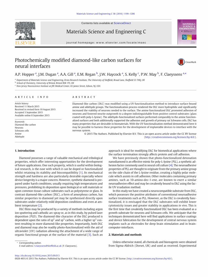
3.2.3. Primary Schwann cell culturePrimary Schwann cells were cultured upon glass, pristine DLC,
DLC-NH2 and PLL surfaces for 21 days after which they were stainedfor the glial marker S100β. Fluorescence micrographs are shown inFig. 6.
Clear differences in Schwann cell morphology were observed acrossthe different surfaces. In contrast to the NG108-15 neuronal cells, aconsiderable number of Schwann cells attached to the glass substrate(Fig. 6a). On glass the cell morphology was either bipolar or tripolarwith thin processes that showed no particular orientational preference.A small degree of Schwann cell attachment was also observed on thepristine DLC surfaces (Fig. 6c). However, in contrast to cells on theglass surfaces, the cells on pristine DLC adopted a variety of uncharac-teristic morphologies, some resembling fibroblasts whilst othersadopted a highly spread polygonal morphology. Nonetheless, positiveexpression of the glial marker S100β was still observed on both theglass and pristine DLC surfaces.
In contrast to those on glass and pristine DLC, Schwann cells cultureduponDLC-NH2 and PLL surfaces formed confluentmonolayers and exhib-ited similar morphologies. The cells adopted a bipolar, elongatedmorphology, which is characteristic of Schwann cells, and nearly allprocesseswere aligned alongside adjacent cells. The confluent cell mono-layers appeared stable and showed no sign of delamination from thesubstrate.
gative control). (b) PLL (positive control). (c) DLC. (d) DLC-NH2. (e) DLC-CHO. Cells werePI (blue). Scale bars: 100 μm.

Fig. 5. Image analysis of NG108-15 neuronal cell dendricity from fluorescence micro-graphs of cells growing upon glass, DLC, DLC-CHO, DLC-NH2 and PLL surfaces after 1, 2,5 and 7 days. Values are reported as the mean of three replicate samples ± standarderror. (a) Mean number of neurites per cell. (b) Mean neurite length. Full statistical anal-ysis was carried out, the results of which are given in the supplementary information.
1204 A.P. Hopper et al. / Materials Science and Engineering C 58 (2016) 1199–1206
4. Discussion
Wehave prepared DLC substrateswhichwere subsequentlymodifiedby a UV functionalization method to covalently bond amine andaldehyde-containing molecules to the surface in order to improveneurocompatibility. DLC and related materials, such as CVD diamond,have several interesting properties which support their use as biomate-rials. Firstly they may be readily doped or functionalized to alter theirbulk characteristics, for example electrical properties, to suit the desiredapplication [2,6–10]. Diamond-basedmaterials have also been tested ex-haustively to prove their biocompatibility and low toxicity [11–16]. Withrespect to manufacturing, DLC is also advantageous in that the size orshape of objects to be coated is of little consequence to the coating proce-dure. Most components and devices may be treated swiftly and at rela-tively low expense to create a stable and robust surface layer [2,17].Additionally, although of less relevance in neurosurgical applications,the wear-resistance of DLC could provide comparatively high longevityfor any potential implants, potentially reducing the need for revision sur-gery in certain applications [2].
Hydrogen-terminated diamond-like materials, such as the DLC pro-duced by pulsed laser deposition used in this study, are known to inhibitneuronal growth due to the lack of adequate interaction sites to facili-tate cell adhesion [18]. By suitable surface modification, however, bio-compatibility and bioactivity may be significantly improved. Uponmodification with surface-bound amine and aldehyde groups DLC wasshown to promote adhesion of NG108-15 neuronal cells and primaryrat Schwann cells. Furthermore, the NG108-15 neuronal cells producedlong neurites and in the case of the amine functional DLCwere indistin-guishable from the positive control coating (PLL).
Although there are several variables that dictate the survival, growthand differentiation of neuronal cells in vitro, one of themost important isthe culture substrate [19,20]. Substrates that have been widely used inthe culture of neuronal cells include glass and tissue culture plastic thathave been coated with attachment factors of some kind. The most com-mon attachment factors are cationic polypeptides of basic amino acidssuch as poly-L-lysine (PLL) and poly-L-ornithine [21,22], whilst extracel-lular matrix components including laminin have also been applied, withcollagen and fibronectin also being used to a lesser extent [23–26]. Suchcoatings have become widespread as a pre-treatment for neuronal cellculture since they promote cellular adhesion, survival and neurite out-growth [19,20,22,27].
It is not entirely clear how substances such as PLL, which containamine groups, improve cell attachment and viability. The effect mayarise from interactions with specific receptors on neuronal cell mem-branes enhancing adhesion to the substrate. Alternatively, a cationicpolypeptide may act as a positively charged interlayer, electrostaticallybinding the negatively charged cell membrane to the substrate surface[22,28,29]. Proteins originating from the serum component of the cellmedium have also been identified as assisting cell adhesion, acting ina similarmanner to extracellularmatrix proteins [29]. Substrates coatedwith PLL have been shown to lead to the development of a greater thick-ness of adsorbed proteins during cell culture compared to control sur-faces, these either being secreted by the cells or adsorbed from theconstituents of the culture medium [20]. Proteins which adsorb initiallyto PLL undergo denaturation and do not encourage neuronal adhesion;however subsequent proteins which bind to this denatured layer retaintheir structure intact, thus encouraging subsequent cell adhesion.
Although PLL is often used as the standard attachment factor to sup-port the growth of Schwann cells in vitro, it is not envisaged to be desir-able or permissible to use such synthetic polypeptides for in vivoapplications such as neural prosthetics due to concerns about toxicity[30]. Covalent functionalization of culture substrates is therefore an ap-pealing alternative in developing such applications. It has previouslybeen demonstrated that surface-bound amine groups can support neu-ronal adhesion and differentiation similarly without detrimental effects[31]. Although research in this area is ongoing it is thought that aminesinteract with cells throughmembrane-bound chondroitin sulphate pro-teoglycans (CSPGs). These proteoglycans exert significant influenceover neuronal differentiation via integrin-mediated signalling whichmay explain the neurocompatibility of amines and amine-containingsubstances such as PLL [32,33].
It is possible that by covalently functionalizing surfaces with aminegroups the longevity of the neurocompatibility may be enhanced incomparison to PLL. Indeed it has been shown that significantly highershear stresses were required to detach NG108-15 cells from covalently-modified amine surfaces, in comparison to those functionalized withECM proteins [19]. This was corroborated by further research whichconcluded that covalently bound PLL was far more effective at retainingadhered cells than PLL that was passively adsorbed to the substrate [22].An additional benefit of covalently bound functionalities is their lack ofleaching into solution, contrasting with non-covalently bound PLL thegradual desorption of which can result in cell dissociation [22]. All thesefactors demonstrate the advantages of the UV functionalization approachdemonstrated here.
In addition to amine groups, aldehyde groups were also introducedto the surface of the DLC using a similar UV functionalization method(with undec-10-enal). In the cell culture studies the aldehyde function-al group performed broadly comparably to the amine group with simi-lar levels of neuronal viability, attachment and neurite developmentand indistinguishable Schwann cell attachment and morphology. Al-though relatively little research has been undertaken regarding suchaldehyde-containing functionalities for cell growth, aldehyde terminat-ed surfaces are regarded as being hydrophilic. In addition, plasmapolymerised surfaces formed from aldehyde-containing monomershave been shown to support the growth and attachment of cells to a
Fig. 6. Fluorescence micrographs of primary rat Schwann cells cultured for 21 days. (a) Glass. (b) PLL. (c) Pristine DLC. (d) DLC-NH2. Cells were immunolabelled for S100β. Scale bars:100 μm.
1205A.P. Hopper et al. / Materials Science and Engineering C 58 (2016) 1199–1206
similar degree as those cultured upon tissue culture modified polysty-rene [34].
Aldehyde-functionalized substrates have been shown to be capableof immobilising proteins upon their surface to produce self-assembledmonolayers (SAMs) of protein, indicating that they may be used forapplications within the biosensor research field as well as for direct in-terface with cell and tissues [35]. A similar study has also shown thatsurfaces bearing aldehyde groups may immobilise proteins from solu-tion (in this case bFGF)whichmay then act as ligands for specific recep-tors allowing the capture and adhesion of cells [36]. It seems highlylikely that a similar mechanism could occur in which serum proteinssuch asfibronectin could be captured by surface-bound aldehydes, lead-ing to increased cell adhesion. As it is known that adhesion receptorswithin the cell membrane not only influence the adherence of cellsbut also mediate cellular growth and differentiation, the importance ofengineering substrates that promote cellular adhesion is clear [37,38].Although theDLC-CHO surfaces supportedneuron and Schwann cell ad-hesion and differentiation of NG108 cells, by some measures (neuritelength and viability) these surfaces did not perform as well as theamine-bearing surfaces. The reason for this is not clear but may bedue to differences in both the degree of protein adsorption and the con-formation of adsorbed proteins. Elucidation of this mechanism wouldrequire further study.
5. Conclusions
The surface of DLCwasmodified using a UV functionalizationmethodto introduce amine and aldehyde groups to improve neurocompatibility.The functionalization process rendered the DLC more hydrophilic andsignificantly increased the viability of neuron-like cells seeded to thesurfaces. The amine-functionalized DLC promoted neuron adhesion and
neurite outgrowth to a degree indistinguishable from positive controlsubstrates. The aldehyde-functionalized surfaces performed comparablyto the amine-functionalized surfaces and both additionally supportedthe adhesion and growth of primary rat Schwann cells. With the UVfunctionalization method demonstrated here it may be possible toharness the unique properties of DLC for the development of implantabledevices to interface with the nervous system.
Acknowledgements
The authors are grateful to EPSRC for funding this work in the formof a studentship for AH and PDRA position for JD supported undergrant number EP/K002503/1. The Sheffield Surface Analysis Centreand Kroto Imaging Facility are also gratefully acknowledged.
Appendix A. Supplementary data
Supplementary data to this article can be found online at http://dx.doi.org/10.1016/j.msec.2015.09.013.
References
[1] P.W. May, E.M. Regan, A. Taylor, J. Uney, A.D. Dick, J. McGeehan, Spatially controllingneuronal adhesion on cvd diamond, Diam. Relat. Mater. 23 (2012) 100–104.
[2] E.M. Regan, J.B. Uney, A.D. Dick, Y.W. Zhang, J. Nunez-Yanez, J.P. McGeehan, F.Claeyssens, S. Kelly, Differential patterning of neuronal, glial and neural progenitorcells on phosphorus-doped and UV irradiated diamond-like carbon, Biomaterials31 (2) (2010) 207–215.
[3] B. Sun, P.E. Colavita, H. Kim, M. Lockett, M.S. Marcus, L.M. Smith, R.J. Hamers, Covalentphotochemical functionalization of amorphous carbon thin films for integrated real-time biosensing, Langmuir 22 (23) (2006) 9598–9605.
[4] A.P. Hopper, J.M. Dugan, A.A. Gill, O.J.L. Fox, P.W. May, J.W. Haycock, F. Claeyssens,Amine functionalized nanodiamond promotes cellular adhesion, proliferation andneurite outgrowth, Biomed. Mater. 9 (4) (2014).

1206 A.P. Hopper et al. / Materials Science and Engineering C 58 (2016) 1199–1206
[5] R. Kaewkhaw, A.M. Scutt, J.W. Haycock, Integrated culture and purification of ratSchwann cells from freshly isolated adult tissue, Nat. Protoc. 7 (11) (2012) 1996–2004.
[6] S. Neuville, A. Matthews, A perspective on the optimisation of hard carbon and re-lated coatings for engineering applications, Thin Solid Films 515 (17) (2007)6619–6653.
[7] T.C. Kuo, R.L. McCreery, G.M. Swain, Electrochemical modification of boron-dopedchemical vapor deposited diamond surfaces with covalently bonded monolayers,Electrochem. Solid-State Lett. 2 (6) (1999) 288–290.
[8] S.C.H. Kwok, W. Jin, P.K. Chu, Surface energy, wettability, and blood compatibilityphosphorus doped diamond-like carbon films, Diam. Relat. Mater. 14 (1) (2005)78–85.
[9] S.C.H. Kwok, P.C.T. Ha, D.R. McKenzie, M.M.M. Bilek, P.K. Chu, Biocompatibility of cal-cium and phosphorus doped diamond-like carbon thin films synthesized by plasmaimmersion ion implantation and deposition, Diam. Relat. Mater. 15 (4–8) (2006)893–897.
[10] N. Yang, H. Uetsuka, H. Watanabe, T. Nakamura, C.E. Nebel, Photochemical attach-ment of amine-layers on h-terminated undoped single crystalline cvd diamonds,Diam. Relat. Mater. 17 (7–10) (2008) 1376–1379.
[11] S.J. Yu,M.W. Kang, H.C. Chang, K.M. Chen, Y.C. Yu, Bright fluorescent nanodiamonds: nophotobleaching and low cytotoxicity, J. Am. Chem. Soc. 127 (50) (2005) 17604–17605.
[12] K.K. Liu, C.L. Cheng, C.C. Chang, J.I. Chao, Biocompatible and detectable carboxylatednanodiamond on human cell, Nanotechnology 18 (32) (2007).
[13] A.M. Schrand, L. Dai, J.J. Schlager, S.M. Hussain, E. Osawa, Differential biocompatibil-ity of carbon nanotubes and nanodiamonds, Diam. Relat. Mater. 16 (12) (2007)2118–2123.
[14] W.J. Ma, A.J. Ruys, R.S. Mason, P.J. Martin, A. Bendavid, Z.W. Liu, M. Ionescu, H.Zreiqat, Dlc coatings: Effects of physical and chemical properties on biological re-sponse, Biomaterials 28 (9) (2007) 1620–1628.
[15] C. Hinuber, C. Kleemann, R.J. Friederichs, L. Haubold, H.J. Scheibe, T. Schuelke, C.Boehlert, M.J. Baumann, Biocompatibility and mechanical properties of diamond-likecoatings on cobalt–chromium–molybdenum steel and titanium–aluminum–vanadiumbiomedical alloys, J. Biomed. Mater. Res. Part A 95A (2) (2010) 388–400.
[16] N. Mohan, C.-S. Chen, H.-H. Hsieh, Y.-C. Wu, H.-C. Chang, In vivo imaging and toxic-ity assessments of fluorescent nanodiamonds in Caenorhabditis elegans, Nano Lett.10 (9) (2010) 3692–3699.
[17] S. Kelly, E.M. Regan, J.B. Uney, A.D. Dick, J.P. McGeehan, E.J. Mayer, F. Claeyssens, G.Bristol Biochip, Patterned growth of neuronal cells on modified diamond-like car-bon substrates, Biomaterials 29 (17) (2008) 2573–2580.
[18] T. Lechleitner, F. Klauser, T. Seppi, J. Lechner, P. Jennings, P. Perco, B. Mayer, D.Steinmuller-Nethl, J. Preiner, P. Hinterdorfer, M. Hermann, E. Bertel, K. Pfaller, W.Pfaller, The surface properties of nanocrystalline diamond and nanoparticulate dia-mond powder and their suitability as cell growth support surfaces, Biomaterials 29(32) (2008) 4275–4284.
[19] R.S. Cargill Ii, K.C. Dee, S.Malcolm, Anassessment of the strength of ng108-15 cell ad-hesion to chemicallymodified surfaces, Biomaterials 20 (23–24) (1999) 2417–2425.
[20] A.E. Schaffner, J.L. Barker, D.A. Stenger, J.J. Hickman, Investigation of the factors nec-essary for growth of hippocampal neurons in a defined system, J. Neurosci. Methods62 (1–2) (1995) 111–119.
[21] E. Yavin, Z. Yavin, Attachment and culture of dissociated cells from rat embryo cere-bral hemispheres on polylysine-coated surface, J. Cell Biol. 62 (2) (1974) 540–546.
[22] Y.H. Kim, N.S. Baek, Y.H. Han, M.-A. Chung, S.-D. Jung, Enhancement of neuronal celladhesion by covalent binding of poly-d-lysine, J. Neurosci. Methods 202 (1) (2011)38–44.
[23] T. Elsdale, J. Bard, Collagen substrata for studies on cell behavior, J. Cell Biol. 54 (3)(1972) 626-&.
[24] S. Carbonetto, M.M. Gruver, D.C. Turner, Nerve-fiber growth in culture on fibronec-tin, collagen, and glycosaminoglycan substrates, J. Neurosci. 3 (11) (1983)2324–2335.
[25] P.J. Lein, G.A. Banker, D. Higgins, Laminin selectively enhances axonal growth andaccelerates the development of polarity by hippocampal neurons in culture. Brainresearch, Dev. Brain Res. 69 (2) (1992) 191–197.
[26] A. Lochter, M. Schachner, Tenascin and extracellular-matrix glycoproteins — frompromotion to polarization of neurite growth in-vitro, J. Neurosci. 13 (9) (1993)3986–4000.
[27] C.D. James, R. Davis, M. Meyer, A. Turner, S. Turner, G. Withers, L. Kam, G. Banker, H.Craighead, M. Isaacson, J. Turner, W. Shain, Aligned microcontact printing ofmicrometer-scale poly-l-lysine structures for controlled growth of cultured neuronson planar microelectrode arrays, IEEE Trans. Biomed. Eng. 47 (1) (2000) 17–21.
[28] I. Lieberman, P. Ove, Protein growth factor for mammalian cells in culture, J.Biolumin. Chemilumin. 233 (3) (1958) 637–642.
[29] E.M. Harnett, J. Alderman, T. Wood, The surface energy of various biomaterials coat-edwith adhesionmolecules used in cell culture, Colloids Surf. B: Biointerfaces 55 (1)(2007) 90–97.
[30] A. Thalhammer, R.J. Edgington, L.A. Cingolani, R. Schoepfer, R.B. Jackman, The use ofnanodiamond monolayer coatings to promote the formation of functional neuronalnetworks, Biomaterials 31 (8) (2010) 2097–2104.
[31] J.H. Wang, U.H. Lin, C.H. Lin, Y.C. Chung, C.R. Chen, Y.C. Kao, J.Y. Lai, T.H. Young,Change in neuron aggregation and neurite fasciculation on eval membranes modi-fied with different diamines, J. Biomed. Mater. Res. Part A 94A (2) (2010) 489–498.
[32] S.P. Massia, J.A. Hubbell, Immobilized amines and basic-amino-acids as mimeticheparin-binding domains for cell-surface proteoglycan-mediated adhesion, J.Biolumin. Chemilumin. 267 (14) (1992) 10133–10141.
[33] W.L. Gu, S.L. Fu, Y.X.Wang, Y. Li, H.Z. Lu, X.M. Xu, P.H. Lu, Chondroitin sulfate proteo-glycans regulate the growth, differentiation and migration of multipotent neuralprecursor cells through the integrin signaling pathway, BMCNeurosci. 10 (2009) 15.
[34] K.S. Siow, L. Britcher, S. Kumar, H.J. Griesser, Plasma methods for the genera-tion of chemically reactive surfaces for biomolecule immobilization and cellcolonization — a review, Plasma Process. Polym. 3 (6–7) (2006) 392–418.
[35] C.D. Hahn, C. Leitner, T. Weinbrenner, R. Schlapak, A. Tinazli, R. Tampé, B. Lackner, C.Steindl, P. Hinterdorfer, H.J. Gruber, M. Hölzl, Self-assembledmonolayers with latentaldehydes for protein immobilization, Bioconjug. Chem. 18 (1) (2006) 247–253.
[36] D. Peelen, V. Kodoyianni, J. Lee, T. Zheng, M.R. Shortreed, L.M. Smith, Specific captureof mammalian cells by cell surface receptor binding to ligand immobilized on goldthin films, J. Proteome Res. 5 (7) (2006) 1580–1585.
[37] M.V. Agrez, R.C. Bates, Colorectal-cancer and the integrin family of cell-adhesion re-ceptors — current status and future-directions, Eur. J. Cancer 30A (14) (1994)2166–2170.
[38] N. Basora, P.H. Vachon, F.E. HerringGillam, N. Perreault, J.F. Beaulieu, Relation be-tween integrin alpha 7b beta 1 expression in human intestinal cells and enterocyticdifferentiation, Gastroenterology 113 (5) (1997) 1510–1521.