materials and methods - shodhgangashodhganga.inflibnet.ac.in/bitstream/10603/31863/8/08_chapter...
TRANSCRIPT

40
MATERIALS AND METHODS
41
Chapter-1 Purification of antimicrobial peptide, AMPs LR14
Source of strain used in the study
An environmental isolate of lactic acid bacteria, Lactobacillus plantarum strain
LR/14 isolated from the plant rhizosphere (LR: L, lactic acid bacteria; R, rhizosphere)
was procured from the lab stock. Strain LR/14 was maintained on MRS (de Man-
Rogosa-Sharpe) medium. The culture was raised at 37°C under static conditions.
Micrococcus luteus MTCC 106, which was used as an indicator strain was grown in
NB (nutrient broth) at 37°C and 200 rpm. The medium composition is given in
Annexure-I.
Raising of culture and preparation of culture supernatant
Lb. plantarum LR/14 culture was grown at 37ºC for 24h. The culture supernatant was
obtained by centrifugation at 6000 X g at 4ºC for 10 min. In order to obtain the crude
AMPs LR14, the culture supernatant was filtered through a 0.2 µm membrane filter
(mdi Advanced Microdevices, Ambala, India) to remove any cell contamination.
Antimicrobial activity assay
The main body of this work was to identify and characterize the antimicrobial peptide
(AMPs LR14) produced and externalized by Lb. plantarum strain LR/14. The crude
culture supernatant was tested for its antimicrobial action against M. luteus.
Antimicrobial action was assayed in terms of both qualitative as well as quantitative
methods.
(A) Qualitative estimation:
(i) Agar Well Diffusion Assay (AWDA) - (Tagg et al., 1976): For AWDA, aliquots
of culture supernatants (100µl) were filled in wells (6mm diameter) cut out in cooled
TGYE agar that was overlaid with soft agar (0.8%) inoculated with an indicator strain
(106 cfu/ml). The plate was incubated under optimal conditions for growth of the
target organism. Diameter of the growth inhibition zones (halo) was measured (in
terms of mm) after 24 h of incubation.

42
(B) Quantitative estimation:
(i) Activity unit per milliliter (AU/ml)-(Daba et. al., 1991):
Activity of antimicrobial peptides was quantified by a microtitre plate assay by a
critical-dilution method. Each well of the microtitre plate containing 200µl nutrient
broth (NB) was inoculated with the indicator organism (106
cfu/ml) derived from a
fresh overnight culture. An antimicrobial peptide sample was serially diluted two-fold
(1/2) in 50µl volume of nutrient broth and mixed with the bacterial sample in a
Falcon 96-well microtitre plate (Becton Dickinson Labware, NJ, USA). The
microtitre plate was incubated for 6h at 37ºC prior to assessment of growth inhibition
turbidometrically at 630nm using a Microplate Reader (Bio-Rad, USA). Activity
units are arbitrary units calculated against the standard indicator strain M. luteus. One
activity unit (AU) is derived from the reciprocal of the AMPs LR14 required to
inhibit the growth of the indicator strain by 50% in comparison to the untreated
control, and represented as per ml.
(ii) Percentage inhibition
The antimicrobial activity was also checked in terms of percentage of growth
inhibition of indicator organism. The nutrient broth was inoculated with indicator
strain (initial OD630 ~0.02) along with different concentrations of AMPs, as described
later, and incubated at 37°C for overnight. Growth in terms of cfu/ml was obtained by
plating an appropriately diluted cell suspension on nutrient agar (NA). This was
compared with the untreated control. The latter was considered as 100% growth and
the percentage of growth inhibition, if any, was calculated.
Protein estimation
Following methods were used to estimate the protein concentration in the culture
supernatant/AMPs solution:
BCA method (Bicinchoninic acid assay kit, Sigma-Aldrich, USA): This method
was used to estimate the protein concentration as per the manufacturer‘s instructions.
The principle of the BCA assay relies on the formation of a Cu2+
-protein complex
43
under alkaline conditions, followed by reduction of the Cu2+
to Cu+. The amount of
reduction is proportional to the protein present. Briefly, 2 ml of BCA working
reagent (provided with the kit) was added to 0.1 ml of each BSA protein standard,
blank and unknown sample. The solutions were vortexed gently for thorough mixing
and incubated at 37ºC for 30 minutes. The tubes were allowed to cool at room
temperature. The reaction solutions were transferred into a cuvette. The absorbance of
the solution was measured at 562 nm. A standard curve based on the BSA protein
standard concentration was drawn. The protein concentration was estimated by
comparing the absorbance of the unknown samples to the standard curve.
Protein estimation by UV absorption: The protein present in the crude AMPs
sample was measured using UV-visible spectrophotometer (Bio-Rad, USA). The
standard curve for the BSA was prepared in the same way and accordingly the
concentration of unknown protein sample was determined.
Protein activity: The antimicrobial activity of protein was measured by activity units
per milliliter (AU/ml) as mentioned earlier.
Precipitation and extraction of the protein by three phase partitioning
The optimum concentration for precipitation of AMPs was standardized by adding
different amounts of ammonium sulfate, so as to achieve 40%, 60% and 90%
saturation. The culture supernatant with added ammonium sulfate was constantly
mixed on a stirrer at 4ºC to aid in the precipitation of protein. This solution was
mixed well with tertiary butanol (Fisher scientific) in a sterile bottle in a ratio of 1:1,
finally poured into a separating funnel, and was allowed to stand till three layers were
distinctly visible. The lowermost layer was the aqueous layer, uppermost organic
layer and in between the protein rich interfacial layer was observed. The interfacial
layer was separated from the other two layers, washed a few times and the protein
was dissolved in autoclaved distilled water. It was concentrated in a Speed Vac
(Thermofisher Scientific, USA). The antimicrobial activity in terms of activity units
44
per ml (AU/ml) was checked in all the three fractions. This was taken as the partially-
purified fraction and stored at 4ºC for future use.
Gel-filtration Chromatography
To get rid of the salt present in the partially-purified fraction, Sephadex G-25
Desalting columns, (pre-packed disposable PD-10 columns, GE-Healthcare Bio-
Sciences, USA) were used.
Column Equilibration : The column was equilibrated with autoclaved distilled water
with three times its bed volume at a flow rate of 1ml/min.
Sample application : The partially-purified protein solution was passed through a
0.2µm syringe filter (mdi Advanced Microdevices, Ambala, India) to get rid of any
contaminants. The sample was loaded on the column at the rate of 250µg/ml/min to
ensure effective binding with the column matrix.
Elution : The elution was done using 20 ml of autoclaved distilled water and the
desalted protein was collected in sterile eppendorff tubes at the rate of 1ml/min on a
fraction collector. Each sample was checked for antimicrobial activity against M.
luteus and protein concentration. While the amount of protein present was determined
by BCA method (Bicinchoninic acid assay kit, Sigma), AWDA as described above
was carried out to look for the presence of any antimicrobial activity. The active
proteinaceous fractions were lyophilized (FD-5, Allied Frost, India) and resuspended
in 10 ml of autoclaved distilled water.
Reverse phase-High performance liquid chromatography (RP-HPLC)
The purity of peptide/peptides obtained after gel filtration chromatography was
analyzed on HPLC using CLASS-VP software (Shimadzu, Japan). A C18 reverse
phase HPLC column (250 X 4.6 mm, Synergy, 4µ hydro RP, Phenomenex, Japan)
was used. The solvents comprised, solvent A which was composed of HPLC grade
water + 0.1% TFA (Trifluoroacetic acid) and solvent B that contained acetonitrile +
0.1% TFA. These solvents were filtered using a filtration unit attached to a vaccum
45
pump and degassed in a bath sonicator. The temperature of the column oven was set
at 40ºC. The column (C18 5µ 250 X 4.6 mm, Phenomenex) was washed with 50%
methanol and then equilibrated with a gradient of solvent A and solvent B. After the
emergence of the baseline, 1 mg/ml of sample was loaded. The peptides were eluted
with the solvent gradient. For standardization, the chromatogram was analyzed at
different flow rates - 0.1ml/min, 0.2ml/min, 0.35ml/min, 0.5ml/min, 1ml/min and
1.5ml/min. However, maximum resolution was obtained at 0.35ml/min, so it was
finally standardized using the following program. The analysis of the sample was
done using a UV detector at a wavelength of 226 nm with CLASS-VP software
(Shimadzu, Japan). After the analysis, column was washed again with methanol and
stored at room temperature. The HPLC program used:
Time (minutes) A solvent concentration B solvent concentration
0-2 80% 20%
2-6 60% 40%
6-12 40% 60%
12-15 100% 0%
Induction effect
Two peaks were resolved by HPLC, which were collected separately. Since the
HPLC profile showed up a minor peak with low activity, studies were carried out to
assess if this peptide as an inducer peptide. For this, three sets were employed and a
time-course study (4h, 8h, 12h, 16h, 20h, 24h) was carried out. While set A
represented the uninduced LR/14 control grown in MRS, in set B, Lb. plantarum
LR/14 culture was grown in the presence of 200 AU/ml of minor peak protein, and
the set C, where culture was raised in the presence of crude culture supernatant of
strain LR/14 containing 200 AU/ml. These cultures were allowed to incubate for 18h,
and the net activity units (AU/ml) was calculated for all the sets aliquoted at different
time points.
46
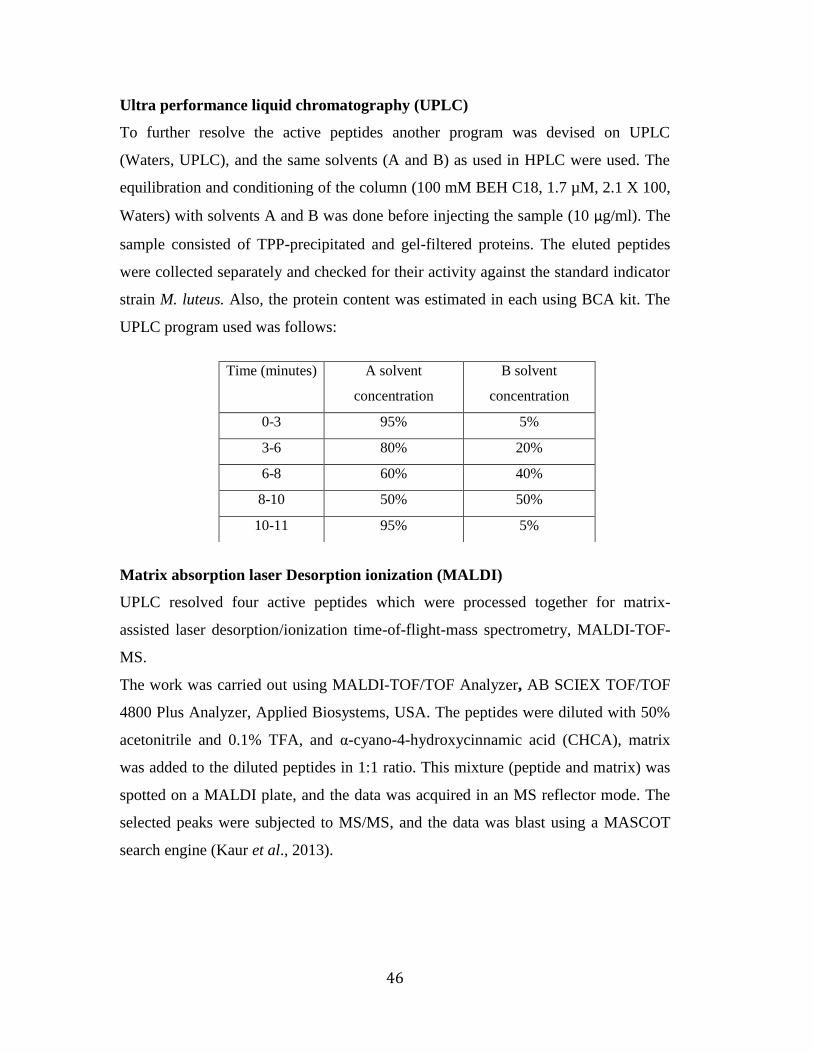
Ultra performance liquid chromatography (UPLC)
To further resolve the active peptides another program was devised on UPLC
(Waters, UPLC), and the same solvents (A and B) as used in HPLC were used. The
equilibration and conditioning of the column (100 mM BEH C18, 1.7 µM, 2.1 X 100,
Waters) with solvents A and B was done before injecting the sample (10 µg/ml). The
sample consisted of TPP-precipitated and gel-filtered proteins. The eluted peptides
were collected separately and checked for their activity against the standard indicator
strain M. luteus. Also, the protein content was estimated in each using BCA kit. The
UPLC program used was follows:
Matrix absorption laser Desorption ionization (MALDI)
UPLC resolved four active peptides which were processed together for matrix-
assisted laser desorption/ionization time-of-flight-mass spectrometry, MALDI-TOF-
MS.
The work was carried out using MALDI-TOF/TOF Analyzer, AB SCIEX TOF/TOF
4800 Plus Analyzer, Applied Biosystems, USA. The peptides were diluted with 50%
acetonitrile and 0.1% TFA, and α-cyano-4-hydroxycinnamic acid (CHCA), matrix
was added to the diluted peptides in 1:1 ratio. This mixture (peptide and matrix) was
spotted on a MALDI plate, and the data was acquired in an MS reflector mode. The
selected peaks were subjected to MS/MS, and the data was blast using a MASCOT
search engine (Kaur et al., 2013).
Time (minutes) A solvent
concentration
B solvent
concentration
0-3 95% 5%
3-6 80% 20%
6-8 60% 40%
8-10 50% 50%
10-11 95% 5%

47
N-terminal sequencing
One of the UPLC-purified peptides was digested with CNBr and then subjected to
Edman degradation and sequence was obtained on ABI‘s ―Procise-492‖ model. The
sequencing was done at the Protein Sequencing Facility at INTAS
Biopharmaceuticals Ltd., Ahmedabad, India.
Identification of AMPs LR14 with AMPs produced by clones A5 and A6
The plantaricin operon of strain LR/14 has been characterized in the lab, and five
structural genes, pln E, F, J, K, and N were identified. In the same study, clones, A5
and A6, were generated which were shown to have suffered large deletions (Nitika
Ghosh, unpublished results). In order to see if AMPs identified during present
investigation were in any way affected, the same were purified from the culture
filtrate of both the clones by TPP method as described above. These were further
resolved on UPLC and UPLC chromatograms were obtained following the same
program as described above.
Similarly to compare these AMPs with plantaricins from strain LR/14, commercially
synthesized plantaricins viz. Pln E, Pln F and Pln A were subjected to UPLC after
mixing them in a ratio of 1:1 v/v.
Characterization of purified AMPs LR14
The purified AMPs LR14 were characterized for different parameters:
Sensitivity to heat, pH, storage conditions and hydrolytic enzymes
Aliquots of purified AMPs LR14 samples were exposed to heat treatment at 60°, 80°
and 100°C for 30 min in a water bath, and one aliquot was autoclaved (121°C at 15
psi, for 15 min). The treated samples were tested for residual antimicrobial activity, in
terms of AU/ml.
In parallel, aliquots of AMPs LR14 samples were adjusted to pH values ranging from
2.0 to 8.0 in 1:1 ratio of different buffers (HCl-KCl 50 mM, pH 2.0, and 4.0;

48
phosphate buffer 50 mM, pH 6.0, 7.0, and 8.0), incubated at 37°C for 4 h, and tested
for residual antimicrobial activity (AU/ml).
To determine the durability, the AMPs LR14 sample was stored at different
temperatures (-20°, 4°, R.T {room temperature: fluctuating between 10° to 30°C
night/day temperature}, and 37°C). Its subsequent activity was checked at an interval
of one month up to one year, by AU/ml, and compared with the fresh preparation.
In a separate experiment, the sensitivity of the AMPs LR14 to proteolytic enzymes
(proteinase K, pepsin, papain, α-chymotrypsin and protease), as also to lipase and α-
amylase (Sigma-Aldrich, USA) was tested at a final concentration of 1 mg/ml, at
37°C for 2 h. After incubation, the enzymes were heat-inactivated (5 min at 100°C),
and the samples were tested for the antimicrobial activity in terms of AU/ml.
Untreated samples were taken as controls in each case.
Chapter-2 Mode of action and Inhibition spectrum of AMPs LR14
Sources of strains used in the study
Micrococcus luteus and Escherichia coli K12 were available in our Lab Stock.
Listeria monocytogenes and Yersinia enterocolitica were obtained from Prof. J.S.
Virdi, Department of Microbiology, UDSC, New Delhi. The fungal stocks were
procured from Indian Agricultural Research Institute (IARI), New Delhi. These were,
Aspergillus niger ITCC 321, Rhizopus nigricans ITCC 4416, Mucor racemosus ITCC
4392, Penicillium chrysogenum ITCC 419. Plasmodium falciparum 3D7 and
Mycobacterium tuberculosis H37Rv were kindly provided by Prof. P.C. Ghosh and
Prof. Anil Tyagi, respectively, Department of Biochemistry, UDSC, New Delhi. The
work against some clinically important pathogens (Pseudomonas, Acinetobacter,
Klebsiella, Enterobacter, Salmonella typhi, Staphylococcus, and VRE) was carried
out in Dr. Rajni Gaind‘s lab, Department of Microbiology, Safdarjung Hospital, New
Delhi. The clinical pathogens were revived on Mac Conkey agar medium and
maintained on Mueller Hinton Infusion agar (MHI) medium, both obtained from
HiMedia, India.
49
The media compositions are detailed in Annexure I.
Maintenance of culture
E. coli K-12 and M. luteus were maintained in Luria broth (LB) and Nutrient broth
(NB), respectively. The cultures were raised at 37°C, 200 rpm, sub-cultured to an
initial A600 of 0.02 (~106 cfu/ml) and grown for 18h in all experiments, unless
otherwise specified. All the cultures were purified by single colony isolation on their
respective agar medium. Colonies from such plates were inoculated to raise the
cultures, depending upon experimental requirements, as described later. All cultures
were maintained on specific media and sub-cultured every 2-3 weeks. All chemicals
used were of analytical grade, obtained from Sigma-Aldrich (USA), and media were
purchased from HiMedia (Mumbai, India). The composition of the media and
solutions are detailed in Annexure I.
Preparation of Glycerol Stocks
For long-term storage of the cultures, glycerol stocks were prepared (Sambrook et al.,
1989). To 0.5 ml of overnight culture, 0.5 ml of sterile 40% glycerol was mixed and
poured in a cryo-vial, and kept at -80ºC. For reviving the bacterial strains, the frozen
surface of stock was scraped with a sterile inoculation needle and immediately
streaked on the medium plate. The plates were incubated at 37ºC.
Growth
Growth was determined by two methods:
Absorbance
An inoculum size (A600 ~0.02) of an exponentially grown culture was transferred to a
fresh medium under sterile conditions. The cultures were raised under specific
conditions for a designated period of time. The change in growth was monitored
spectrophotometrically by measuring absorbance/optical density (A/OD) at 600 nm
for both net growth as well as growth at different time intervals.

50
Determination of colony forming units (cfu/ml)
For this, 0.1 ml cell suspension was serially diluted in 0.9 ml sterile normal saline
(0.85% NaCl) and vortexed. Then 0.1 ml from the appropriate dilutions was plated on
the required agar medium and incubated at specific temperature for overnight. Viable
counts were determined as colony-forming units per ml (cfu/ml) of the culture.
Percentage inhibition
The antimicrobial activity was also checked in terms of percentage of growth
inhibition of indicator organism as described earlier.
Cell viability assay
The two strains were grown in their respective media. An inoculum of ~0.02 OD600
(~106 cfu/mL) both of M .luteus and E. coli were treated with different concentrations
of crude AMPs LR14 for 18h. The concentrations used for M .luteus were 40, 80,
120, 140, and 160 AU/ml and for E. coli 200, 400, 600, and 1000 AU/ml. Cells
without any treatment were taken as control. Cells from different treatments and the
control were serially diluted in 0.85% saline, and viability was monitored in terms of
cfu/ml after overnight incubation at 37ºC. The percent inhibition was then calculated
for each treatment set with respect to control. From this, the effective concentration
was standardized, and the subsequent studies were carried out at 120 (IC50) and 140
(IC99) AU/ml in case of M. luteus, and 400 (IC50) and 600 (IC99) AU/ml in case of
E.coli. Since, the activity units (AU/ml) is an arbitrary unit and varies from organism
to organism, the same were converted into concentration of protein. Thus, 120, 140,
400, and 600 AU/ml used in the study amounted to 75, 100, 250, and 450 µg/ml of
protein, respectively.
Light microscopy : The cells of treated (IC50 and IC99 concentrations) and control
sets of the two strains were each harvested by centrifugation (6000 X g for 10min)
and washed twice with 0.1M phosphate buffer pH 7.4. The cells were stained with
safranine, mounted on a clean glass slide, and visualized (magnification 100 X) under
a light microscope, (Olympus, BX51, Japan).
51
Dual staining method: To further distinguish the resulting growth inhibition, dual
staining (two vital dyes viz. calcein AM and propidium iodide are known to provide
green and red fluorescence to live and dead cells, respectively) of AMPs LR14-
treated and control cells was carried out, as per the manufacturer‘s instructions (Live-
Dead Cell Dual Staining kit – Sigma, USA). In brief, 10 µl solution A and 5 µl
solution B was added to 5 ml PBS to prepare assay solution. The cell suspension was
centrifuged and the cell pellet was washed with PBS several times. Prepared cell
suspension in PBS was prepared at a density of 105 to 10
6 cells/ml. 200 µl of cell
suspension and 100 µl assay solution was mixed and incubated at 37°C for 15 min.
The fluorescence of viable and dead cells was monitored simultaneously at an
excitation wavelength of 490 nm through a fluorescence microscope, Olympus,
BX51, Japan (magnification 100X).
Estimation of Membrane potential
The protocol of Breeuwer and Abee (2004) was followed. Briefly, ~107 cells of M.
luteus and E. coli were centrifuged at 2800 X g for 10 min, washed and resuspended
in potassium phosphate buffer (pH 7). The cells were stored on ice until use.
Following stock solutions were prepared: florescent probe DiSC3 (3mM in DMSO)
stored in freezer, and valinomycin (3mM in ethanol). The cuvette was cleaned with
70% ethanol and air-dried. 3 ml cell suspension was added to the cuvette and mixed
using a magnetic stirrer. Then, 5 µl DiSC3 (final concentration of 5 µM) was added
and the spectrofluorimeter measurements were done at excitation wavelength 643 nm,
slit width 10 nm, and emission wavelength 666 nm, with slit width 10 nm. Once the
fluorescence signal was stable, ~15 min, 10 mM glucose was added to energize the
cells. After another 5 min of incubation, crude and purified AMPs LR14 were added
separately, and waited again until the fluorescence signal was stable. The
concentratons used were: for M. luteus cells, crude AMPs LR14 – 75 µg/ml and
purified AMPs LR14 - 65 µg/ml, and for E. coli, crude AMPs LR14 - 250 µg/ml and
purified AMPs LR14 – 100 µg/ml). In control, 10 µl valinomycin was added and the
fluorescence signal was recorded.

52
Mode of action
AMPs produced by lactic acid bacteria are known to be membrane-active compounds.
In order to see whether killing effect of AMPs LR14 is also due to this property, the
cells of M. luteus and E. coli were treated with 75, 250 (IC50) and 100, 450 (IC99)
concentrations, respectively for 18h. The culture supernatant was prepared to assess
various parameters. An untreated control was maintained in all the cases.
Preparation of culture supernatant
The treated and untreated cultures (grown at 37°C for 18h) of two strains were
centrifuged at 6000 X g at 4°C for 10 min. The spent culture filtrate was filtered
through a 0.2 µm membrane and stored at 4°C for further use.
K+ release
The culture supernatant from both the samples was analyzed to detect the
extracellular K+ using Atomic Absorption Spectrophotometer-Inductively Coupled
Plasma Optical Emission Spectrometry (ICP-OES) at 766.5 nm with an Optima 4200
instrument (Perkin Elmer, USA) and compared with untreated control. Standard curve
was made using 1 to 5 ppm of potassium chloride. The values obtained were
expressed as µmol/l.
Measurement of inorganic phosphate (ATPase assay)
The phosphate content in the culture broth was determined according to Heinonen
and Lahti (1981). Briefly, to 1ml of culture supernatant, 2ml of freshly prepared
AAM solution (acetone: 5N H2SO4: 10mM ammonium molybdate in a ratio of 2:1:1
v/v) was added and thoroughly mixed. Then 0.1ml of 1M citric acid was added,
mixed well, and the absorbance was recorded in a UV-Vis Spectophotometer
(Shimadzu, Japan) at 355nm. A standard curve of phosphate was prepared, from a
stock solution of KH2PO4 (2,000µg/ml) in distilled water. A dilution series ranging
between 50 µg/ml and 2,000 µg/ml was prepared from the stock solution. The values
obtained were expressed as µmol/l.
53
ATP release
To determine the leakage of adenosine 5‘-triphosphate (ATP) in the culture
supernatant of the cells treated with AMPs LR14, ATP detection kit (Sigma Aldrich,
USA) was used. The assay was based on the amount of light emitted when firefly
luciferase consumes ATP to catalyze the oxidation of D-luciferin. The readings were
measured using a luminometer, (Turner Biosystems, Sunnyvate, CA). These were
represented as nanomoles of ATP released, based on the standard curve of ATP (ATP
standard was made by diluting with the dilution buffer provided in the kit) in the
range of 10-3
to 10-5
mmoles/l and assayed similarly.
Release of UV absorbing material
Leakage of UV light-absorbing material was used as an indicator for the loss of cell
membrane integrity. M. luteus and E. coli cells corresponding to OD600 of ~0.02 were
treated with AMPs LR14 at their IC50 and IC99 concentrations, respectively in 0.85%
saline and incubated at 37ºC. Samples were removed after overnight incubation,
centrifuged, and culture supernatant filtered through 0.2 μm membrane (mdi
Advanced Microdevices, India). The absorbance of the filtrates was measured at 260
nm in a 96-well microtiter-plate, keeping normal saline as blank using a Microplate
Reader (Ultramark, Microplate imaging system, Biorad, USA).
Transmission electron microscopy
Untreated and treated cells of M. luteus and E. coli harvested by centrifugation (6000
X g for 10min) and washed twice with 0.1 M phosphate buffer pH (7.4), were pre-
fixed in 2.5% (v/v) glutaraldehyde and 2% (v/v) paraformaldehyde in 0.1 M
phosphate buffer pH 7.4 for 4h. Such cells were then post-fixed in 1% (w/v) osmium
tetraoxide for 1h. Following each fixation step, excess fixative was washed with 0.1
M phosphate buffer pH 7.4. The samples dehydrated using graded ethanol series (50-
100%) and infiltrated with a resin, were placed in an embedding mould, and
polymerized in an oven at 60ºC for 24 hours. Thin sections were cut by an
ultramicrotome and mounted on grids, covered with collodion film, and stained with
2% uranyl acetate in Reynold‘s lead citrate solution. Preparations were observed
54
using a transmission electron microscope (TEM) (FEI, Philips, Morgagni 268 D,
Holland) at 70 kV.
Assessment of resistance in M.luteus and E. coli against AMPs LR14
Approximately 107 cells of M. luteus and E. coli were treated with 100 µg/ml and 450
µg/ml (IC99 concentrations) of AMPs LR14, respectively for 18h. Different dilutions
of the cultures were plated on NB and LB agar plates, respectively and allowed to
incubate for 24h. A master plate each for treated M. luteus and E.coli cells was made
by patching the viable cells. These cells were again subjected to AMPs LR14
treatment using same concentrations for overnight and plated to check the viable cell
count. The treatment was repeated for seven successive cycles and in each viability
count was monitored.
Studies on fungi
This part of the work was directed to study the effect of AMPs LR14 against food-
spoilage fungi so as to implicate its role as a food biopreservative.
Sources of strains and maintenance of culture
Isolates of fungi viz., Aspergillus niger ITCC-321, Rhizopus nigricans ITCC-4416,
Mucor racemosus ITCC-4392, and Penicllium chrysogenum ITCC-419 were
procured from ITCC, IARI, New Delhi. All the strains were maintained on PDA
(Peptone Dextrose agar) medium. The cultures were raised at 30°C under static
conditions and sub-cultured every week. All chemicals used were of analytical grade,
and medium was purchased from HiMedia (Mumbai, India). The composition of the
media and solutions are detailed in Annexure I.
Counting of spores
Spores from a fresh plate of culture were picked up with a sterile inoculation loop
suspended in 0.85% saline and vortexed to get homogenous suspension. Spore
counting was done on a Neubaur‘s chamber or Haemocytometer.

55
Screening of antifungal activity
These experiments were carried out with all the four fungal strains selected. The
antifungal effect was visualized by dual culture assay. Lb. plantarum LR/14 was
streaked on MRS plates in two vertical streaks and allowed to incubate at 30ºC for 2
days. Approximately 105 spores/ml were mixed with soft PDA. This suspension was
poured over the LR/14 grown MRS plates and left for incubation at 30ºC for another
2 days. Any zone of growth inhibition observed was qualified as the antifungal
activity.
In a separate experiment, the antifungal effect was further confirmed by growing
these fungi on AMPs LR14-supplemented PDA plates and incubated for 48h at 30ºC.
Fungal discs (1cm X 1cm) from a fully grown colony, representing different stages of
growth were cut and kept on these petriplates. Fungi grown only on PDA without
AMPs LR14 served as controls.
Standardization of effective/lethal dose for causing fungal inhibition
Different concentrations ranging from 50, 100, 250, 500, 1,000, 2,000, 3,000 and
5,000 µg/ml of crude and purified AMPs LR14 were tested against different fungi as
mentioned above. Approximately 105 spores/ml of Aspergillus niger (taken as a test
case) were added to Peptone Dextrose (PD) medium along with the above mentioned
concentrations of AMPs LR14 in Erlenmeyer flasks. Another set of flasks with fungal
spores but without AMPs LR14 were kept as untreated control. The control and
treated cultures were incubated at 30ºC for 48h. The biomass was filtered through a
pre-weighed Whatman No. 1 filter paper pre-dried at 80ºC in an oven for 4h. The
filter papers containing the biomass were dried, weighed and dry biomass was
quantified in terms of the net weight. Percentage growth inhibition was calculated by
comparing the biomass obtained in the untreated and treated samples. For further
experiments, 3 and 5 mg/ml of crude and 1 and 2 mg/ml of purified AMPs LR14
were standardized as the lethal doses.
56
Effect of AMPs LR14 on different growth phases of fungi
For this, fungal spores were treated with crude AMPs LR14 at the standardized
concentrations (3 mg/ml and 5 mg/ml). The samples were stained with cotton blue
and mounted in lactophenol. Microscopy was done at different time intervals so as to
track the different phases of life cycle including: spore germination, hyphae formation
and mycelial ramifications and sporulation. In these experiments, biomass dry weight
was measured after 48h of incubation.
Inhibition of fungal spore germination
The cell free supernatant containing AMPs LR14 was screened against the spores of
selected fungi. Crude AMPs LR14 concentrations of 3 and 5 mg/ml were added to PD
broth containing ~105 spores/ml. The control set involved the addition of sterile MRS
broth only to the fungal spore suspension. The control and the experimental flasks
were kept at 30ºC for 48h. During this period, samples from the flasks were
withdrawn at different time intervals depending upon the time taken for various
stages of growth (under control conditions) of each fungus. After staining with cotton
blue, microscopic visualization (60X) was done for each of the treated as well as the
untreated controls. Another set comprising purified AMPs LR14 concentrations of
1mg/ml and 2 mg/ml were also tested with the spore suspension (~105 spores/ml) in
PD broth, keeping an untreated control alongwith. After 48h of incubation, biomass
dry weight was measured.
Inhibition of fungal growth (hyphae formation and mycelial ramifications)
In this assay, independent sets consisting of Erlenmeyer flasks containing ~ 105
spores per ml added in 10ml PD medium were treated with 3 and 5 mg/ml of crude
AMPs LR14, and 1 and 2 mg/ml of purified AMPs LR14 at different times. While in
one set AMPs were added at the start of the experiment, further treatments were done
after 8h for A. niger, R. nigricans and M. racemosus and after 12h for P.
chrysogenum for tracking hyphal growth, and after 16h for A. niger, R. nigricans and
M. racemosus and 24h for P. chrysogenum for tracking full vegetative mycelial
57
growth. In the control experiment MRS was added to PD broth containing ~105
spores in place of AMPs LR14. The flasks were incubated at 30ºC till 48h.
While the effect on spore germination was assessed microscopically, the percentage
inhibition of dry weight was employed for comparing the growth of the control with
the other treated sets. The dry weight of the mycelium was determined after 48h of
incubation in all cases, as described earlier. The percent inhibition was calculated
using the formula:
(R1 – R2) / R1 x 100
Where, R1 is the dry weight of control and R2 is the dry weight of the treated fungal
biomass.
Fungi-static/cidal effect of purified AMPs LR14
To confirm the antifungal nature of AMPs LR14, it was important to test the viability
of all the fungi tested. An aliquot of culture (100 µl) after 18h of treatment from each
set (treatment at 1 mg/ml and 2 mg/ml and an untreated control) for all the four fungi
(A. niger, R. nigricans, M. racemosus, and P. chrysogenum) was plated on PDA
medium and incubated at 30°C for 48h. The colony growth was monitored.
Effect of AMPs LR14 against various food-spoilage bacteria
Log phase cells (~ 106 cfu/ml) of M. luteus, E. coli, Listeria monocytogenes and
Yersinia enterocolitica were added to NB/LB. E. coli was taken as a representative
Gram-negative bacteria. To this medium, 25, 50 and 100 µg/ml of purified AMPs
LR14 was added. The loss in cell viability (log10 cfu/ml) was monitored by plating
the cells on their respective media.
Evaluation of anti-plasmodial activity
Plasmodium falciparum strain 3D7, obtained from National Institute of Malaria
Research, New Delhi, was grown in RPMI-1640 with 10% human serum
(Invitrogen). The strain was maintained by serial passages in human erythrocytes
cultured at 4% hematocrit in RPMI-1640 medium (Life Technologies) supplemented
58
with 10% human serum and incubated at 37°C under the atmosphere of mixed gas
(containing 5% CO2, 5%O2, and 90% N2) in a plastic chamber. Heparinized whole O+
blood was collected from Rotary Blood Bank, New Delhi, and RBCs were separated
under sterile conditions by centrifugation to remove serum and buffy coat. The levels
of parasitemia were routinely monitored on blood smear with 5% Giemsa-azure type
B stain in phosphate buffer (20mM, pH 7.2).
Anti-plasmodial effect of the crude and purified AMPs LR14 was monitored by
studying the incorporation of [3H]-hypoxanthine in the nucleic acid of the parasite. In
brief, AMPs LR14 was serially diluted (stock concentration of crude AMPs LR14 –
1.6 mg/ml and purified AMPs LR14 – 85 µg/ml) and added to P. falciparum infected
erythrocyte suspension (2% final hematocrit and 1% parasitemia) in a 96-well tissue
culture plate along with an untreated control. After 24 h of incubation at 37°C, 20 μl
of 0.2 μCi/well of [3H]-hypoxanthine (American Radiolabeled Chemicals, Inc. with
specific activity of 25 Ci/mmol) was added to each well containing unsynchronized
parasite culture. After 18h of incubation, the cells were harvested onto a glass-fibre
filter paper using a Skatron Semi-automated cell harvester. The paper discs were
placed into 5 ml scintillation cocktail. The scintillation cocktail (1 litre) composed of
0.1 gm POPOP (1,4, bis 2-5 phenyl oxazolyl benzene), 4 gm PPO (2-5 diphenyl
oxazole), 300 ml ethanol and 700 ml toluene was kept for overnight stirring before
use. [3H]-hypoxanthine incorporation in nucleic acid was measured in a liquid
scintillation β-counter (Model: Tri-Carb 2900 TR, Perkin Elmer, USA), and
inhibition of growth was calculated by comparison with control (Control consisted of
complete medium as a substitute for the test molecule.) All data points were collected
in triplicate for each experiment. The IC50 (concentration of AMPs required to inhibit
50% of growth of parasite as measured by incorporation of [3H]-hypoxanthine) was
generated as described by Mustafa et al. (2011).
Assessment of Hemolytic Activity
In order to show whether antiplasmodial activity of AMPs LR14 is not due to the
lysis of the erythrocytes, cultured infected and uninfected erythrocytes were treated
59
with AMPs LR14 and absorbance of Hb at 540 nm was measured as per the protocol
of Aditya et al. (2010). Briefly, to prepare the negative control, a volume of 100µl
cell suspension was added to 3ml normal saline solution (normotonic conditions; no
lysis expected). Likewise, 100 µl of cells were added to a second test tube and the
volume was made up to 3ml with double-distilled water (hypotonic condition; 100%
haemolysis expected). A few concentrations of AMPs LR14 (25-100 µg/ml) were
added to P. falciparum infected (2% hematocrit and 1% parasitemia) and uninfected
erythrocytes (2% hematocrit) along with an untreated control, all in PBS for 1h at
37°C. The reaction was stopped by addition of 50 µl of 2.5% glutaraldehyde. The
blood samples were then centrifuged at 3000 X g for 15 min and the absorbance of
supernatant was measured at 540 nm using a UV/VIS spectrophotometer (UV 1800
Shimadzu).
Effect against Mycobacterium smegmatis and M. tuberculosis
Mycobacteria were grown in 7H9 broth (Difco Middlebrook) and 7H11 Agar (Difco
Mycobacteria). Approximately 107 cfu/ml of M. smegmatis cells were taken in
different sets. Three concentrations: 1 µg/ml, 10 µg/ml, 40 µg/ml of AMPs LR14 was
added to different sets. Isoniazid at a concentration of 5 µg/ml was used as a positive
control for the inhibition studies. A set without AMPs or antibiotic served as a
negative control. The incubation of cells was done at 37°C and the time of treatment
was 20h. Aliquots were withdrawn at different time-points and the cells were plated
on 7H11 medium to check for the viability.
To study the inhibition of M. tuberculosis by AMPs LR14, sterile filter paper discs
were impregnated with 50, 100, 200 µg of AMPs LR14, isoniazid, and nisin (a
commercially available bacteriocin known to cause inhibition of M. tuberculosis). M.
tb cells (~ 107 cfu/ml) were suspended in 0.85% saline, and the cell suspension was
swabbed on 7H11 plates. The discs containing AMPs LR14, isoniazid, and nisin were
placed on these plates and incubated at 37°C for three weeks. Zones of inhibition, if
produced, were recorded.

60
Effect of AMPs LR14 against clinical isolates of Gram-positive and Gram-
negative bacteria
This study was carried out at Deptt. of Microbiology, Safdarjung Hospital. A few
pathogenic strains viz. E. coli 1491, Klebsiella pneumoniae 1459, Acinetobacter sp.
1476, Staphylococcus aureus 1492, Enterobacter sp. 1481, Pseudomonas sp. 383,
Vancomycin Resistant Enterococci (VRE) 408, and Salmonella typhi, isolated from
blood and sputum of patients were employed. These were revived on Mac Conkey
Agar (HiMedia, India). Crude and purified AMPs LR14 were impregnated on
sterilized filter paper discs such that the final concentration was 3 mg and 30 mg for
purified and crude AMPs, respectively. The discs were air-dried.
Further, a loopful of cells from all the eight strains was homogenously suspended in
0.85% saline. Sterilized swabs were dipped in each of the culture solutions and
swabbing was done on Mueller Hinton Infusion (MHI) Agar (HiMedia, India) plates.
The discs impregnated with purified and crude AMPs LR14 were kept on each of
these plates. The plates were incubated at 37°C incubator for 24h. The zones of
inhibition around the discs was measured.
Chapter-3 Cytotoxicity and Applications of AMPs LR14
This part of the study was developed to test the toxicity of AMPs LR14. This
comprised both the in-vitro tests as well as those carried out with different model
systems.
MTT assay
Cell viability was measured by the colorimetric MTT (3-[4,5-dimethylthiazol-2-yl]-
2,5-diphenyltetrazolium bromide) (Sigma Aldrich, USA) dye reduction assay using
the protocol of Cruz-Chamorro et al. (2006). Approximately 105 Chinese hamster
ovary (CHO pro-) cells were seeded in Dulbecco Modified Eagle‘s Minimal Essential
medium (DMEM - medium composition given in Annexure-I) in each well of a 96-
well microtiter plate. Cells were grown in a humidified incubator at 37°C, under 5%

61
CO2 and 95% air atmosphere. Different concentrations (10 µg/ml to 1 mg/ml) of
purified AMPs LR14 were added to these cells. After incubation for 48h, 20 µl of
MTT at a concentration of 5 mg/ml in phosphate-buffered saline (PBS, pH 7.4) was
added to each well. After 4 h of incubation at 37ºC in a humidified atmosphere with
5% CO2, the formazan precipitate formed was solubilized by the addition of DMSO
at room temperature for 10min. The absorbance was measured by a microplate reader
(BioRad, USA), at wavelength of 550 nm. The absorbance of the untreated cells was
set at 100%, and results were expressed as percent cell viability under different
treatments.
Hemolytic activity
The test was carried out to analyze the hemolytic activity of AMPs LR14. The
experiment was done in accordance with a method used by Aditya et al. (2010). For
this, to 5ml of whole blood sampled from a healthy volunteer the anticoagulant
EDTA was added at a concentration of 1.8 mg/ml. Blood sample was centrifuged at
3000 X g for 20 min (Sigma 2-16K, Germany). Buffy coat was removed and the
packed cells were washed thrice with normal saline. To prepare the negative control,
100 µl of cell suspension was added to 3ml normal saline. Likewise, a suspension of
cells (100 µl) was taken in a second test tube and the volume was made upto 3 ml
with double-distilled water, to serve as a positive control. Different concentrations
(100, 500, 1,000, 5,000, 10,000 and 15,000 µg/ml) of the purified preparation of
AMPs LR14 were added to 100 µl of cell suspension. All the samples were incubated
at 37°C for 1h. The reaction was terminated by addition of 50 µl of 2.5%
glutaraldehyde, since it stops the hemolysis. The blood samples were then centrifuged
at 3000 X g for 15min and the absorbance of supernatant was measured at 540 nm
using a spectrophotometer (UV-1800 Shimadzu, Japan). The experiment was
performed in three independent sets.
Cytotoxicity of AMPs LR14 on onion root tip as a model system
To study the chromosomal aberrations/anomalies (if any) caused by AMPs LR14, the
studies were done on a plant root system i.e., onion. A stock solution of 10 mg/ml
62
concentration of purified AMPs LR14 was used from which different concentrations
ranging from 2.0, 1.0, 0.5, 0.25 mg/ml were taken in different beakers. Water alone
served as a control. Onion bulbs were kept on these beakers so as to effect the
germination and root tip formation. The germination was allowed for 2-3 days. The
observations suggested that the AMPs LR14 (1 mg/ml) inhibited the formation of root
tips. Therefore in the next study the onion bulb was allowed to germinate in tap water
and once the roots of 1-2 cm in length were formed they were dipped in AMPs LR14
solution (1 mg/ml) for 3h. The root tips were fixed with Carnoy‘s fixative (glacial
acetic acid : absolute alcohol in 1:3 ratio) for overnight. Staining was done with 1%
acetocarmine and a squash preparation was made and then visualized under a bright
field microscope for different stages of mitosis.
Evaluation of in-vivo toxicity of AMPs LR14
I – Drosophila as a model system
The work was further extended to study the toxic effects of AMPs LR14 and
Drosophila melanogaster was chosen as a model system. Drosophila has been a
powerful model organism to study several aspects of development and diseases. Its
relatively small life cycle with distinct developmental stages, simple tissue
organization, and availability of sophisticated genetic tools are some of the
advantages using this insect as a model system. It is also turning out to be a good
model system for study of many other biological phenomena including human
intestinal diseases.
Fruit fly (Drosophila melanogaster): Life Cycle
Distinctive stages of the life cycle of D. melanogaster along with their associated
features are discussed below (Fig. I):
Egg: The egg of D. melanogaster is about 0.5 mm long. A female may lay as many as
400 eggs in a favorable egg-laying ground. Within 24 hours of laying, the eggs hatch
into 1st
instar larvae. At room temperature, this hatching time is as short as 15h. When
kept at room temperature, a Drosophila egg requires 8-10 days to develop into an
adult; whereas in temperatures higher than this, development time is less.

63
Larva: The larval stage of Drosophila consists of three instars. Within 24h of
hatching, the 1st instar larva molts to develop into 2
nd instar larva. Again after 24h
(i.e. 48 hours after egg hatching), the 2nd
instar larva molts and matures to 3rd
instar.
During these molting stages, the larva loses its spiracles, mouth, and hooks and
acquires new ones.
During larval development, tissues known as imaginal discs grow inside the larva.
Imaginal discs develop to form most structures of the adult body, such as the head,
legs, wings, thorax and genitalia. Cells of the imaginal discs are set aside during
embryogenesis and continue to grow and divide during the larval stages—unlike most
other cells of the larva, which differentiate to perform specialized functions and grow
without further cell division.
Pupa: After 4 days of voracious feeding, the 3rd
instar larva encapsulates itself inside
a hard and dark-colored puparium. It is in this pupal stage, where the metamorphosis
of D. melanogaster take place, giving rise to wings and legs. At room temperature,
the duration of metamorphosis is ~4 days.
Adult: The adult D. melanogaster emerges through the operculum of the puparium,
and the female flies are receptive within 8 – 12h of emergence. Following mating, the
female Drosophila stores the sperms, which can be utilized for fertilization for
several days.

64
Figure I. Representation of life cycle of Drosophila melanogaster
Drosophila stock
Drosophila melanogaster Oregon R+
stocks were maintained in Sussex culture
medium (Graf et al. 1992) at 24 ± 1ºC in a 250 ml culture bottles. The medium
composition is given in Annexure I.
AMPs LR14 treatment
Different concentrations were prepared from a stock of 30 mg/ml of purified AMPs
LR14. Though different concentrations (2, 5, 7 mg/ml) were tried, the final
concentrations used to study toxicity were 10 mg/ml, 15 mg/ml and 20 mg/ml). These
concentrations were mixed with the food for exposing to D. melanogaster in different
sets. To extend the above study, the food mixed with AMPs LR14 was poured in
petriplates (60 mm in diameter) and utilized for further rearing the flies at various
concentrations. Adult flies (50) were raised on these concentrations and survival was
monitored. Subsequently, 50 eggs were placed on this food supplemented with
65
various concentrations of peptide, and developmental pattern was monitored over a
period of 20 days. A total of 50 eggs were used for each experimental set up, and
each experiment was replicated three times.
Staining procedure
Phalloidin-TRITC and DAPI
To study the effect on different tissues and organs, salivary glands from late third
instar larva, and ovaries from the adult flies were dissected out from control as well as
treated (10 mg/ml) individuals. The tissues were washed thoroughly with phosphate
buffer (pH 7.4) and fixed in 4% para-formaldehyde at 4ºC for 20 minutes. Following
several washes with PBS, staining was done as described by Lee et al. (2007), at 4ºC
for overnight with 1µg/ml of phalloidin-TRITC (tetramethylrhodamine B
isothiocyanate) (Sigma-Aldrich, USA), prepared in 1X PBS. The tissues were then
washed with PBS and stained with 4', 6-diamidino-2-phenylindole (DAPI, 1µg/ml
prepared in 1X PBS) for 10 min. The tissues were washed again in PBS and mounted
in 80% glycerol. Stained samples were studied under confocal microscope (Leica
TCS SP5 Version LAS AF 2.5.1). UV diode laser (excitation 405 nm) and DPS Red
lasers (excitation 561 nm) were used to excite samples and fluorescence was collected
at 420 nm and 570 nm (λmax) for DAPI and phalloidin-TRITC, respectively. DAPI
stained preparations were also visualized under a fluorescence microscope (Olympus
BX51, Japan), separately. The experiment was repeated twice.
Staining with acridine orange
In addition, acridine orange staining was performed to study DNA fragmentation and
apoptosis. Acridine orange is a cell permeable cationic dye used for identifying
apoptotic cells with an excitation maximum at 502 nm and an emission maximum at
525 nm (green). The staining was carried out as described previously (Ianella et al.
2008) with acridine orange (1µg/ml prepared in 1X PBS) for 5-10 min. The tissues
were washed with PBS to remove unbound dye. Stained samples were individually
studied under fluorescence microscope (Olympus BX51, Japan).

66
In all the above experiments, Oregon R+ flies reared on normal diet and incubated at
24°C were utilized as untreated controls.
II – A Mammalian system
Acute oral toxicity test of AMPs LR14 was carried out at Shriram Institute for
Industrial Research, Delhi. The studies have been conducted in compliance with
Good Laboratory Practices (G.L.P) in accordance to the ‗OECD Guidelines for
testing of chemicals‘ for non-clinical laboratory studies.
Experimental Design :
A batch consisting of three female Wistar rats (Rattus rattus albanicus), each
weighing 160-180 gm were used for each test with different concentration of AMPs
LR14. The selection of animals was random and was carried out at animal facility,
Shriram Institute, New Delhi. Initially an acclimatization period of 5 days was given
to the animals. The animals were administered with the single dose of test substance
(purified AMPs LR14). One control group with vehicle i.e. distilled water was also
included in the plan of work.
Identification of animals
Each cage was tagged having the description of study number, study name, dose,
animal number, date of initiation and date of completion of the experiment. The
animals were also marked with the help of marking ink.
Husbandry and diet
All animals were randomly selected and caged in a group of 3 according to sex in
polypropylene cages fitted with wire mesh tops and having sterilized paddy husk
bedding. The room temperature was maintained at 22±3ºC with 30-70% relative
humidity. The room was ventilated at the rate of approximately 15 air changes per
hour. Lighting was controlled to give 12h artificial light (8 am- 8 pm) each day.
67
Water and standard pelleted feed (Amrut feeds Ltd.) was made freely available to the
experimental animals. There were no known contaminants in the feed and the water at
levels that would have interfered with the experimental results obtained.
Method and frequency of administration
The animals were fasted overnight prior to dosing and for 4h after dosing. A batch
consisting of three female rats was administered with the single dose of AMPs LR14
solution orally at a dose level of 50 mg/kg body weight with the help of cannula
attached with syringe. Similarly, one control group was also administered with the
vehicle i.e. distilled water. Similarly second, third and fourth doses of 300, 1000,
2000 mg/kg body weight, respectively were given to different batches of three rats
each. The test substance (AMPs LR14) was administered once only following
overnight fasting.
Sacrifice and necropsy
All the animals were subjected to necropsy at the end of the observation period, and
all the findings were duly recorded.
Statistical analysis
The test was done in replicates of three animals per batch. Parameters such as body
weight changes were tabulated and analyzed by student‘s ‗t‘- test.
Studies on generation of immune response of AMPs LR14
Purified preparation of the peptide (200 μg/ml) was used to immunize rabbit. Booster
doses (100 μg/ml) were given at an interval of four weeks. AMPs LR14 as antigen
was injected subcutaneously and the rabbit was bled after four months. Blood
collected from the animal was subjected to Enzyme linked immunosorbent assay
(ELISA) in order to detect the formation of antibodies. Different dilutions (10 ng/ml,
100 ng/ml, 1 µg/ml, 10 µg/ml) of antigen (purified AMPs LR14) were added to a
microtiter plate and kept for incubation at 4°C for overnight. The plate was washed
with 0.01 M phosphate buffer pH 7.2. Casein was added to all the wells and incubated
68
at room temperature for 1h. Casein was removed from the wells and washed with
0.01M PBS. The plate was tapped gently on a blotting sheet.
Next, primary antibodies were added row-wise, where 1/10 dilution was added in row
A, 1/100 in row B, 1/500 in row C, 1/1000 in row D, 1/2000 in row E, 1/5000 in row
F and 1/10,000 in row G. In row H, 1/10 pre-bled antiserum was taken as control.
Washing was done again with PB thrice and tapped gently every time. Further,
secondary antibodies (goat antirabbit IgG - Horse radish peroxidase (HRP) conjugate)
were added and the plates were incubated for 1h at 37°C. Again, washing was done
with PB thrice. The substrate o-Phenylenediamine (OPD) at a concentration of
10mg/ml was added to each well and plate was incubated at room temperature for 30
min. Absorbance was taken at 490 nm.
Treatment of wheat seeds with AMPs LR14
The role of AMPs LR14 as a preservative was studied on wheat grains, Triticum
aestivum var. HD 2824, procured from IARI. The grains were sterilized with 0.1%
HgCl2 followed by thorough washing with autoclaved distilled water (3-4 times).
After air drying, they were dipped in AMPs LR14 solution (purified- 1 mg/ml and 2
mg/ml) and another set was kept as untreated control. The time of treatment was
standardized to be 3h. The grains were kept for germination in sterile petriplates laid
with wet Whatman sheets/ moist cotton pads. Sterile distilled water was added
regularly for germination. The plates were kept in an incubator at 25ºC with 60%
humidity.
Following parameters were undertaken to study the effect of AMPs LR14 on wheat
grains:
(1) Seed germination / vigor; (2) Seed viability; (3) Seed health; (4) Seed
carbohydrate and protein content; (5) Residual effect of the peptide
Seed Germination – Twenty wheat grains were kept on Whatman sheets/sterilized
moist cotton pads in a petriplate and checked for germination. The grains were

69
marked as normal, abnormal (sprouting but radicles not growing further), and dead
(non-germinating).
Seed viability – The viability of seeds was arrived at by two methods :
Viability Index I = Germination (%) X seedling length (cm)
Viability Index II = Germination (%) X Dry weight (gm)
Another method used for checking the seed viability was by staining them with
tetrazolium chloride solution. One percent solution of tetrazolium salt in 0.1M
phosphate buffer (pH 7.0-7.2) was prepared and stored in a dark place in a
refrigerator to prevent photo-oxidation.
Wheat grains (treated and untreated) were kept over moist filter paper for 18-24h at
25 ± 1ºC. The grains were bisected longitudinally. The excised grains/embryos were
incubated in 1% tetrazolium chloride solution for 3h in dark such that the seed was
completely immersed into it. The excess solution was drained off, seeds were washed
in distilled water and soaked in 10ml of acetone at 35-40ºC with occasional stirring
till the extraction of colored compound was complete. The seeds were visualized for
stained embryo.
Seed Health – The untreated and treated wheat grains (concentrations: crude AMPs
LR14 - 3 and 5 mg/ml; purified AMPs LR14 - 1 and 2 mg/ml) were kept on PDA
agar plates for 5 days in an incubator at 25 ± 1ºC. The seeds were then screened for
growth of the fungus on or around the grains.
Seed Quality – The total sugar estimation was done by Anthrone method as
described by Brink et al. (1960). Anthrone solution (0.2% in conc. H2SO4) was added
to 500 µl of treated (1 mg/ml and 2 mg/ml) or untreated crushed seeds in 125 ml of
50% ethanol. The solution was heated at 100°C for 7-10 min. It was cooled and OD
was measured at 620 nm.
70
The protein was measured by BCA, as described earlier.
Residual activity of the peptide (AMPs LR14)
Treated seeds of wheat were stored in dry containers at room temperature and the
residual activity was checked after two and a half years. The treated grains were kept
on PDA plates for 6 days, and fungal growth was monitored in comparison to the
untreated control.
Application of AMPs LR14 on mungbean sprouts
The effect of AMPs LR14 on mungbean sprouts was tested as a test case. Mungbean
sprouts were prepared, sterilized with 0.1% HgCl2 and washed thoroughly with sterile
distilled water. Control sets included untreated and uninfected sprouts, and in others
sprouts were infected with ~107 cells/ml of M. luteus and sprouts infected with ~10
5
spores/ml of A. niger for 10 min. For this, the sprouts were suspended in M. luteus
culture and A. niger spore suspension, separately. In different sets, treatment sets
included sprouts treated with purified AMPs LR14 (final concentration – 2 mg/ml)
and treated sprouts were further challenged with M. luteus and A. niger. These sprouts
were kept in different zipper pouches and stored at 4ºC. After 6 days, the untreated
fungus infected and AMPs-treated mungbean sprouts were kept on PDA medium.
The sprouts from different sets were also stored (for 3 months) and the status of
infection was visually monitored as described by Bennik et al. (1999).
Role of AMPs LR14 as free radical scavengers (antioxidants)
Antioxidant compounds scavenge free reactive radicals and thus inhibit the oxidative
mechanisms that may lead to food deterioration. For the assay, 0.8 ml of crude or
purified AMPs LR14 (concentrations ranging from 100 µg/ml to 50 mg/ml) was
added to 1 ml of freshly prepared α, α- Diphenyl- β- picrylhydrazyl radical [DPPH
0.2mM in methanol]. The reaction was allowed to occur for 30 min. The blank was
made by adding HPLC (de-ionized) grade water to DPPH solution. The absorbance
was measured at 517 nm in a UV-Vis Spectrophotometer (Smart-spec plus, Bio-rad,
USA). The scavenging activity of AMPs LR14 was calculated by the formula given
below :

71
Scavenging ability = [1-A517 (sample)/ A517 (blank)] X 100%
A standard curve was made with 6-hydroxy-2,5,7,8-tetramethylchroman-2-carboxylic
acid (Trolox), a derivative of vitamin E, in the range of 50 µg/ml to 5 mg/ml and the
antioxidant potential of AMPs LR14 and Trolox was compared as described by
Prakash, (2001).
Stability of AMPs LR14 in simulated gastric passage
The protocol of Ghosh et al. (2008) was followed. To simulate the dilution and
possible hydrolysis of AMPs in the human oral cavity, the purified AMPs LR14
(50µg/ml – 850 AU/ml) was diluted 1:1 with a sterile electrolyte solution (described
in Annexure I) to which lysozyme was added at a final concentration of 100 ppm and
incubated for 5 min at 37ºC. One milliliter aliquot was removed, serially diluted and
its activity units were calculated (as described earlier). The rest of the sample was
subsequently diluted 3:5 with an artificial gastric fluid, consisting of the electrolyte
solution, as described in Annexure I (adjusted to pH 2.0) and to which 0.3% pepsin
was added. After 1h of incubation at 37ºC, another 1 ml aliquot was removed, and
activity units calculated. To simulate the digestion in small intestine, the remaining
sample was diluted 1:4 using an artificial duodenal secretion (pH 7.2). One milliliter
aliquots were again removed after 3h, and activity units calculated to determine the
activity (if any) of AMPs LR14. The dilution of AMPs LR14 and the effect of all
lytic enzymes on its own at each step was taken into consideration while checking the
activity of control sets. Also, in the control set, lysozyme was heat inactivated so as to
nullify the inhibitory effect of lysozyme against the indicator strain, M. luteus.
Control solution devoid of pepsin was adjusted to pH 7.0.
Effect of preservative agents (in combination) on the activity of AMPs LR14
The survival of target organisms was monitored in the presence of AMPs LR14 when
combined with salt or sucrose. Approximately 107 cells of M. luteus or E. coli
cultures were grown in NB and LB medium, respectively. Different concentrations of
sucrose or NaCl (2%, 5%, 10%) with or without purified AMPs LR14 (100 µg/ml)
72
were added to these cultures. The latter served as the respective controls. Incubation
was done at 37°C for 18h. Colony forming units/ml (cfu/ml) was estimated for all the
sets. The results were expressed in terms of log10 cfu/ml.
Establishment of Lb. plantarum strain LR/14 in Drosophila gut
Sussex medium was autoclaved and poured in sterile test tubes. Oregon R+ flies were
transferred to the sterile food and incubated at 24°C. The insects were allowed to
complete the generation with visual monitoring everyday. The parental flies were
transferred to another tube to separate them from the next generation flies. From the
offspring, three flies were taken out, their gut was isolated, resuspended and crushed
in phosphate buffer (PBS, pH= 7.4). Hundred microlitre of the suspension was plated
on three media, viz., MRS, LB, and BHI (Brain Heart Infusion, HiMedia), separately.
The plates were incubated at 37°C till the colonies were visible. Some of these flies
were transferred to another tube and raised on sterile Sussex medium for the second
successive generation. Three flies from this generation were subjected to the
dissection and crushing of gut and subsequently plating of the contents on the three
media.
To the autoclaved Sussex medium, Lb. plantarum strain LR/14 cells (~107 cfu/ml)
were added and flies from the last generation were reared on this food. The flies were
visually monitored on a daily basis to look for any changes in their behavior. After
the completion of the generation, three flies were again taken out, their gut isolated
and crushed and plated on MRS medium. Parallely, a control was also taken which
included flies reared on a normal (un-autoclaved) Sussex medium. The gut isolation
and plating of the suspension on MRS medium was done in a similar manner as
described above. The plates were allowed to incubate at 37°C for 2 days. A single
colony was picked from each plate and patched on a master plate. To ensure that the
bacterial colonies appearing on MRS were due to the presence of associated bacteria
only to the gut, a non-target organ, such as ovary was also dissected and crushed and
plated on the media (LB, MRS, BHI).
73
Next, genomic DNA was isolated, using Gen Elute Bacterial Genomic DNA isolation
kit (Sigma-Aldrich, USA) for both sets of gut bacterial colonies as well as Lb.
plantarum strain LR/14 as a control. They were labeled as DNA from :
(A) – Bacteria isolated from gut of Drosophila reared on normal unautoclaved food
(B) - Bacteria isolated from gut of Drosophila reared on strain LR/14-containing
food
(C) – Lb. plantarum strain LR/14
A PCR was set up using six different sets of primers. These comprised: bile salt
hydrolase (bsh), 16S rRNA gene, ABC transporter gene, mannose binding protein
domain gene (mub), pln EF, and pln JK (Table I, II, III). The amplicons were
resolved on 1% agarose gel made in 1X TAE buffer containing EtBr. The gel was run
at 70 Volts for 1.5h. The bands were visualized in a UV transilluminator and
photographed (BioRad, USA). The reaction components and conditions used are
listed in Table I and II. PCR amplification was done for 30 cycles in PCR machine
(BioRad, USA).
Table I. PCR reaction components added in 25 µl volume
Reagent Amount Final
concentration
Water Variable -
10X PCR Buffer 2.5 µl 1X
25 mM MgCl2 1 µl 1.0 mM
2.5 mM dNTP mix 0.2 µl 250 µM each dNTP
10 pmol/µl Forward primer 1 µl 0.4 pmol/µl
10 pmol/µl Reverse primer 1 µl 0.4 pmol/µl
DNA template 20 ng 2 ng/µl
DMSO 1.25 µl 5%
Taq polymearse 0.1 µl 0.05 units/µl
74
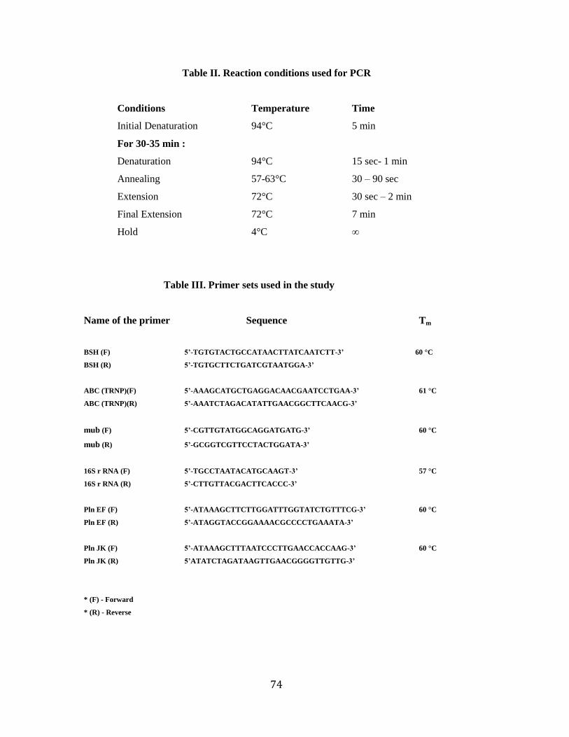
Table II. Reaction conditions used for PCR
Conditions Temperature Time
Initial Denaturation 94°C 5 min
For 30-35 min :
Denaturation 94°C 15 sec- 1 min
Annealing 57-63°C 30 – 90 sec
Extension 72°C 30 sec – 2 min
Final Extension 72°C 7 min
Hold 4°C ∞
Table III. Primer sets used in the study
Name of the primer Sequence Tm
BSH (F) 5‟-TGTGTACTGCCATAACTTATCAATCTT-3‟ 60 °C
BSH (R) 5‟-TGTGCTTCTGATCGTAATGGA-3‟
ABC (TRNP)(F) 5‟-AAAGCATGCTGAGGACAACGAATCCTGAA-3‟ 61 °C
ABC (TRNP)(R) 5‟-AAATCTAGACATATTGAACGGCTTCAACG-3‟
mub (F) 5‟-CGTTGTATGGCAGGATGATG-3‟ 60 °C
mub (R) 5‟-GCGGTCGTTCCTACTGGATA-3‟
16S r RNA (F) 5‟-TGCCTAATACATGCAAGT-3‟ 57 °C
16S r RNA (R) 5‟-CTTGTTACGACTTCACCC-3‟
Pln EF (F) 5‟-ATAAAGCTTCTTGGATTTGGTATCTGTTTCG-3‟ 60 °C
Pln EF (R) 5‟-ATAGGTACCGGAAAACGCCCCTGAAATA-3‟
Pln JK (F) 5‟-ATAAAGCTTTAATCCCTTGAACCACCAAG-3‟ 60 °C
Pln JK (R) 5‟ATATCTAGATAAGTTGAACGGGGTTGTTG-3‟
* (F) - Forward
* (R) - Reverse
75
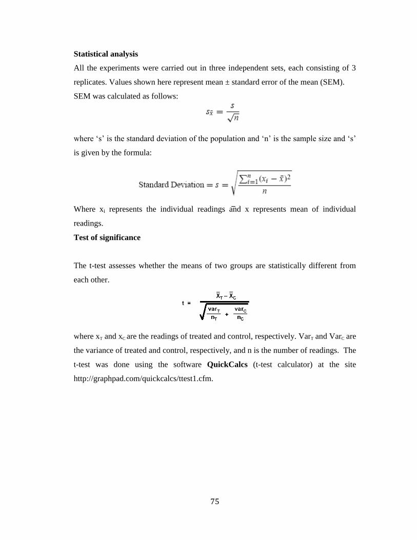
Statistical analysis
All the experiments were carried out in three independent sets, each consisting of 3
replicates. Values shown here represent mean ± standard error of the mean (SEM).
SEM was calculated as follows:
where ‗s‘ is the standard deviation of the population and ‗n‘ is the sample size and ‗s‘
is given by the formula:
Where xi represents the individual readings and x represents mean of individual
readings.
Test of significance
The t-test assesses whether the means of two groups are statistically different from
each other.
where xT and xC are the readings of treated and control, respectively. VarT and VarC are
the variance of treated and control, respectively, and n is the number of readings. The
t-test was done using the software QuickCalcs (t-test calculator) at the site
http://graphpad.com/quickcalcs/ttest1.cfm.