masticatory function in man - schweitzerdental.com
TRANSCRIPT

MASTICATORY FUNCTION IN MAN
Mandibular Repositioning
JEROME M. SCHWEITZER, D.D.S." New York University, College of Dentistry, New York, N. Y.
T HE MANDIBLE can be repositioned vertically, but evidence substantiating its ability to maintain its new vertical position is difficult to obtain. Too often
the musculature reverts to its previous position by intruding the teeth, destroying bony structures, or displacing soft structures or by a combination of these changes.
Evidence that the mandible has been repositioned in a lateral direction is also difficult to obtain. Because of the projection, mandibular changes must be measured in the frontal or the horizontal plane. Both of these projections make accurate measurement questionable.
The mandible seems to change its position in diseases such as acromegaly. This repositioning is due to uneven bone growth, and the apparent positional changes are induced by dimensional changes.
Colignonl and Barrett2 have described mandibular repositioning in lCyear- old girls. Barrett maintained that after the “bite plate” was placed in the mouth and the posterior teeth were taken out of occlusion, the masseter muscle caused the gonial angle to become more obtuse. Therefore, the jaw became elongated in shape to give the desired results.
Breitne9 described three methods by which mandibular repositioning could occur : (1) by tooth movement, (2) by reorientation of the glenoid fossa, and (3) by growth of the condyloid process. These methods could occur simultaneously. He made a special model for demonstrating them.
Forward positioning of the mandible has occurred in patients wearing com- plete dentures. The patients were usually elderly people who had worn dentures for a long time. In most patients, there was an increase of the gonial angle. Merkeley* has called attention to mandibular changes resulting from an in- creased gonial angle.
Sears6 described a method of repositioning the mandible by means of molar pivots. He claims to have changed the position of the condyles in the mandibular .fossae in as short a period as 2 days.
FORMS OF REPOSITIONING
There are two forms of repositioning of the mandible. In one form, the mandible is forced to assume a position which is not its true position with refer-
Read before the Greater New York Academy of Prosthodontics in New York, N. Y., and the Academy of Denture Prosthetics in Minneapolis, Minn.
*Clinical Professor of Denture Prosthesis.
262
263
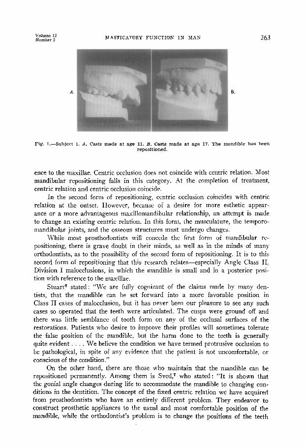
Fig. I.-Subject 1. A, Casts made at age 11. I?, Casts made at age 17. The mandible has been repositioned.
ence to the maxillae. Centric occlusion does not coincide with centric relation. Most mandibular repositioning falls in this category. At the completion of treatment, centric relation and centric occlusion coincide.
In the second form of repositioning, centric occlusion coincides with centric relation at the outset. However, because of a desire for more esthetic appear- ance or a more advantageous maxillomandibular relationship, an attempt is made to change an existing centric relation. In this form, the musculature, the temporo- mandibular joints, and the osseous structures must undergo changes.
While most prosthodontists will concede the first form of mandibular re- positioning, there is grave doubt in their minds, as well as in the minds of many orthodontists, as to the possibility of the second form of repositioning. It is to this second form of repositioning that this research relates-especially Angle Class II! Division I malocclusions, in which the mandible is small and in a posterior posi- tion with reference to the maxillae.
Stuart6 stated : “We are fully cognizant of the claims made by many den- tists, that the mandible can be set forward into a more favorable position in Class II cases of malocclusion, but it has never been our pleasure to see any such cases so operated that the teeth were articulated. The cusps were ground off and there was little semblance of tooth form on any of the occlusal surfaces of the restorations. Patients who desire to improve their profiles will sometimes tolerate the false position of the mandible, but the harm done to the teeth is generally quite evident . . . . We believe the condition we have termed protrusive occlusion to be pathological, in spite of any evidence that the patient is not uncomfortable, or conscious of the condition.”
On the other hand, there are those who maintain that the mandible can be repositioned permanently. Among them is Sved,7 who stated: “It is shown that the gonial angle changes during life to accommodate the mandible to changing con- ditions in the dentition. The concept of the fixed centric relation we have acquired from prosthodontists who have an entirely different problem. They endeavor to construct prosthetic appliances to the usual and most comfortable position of the mandible, while the orthodontist’s problem is to change the positions of the teeth

264 SCHWEITZER J. Pros. Den. March-April, 1962
and the position of the mandible to establish normal occlusion . . . . It is very fortunate that there is an adequate compensating mechanism to alter the dimensions of the mandible to meet the requirements which at times may be very severe . . . . We have repositioned the mandible very many times in the past, notwithstanding statements to the contrary.
“Everything known about the adaptability of the mandible points to the possibility of the permanent repositioning of the mandible. I, personally, believe that the mandible adjusts itself to major changes in occlusion and I cannot recon- cile myself to the idea that the position of the lower jaw is fixed permanently and cannot be changed by orthodontic means.”
Fig. 2.
Fig. 4.
Fig. Z.-The posterior position of the mandible in subject 2 at age 16 represents a failure to reposition the mandible.
Fig. 3.-The anterior position of the mandible in subject 2. Fig. 4.-A pernicious habit of thumbsucking is given as a cause of failure in mandibular
repositioning. Note the severe nailbiting habit of this subject.

hIASTICATORY FUXCTION IN MAX 265
Fig. &-The left temporomandibular roentgenograms of subject 2 show condylar positions with different positions of tooth contact. A, Left anterior contact. B, Left posterior contact. C, Left rest position.
SELECTION OF SUBJECTS
Five patients with “dual closure patterns” were selected. The first 3 had been treated orthodontically, the other 2 by means of occlusal reconstruction.
SUBJECT 1
Orthodontic treatment was started at age 11 and concluded at age 14 (Fig. 1). The mandible had been brought forward, and the original centric relation had been changed. The subject is now 17 years old, and the positions of the casts represent the true centric maxillomandibular relationship. The mandible has been permanently repositioned in an anterior direction. (A motion picture also demonstrates the inability of this subject to further retrude the mandible.)

266 SCHWEITZER J. Pros. Den. March-April, 1962
SUBJECT 2
This patient was a 16-year-old girl (Figs. 2 and 3) who had been treated by two different orthodontists for Se years. The second orthodontist claims to have repositioned the mandible when his treatment was completed at the age of 14. Her treatment started when she was 9 years of age.
The treatment failed inasmuch as she has established dual closure patterns. The patient still persists in thumbsucking (Fig. 4)) which, together with failure to wear her retaining restoration, is given as the cause of her lack of
Fig. 6 .-Casts of subject 3 made at age 9.
progress. The temporomandibular joint roentgenograms show the difference be- tween the anterior and posterior condylar positions (Fig. 5). Her power chew- ing is done with the mandible in its posterior interocclusal position. (A motion picture demonstrates her various anteroposterior interocclusal relationships.)
SUBJECT 3
The third subject was a man 24 years of age whose orthodontic treatment started at the age of 9 (Fig. 6). If an attempt had been made to reposition the mandible, it was unsuccessful (Figs. 7 through 10).
Fig. 7.- The anterior position of the mandible of subject 3 at age 24. Fig. S.-The posterior position of the mandible of subject 3.

i?ltEr i2 MASTICATORY FUNCTION IN MAN 267
the con1
Fig. O.-Left temporomandibular joint roentgenograms of subject 3 indicate the position condyle for various positions of tooth contact. A, Left posterior contact. B, Left anteri tact. C, Left lateral contact. D, Left protrusive contact.
of ior
Fig. IO.--Right temporomandibular joint roentgenograms of subject 3. A, Right anterior contact. B, Right posterior contact. C, Right rest position. D, Right open position.

268 SCHWEITZER J. Pros. Den. March-April, 1962
Methods.-Styluses were placed on the maxillary and mandibular incisors (Fig. 11). A motion picture was made in three planes while the subject carried out a total envelope of movement (Fig. 12) and then chewed large pieces of banana and of a hard roll sandwich. (Only the diagrams made in the horizontal and sagittal planes have been analyzed.)
Results.-Subject 3 started chewing from the most comfortable position and ended with the mandible in its posterior contact position. This latter position was slightly in advance of the posterior border position when the diagrams of functional chewing were superimposed upon the total envelope of movement (Fig. 13).
Sixteen diagrams were made of subect 3 chewing a hard roll sandwich. In two of these cycles (Figs. 3 and 6), the mandible reached its posterior border path in the closing stroke (Figs. 14 and 15). The greatest majority of chewing was well within the total envelope of movement. The analysis indicated that the mandible reached its posterior interocclusal position at the end of each chewing
Fig. Il.-The styluses are attached to the upper and lower incisors of subject 3 by means of metal labial castings.
cycle, except at the start and finish of chewing. There was also evidence of antero- posterior chewing probably because of the wide anteroposterior range afforded by the dual closure patterns. The opening stroke was anterior to the closing stroke in fifteen of the sixteen chewing cycles.
Three total envelopes of movement were drawn in the horizontal plane by following the three markers placed upon the mandibular stylus (Fig. 16). Each tracing differs from the other according to its position in space. None of these tracings have the same dimensions or shape as the total envelope within the oral cavity in which this movement is taking place. However, the true size may be computed by pantographs and mathematics.
The mandible started from its protruded contact position and ended in its retruded contact position when the subject chewed a large piece of banana, as viewed in the horizontal plane (Fig. 17). In several of these horizontal projec- tions, the entire cycle seemed to be within the borders of the total envelope of

Volume 12 Number 2 MASTICATORY FUNCTION IN MAN 269
movement at the interocclusal level (Fig. 18). However, this is an illusion that re- sults from viewing a three dimensional figure in only one dimension. Actually, a great part of the chewing cycle took place below the interocclusal contact level. Only a small part and often none of the chewing was performed on the horizontal plane at the interocclusal level because of a straight vertical stroke. The diagrams of the frontal and sagittal projections demonstrate the correctness of these statements.
In the last cycle of chewing the banana (Fig. 19), the mandible was located in its retruded contact position. From there, the subject automatically moved the
Fig. 13-A diagram of subject 3 in the sagittal plane. Styluses are attached to the maxil- lary and mandibular incisors. With the mouth empty, the subject made two total envelopes of movement. The movement started at A with the mandible forward and reached the retruded position at B. Then the mandible moved forward again, and the movement ended at A.
jaw forward in preparation for the start of chewing a hard roll sandwich. There was little difference between the diagrams made of chewing of the banana and those made of chewing of the hard roll sandwich. The sandwich required more chewing cycles to prepare it for deglutition.
Electromyographic Studies.-Single styluses were placed upon the maxillary and mandibular anterior teeth of subject 3. Surface electrodes were placed on the skin overlying the masseter muscles to record the electrical activity from these
270 SCHWEITZER J. Pros. Den. March-April, 1962
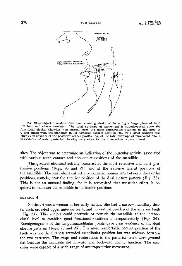
Fig. 13.-Subject 3 made a functional chewing stroke while eating a large piece of hard roll ham and cheese sandwich. The total envelope of movement is superimposed upon the functional stroke. Chewing was started from the most comfortable position in the area of C and ended with the mandible in its posterior contact position (B). This latter position was slightly in advance of the posterior border position (A) of the total envelope of movement. There is evidence of anteroposterior chewing very close to the interocclusal contact level.
sites. The object was to determine an indication of the muscular activity associated with various tooth contact and noncontact positions of the mandible.
The greatest electrical activity occurred at the most retrusive and most pro- trusive positions (Figs. 20 and 21) and at the extreme lateral positions of the mandible. The least electrical activity occurred somewhere between the border positions, namely, near the anterior position of the dual closure pattern (Fig. 22). This is not an unusual finding, for it is recognized that muscular effort is re- quired to maintain the mandible in its border positions.
SUBJECT 4
Subject 4 was a woman in her early sixties. She had a narrow maxillary den- tal arch, crowded upper anterior teeth, and no vertical overlap of the anterior teeth (Fig. 23). This subject could protrude or retrude the mandible at the interoc- clusal level to establish good functional positions anteroposteriorly (Fig. 24). Roentgenograms of the temporomandibular joints gave clear evidence of the dual closure patterns (Figs. 25 and 26). The most comfortable contact position of the teeth was not the furthest retruded mandibular position but was midway between the two extremes. The cusps and restorations in the posterior teeth were ground flat because the mandible slid forward and backward during function. The con- dyles were capable of a wide range of anteroposterior movement.
Volume 12 Number 2
MASTICATORY FUNCTION IN MAN 271
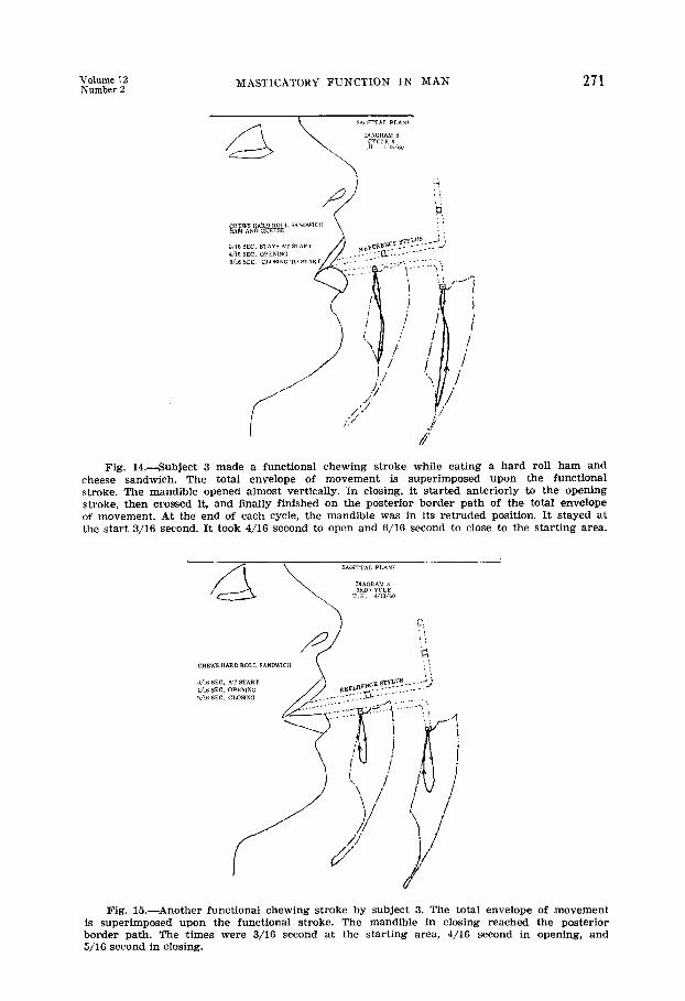
Fig. 14.-Subject 3 made a functional chewing stroke while eating a hard roll ham and cheese sandwich. The total envelope of movement is superimposed upon the functional stroke. The mandible opened almost vertically. In closing, it started anteriorly to the opening stroke, then crossed it, and finally finished on the posterior border path of the total envelope of movement. At the end of each cycle, the mandible was in its retruded position. It stayed at the start 3/16 second. It took 4/16 second to open and 6/16 second to close to the starting area.
Fig. 15.-Another functional chewing stroke by subject 3. The total envelope of movement is superimposed upon the functional stroke. The mandible in closing reached the posterior border path. The times were 3/16 second at the starting area, 4/16 second in opening, and 5/16 second in closing.
272 SCHWEITZER J. Pros. Den. Narch-April, 1962
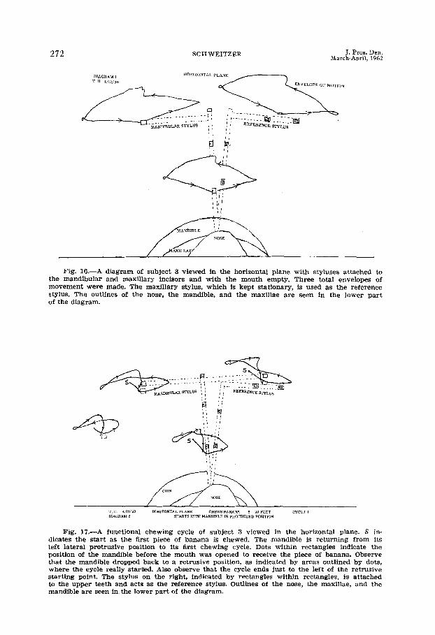
Fig. 16.-A diagram of subject 3 viewed in the horizontal plane with styluses attached to the mandibular and maxillary incisors and with the mouth empty. Three total envelopes of movement were made. The maxillary stylus, which is kept stationary, is used as the reference stylus. The outlines of the nose, the mandible, and the maxillae are seen in the lower part of the diagram.
Fig. 17.-A functional chewing cycle of subject 3 viewed in the horizontal plane. S in- dicates the start as the first piece of banana is chewed. The mandible is returning from its left lateral protrusive position to its first chewing cycle. Dots within rectangles indicate the position of the mandible before the mouth was opened to receive the piece of banana. Observe that the mandible dropped back to a retrusive position, as indicated by areas outlined by dots, where the cycle really startled. Also observe that the cycle ends just to the left of the retrusive starting point. The stylus on the right, indicated by rectangles within rectangles, is attached to the upper teeth and acts as the reference stylus. Outlines of the nose, the maxillae, and the mandible are seen in.the lower part of the diagram.

hi.4STICATORY FUNCTION IN MAN 273
Fig. 13-A functional chewing cycle of subject 3 viewed in the horizontal plane. The stylus on the right is attached to the maxillary teeth and acts as a reference stylus. That on the left is attached to the mandibular teeth and is the moving stylus which makes the diagrams. A piece of banana is being chewed. The total envelope of movement has been superimposed upon the functional chewing cycle. The functional chewing cycle seems to be within the total envelope of movement in this plane, although it may be the projection that makes this appear to be true. Much of functional chewing is done below the interocclusal contact level. If this functional cycle is viewed in the frontal and sagittal projections, a true picture of the action may be obtained. This entire cycle took 1 lo/16 seconds from start to finish.
Fig. lg.-The last chewing cycle, in which subject 3 chewed a banana, as viewed in the horizontal plane. The functional stroke ended in the retruded position of the mandible at the rectangles with an x. Next the subject prepared to chew a piece of a hard roll sandwich. Note that the mandible came forward from X to the dotted square, which is a protrusive position, and starts the new functional chewing cycle from there. The reference stylus is at- tached to the maxillary teeth.
274 SCHWEITZER J. Pros. Den. March-April, 1962
A complete oral reconstruction was completed for this subject. An occlusion was developed that would permit function in any anteroposterior or lateral position that the neuromuscular system dictated (Figs. 27 and 28). Maximal contact of the teeth was obtainable in several anteroposterior positions.
Styluses were attached to the maxillary and mandibular teeth (Fig. 29)) and mandibular movements were recorded in the horizontal and sagittal planes.
Results in the Horizontal Plane.--Diagrams of jaw movements of subject No. 4 indicate that the protrusive movement differs from the return or backward pro- trusive movement in the total envelope of movement (Fig. 30). Also, the radial lateral strokes differ from the medial lateral strokes.
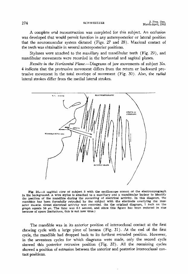
Fig. 20.-A sagittal view of subject 3 with the oscilloscope screen of the electromyograph in the background. A wire stylus is attached to a maxillary and a mandibular incisor to identify the pesition of the mandible during the recording of electrical activity. In this diagram, the mandible has been forcefully retruded by the subject with the electrode overlying the mas- seter muscle. Great electrical activity was recorded. (In the original diagram, 1 inch on the graph equals 50 bv. The time was 0.1 second, and since this figure has been reduced in size because of space limitations, this is not now true.)
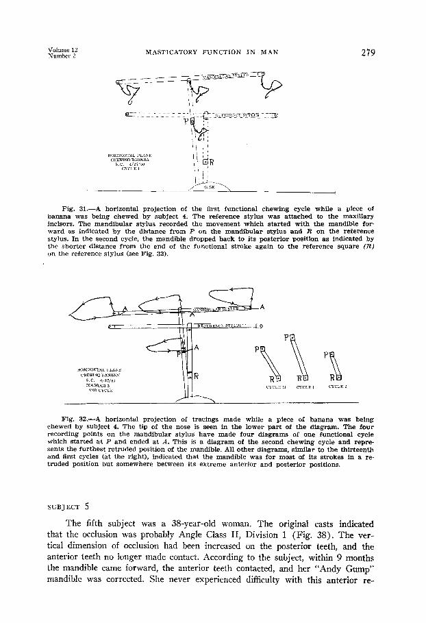
The mandible was in its anterior position of interocclusal contact at the first chewing cycle with a large piece of banana (Fig. 31). At the end of the first cycle, the mandible had dropped back to its furthest retruded position. However, in the seventeen cycles for which diagrams were made, only the second cycle showed this posterior retrusive position (Fig. 32). All the remaining cycles showed a position of retrusion between the anterior and posterior interocclusal con- tact positions.

Volume 12 Number 2 MASTICATORY FUNCTION IN MAN 275
Fig. ZL-Subject 3 with the oscilloscope screen of the electromyograph in the background. The wire stylus attached to the lower teeth, when compared to that attached to tkae upper teeth, indicates that the mandible is being forcefully protruded. Great electrical activity is apparent.
Fig. 22.-Subject 3 with the oscilloscope screen of the electromyograph, in the background. The stylus attached to the mandibular teeth, as compared with the maxillary stylus, indicates that the mandible is in the comfortable position. This position fs between the two border positions, as demonstrated in Figs. 20 and 21. Much less electrical activity is observed in this position. The electrode is overlying the masseter muscle.

276 SCHWEITZER J. Pros. Den March-April, 1962
Fig. 23. Fig. 24.
Fig. 23.-The mandible of subject 4 is in the forward position of the dual closure pattern. Fig. 24.-The mandible of subject 4 is in the posterior position of the dual closure pattern.
Fifteen diagrams were made while the subject chewed a large piece of a hard roll sandwich (ham and cheese). Once again, the mandible started in its forward position and ended in a retruded position (Fig. 33). All of the remaining thirteen diagrams indicated that functional chewing occurred anteriorly to the most retruded position, but not as far forward as the starting position.
Fig. 25.- The right temporomandibular roentgenograms of subject 4 show the condyle positions for various closure patterns. A, Right posterior contact. B, Right lateral contact. C, Right anterior contact (midway). D, Right anterior contact (small overjet).

MASTICATORY FUNCTIOX IN MAN 277
Fig. 26.-The left temporomandibular roentgenograms of subject 4 show the condyle position for various closure patterns. A, Left posterior contact. B, Left lateral contact. C, Left anterior contact (midway). D, Left anterior contact (small overjet).
The wide range of lateral movement was demonstrated in chewing of the hard roll sandwich (Fig. 34). However, this lateral movement did not take place at the interocclusal contact level, as can be seen in the frontal projection.
Fig. 27. Fig. 28.
Fig. W.-The intercuspal contacts of subject 4 are coordinated for the forward position of the mandible.
Fig. 28.-The intercuspal contacts of subject 4 are coordinated for the retruded position of the mandible.

278 SCHWEITZER J. Pros. Den. March-April, 1962
Fig. 29.-The styluses are attached to the maxillary and mandibular incisors of subject 4 to record mandibular movements. The maxillary stylus acts as the reference stylus.
Fig. 30.-Subject 4. A stylus was attached to the mandibular teeth to record movements of the mandible. The reference stylus was attached to the maxillary teeth. This is a horizonta1 projection. The nose is shown in the lower part of the figure. The subject has made extreme lateral and protrusive movements. The four points on the mandibular stylus have made four recordings all of which are dissimilar because of their different positions on the stylus. However, the outgoing or radial stroke differs from the return or medial stroke in ten of the twelve movements. (In Fig. 42, subject 5, eight of twelve movements were different.)
Results in the SagittaE Plane.-Diagrams were made of two total envelopes of movement in the sag&al plane (Figs. 35 and 36). The diagrams are not identical because the subjects were not completely aware of the procedure and they could not carry out two identical envelopes of movement. The double apex of the first envelope indicates the anterior and the posterior closing positions, and the straight upward protrusive line indicates a lack of incisal overlap of the anterior teeth. The range of anteroposterior interocclusal contact indicates that the comfortable interocclusal contact position is midway between the extreme positions (Fig. 37).
MASTICATORY FUNCTIOK IN MAN 279
Fig. 31.-A horizontal projection of the first functional chewing cycle while a piece of banana was being chewed by subject 4. The reference stylus was attached to the maxillary incisors. The mandibular stylus recorded the movement which started with the mandible for- ward as indicated by the distance from P on the mandibular stylus and R on the reference stylus. In the second cycle, the mandible dropped back to its posterior position as indicated by the shorter distance from the end of the functional stroke again to the reference square (R) on the reference stylus (see Fig. 32).
Fig. 33-A horizontal projection of tracings made while a piece of banana was being chewed by subject 4. The tip of the nose is seen in the lower part of the diagram. The four recording points on the mandibular stylus have made four diagrams of one functional cycle which started at P and ended at A. This is a diagram of the second chewing cycle and repre- sents the furthest retruded position of the mandible. All other diagrams, similar to the thirteenth and first cycles (at the right), indicated that the mandible was for most of its strokes in a re- truded position but somewhere between its extreme anterior and posterior positions.
SUBJECT 5
The fifth subject was a 38-year-old woman. The original casts indicated that the occlusion was probably Angle Class II, Division 1 (Fig. 38). The ver- tical dimension of occlusion had been increased on the posterior teeth, and the anterior teeth no longer made contact. According to the subject, within 9 months the mandible came forward, the anterior teeth contacted, and her “Andy Gump” mandible was corrected. She never experienced difficulty with this anterior re-

280 SCHWEITZER J. Pros. Den. March-April, 1962
Fig. 33 .-The first chewing cycle of subject 4 shown in a horizontal projection. The subject was chewing a large piece of a hard roll sandwich. The mandibular stylus starts with the mandible protruded, but the functional stroke ends with the mandible in retrusion at A. Note that the distance from P to R is greater than the distance from A to R. R is on the reference stylus attached to the maxillary teeth. This entire functional chewing cycle took 9/16 second.
Fig. 34.-A horizontal projection which shows the wide lateral range of chewing by sub- ject 4. This is the second cycle during which a large piece of a hard roll ham and cheese sand- wich was being chewed. The distance P to R is less in this diagram than in Fig. 33. This indicates that the mandible is now chewing in a more retruded position than in its first cycle. The reference stylus is attached to the maxillary incisors and is stationary.
positioning. In fact, she was pleased with her appearance. The mandible had been in the forward position approximately le years.
Roentgenographic Findings.- Temporomandibular roentgenograms indicated a posterior positioning of the condyles in the glenoid fossae when the mandible was retruded (Figs. 39 and 40). The condyles were considerably forward and in- ferior in respect to their former position and the articular eminence when the mandible was in the anterior contact position.
Methods.-A crossbar stylus was constructed for the lower anterior teeth (Fig. 41)) and recordings were made of mastication.
“N$g: ‘?” MASTICATORY FUNCTION IN MAX 281
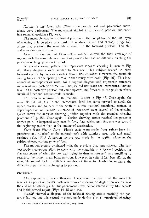
Results in the Horizontal Platte.---Extreme lateral and protrusive move- ments were performed. The movement started in a forward position but ended in a retruded position (Fig. 42).
The mandible was in a retruded position at the completion of the final cycle
of chewing a large piece of a hard roll sandwich (ham and cheese) (Fig. 43). From that position, the mandible advanced to the forward position. The chin and nose also moved forward.
Results i?z the Sagittal PlapLe.-The subject started the total envelope of motion with the mandible in an anterior position but had no difficulty reaching the posterior or hinge position (Fig. 44).
A typical chewing pattern that represents forward chewing is seen in Fig. 45. Many diagrams were similar to this one. This subject wished to chew forward even if by conscious rather than reflex chewing. However, the mandible swung back after the opening stroke in the twenty-third cycle (Fig. 46). This is an abnormal anteroposterior width for a sagittal diagram and represents extensive movement in a posterior direction, The jaw did not reach the interocclusal contact level in the posterior position but came upward and forward to the position where maximal functional contact could Ire made.
The extreme retrusion of the mandible is seen in Fig. 47. Once again, the mandible did not close to the interocclusal level but came forward to avoid the upper molars and to permit the teeth to attain maximal functional contact. A superimposition of the total envelope of movement over several of the chewing cycles shows the anterior chewing position together with the retruded chewing positions (Fig. 48). Once again, a closing chewing stroke reached the posterior border path. [t happened only once in forty-four cycles, and this one was toward the beginning rather than at the ending of mastication.
Tests With Plastic Casts.-Plastic casts were made from rubber-base im- pressions and attached to the natural teeth with stainless steel rods and metal castings (Fig. 49) .* A motion picture was made in the sagittal plane as the subject chewed various types of food.
The motion picture confirmed what the previous diagrams showed. The sub- ject made a conscious effort to chew with the mandible in a forward position, for she was aware of what the test was trying to demonstrate and was unwilling to return to the former mandibular position. However, in spite of her best efforts, the mandible moved back a sufficient number of times to clearly demonstrate the difficulty of permanently changing its position.
DISCVSSION
The exponents of some theories of occlusion maintain that the mandible reaches its posterior border path when power chewing or deglutition occurs near the end of the chewing act. This phenomenon was demonstrated in my first report” and in this second report (Figs. 14, 15, and 48).
PosseW showed a diagram of the habitual closing stroke reaching the pos- terior border, but this record was not made during normal functional chewing.
*P. Christensen: Personal rommuniration, May, 1958.
282 SCHWEITZER J. Pros. Den. March-April, 1962
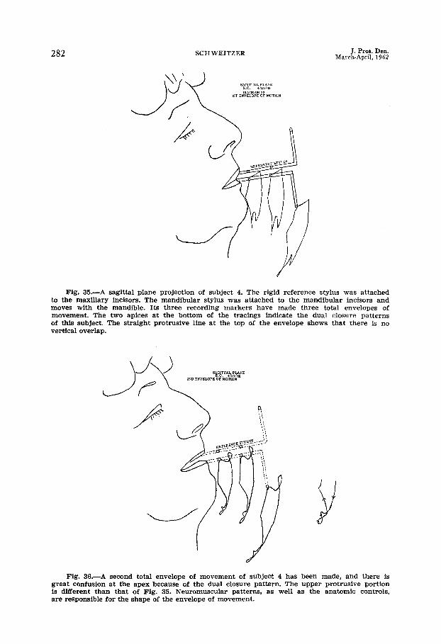
Fig. 35.-A sagittal plane projection of subject 4. The rigid reference stylus was attached to the maxillary incisors. The mandibular stylus was attached to the mandibular incisors and moves with the mandible. Its three recording markers have made three total envelopes of movement. The two apices at the bottom of the tracings indicate the dual closure patterns of this subject. The straight protrusive line at the top of the envelope shows that there is no vertical overlap.
Fig. 36.-A second total envelope of movement of subject 4 has been made, and there is great confusion at the apex because of the dual closure pattern. The upper protrusive portion is different than that of Fig. 35. Neuromuscular patterns, as well as the anatomic controls, are responsible for the shape of the envelope of movement.
“N%Er ‘2” MASTICATORY FUNCTION IN MAN 283
His subject was in the dorsal recumbent position, and habitual opening and closing movements were performed with the mouth empty.
Two of the sixteen chewing cycles of my third subject reached the posterior path. Thus, the mandible did reach this position but did so at the beginning of chewing of the bolus rather than at the end.
Traditional concepts of dynamic occlusion stress wide range interarch cuspal contacts on the horizontal plane at the interocclusal level. Thus, the diagrams of functional chewing cycles should show lines within the confines of the sea gull-shaped tracing at this level of cuspal contact. However, they do not.
The majority of the forward protrusive and lateral movements differed from the backward protrusive and lateral movements in subjects 4 and 5. According to some theories of occlusion, the vertical axes of the articulator are indirectly ad- justed by means of these lateral pantographs. While either set of outward or in- ward strokes can be used in locating the vertical axes, the constancy of the condyle path is open to question. This difference between the radial lateral and medial lateral strokes is probably the reason why the gnathologists pull the pins of all the styluses, after the radial lateral and outward protrusive strokes have been made, while re- cording jaw movements by means of the gnathograph.
The border movement that could be followed with relative accuracy both in opening and closing movements of the mandible was the path of the posterior transverse hinge axis line. The boundary lines of this total envelope consist of border movements which are controlled primarily by ligaments and tendons aided
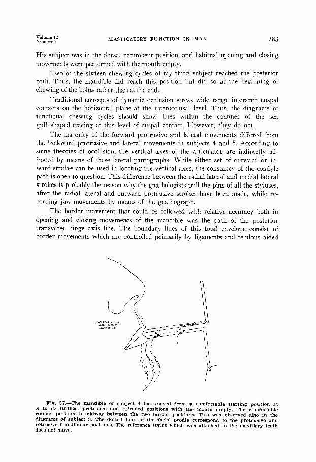
Fig. 37.-The mandible of subject 4 has moved from a comfortable starting position at A to its furthest protruded and retruded positions with the mouth empty. The comfortable contact position is midway between the two border positions. This was observed also in the diagrams of subject 3. The dotted lines of the facial profile correspond to the protrusive and retrusive mandibular positions. The reference stylus which was attached to the maxillary teeth does not move.
284 SCHWEITZER J. Pros. Den. March-April, 1962
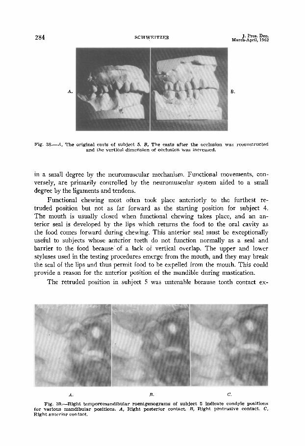
Fig. 38.--A, The original casts of subject 5. B, The casts after the occlusion was reconstructed and the vertical dimension of occlusion was increased.
in a small degree by the neuromuscular mechanism. Functional movements, con- versely, are primarily controlled by the neuromuscular system aided to a small degree by the ligaments and tendons.
Functional chewing most often took place anteriorly to the furthest re- truded position but not as far forward as the starting position for subject 4. The mouth is usually closed when functional chewing takes place, and an an- terior seal is developed by the lips which returns the food to the oral cavity as the food comes forward during chewing. This anterior seal must be exceptionally useful to subjects whose anterior teeth do not function normally as a seal and barrier to the food because’ of a lack of vertical overlap. The upper and lower styluses used in the testing procedures emerge from the mouth, and they may break the seal of the lips and thus permit food to be expelled from the mouth. This could provide a reason for the anterior position of the mandible during mastication.
The retruded position in subject 5 was untenable because tooth contact ex-
A. B. c.
Fig. 39.-Right temporomandibular roentgenograms of subject 5 indicate condyle positions for various mandibular positions. A, Right posterior contact. B, Right protrusive contact. C, Right anterior contact.
Volume 12 1Vumber 2
MASTICATORS FUh-CTION IN IIAn‘ 2x5
isted only on the last molars when the mandible was in this position. The vertical dimension of occlusion was increased by this deflective occlusal contact of the molars. In addition, in functional chewing the mandible came forward to the position of maximal interocclusal contact. According to traditional concepts, this mandible was never at rest. There was neuromuscular activity at all times, but the subject had no temporomandibular joint symptoms and stated that she was never uncomfortable. In fact, she wished to keep the mandible in this forward position and made every voluntary effort to chew in it. However, in the course of many chewing cycles, especially with hard food, it is difficult to maintain anv un- natural chewing position.
Some theories of occlusion maintain that the normal interocclusal position is a border position. However, it is highly improbable that the temporomandibular joint would not have a range of adaptation or safety as do all other joints. There is a thickness of tissue in the temporomandibular joints which permits this func- tional range.
Seemingly, the teeth seldom contact during reflex functional movements of chewing in most of the subjects until final intercuspation takes place. When this in- terocclusal level is reached, chewing either ceases or pauses before another cycle
A. B. c.
D. E.
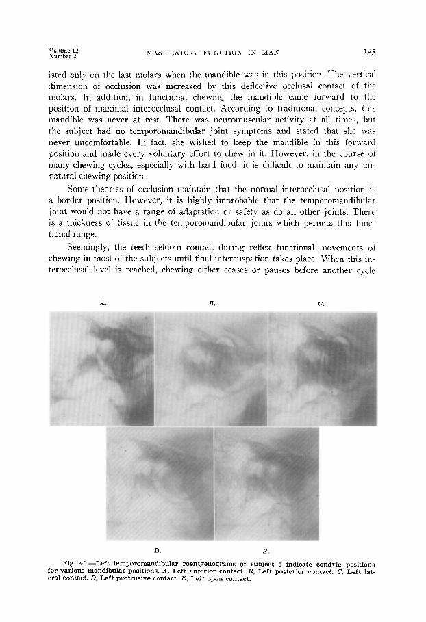
Fig. 40.-Left temporomandibular roentgenograms of subject 5 indicate condyle positions for various mandibular positions. A, Left anterior contact. B, Left posterior contact. C, Left lat- eral contact. D, Left protrusive contact. E, Left open contact.
286 J. Pros. Den. March-April, 1962
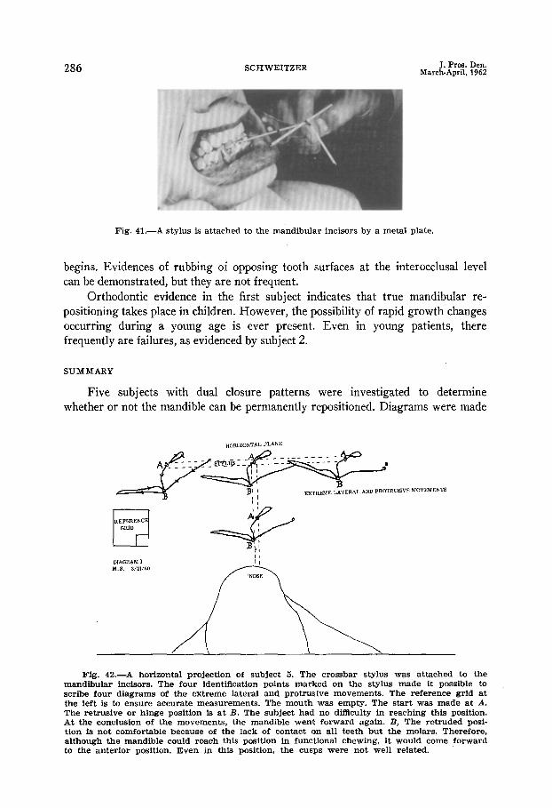
Fig. 41.-A stylus is attached to the mandibular incisors by a metal plate.
begins. Evidences of rubbing of opposing tooth surfaces at the interocclusal level can be demonstrated, but they are not frequent.
Orthodontic evidence in the first subject indicates that true mandibular re- positioning takes place in children. However, the possibility of rapid growth changes occurring during a young age is ever present. Even in young patients, there frequently are failures, as evidenced by subject 2.
SUMMARY
Five subjmects with dual closure patterns were investigated to determine whether or not the mandible can be permanently repositioned. Diagrams were made
Fig. 42.-A horizontal projection of subject 5. The crossbar stylus was attached to the mandibular incisors. The four identification points marked on the stylus made it possible to scribe four diagrams of the extreme lateral and protrusive movements. The reference grid at the left is to ensure accurate measurements. The mouth was empty. The start was made at A. The retrusive or hinge position is at B. The subject had no difficulty in reaching this position. At the conclusion of the movements, the mandible went forward again. B, The retruded posi- tion is not comfortable because of the lack of contact on all teeth but the molars. Therefore, although the mandible could reach this position in functional chewing, it would come forward to the anterior position. Even in this position, the cusps were not well related.

Volume 12 Number 2
MASTICATORY FUNCTION IN MAN 287
of the chewing cycles in the frontal, sagittal, and horizontal planes. Temporo- mandibular roentgenograms were made, and the orthodontic history and casts of the teen-age subjects were obtained.
FINDINGS
1. The majority of the records of radial lateral strokes differed from medial lateral strokes in the total envelope of movement on the horizontal plane.
2. Some functional chewing strokes reached the posterior border path at a distance below the interocclusal level and reached the interocclusal level. However, these strokes took place during the functional chewing cycle and not at the end of the period of mastication.
Fig. 43.-A horizontal projection of subject 5 at the completion of the last chewing stroke when the subject chewed a large piece of a hard roll ham and cheese sandwich. Chewing ended at B, which was retrusive. Then the mandible moved forward to A, which was protrusive and an eccentric position. The reference grids enable more accurate measurement. The nose, in the foreground, also came forward to the dotted lines when the mandible advanced. The stylus was attached to the mandible.
3. The normal interocclusal contact position of the mandible was not found to be a border position.
4. The greatest proportion of the masticatory cycles are carried on an- teriorly to the most retruded mandibular position.
5. The mandible maintains its most comfortable interocclusal position some- where between the extremes of its anterior and posterior positions in subjects with dual closure patterns.
6. The electromyographic results indicated that the mandible was in its
most comfortable contact and relational position when it was anterior to its re- truded position and that great muscular activity was developed in the masseter muscle in the border positions.
288 SCHWEITZER J. Pros. Den. March-April, 1962
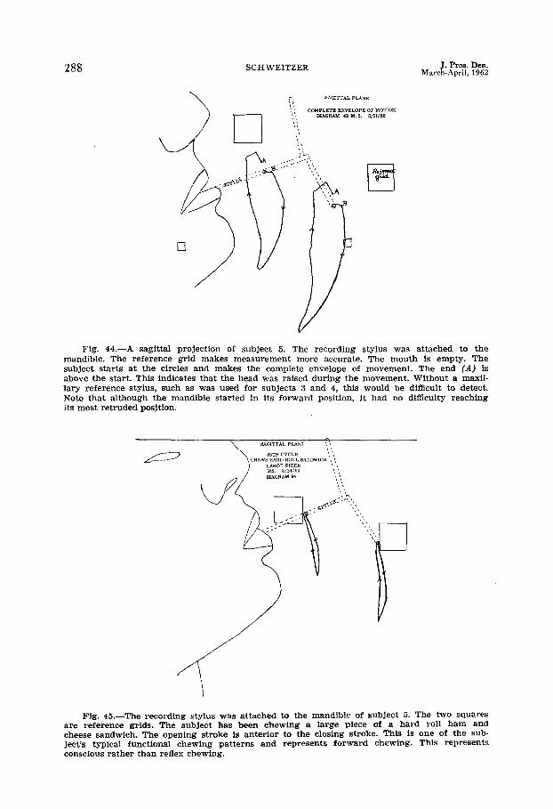
Fig. 44.-A sagittal projection of subject 5. The recording stylus was attached to the mandible. The reference grid makes measurement more accurate. The mouth is empty. The subject starts at the circles and makes the complete envelope of movement. The end (A) is above the start. This indicates that the head was raised during the movement. Without a maxil- lary reference stylus, such as was used for subjects 3 and 4, this would be difficult to detect. Note that although the mandible started in its forward position, it had no difficulty reaching its most retruded posjtion.
Fig. 45.- The recording stylus was attached to the mandible of subject 5. The two squares are reference grids. The subject has been chewing a large piece of a hard roll ham and cheese sandwich. The opening stroke is anterior to the closing stroke. This is one of the sub- ject’s typical functional chewing patterns and represents forward chewing. This represents conscious rather than reflex chewing.

MASTICATORY FUNCTION IN MAN 289
Fig. 46.-- Subject 5 is chewing a large piece of a hard roll ham and cheese sandwich. Here the mandible opens in its forward position but then swings back an abnormal distance until it reaches its posterior retrusive position. Note the difference in the measurable distance from the depth of the cleft in the chin in Figs. 45 and 46 to realize how much further back the mandible is in Fig. 46 than in Fig. 45. Also, note that the mandible did not reach the inter- occlusal contact surface but came forward while it traveled upward to reach its protruded eccentric position. Two points on the stylus attached to the lower teeth have been recorded. The reference squares are for measurements. This functional cycle took 5/16 second in opening, 2/16 second in closing to extreme left, and another S/16 second to complete its closing to the starting area.
Fig. 4’7.-Subject 5 is chewing a large piece of a hard roll sandwich. The reference grids are shown. The stylus is attached to the mandible. The opening starts with the mandible in a forward position. The mandible is then extremely retruded but does not close to the interoc- clusal contact level. Only limited contact is attained here so that the mandible must come for- ward to a protrusive position to attain maximum functional contact. While in its retruded position, it is shown to be opening and closing below the Interocclusal contact level. This may indicate a large increase in the vertical dimension, while the mandible is in retrusion with limited contact. The opening stroke took f3/16 second. Closing to A took 7/16 second, opening to B took 7/16 second, and closing to C took 3/16 second.

290 SCHWEITZER J. Pros. Den. March-April, 1962
7. The mandible rarely opens or closes in a straight line. These move- ments are usually made on a curved line to one side or the other of the midsagittal plane.
CONCLUSIONS
1. Because of the difference of the outward and inward lateral strokes, as well as the protrusive strokes of the stylus on the horizontal plane, the constancy of the condyle path is open to question.
2. Reports by orthodontists indicate that true mandibular repositioning is possible in children,
3. Repositioning the mandible in an adult in whom the musculature must be reoriented to new positions is difficult. The condyles are usually in their correct positions in the mandibular fossae during physiologic rest of the mandible. The incorrect position of the condyles in patients with malocclusion is assumed only when the teeth are in contact. In addition, the mandible is retruded in deglutition,
Fig. 48.- Subject 5 is chewing a large piece of a hard roll sandwich. The total envelope of movement is superimposed upon the composite of several chewing strokes. The mandible starts to function in its forward position but immediately reaches its retruded position and its pos- terior border path. Here it functions below the interocclusal contact level mainly because it is impossible to reach that level. It frequently comes forward and upward to start over again. A purely protrusive functional stroke is also shown superimposed upon this diagram of the total envelope. With soft foods, the subject can readily function with the mandible in this forward position, but it would be extremely difficult for her to hold it forward when chewing hard foods in large pieces. These require a bracing of the mandible against the posterior in- ferior portion of the articular eminence. The diagrams to the right indicate a functional pro- trusive cycle and a cycle in which the mandible starts forward, goes back, and ends forward.

Volume 12 Xumber 2 MASTICATORY FUNCTION IN MAN 291
Fig. 49.-Metal labial and lingual plates were made to fit the stone casts of subject 5 shown at the right. Duplicate plastic casts were made from alginate impressions. These are on the left. They were attached to the metal plates by means of stainless steel rods. When the metal plates are wired to the upper and lower teeth in the mouth, the patient’s chewing patterns can be observed outside the mouth.
and in lateral chewing, the condyles return to their most retruded positions in the fossae. Thus, during functional chewing, it is most difficult to cause any but the slowest of changes in the temporomandibular joints.
4. The mandible usually can be retruded to its former position when true mandibular repositioning in adults is claimed. Evidences of changes in the condyle to fossa relationship record were found in either a static protrusive relationship of maximal functional interocclusal contact or a downward and forward positioning of the condyle caused by an increased vertical dimension, or in a combination of both.
I wish to thank Dr. Harold Schwartz and Dr. Harry Shpuntoff for their assistance in photographing the subjects and Dr. Shpuntoff for his guidance in the electromyographic study.
REFERENCES
1. Colignon, J.: _-. _- Case of -Prominence of the Upper Jaw, D. Cosmos 29:318-325, 1887. 2. Barrett, W. H. : Modified Occlusion, D. Cosmos 29:477-479, 1887. 3. Breitner, C.: Alteration of Occlusal Relations Induced by Experimental Procedure, Am.
J. Orthodontics 29:277-289, 1943. 4. Merkeley, H. J.: Temporomandibular Joint Disturbances as Related to an Increasing Angle
of the Jaw, J. PROS. DEN. 9:336-339, 1959. 5. Sears, V. H.: Mandibular Condyle Migrations as Influenced by Tooth Occlusions, J.A.D.A.
45:179-192, 1952. 6. Stuart, C. E.: Articulation of Human Teeth. II. A Research Report, D. Items Interest
61:1029-1037, 1142-1154, 1939; 62:8-17, 106-112, 1940. 7. Sved, A. : The Mesial Drift of Teeth During Growth, Am. J. Orthodontics 41:539-553, 19.55. 8. Schweitzer, J. M.: Masticatory Function in Man, J. PROS. DEN. 2:625-647, 1961. 9. Posselt, U.: Studies in the Mobility of the Human Mandible, Acta odont. scandinav.
1O:suppl. 10 :19-160, 1952.
730 FIFTH AVE. NEW YORK 19, N. Y.