massive rotator cuff tears: definition and treatment
TRANSCRIPT
ORIGINAL PAPER
Massive rotator cuff tears: definition and treatment
Alexandre Lädermann1,2,3& Patrick J. Denard4,5
& Philippe Collin6
Received: 6 April 2015 /Accepted: 6 April 2015 /Published online: 1 May 2015# SICOT aisbl 2015
AbstractPurpose The aim of this review is to summarise tear patternclassification and management options for massive rotatorcuff tears (MRCT), as well as to propose a treatment paradigmfor patients with a MRCT.Method Data from 70 significant papers were reviewed inorder to define the character of reparability and the possibilityof alternative techniques in the management of MRCT.Results Massive rotator cuff tears (MRCT) include a widepanoply of lesions in terms of tear pattern, functional impair-ment, and reparability. Pre-operative evaluation is critical tosuccessful treatment. With the advancement of medicaltechnology, arthroscopy has become a frequently used meth-od of treatment, even in cases of pseudoparalytic shoulders.Tendon transfer is limited to young patients with an irrepara-ble MRCT and loss of active rotation. Arthroplasty can be
considered for the treatment of a MRCT with associatedarthritis.Conclusion There is insufficient evidence to establish anevidence-based treatment algorithm for MRCTs. Treat-ment is based on patient factors and associated pathol-ogy, and includes personal experience and data from caseseries.
Keywords Shoulder function .Massive rotator cuff repair .
Cuff teararthropathy .Scores .Tendon transfer .Arthroscopy .
Pseudoparalysis . Reverse shoulder arthroplasty . Outcome
Introduction
Massive rotator cuff tears (MRCT) comprise approximately20 % of all cuff tears and 80 % of recurrent tears [1, 2]. Thiscondition can be treated with various approaches, according toclinical factors, characteristics of the tear and biological fac-tors [3]. Advances during the last 15 years of arthroscopic andprosthetic techniques, and better understanding of patho-anatomy have opened new frontiers in management of thiscondition, such that some of the previous definitions and treat-ment options are no longer valid.
Few articles have been published about the proper manage-ment of MRCT [4–11]. This article provides a comprehensivereview of current concepts pertaining to MRCT, including acontemporary definition and classification of this lesion, areview of pertinent biomechanical changes induced by thiscondition, and clinical, radiological and electromyographic(EMG) implications. Lastly, this article presents the authors’preferred options and their treatment algorithm to provide thebest functional outcome.
* Alexandre Lä[email protected]
1 Division of Orthopaedics and Trauma Surgery, La Tour Hospital,Rue J.-D. Maillard 3, 1217 Meyrin, Switzerland
2 Faculty of Medicine, University of Geneva, Rue Michel-Servet 1,1211 Geneva 4, Switzerland
3 Division of Orthopaedics and Trauma Surgery, Department ofSurgery, GenevaUniversity Hospitals, RueGabrielle-Perret-Gentil 4,1211 Geneva 14, Switzerland
4 Southern Oregon Orthopedics, Medford, OR, USA5 Department of Orthopaedics and Rehabilitation, Oregon Health &
Science University, Portland, OR, USA6 Saint-Grégoire Private Hospital Center, Boulevard Boutière 6,
35768 Saint-Grégoire Cedex, France
International Orthopaedics (SICOT) (2015) 39:2403–2414DOI 10.1007/s00264-015-2796-5
Definition and classification
Massive rotator cuff tear
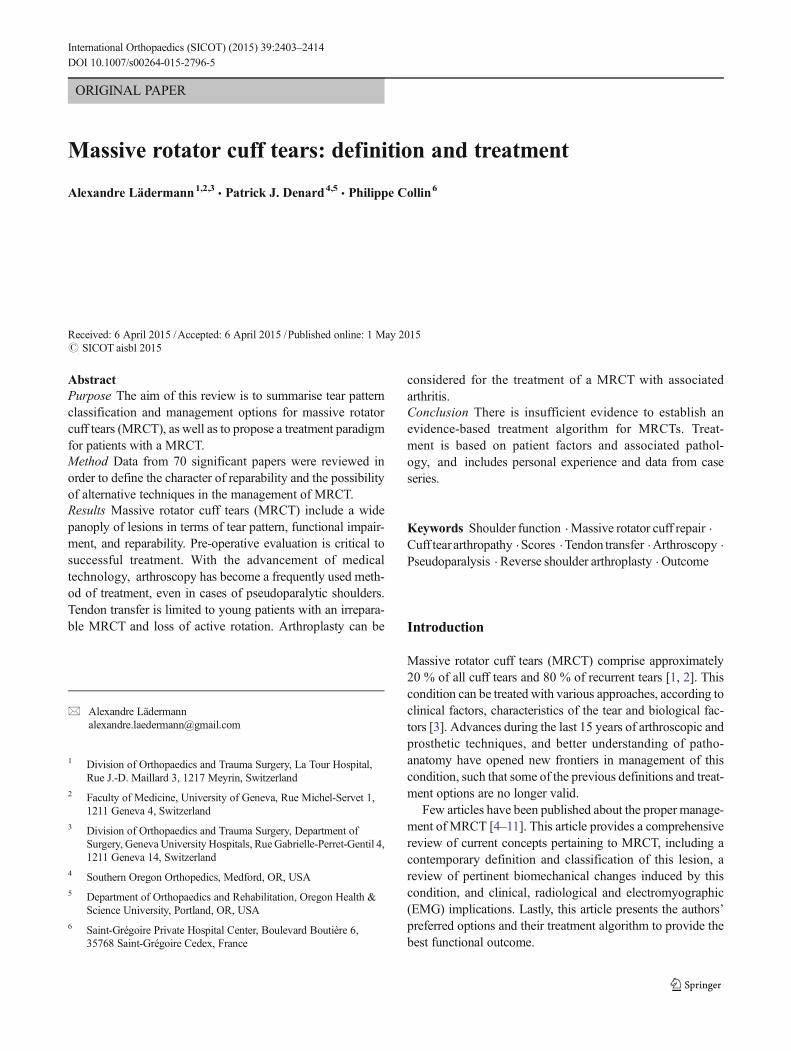
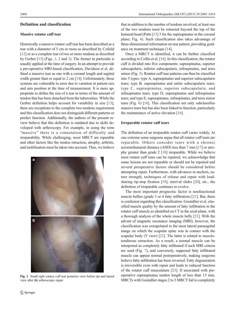
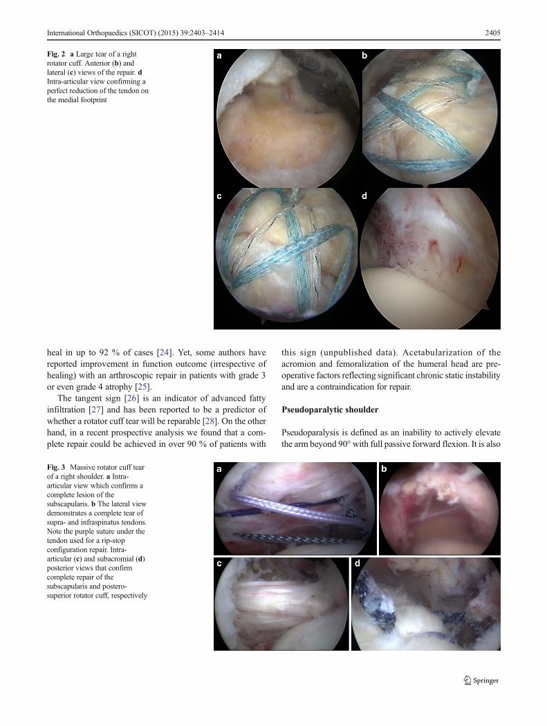
Historically a massive rotator cuff tear has been described as atear with a diameter of 5 cm or more as described by Cofield[12] or as a complete tear of two or more tendons as describedby Gerber [13] (Figs. 1, 2 and 3). The former in particular isusually applied at the time of surgery. In an attempt to providea pre-operative MRI-based classification, Davidson et al. de-fined a massive tear as one with a coronal length and sagittalwidth greater than or equal to 2 cm [14]. Unfortunately, thesesystems are vulnerable to error due to variation in patient sizeand arm position at the time of measurement. It is more ap-propriate to define the size of a tear in terms of the amount oftendon that has been detached from the tuberosities. While theGerber definition helps account for variability in size [13],there are exceptions to the complete two-tendons requirementand this classification does not distinguish different patterns orpredict function. Additionally, the authors of the present re-view believe that this definition is outdated due to skills de-veloped with arthroscopy. For example, in using the termBmassive^ there is a connotation of difficulty andirreparability. While challenging, most MRCT are reparableand other factors like the tendon retraction, atrophy, arthritis,and mobilisation must be taken into account. Thus, we believe
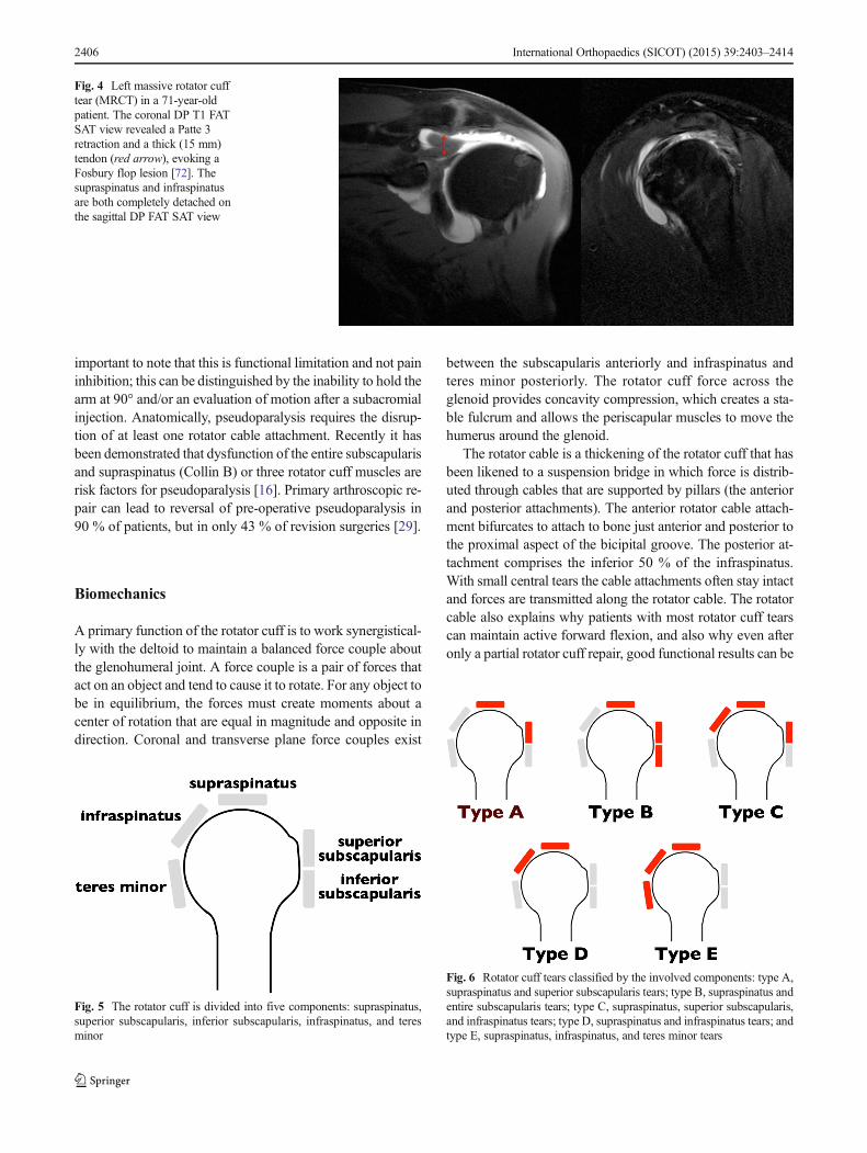
that in addition to the number of tendons involved, at least oneof the two tendons must be retracted beyond the top of thehumeral head (Patte [15] 3 for the supraspinatus in the coronalplane; Fig. 4). Such classification also takes advantage ofthree-dimensional information on tear pattern, providing guid-ance on treatment technique [14].
Once a MRCT is identified, it can be further classifiedaccording to Collin et al. [16]. In this classification, the rotatorcuff is divided into five components: supraspinatus, superiorsubscapularis, inferior subscapularis, infraspinatus, and teresminor (Fig. 5). Rotator cuff tear patterns can then be classifiedinto 5 types: type A, supraspinatus and superior subscapularistears; type B, supraspinatus and entire subscapularis tears;type C, supraspinatus, superior subscapularis, andinfraspinatus tears; type D, supraspinatus and infraspinatustears; and type E, supraspinatus, infraspinatus, and teres minortears (Fig. 6) [16]. This classification not only subclassifiesmassive tears but has also been linked to function, particularlythe maintenance of active elevation [16].
Irreparable rotator cuff tears
The definition of an irreparable rotator cuff varies widely. Atone extreme some surgeons argue that all rotator cuff tears arereparable . Others consider tears wi th a chronicacromiohumeral distance (AHD) less than 7 mm [17] or atro-phy greater than grade 2 [18] irreparable. While we believemost rotator cuff tears can be repaired, we acknowledge thatsome lesions are not reparable or should not be repaired andseveral preoperative factors should be considered beforeattempting repair. Furthermore, with advances in anchors, su-ture strength, techniques of release and repair with load-sharing rip-stop fixation [19], interval slides [20], etc., thedefinition of irreparable continues to evolve.
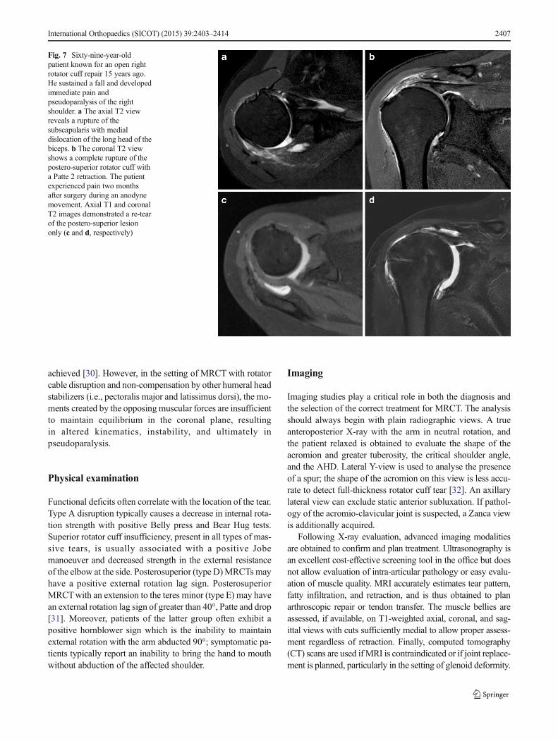
The most important prognostic factor is nonfunctionalmuscle bellies (grade 3 or 4 fatty infiltration) [21]. But, thereis confusion regarding this classification. Goutallier et al. clas-sified muscle quality by the amount of fatty infiltration in therotator cuff muscle as identified on CT in the axial plane, witha thorough analysis of the whole muscle belly [21]. With theadvent of magnetic resonance imaging (MRI), however, theclassification was extrapolated to the most lateral parasagittalimage on which the scapular spine was in contact with thescapular body (Y view) [22]. The latter is related to musclo-tendinous retraction. As a result, a normal muscle can beinterpreted as completely fatty infiltrated if such MRI criteriaare used (Fig. 7), and conversely, supposed fatty infiltratedmuscle can appear normal postoperatively, making surgeonsbelieve fatty infiltration has been reversed. Fatty degenerationis irreversible even with repair and leads to reduced functionof the rotator cuff musculature [23]. If associated with pre-operative supraspinatus tendon length of less than 15 mm,MRCTswith Goutallier stages 2 to 3MRCT fail to completely
Fig. 1 Small right rotator cuff tear posterior view before (a) and lateralview after (b) arthroscopic repair
2404 International Orthopaedics (SICOT) (2015) 39:2403–2414
heal in up to 92 % of cases [24]. Yet, some authors havereported improvement in function outcome (irrespective ofhealing) with an arthroscopic repair in patients with grade 3or even grade 4 atrophy [25].
The tangent sign [26] is an indicator of advanced fattyinfiltration [27] and has been reported to be a predictor ofwhether a rotator cuff tear will be reparable [28]. On the otherhand, in a recent prospective analysis we found that a com-plete repair could be achieved in over 90 % of patients with
this sign (unpublished data). Acetabularization of theacromion and femoralization of the humeral head are pre-operative factors reflecting significant chronic static instabilityand are a contraindication for repair.
Pseudoparalytic shoulder
Pseudoparalysis is defined as an inability to actively elevatethe arm beyond 90° with full passive forward flexion. It is also
Fig. 2 a Large tear of a rightrotator cuff. Anterior (b) andlateral (c) views of the repair. dIntra-articular view confirming aperfect reduction of the tendon onthe medial footprint
Fig. 3 Massive rotator cuff tearof a right shoulder. a Intra-articular view which confirms acomplete lesion of thesubscapularis. b The lateral viewdemonstrates a complete tear ofsupra- and infraspinatus tendons.Note the purple suture under thetendon used for a rip-stopconfiguration repair. Intra-articular (c) and subacromial (d)posterior views that confirmcomplete repair of thesubscapularis and postero-superior rotator cuff, respectively
International Orthopaedics (SICOT) (2015) 39:2403–2414 2405
important to note that this is functional limitation and not paininhibition; this can be distinguished by the inability to hold thearm at 90° and/or an evaluation of motion after a subacromialinjection. Anatomically, pseudoparalysis requires the disrup-tion of at least one rotator cable attachment. Recently it hasbeen demonstrated that dysfunction of the entire subscapularisand supraspinatus (Collin B) or three rotator cuff muscles arerisk factors for pseudoparalysis [16]. Primary arthroscopic re-pair can lead to reversal of pre-operative pseudoparalysis in90 % of patients, but in only 43 % of revision surgeries [29].
Biomechanics
A primary function of the rotator cuff is to work synergistical-ly with the deltoid to maintain a balanced force couple aboutthe glenohumeral joint. A force couple is a pair of forces thatact on an object and tend to cause it to rotate. For any object tobe in equilibrium, the forces must create moments about acenter of rotation that are equal in magnitude and opposite indirection. Coronal and transverse plane force couples exist
between the subscapularis anteriorly and infraspinatus andteres minor posteriorly. The rotator cuff force across theglenoid provides concavity compression, which creates a sta-ble fulcrum and allows the periscapular muscles to move thehumerus around the glenoid.
The rotator cable is a thickening of the rotator cuff that hasbeen likened to a suspension bridge in which force is distrib-uted through cables that are supported by pillars (the anteriorand posterior attachments). The anterior rotator cable attach-ment bifurcates to attach to bone just anterior and posterior tothe proximal aspect of the bicipital groove. The posterior at-tachment comprises the inferior 50 % of the infraspinatus.With small central tears the cable attachments often stay intactand forces are transmitted along the rotator cable. The rotatorcable also explains why patients with most rotator cuff tearscan maintain active forward flexion, and also why even afteronly a partial rotator cuff repair, good functional results can be
Fig. 4 Left massive rotator cufftear (MRCT) in a 71-year-oldpatient. The coronal DP T1 FATSAT view revealed a Patte 3retraction and a thick (15 mm)tendon (red arrow), evoking aFosbury flop lesion [72]. Thesupraspinatus and infraspinatusare both completely detached onthe sagittal DP FAT SAT view
Fig. 5 The rotator cuff is divided into five components: supraspinatus,superior subscapularis, inferior subscapularis, infraspinatus, and teresminor
Fig. 6 Rotator cuff tears classified by the involved components: type A,supraspinatus and superior subscapularis tears; type B, supraspinatus andentire subscapularis tears; type C, supraspinatus, superior subscapularis,and infraspinatus tears; type D, supraspinatus and infraspinatus tears; andtype E, supraspinatus, infraspinatus, and teres minor tears
2406 International Orthopaedics (SICOT) (2015) 39:2403–2414
achieved [30]. However, in the setting of MRCT with rotatorcable disruption and non-compensation by other humeral headstabilizers (i.e., pectoralis major and latissimus dorsi), the mo-ments created by the opposingmuscular forces are insufficientto maintain equilibrium in the coronal plane, resultingin altered kinematics, instability, and ultimately inpseudoparalysis.
Physical examination
Functional deficits often correlate with the location of the tear.Type A disruption typically causes a decrease in internal rota-tion strength with positive Belly press and Bear Hug tests.Superior rotator cuff insufficiency, present in all types of mas-sive tears, is usually associated with a positive Jobemanoeuver and decreased strength in the external resistanceof the elbow at the side. Posterosuperior (type D)MRCTsmayhave a positive external rotation lag sign. PosterosuperiorMRCTwith an extension to the teres minor (type E) may havean external rotation lag sign of greater than 40°, Patte and drop[31]. Moreover, patients of the latter group often exhibit apositive hornblower sign which is the inability to maintainexternal rotation with the arm abducted 90°; symptomatic pa-tients typically report an inability to bring the hand to mouthwithout abduction of the affected shoulder.
Imaging
Imaging studies play a critical role in both the diagnosis andthe selection of the correct treatment for MRCT. The analysisshould always begin with plain radiographic views. A trueanteroposterior X-ray with the arm in neutral rotation, andthe patient relaxed is obtained to evaluate the shape of theacromion and greater tuberosity, the critical shoulder angle,and the AHD. Lateral Y-view is used to analyse the presenceof a spur; the shape of the acromion on this view is less accu-rate to detect full-thickness rotator cuff tear [32]. An axillarylateral view can exclude static anterior subluxation. If pathol-ogy of the acromio-clavicular joint is suspected, a Zanca viewis additionally acquired.
Following X-ray evaluation, advanced imaging modalitiesare obtained to confirm and plan treatment. Ultrasonography isan excellent cost-effective screening tool in the office but doesnot allow evaluation of intra-articular pathology or easy evalu-ation of muscle quality. MRI accurately estimates tear pattern,fatty infiltration, and retraction, and is thus obtained to planarthroscopic repair or tendon transfer. The muscle bellies areassessed, if available, on T1-weighted axial, coronal, and sag-ittal views with cuts sufficiently medial to allow proper assess-ment regardless of retraction. Finally, computed tomography(CT) scans are used ifMRI is contraindicated or if joint replace-ment is planned, particularly in the setting of glenoid deformity.
Fig. 7 Sixty-nine-year-oldpatient known for an open rightrotator cuff repair 15 years ago.He sustained a fall and developedimmediate pain andpseudoparalysis of the rightshoulder. a The axial T2 viewreveals a rupture of thesubscapularis with medialdislocation of the long head of thebiceps. b The coronal T2 viewshows a complete rupture of thepostero-superior rotator cuff witha Patte 2 retraction. The patientexperienced pain two monthsafter surgery during an anodynemovement. Axial T1 and coronalT2 images demonstrated a re-tearof the postero-superior lesiononly (c and d, respectively)
International Orthopaedics (SICOT) (2015) 39:2403–2414 2407
Suprascapular nerve neuropathy and MRCT
Recently there has been growing interest in the relationshipbetween suprascapular neuropathy and MRCTs. Theoretical-ly, medial retraction of posterosuperior rotator cuff tears canplace excessive traction on the suprascapular nerve [33].However, clinical diagnosis is beset with uncertainties as thepotential symptoms of suprascapular nerve neuropathy, name-ly, pain, weakness, and atrophy, are inseparable from those ofMRCT. There is actually no support for routine suprascapularnerve release when MRCT repair is performed for severalreasons. First, it is clearly demonstrated that repair of MRCTwithout release leads to satisfactory results. Moreover, theprevalence of suprascapular nerve neuropathy in case ofMRCT in a recent prospective study was low (2 %) [34].
Treatment options
It should be remembered that nonoperative treatment is suc-cessful in many cases. When surgery is indicated, the primaryaim is restoration of force couples and anatomic or partialrepair of the rotator cuff to its footprint. However, a numberof factors (refusal of the patient, biologic factors, characteris-tics of the tear, etc.) can make these goals difficult, impossible,or unwanted to achieve. Fatty infiltration, rotator cuff retrac-tion, and poor tendon compliance are common in patients withMRCT. In these situations, other approaches have been advo-cated, with varying degrees of success [35]. These includephysical therapy [36, 37], subacromial decompression andpalliative biceps tenotomy (subacromial debridement) [38],muscle transfer [39], and reverse shoulder arthroplasty [40].However, there are no randomized controlled trials comparingthese various options and recommendations are mainly basedon retrospective case series and the surgeon’s ownexperiences.
Conservative treatment
Many patients with MRCT respond favourably to nonsurgicaltreatment. Nevertheless, patients must be aware that despiteclinical improvement, future treatment may be impacted byprogression of glenohumeral osteoarthritis and fatty infiltra-tion as well as narrowing of the AHD. In a series of 19 patientswith MRCTs treated nonoperatively the average Constantscore was 83 % at a mean follow-up of 48 months. However,50 % of Breparable^ tears became Birreparable^ during thisperiod [37].
The mainstay of nonoperative treatment includes nonste-roidal anti-inflammatory drugs, subacromial corticosteroid in-jections, and physical therapy. The protocol of rehabilitationfocused habitually on global deltoid reconditioning and
periscapular strengthening. Although certain authors pro-posed that re-education of the anterior deltoid muscle to com-pensate for a deficient rotator cuff is the cornerstone, we attachmore importance to solicitation of stabilizing muscles of theglenohumeral joint with an approach based on exercises inhigh position. In this position, the deltoid, which acts syner-gistically with the remaining rotator muscles, has no upwardcomponent and participates in the articular coaptation [36].
In general, nonoperative management is attempted forsix months before considering surgery. Younger patients(<60 years of age), however, may be immediate candidatesfor surgery based on the high risk for progression with con-servative treatment. If after six months, symptoms have notimproved, the chances of success with further nonoperativetreatment decreases and operative treatment may be consid-ered for older patients. It is unclear if it is exercise alone orexercise in combination with other interventions during therecovery process that offers the greatest benefit. In a recentprospective cohort of 45 patients suffering frompseudoparalysis with a radiographically confirmed MRCT,Collin and al. found after a follow-up of 48 months that themean Constant score improved from 43 to 56 points and themean forward flexion improved from 76° to more than 160°after completion of the program [36]. They also demonstratedthat effectiveness of physical therapy is related to the size andlocation of the lesion; if the tear involved the posterosuperiorrotator cuff (B type), or only two tendons or less, most patientsregained active anterior elevation that persisted for 48 months[36]. The anterior rotator cuff is the key of anterior activeelevation as only 20 % of patients with MRCTs, but an intactsubscapularis, develop pseudoparalysis [16].
Operative treatment
For older patients surgery is considered when nonoperativetreatment fails. Additionally, we often consider surgery as firstline treatment in young patients because there is a highrate of progression with conservative treatment and fortears involving the anterior rotator cable since this areais most important to maintenance of forward elevation aspreviously noted.
A primary or revision approach, either open or arthroscop-ic, should be discussed if the rotator cuff is still reparable [41,42]. If the tear is irreparable a variety of other options havebeen proposed. These include debridement with or without abiceps tenotomy/tenodesis [38] or acromioplasty/tuberoplasty[43, 44], partial rotator cuff repair [45], tendon transfers [39],graft or biodegradable spacer interposition [46], superior cap-sule reconstruction [47] and arthroplasty [48]. Although theyare consistently proposed in review articles as part of a treat-ment algorithm, we feel that simple subacromial debridement,isolated tuberoplasty, deltoid flap and hemiarthroplasties have
2408 International Orthopaedics (SICOT) (2015) 39:2403–2414
very limited and primarily historical roles because the resultsof the procedures have been disappointing over time. On theother hand, recent innovativemethods, such as trapezius trans-fer to improve external rotation, superior capsular reconstruc-tion, and insertion of a biodegradable spacer, are in their earlystages and will consequently not be discussed in this reviewarticle.
Arthroscopic rotator cuff repair
Our approach is to repair all of the rotator cuff that can rea-sonably be brought back to the tuberosities without excessivetension, and to address all potential causes of persistent pain orfactors threatening the repair. The goal of a repair, even ifpartial, is to restore force couples [45] and to re-establish theBsuspension bridge^ [49]. In this theory, complete closure ofthe defect is less important than restoration of a stable fulcrumfor normal shoulder kinematics. Although shoulder strengthmay not improve after this intervention, function is usuallyenhanced because of relief from pain caused by mechanicalimpingement. Additionally, although complete healing ofmassive tears is not always achievable, we believe that partialhealing of the cuff may prevent secondary extension of thetear.
Repair techniques have been previously thoroughly de-scribed [50]. Mobilization techniques vary based on the typeof lesion [14] and surgeons’ preferences.Margin convergence,interval slides [20] and reinforcement by biological and syn-thetic grafts [51] have all been suggested but it is not clearwhen each becomes beneficial.
The acromion and biceps
Complete anterior acromioplasty is not advisable in the settingof a massive tear as it may lead to postoperative anterosuperiormigration of the humeral head. The acromio-humeral arch isprobably a component of human evolution used to compen-sate the deficiency of the superior rotator cuff [52]. However,the lateral acromion might be responsible for more impinge-ment than the anterior part [53] and may increase stress on therepaired rotator cuff [54]. Consequently, we recommendadding a lateral acromioplasty to any postero-superior rotatorcuff repair if the critical shoulder angle is above 35° [55, 56].This is done in an arthroscopic fashion, but bevelling the un-dersurface of the acromion with care taken to not disrupt thedeltoid insertion.
We almost consistently performed a tenotomy or tenodesisof the long head of the biceps in the setting of a massiverotator cuff tear. There is evidence suggesting that the longhead of the biceps tendon may be a source of pain and con-tributes to the discomfort associated with symptomaticMRCT[38]. In a large series, Walch et al. observed an increase in the
Constant score from 48.4 preoperatively to 67.6 after arthro-scopic biceps tenotomy. At last follow-up, 87 % of patientswere satisfied or very satisfied with the result. However, theacromiohumeral interval decreased by a mean of 1.3 mm dur-ing the follow-up period.
There is one exception with B type MRCTs; the tenotomyis not recommended as it could aggravate the situation.
Repair techniques
Unfortunately, even if reinsertion of the tendon on the bone isachievable, it is often difficult to reliably achieve long-termhealing with a structurally intact repair [57]. In the setting of amassive tear, a double-row repair improves long-term func-tional outcome [41, 58, 59]. However, this should not be per-formed at the expense of over-tensioning as application of adouble-row repair to a tendon with poor tendon length andexcursion may lead to medial failure [24]. On the other hand,poor-quality tendon can be managed with load-sharing rip-stop fixation construct [19]. This technique has demonstratedsuperior fixation strength in cadaveric studies. However, noclinical studies have been reported on healing following thisrepair.
Augmentation
Graft augmentation may improve healing in massive rotatorcuff tears [60] but add significant cost and time to the proce-dure. The choice of graft is influenced by several factors in-cluding mechanical properties, host response and potential foringrowth. Scaffolds provide mechanical support and have bi-ological properties that may favourably influence cell prolif-eration and differentiation, hopefully improving tendon-to-bone healing. Currently, scaffolds derived from dermis, smallintestinal submucosa, skin, fascia lata, and pericardium havebeen processed and marketed for augmentation in the repair ofmassive tears. We prefer biological grafts, when compared tosynthetic grafts, due to the unknown host response to syntheticgrafts. An important factor in the longevity and strength of agraft is the amount of ingrowth.
Results
We have previously reported our results following arthroscop-ic repair of MRCTs [29, 42]. For primary repair, improve-ments were observed in forward flexion (132° vs 168°), pain(6.3 vs 1.3), UCLA score [61] (15.7 vs 30.7), and AmericanShoulder and Elbow Surgeons score [62] (41.7 vs 85.7)(P<0.001). A good or excellent outcome was obtained in78 % of cases. Similar results were noticed after repair of typeA, B and C MRCT [63]. After revision of MRCT repair [42],mean active forward elevation improved by 15°, from 136.0°
International Orthopaedics (SICOT) (2015) 39:2403–2414 2409
±51.9° (range, 30–180°) at baseline to 151.4°±41.5° (range,30–180°) at final follow-up (P=0.019). The mean pain scoreimproved by 3.1 points, from 5.0±2.4 points at baseline to 1.9±2.3 points at final follow-up (P<0.001). The mean ASESscore improved from 45.7±17.8 at baseline to 75.5±20.3 atfinal follow-up (P<0.001). The mean UCLA score also im-proved, from 16.7±4.9 at baseline to 26.4±6.9 at final follow-up (P<0.001). According to the UCLA score, functional re-sults were excellent in 15 % of cases, good in 35 %, fair in25 %, and poor in 25 %. Seventy-nine percent of the patientswere satisfied, and 32 patients (60 %) returned to their previ-ous activities [42].
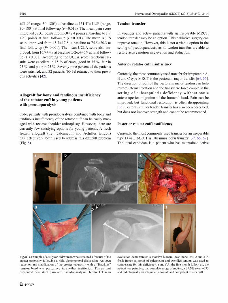
Allograft for bony and tendinous insufficiencyof the rotator cuff in young patientswith pseudoparalysis
Older patients with pseudoparalysis combined with bony andtendinous insufficiency of the rotator cuff can be easily man-aged with reverse shoulder arthroplasty. However, there arecurrently few satisfying options for young patients. A freshfrozen allograft (i.e., calcaneum and Achilles tendon)has effectively been used to address this difficult problem(Fig. 8).
Tendon transfer
In younger and active patients with an irreparable MRCT,tendon transfer may be an option. This palliative surgery canimprove rotation. However, this is not a viable option in thesetting of pseudoparalysis, as no tendon transfers are able torestore active motion in elevation and abduction.
Anterior rotator cuff insufficiency
Currently, the most commonly used transfer for irreparable A,B and C type MRCT is the pectoralis major transfer [64, 65].The direction of pull of the pectoralis major tendon can helprestore internal rotation and the transverse force couple in thesetting of subscapularis deficiency without staticanterosuperior migration of the humeral head. Pain can beimproved, but functional restoration is often disappointing[65]. Pectoralis minor tendon transfer has also been described,but does not improve strength and cannot be recommended.
Posterior rotator cuff insufficiency
Currently, the most commonly used transfer for an irreparabletype D or E MRCT is latissimus dorsi transfer [39, 66, 67].The ideal candidate is a patient who has maintained active
Fig. 8 a Example of a 44-year-oldwomanwho sustained a fracture of thegreater tuberosity following a right glenohumeral dislocation. An openreduction and stabilization of the greater tuberosity with a BHawkins^tension band was performed in another institution. The patientpresented persistent pain and pseudoparalysis. b The CT scan
evaluation demonstrated a massive humeral head bone loss. c and d Afresh frozen allograft of calcaneum and Achilles tendon was used tocompensate for this deficiency. e and f At the five-month follow-up, thepatient was pain free, had complete range of motion, a SANE score of 95and radiologically an integrated allograft and competent rotator cuff
2410 International Orthopaedics (SICOT) (2015) 39:2403–2414
anterior elevation, but lacks control of the arm in space inexternal rotation (simple weakness in external rotation is nota sufficient indication for surgery), and who also has an intactsubscapularis and no glenohumeral arthritis. Results are dis-appointing in patients with subscapularis insufficiency [39]. Inaddition, results have been disappointing in the setting of pre-operative teres minor tears or atrophy [39]. Gerber et al. [39]reported long-term results at a mean of 147 months. The meanSSV in 46 shoulders increased from 29 % preoperatively to70 %, the relative Constant score improved from 56 % to80 %, and the pain score improved from 7 to 13 points(P<0.0001 for all). However, there is no proof that latissimusdorsi transfer gives better long-term results than a simple par-tial rotator cuff repair. Effectively, 60 % of type E MRCTs donot lose control of the arm in external rotation [31]. Conse-quently and despite large series recently published, indicationsfor this type of surgery are rare.
Reverse shoulder arthroplasty
A hemiarthroplasty or an anatomical total shoulderarthroplasty is contraindicated in the absence of a functionalrotator cuff because loss of a balanced coronal force couple,leading to either limited goal prosthesis or to glenoid compo-nent loosening, respectively. Reverse shoulder arthroplastyhas recently emerged as a treatment for MRCT [48]. Whileprimarily used in the setting of glenohumeral arthritis Hamada
4–5, its implantation might be discussed in certain cases ofglenohumeral arthritis Hamada 1–3 [68], particularly in olderpatients or those with chronic pseudoparalysis (as opposed toacute pseudoparalysis which responds well to arthroscopictreatment).
The reverse ball-and-socket relationship of the prosthesisrestores stability to the glenohumeral joint. The glenosphereposition medializes and lowers the glenohumeral center ofrotation, thereby increasing the lever arm of the deltoid mus-cle. Deltoid tension, produced by the lowered centre of rota-tion, increases muscle fibre recruitment of the anterior andposterior deltoid that compensates for a deficient rotator cuff.While initial results were associated with a substantial rate ofclinical and radiological complications [69], tremendous ef-forts have been made to better understand biomechanics of theprosthesis and to lower prevalence of various complicationsincluding scapular notching, learning curve effect [70],lengthening of the arm [71], surgical approach, etc. Thesehave all led to improvement in outcomes and decreased com-plications. While RSA is technically easier than arthroscopicrepair, we usually do not recommend reverse shoulderarthroplasty as the first line of treatment for massive rotatorcuff tears with minimal arthritis. Indications for MRCTs re-main limited in our hands to (1) tears with advanced atrophyand with chronic pseudoparalysis, (2) type B irreparableMRCT, (3) adaptive changes of the proximal humerus (classicrotator cuff arthropathy) and (4) failure of revision rotator cuffrepair [29].
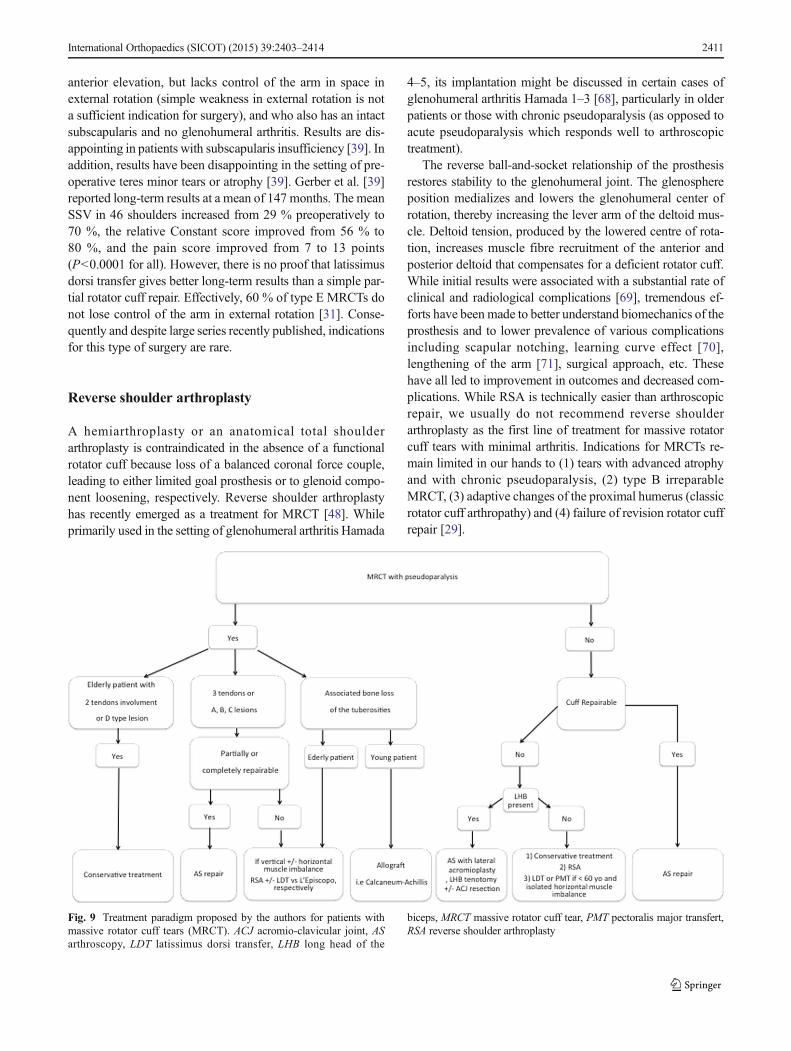
Fig. 9 Treatment paradigm proposed by the authors for patients withmassive rotator cuff tears (MRCT). ACJ acromio-clavicular joint, ASarthroscopy, LDT latissimus dorsi transfer, LHB long head of the
biceps, MRCT massive rotator cuff tear, PMT pectoralis major transfert,RSA reverse shoulder arthroplasty
International Orthopaedics (SICOT) (2015) 39:2403–2414 2411
Author’s preferred paradigm treatment for MRCT
Unfortunately, the scientific literature does not contain enoughdata to allow establishment of an evidence-based treatmentalgorithm. Treatment is based on patient factors and associat-ed pathology as previously discussed and therefore includespersonal experience and scientific data. The following criteriahave proven helpful in the assessment of the key parameters inthe decision-making process for MRCT in our experience andare offered for consideration. In general, for patients that havea complete or a partially reparable MRCT that resists conser-vative treatment, whatever the age, we offer a primary cuffrepair. If the tear is only partially reparable, the goal is totransform a B or C type into an A type, and an E type in a Dtype. Associated procedures are systematically performed inorder to relieve all potential sources of pain. For patients underthe age of 65 that have a loss of control in space of externaland internal rotation, we consider tendon transfer if there is noglenohumeral arthritis. For pseudoparalysis, the first line oftreatment is conservative in elderly patients with two-tendoninvolvement or a type D lesion [36]. In the setting of youngpatients (<65 years) or type A, B, C and E lesions (3 tendonsor anterior involvement), we recommend an attempt at anarthroscopic approach. It is important to remember that arthro-scopic treatment does not compromise subsequent RSA if thisis needed; particularly in young patients, we believe it is betterto attempt an arthroscopic repair than to proceed directly toRSA. On the basis of the aforementioned elements, we use atreatment paradigm for all patients with MRCT (Fig. 9).
Disclaimer One author of this study (PC) is a paid consultant fromTornier and Smith and Nephew and received royalties from Tornier, Storzand Advanced Medical Application. Another author (PJD) is a paid con-sultant for Arthrex.
Ethical committee approval NA
References
1. Burkhart SS, Danaceau SM, Pearce CE Jr (2001) Arthroscopicrotator cuff repair: analysis of results by tear size and by repairtechnique-margin convergence versus direct tendon-to-bone repair.Arthroscopy 17:905–912. doi:10.1053/jars.2001.26821
2. Lo IK, Burkhart SS (2004) Arthroscopic revision of failed rotatorcuff repairs: technique and results. Arthroscopy 20:250–267. doi:10.1016/j.arthro.2004.01.006
3. Favard L, Berhouet J, Colmar M, Boukobza E, Richou J, SonnardA, Huguet D, Courage O (2009) Massive rotator cuff tears in pa-tients younger than 65 years. What treatment options are available?Orthopaed Traumatol Surg Res OTSR 95:S19–S26. doi:10.1016/j.otsr.2009.03.005
4. Ainsworth R, Lewis JS (2007) Exercise therapy for the conserva-tive management of full thickness tears of the rotator cuff: a sys-tematic review. Br J Sports Med 41:200–210. doi:10.1136/bjsm.2006.032524
5. Bedi A, Dines J, Warren RF, Dines DM (2010) Massive tears of therotator cuff. J Bone Joint Surg Am 92:1894–1908. doi:10.2106/JBJS.I.01531
6. Elhassan B, Endres NK, Higgins LD, Warner JJ (2008) Massiveirreparable tendon tears of the rotator cuff: salvage options. InstrCourse Lect 57:153–166
7. Franceschi F, Papalia R, Vasta S, Leonardi F, Maffulli N, Denaro V(2015) Surgical management of irreparable rotator cuff tears. KneeSurg Sports Traumatol Arthrosc 23(2):494–501. doi:10.1007/s00167-012-2317-7
8. Gerber C, Wirth SH, Farshad M (2011) Treatment options for mas-sive rotator cuff tears. J Shoulder Elbow Surg 20:S20–S29. doi:10.1016/j.jse.2010.11.028
9. Huffman GR, Romeo AA (2013) Massive rotator cuff tear.Orthopedics 36:625–627. doi:10.3928/01477447-20130724-08
10. Neri BR, Chan KW, Kwon YW (2009) Management of massiveand irreparable rotator cuff tears. J Shoulder Elbow Surg 18:808–818. doi:10.1016/j.jse.2009.03.013
11. Singh A, Jawa A, Morman M, Sanofsky B, Higgins L (2010)Massive rotator cuff tears: arthroscopy to arthroplasty. InstrCourse Lect 59:255–267
12. Cofield RH (1982) Subscapular muscle transposition for repair ofchronic rotator cuff tears. Surg Gynecol Obstet 154:667–672
13. Gerber C, Fuchs B, Hodler J (2000) The results of repair of massivetears of the rotator cuff. J Bone Joint Surg Am 82:505–515
14. Davidson J, Burkhart SS (2010) The geometric classification ofrotator cuff tears: a system linking tear pattern to treatment andprognosis. Arthroscopy 26:417–424. doi:10.1016/j.arthro.2009.07.009
15. Patte D (1990) Classification of rotator cuff lesions. Clin OrthopRelat Res 254:81–86
16. Collin P, Matsumura N, Lädermann A, Denard PJ, Walch G (2014)Relationship between massive chronic rotator cuff tear pattern andloss of active shoulder range of motion. J Shoulder Elbow Surg23(8):1195–202. doi:10.1016/j.jse.2013.11.019
17. Nove-Josserand L, Edwards TB, O’Connor DP, Walch G (2005)The acromiohumeral and coracohumeral intervals are abnormal inrotator cuff tears with muscular fatty degeneration. Clin OrthopRelat Res 433:90–96
18. Warner JJ, Higgins L, Parsons IM, Dowdy P (2001) Diagnosis andtreatment of anterosuperior rotator cuff tears. J Shoulder ElbowSurg 10:37–46. doi:10.1067/mse.2001.112022
19. Burkhart SS, Denard PJ, Konicek J, Hanypsiak BT (2014)Biomechanical validation of load-sharing rip-stop fixation for therepair of tissue-deficient rotator cuff tears. Am J Sports Med 42:457–462. doi:10.1177/0363546513516602
20. Lo IK, Burkhart SS (2004) Arthroscopic repair of massive,contracted, immobile rotator cuff tears using single and doubleinterval slides: technique and preliminary results. Arthroscopy 20:22–33. doi:10.1016/j.arthro.2003.11.013
21. Goutallier D, Postel JM, Bernageau J, Lavau L, Voisin MC (1994)Fatty muscle degeneration in cuff ruptures. Pre- and postoperativeevaluation by CT scan. Clin Orthop Relat Res 304:78–83
22. Fuchs B, Weishaupt D, Zanetti M, Hodler J, Gerber C (1999) Fattydegeneration of the muscles of the rotator cuff: assessment by com-puted tomography versus magnetic resonance imaging. J ShoulderElbow Surg 8:599–605
23. Gladstone JN, Bishop JY, Lo IK, Flatow EL (2007) Fatty infiltra-tion and atrophy of the rotator cuff do not improve after rotator cuffrepair and correlate with poor functional outcome. Am J SportsMed 35:719–728. doi:10.1177/0363546506297539
24. Meyer DC, Farshad M, Amacker NA, Gerber C, Wieser K (2012)Quantitative analysis of muscle and tendon retraction in chronicrotator cuff tears. Am J Sports Med 40:606–610. doi:10.1177/0363546511429778
2412 International Orthopaedics (SICOT) (2015) 39:2403–2414
25. Burkhart SS, Barth JR, Richards DP, Zlatkin MB, Larsen M (2007)Arthroscopic repair of massive rotator cuff tears with stage 3 and 4fatty degeneration. Arthroscopy 23:347–354. doi:10.1016/j.arthro.2006.12.012
26. Zanetti M, Gerber C, Hodler J (1998) Quantitative assessment ofthe muscles of the rotator cuff with magnetic resonance imaging.Invest Radiol 33:163–170
27. Williams MD, Lädermann A, Melis B, Barthelemy R, Walch G(2009) Fatty infiltration of the supraspinatus: a reliability study. JShoulder Elbow Surg 18:581–587. doi:10.1016/j.jse.2008.12.014
28. Kissenberth MJ, Rulewicz GJ, Hamilton SC, Bruch HE, HawkinsRJ (2014) A positive tangent sign predicts the repairability of rota-tor cuff tears. J Shoulder Elbow Surg 23:1023–1027. doi:10.1016/j.jse.2014.02.014
29. Denard PJ, Lädermann A, Jiwani AZ, Burkhart SS (2012)Functional outcome after arthroscopic repair of massive rotator cufftears in individuals with pseudoparalysis. Arthroscopy 28:1214–1219. doi:10.1016/j.arthro.2012.02.026
30. Burkhart SS, Nottage WM, Ogilvie-Harris DJ, Kohn HS, PachelliA (1994) Partial repair of irreparable rotator cuff tears. Arthroscopy10:363–370
31. Lädermann A, Collin P, Walch G (2014) Assessment of teres minorin massive rotator cuff tears. Swiss MedWkly 144(Suppl 204):40S
32. Hamid N, Omid R, Yamaguchi K, Steger-MayK, Stobbs G, KeenerJD (2012) Relationship of radiographic acromial characteristics androtator cuff disease: a prospective investigation of clinical, radio-graphic, and sonographic findings. J Shoulder Elbow Surg 21:1289–1298. doi:10.1016/j.jse.2011.09.028
33. Albritton MJ, Graham RD, Richards RS 2nd, Basamania CJ (2003)An anatomic study of the effects on the suprascapular nervedue to retraction of the supraspinatus muscle after a rotatorcuff tear. J Shoulder Elbow Surg 12:497–500. doi:10.1016/S1058274603001824
34. Collin P, Treseder T, Lädermann A, Benkalfate T, Mourtada R,Courage O, Favard L (2014) Neuropathy of the suprascapular nerveand massive rotator cuff tears: a prospective electromyographicstudy. J Shoulder Elbow Surg 23:28–34. doi:10.1016/j.jse.2013.07.039
35. Berhouet J, Collin P, Benkalfate T, Le Du C, Duparc F, Courage O,Favard L (2009) Massive rotator cuff tears in patients younger than65 years. Epidemiology and characteristics. Orthopaed TraumatolSurg Res OTSR 95:S13–S18. doi:10.1016/j.otsr.2009.03.006
36. Collin P, Gain S, Nguyen Huu F, Lädermann A (2015) Is rehabili-tation efficient in massive rotator cuff tears? Orthop Traumatol SurgRes. doi:10.1016/j.otsr.2015.03.001
37. Zingg PO, Jost B, Sukthankar A, Buhler M, Pfirrmann CW, GerberC (2007) Clinical and structural outcomes of nonoperative manage-ment of massive rotator cuff tears. J Bone Joint Surg Am 89:1928–1934. doi:10.2106/JBJS.F.01073
38. Walch G, Edwards TB, Boulahia A, Nove-Josserand L, Neyton L,Szabo I (2005) Arthroscopic tenotomy of the long head of thebiceps in the treatment of rotator cuff tears: clinical and radiograph-ic results of 307 cases. J Shoulder Elbow Surg 14:238–246. doi:10.1016/j.jse.2004.07.008
39. Gerber C, Rahm SA, Catanzaro S, Farshad M, Moor BK (2013)Latissimus dorsi tendon transfer for treatment of irreparableposterosuperior rotator cuff tears: long-term results at a minimumfollow-up of ten years. J Bone Joint Surg Am 95:1920–1926. doi:10.2106/JBJS.M.00122
40. Wall B, Nove-Josserand L, O’Connor DP, Edwards TB, Walch G(2007) Reverse total shoulder arthroplasty: a review of results ac-cording to etiology. J Bone Joint Surg Am 89:1476–1485. doi:10.2106/JBJS.F.00666
41. Denard PJ, Jiwani AZ, Lädermann A, Burkhart SS (2012) Long-term outcome of arthroscopic massive rotator cuff repair: the
importance of double-row fixation. Arthroscopy 28:909–915. doi:10.1016/j.arthro.2011.12.007
42. Lädermann A, Denard PJ, Burkhart SS (2011) Midterm outcome ofarthroscopic revision repair of massive and nonmassive rotator cufftears. Arthroscopy 27:1620–1627. doi:10.1016/j.arthro.2011.08.290
43. Neer CS 2nd (1972) Anterior acromioplasty for the chronic im-pingement syndrome in the shoulder: a preliminary report. J BoneJoint Surg Am 54:41–50
44. Scheibel M, Lichtenberg S, Habermeyer P (2004) Reversed ar-throscopic subacromial decompression for massive rotatorcuff tears. J Shoulder Elbow Surg 13:272–278. doi:10.1016/S1058274604000242
45. Burkhart SS (1994) Reconciling the paradox of rotator cuff repairversus debridement: a unified biomechanical rationale for the treat-ment of rotator cuff tears. Arthroscopy 10:4–19
46. Senekovic V, Poberaj B, Kovacic L, Mikek M, Adar E, Dekel A(2013) Prospective clinical study of a novel biodegradable sub-acromial spacer in treatment of massive irreparable rotator cufftears. Eur J Orthopaed Surg Traumatol Orthoped Traumatol 23:311–316. doi:10.1007/s00590-012-0981-4
47. Mihata T, Lee TQ, Watanabe C, Fukunishi K, Ohue M, TsujimuraT, Kinoshita M (2013) Clinical results of arthroscopic superior cap-sule reconstruction for irreparable rotator cuff tears. Arthroscopy29:459–470. doi:10.1016/j.arthro.2012.10.022
48. Mulieri P, Dunning P, Klein S, Pupello D, Frankle M (2010)Reverse shoulder arthroplasty for the treatment of irreparable rota-tor cuff tear without glenohumeral arthritis. J Bone Joint Surg Am92:2544–2556. doi:10.2106/JBJS.I.00912
49. Burkhart SS, Esch JC, Jolson RS (1993) The rotator crescent androtator cable: an anatomic description of the shoulder’s Bsuspensionbridge^. Arthroscopy 9:611–616
50. Burkhart S, Lo I, Brady P (2006) A cowboy’s guide to advancedshoulder arthroscopy. Lippincott Williams &Wilkins, Philadelphia
51. Barber FA, Aziz-Jacobo J (2009) Biomechanical testing of com-mercially available soft-tissue augmentation materials. Arthroscopy25:1233–1239. doi:10.1016/j.arthro.2009.05.012
52. Voisin JL, Ropars M, Thomazeau H (2014) The human acromionviewed from an evolutionary perspective. Orthopaed TraumatolSurg Res OTSR 100:S355–S360. doi:10.1016/j.otsr.2014.09.011
53. Lädermann A, Chague S, Kolo FC, Charbonnier C (2014)Kinematics of the shoulder joint in tennis players. J Sci MedSport. doi:10.1016/j.jsams.2014.11.009
54. Gerber C, Snedeker JG, Baumgartner D, Viehofer AF (2014)Supraspinatus tendon load during abduction is dependent on thesize of the critical shoulder angle: a biomechanical analysis. JOrthop Res 32:952–957. doi:10.1002/jor.22621
55. Kim JR, Ryu KJ, Hong IT, Kim BK, Kim JH (2012) Can a highacromion index predict rotator cuff tears? Int Orthop 36:1019–1024. doi:10.1007/s00264-012-1499-4
56. Moor BK,Wieser K, Slankamenac K, Gerber C, Bouaicha S (2014)Relationship of individual scapular anatomy and degenerative rota-tor cuff tears. J Shoulder Elbow Surg 23:536–541. doi:10.1016/j.jse.2013.11.008
57. Zumstein MA, Jost B, Hempel J, Hodler J, Gerber C (2008) Theclinical and structural long-term results of open repair of massivetears of the rotator cuff. J Bone Joint Surg Am 90:2423–2431. doi:10.2106/JBJS.G.00677
58. Carbonel I, Martinez AA, Calvo A, Ripalda J, Herrera A (2012)Single-row versus double-row arthroscopic repair in the treatmentof rotator cuff tears: a prospective randomized clinical study. IntOrthop 36:1877–1883. doi:10.1007/s00264-012-1559-9
59. Connelly TM, Shaw A, O’Grady P (2015) Outcome of open mas-sive rotator cuff repairs with double-row suture knotless anchors:case series. Int Orthop. doi:10.1007/s00264-015-2720-z
International Orthopaedics (SICOT) (2015) 39:2403–2414 2413
60. Barber FA, Burns JP, Deutsch A, Labbe MR, Litchfield RB (2012)A prospective, randomized evaluation of acellular human dermalmatrix augmentation for arthroscopic rotator cuff repair.Arthroscopy 28:8–15. doi:10.1016/j.arthro.2011.06.038
61. Ellman H, Hanker G, Bayer M (1986) Repair of the rotator cuff.End-result study of factors influencing reconstruction. J Bone JointSurg Am 68:1136–1144
62. Richards RR, An KN, Bigliani LU, Friedman RJ, Gartsman GM,Gristina AG, Iannotti JP, Mow VC, Sidles JA, Zuckerman JD(1994) A standardized method for the assessment of shoulder func-tion. J Shoulder Elbow Surg 3:347–352. doi:10.1016/S1058-2746(09)80019-0
63. Denard PJ, Jiwani AZ, Lädermann A, Burkhart SS (2012) Long-term outcome of a consecutive series of subscapularis tendon tearsrepaired arthroscopically. Arthroscopy 28(11):1587–1591. doi:10.1016/j.arthro.2012.02.031
64. Gavriilidis I, Kircher J, Magosch P, Lichtenberg S, Habermeyer P(2010) Pectoralis major transfer for the treatment of irreparableanterosuperior rotator cuff tears. Int Orthop 34:689–694. doi:10.1007/s00264-009-0799-9
65. Weening AA, Willems WJ (2010) Latissimus dorsi transfer fortreatment of irreparable rotator cuff tears. Int Orthop 34:1239–1244. doi:10.1007/s00264-010-0970-3
66. Lehmann LJ, Mauerman E, Strube T, Laibacher K, Scharf HP(2010) Modified minimally invasive latissimus dorsi transfer in
the treatment of massive rotator cuff tears: a two-year follow-upof 26 consecutive patients. Int Orthop 34:377–383. doi:10.1007/s00264-009-0782-5
67. Zafra M, Carpintero P, Carrasco C (2009) Latissimus dorsi transferfor the treatment of massive tears of the rotator cuff. Int Orthop 33:457–462. doi:10.1007/s00264-008-0536-9
68. Hamada K, Fukuda H, Mikasa M, Kobayashi Y (1990)Roentgenographic findings in massive rotator cuff tears. A long-term observation. Clin Orthop Relat Res 254:92–96
69. Mélis B, DeFranco M, Lädermann A, Mole D, Favard L, Nerot C,Maynou C, Walch G (2011) An evaluation of the radiologicalchanges around the Grammont reverse geometry shoulderarthroplasty after eight to 12 years. J Bone Joint Surg Br 93:1240–1246. doi:10.1302/0301-620X.93B9.25926
70. Walch G, Bacle G, Lädermann A, Nove-Josserand L, Smithers CJ(2012) Do the indications, results, and complications of reverseshoulder arthroplasty change with surgeon’s experience? JShoulder Elbow Surg 21(11):1470–1477. doi:10.1016/j.jse.2011.11.010
71. Lädermann A, Edwards TB,Walch G (2014) Arm lengthening afterreverse shoulder arthroplasty: a review. Int Orthop 38:991–1000.doi:10.1007/s00264-013-2175-z
72. LädermannA, Denard PJ, Kolo FC (2015) A new tear pattern of therotator cuff and its treatment: Fosbury flop tears. Int J Should Surg9:9–12. doi:10.4103/0973-6042.150217
2414 International Orthopaedics (SICOT) (2015) 39:2403–2414