management of endometrosis
TRANSCRIPT

Introduction
Definition
Incidence
Stage
Pathophysiology
Risk factor
Signs & symptoms

Clinical Evaluation of patient with endometriosis History Examination Diagnosis Treatment Complication Differential diagnosis Prevention Prognosis Follow-up Conclusion


Functional human uterus are receiving the embryo, to give shelter to the foetus during pregnancy and delivering the newborn at term. The uterus is a pear-shaped, muscular, hallow organ with a triple-layered wall:
i. Myometrium (middle tunica mucosa)ii. Perimetrium ( outer tunica serosa)iii. Endometrium (inner most layer)
Endometriosis is originated from the word endometrium.
The endometrium is the layer in which the implantation takes place. It experiences morphologic and functional changes that are closely associated with the cyclic release of sexual hormones.
In the light of the above, if implantation doesn't occur the layer of the endometrium shed and expulsed, leading to menstruation.
Endometriosis is a benign gynaecological disease characterized by the presence of functional endometrial glands and stroma outside the uterus (ectopic).
In a typical patient, the ectopic implants are located
in the dependant portions of the female pelvis:
Because is a chronic oestrogenic-dependant inflammatory disease, it therefore affects approximately 10% of women of reproductive age.
The public health burden of endometriosis remain elusive because of the mode of diagnosis of the disease, giving us a lack of reliable data annually.
Moreover many women are asymptomatic and endometriosis lesions heal spontaneously in them without a diagnosis been previously made.
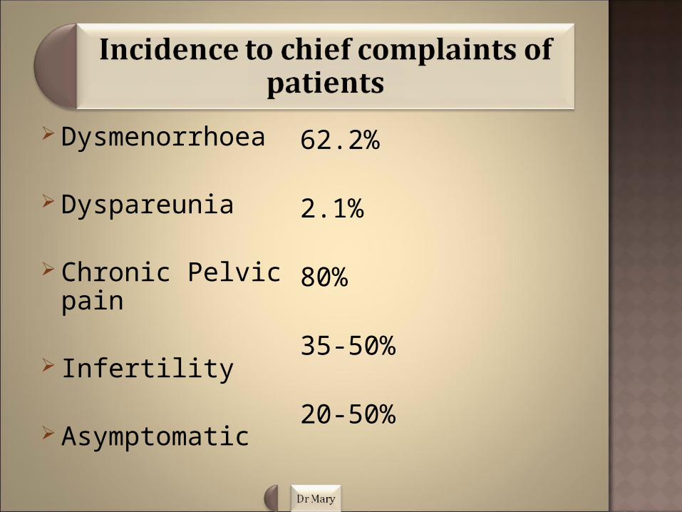
Dysmenorrhoea
Dyspareunia
Chronic Pelvic pain
Infertility
Asymptomatic
62.2%
2.1%
80%
35-50%
20-50%

There is a 10-folds increase incidence in women with an affected first degree relative(family history). And as well as monozygotic twins are markedly concordant for endometriosis.
Rate of endometriosis was found to increase with age from 12% in females ages 11-13 years to 45% in females aged 20-21years and peak incidence between ages of 25 and 35 years.

Endometriosis is classified into one of four stages depending on the following;
Size, location Type Extent Depth of endometriosis implants Presence and severity of adhesions Presence and size of ovarian endometriomas
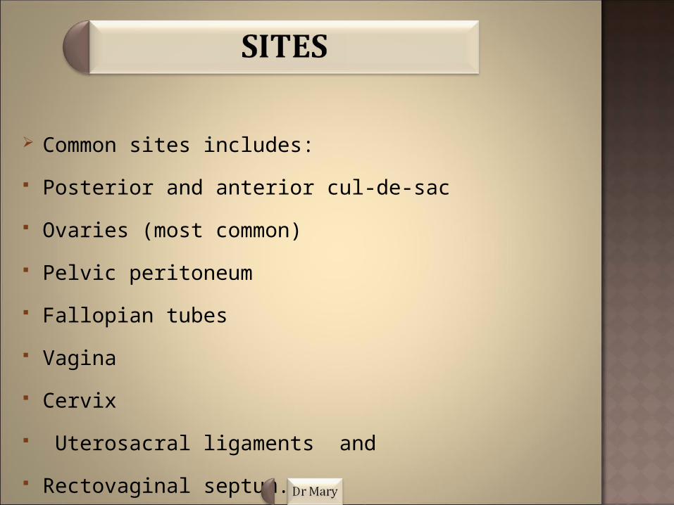
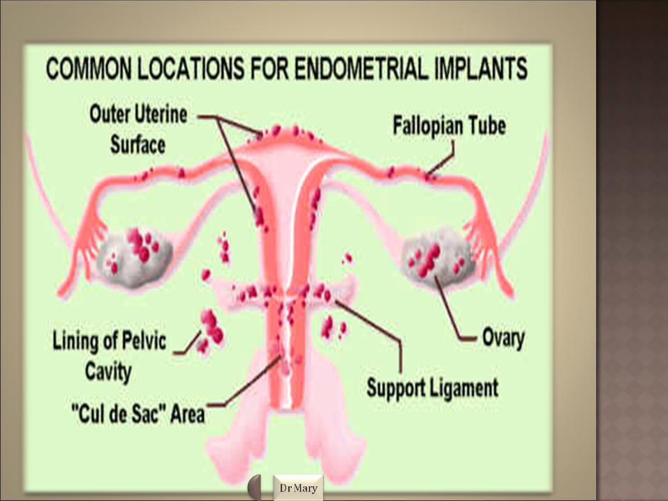
Common sites includes:
Posterior and anterior cul-de-sac
Ovaries (most common)
Pelvic peritoneum
Fallopian tubes
Vagina
Cervix
Uterosacral ligaments and
Rectovaginal septum.

Unusual implantation sites are:
Laparotomy scars Pleura, lung Diaphragm Kidney Spleen Gallbladder Nasal mucosa Spinal canal Stomach.
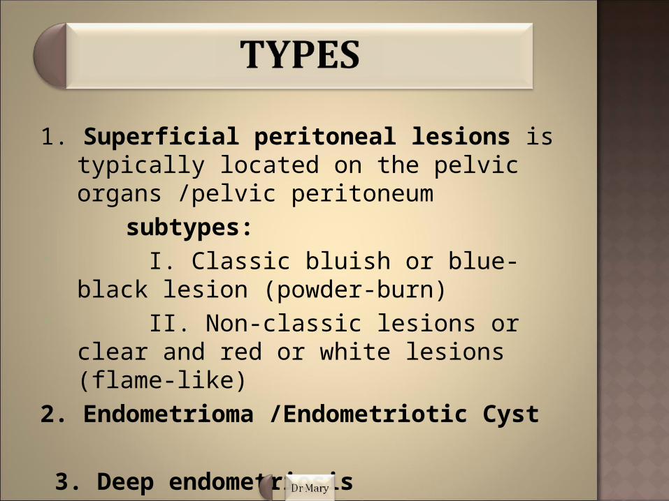
1. Superficial peritoneal lesions is typically located on the pelvic organs /pelvic peritoneum
subtypes: I. Classic bluish or blue-black lesion
(powder-burn) II. Non-classic lesions or clear and red or
white lesions (flame-like) 2. Endometrioma /Endometriotic Cyst
3. Deep endometriosis
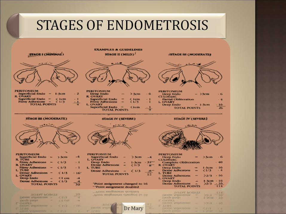
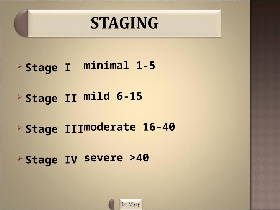
Stage I
Stage II
Stage III
Stage IV
minimal 1-5
mild 6-15
moderate 16-40
severe >40
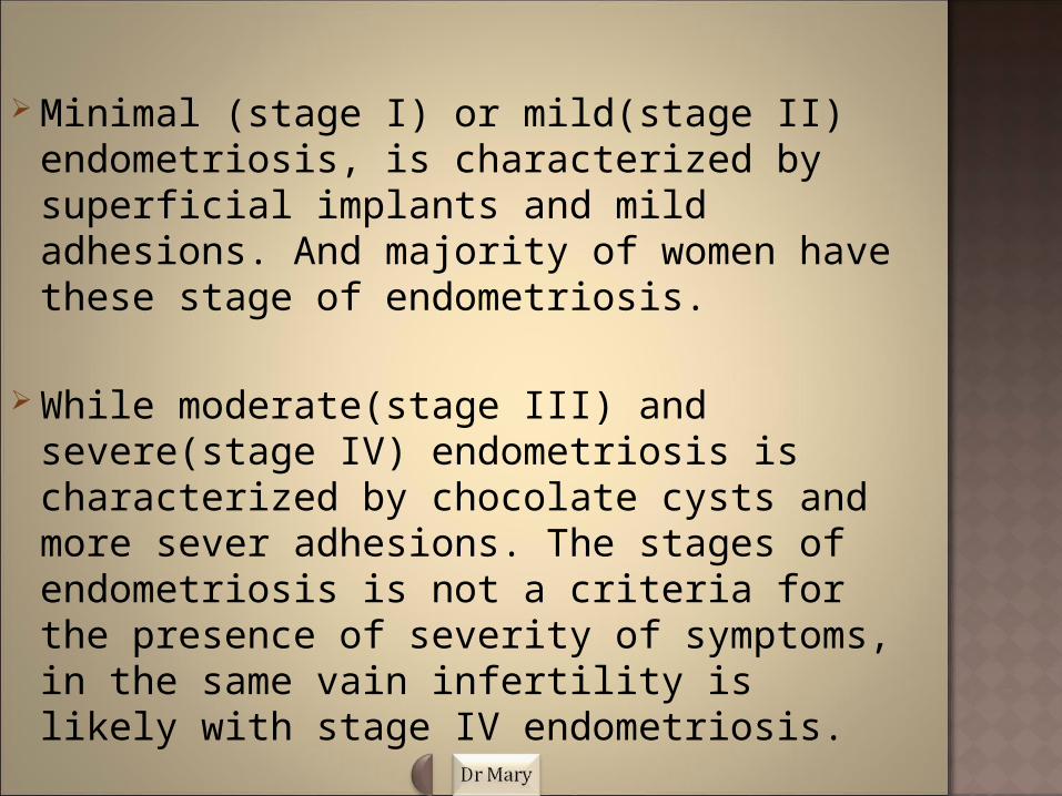
Minimal (stage I) or mild(stage II) endometriosis, is characterized by superficial implants and mild adhesions. And majority of women have these stage of endometriosis.
While moderate(stage III) and severe(stage IV) endometriosis is characterized by chocolate cysts and more sever adhesions. The stages of endometriosis is not a criteria for the presence of severity of symptoms, in the same vain infertility is likely with stage IV endometriosis.


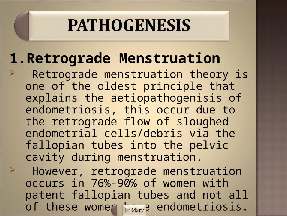
1.Retrograde Menstruation Retrograde menstruation theory is one of the
oldest principle that explains the aetiopathogenisis of endometriosis, this occur due to the retrograde flow of sloughed endometrial cells/debris via the fallopian tubes into the pelvic cavity during menstruation.
However, retrograde menstruation occurs in 76%-90% of women with patent fallopian tubes and not all of these women have endometriosis.

Factors obstructing menstruation are, congenital abnormalities, including imperforate hymen and iatrogenic cervical stenosis etc.
The location of superficial endometriotic lesions in the posterior aspect and left side of the pelvis may be due to the effects of gravity on regurgitated menstruation product and the anatomical position of the sigmoid colon.

2.Coelomic Metaplasia These theory postulates the origin of
endometriosis from metaplasia of specialised cells that are present in the mesothelial lining of the visceral and abdominal peritoneum
Hormonal or immunological factors stimulates the transformation of normal peritoneal tissue/cells into endometrium-like tissue.

These theory clearly explains the occurrence of endometriosis in pre-pubertal girls even thou oestrogen which is the driving force of endometrial growth is not present in them and therefore this condition may be different from endometriosis that is found in women of reproductive age.
According to this theory, residual embryonic cells of the wolffian or mullerian ducts persist and develop into endometriotic lesions that respond to oestrogen. these describes the hormon-dedpendent transformation of peritoneal cells into mullerian-type cells in adolescent.
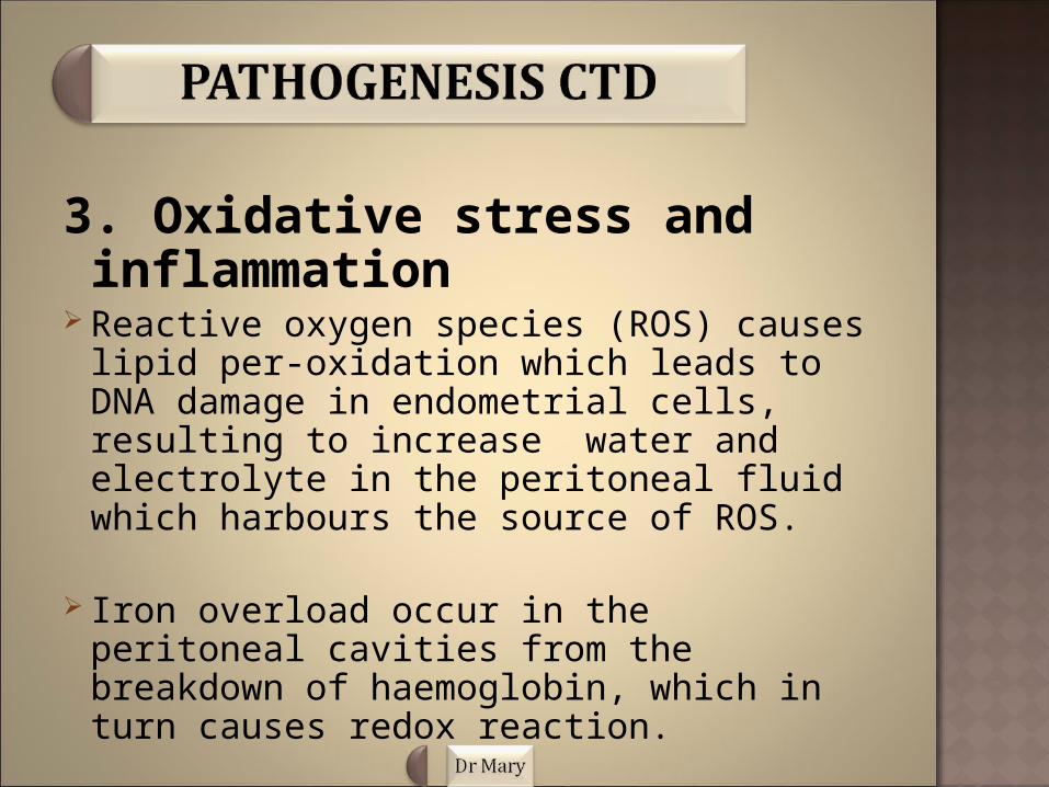
3. Oxidative stress and inflammation
Reactive oxygen species (ROS) causes lipid per-oxidation which leads to DNA damage in endometrial cells, resulting to increase water and electrolyte in the peritoneal fluid which harbours the source of ROS.
Iron overload occur in the peritoneal cavities from the breakdown of haemoglobin, which in turn causes redox reaction.

The release of the pro-inflammatory heam products and the oxidative stress signals generated from the ROS causes inflammation which leads to the recruitment of lymphocytes and activated macrophages producing cytokines that induce oxidizing of enzymes and promotes endothelial growth.

excess proliferation of ROS is accomplished by a decreased level of antioxidants which usually eliminates these molecules.
The resulting accumulation of ROS may contributes to the propagation and maintenance of endometriosis and associated symptoms.
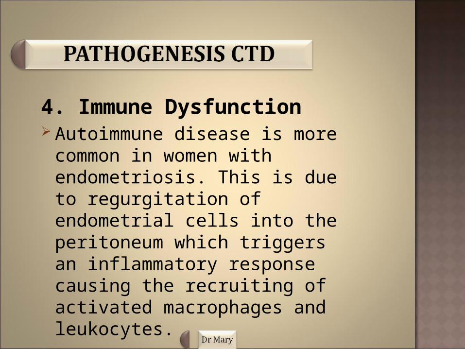
4. Immune Dysfunction Autoimmune disease is more
common in women with endometriosis. This is due to regurgitation of endometrial cells into the peritoneum which triggers an inflammatory response causing the recruiting of activated macrophages and leukocytes.

This inflammatory response leads to a defective immune-surveillance that prevents elimination of the menstrual debris and promotes the implantation and growth of endometrial cells in the ectopic sites.
These theory explains better why women with endometriosis have higher concentration of activated macrophages, decreased cellular immunity as well as a repressed NK cell function.
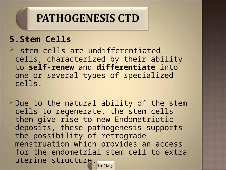
5.Stem Cells stem cells are undifferentiated cells,
characterized by their ability to self-renew and differentiate into one or several types of specialized cells.
Due to the natural ability of the stem cells to regenerate, the stem cells then give rise to new Endometriotic deposits, these pathogenesis supports the possibility of retrograde menstruation which provides an access for the endometrial stem cell to extra uterine structure.
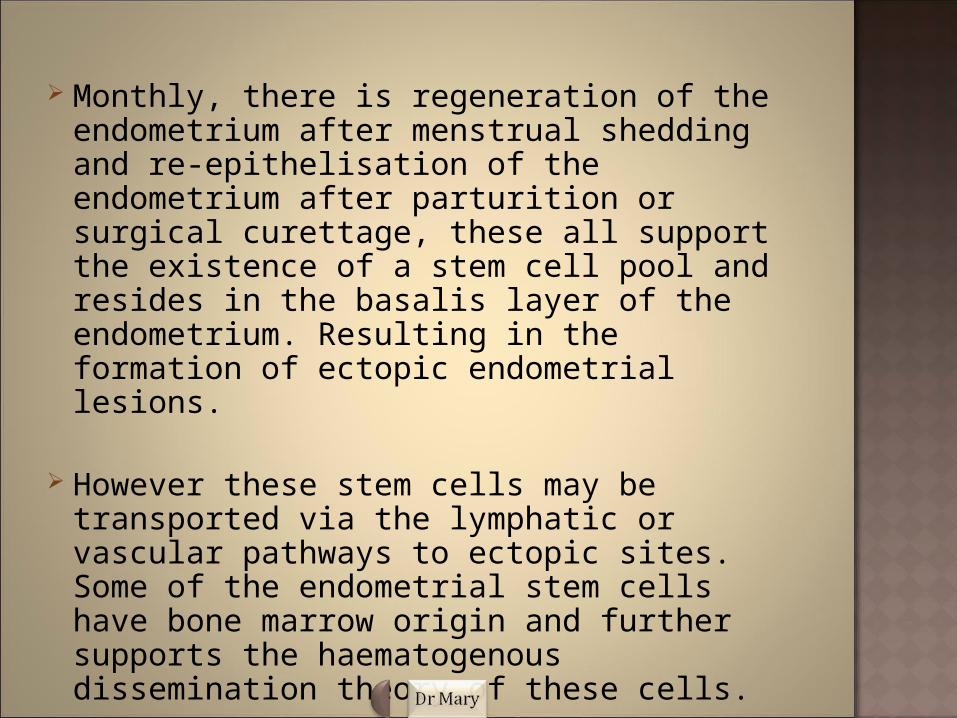
Monthly, there is regeneration of the endometrium after menstrual shedding and re-epithelisation of the endometrium after parturition or surgical curettage, these all support the existence of a stem cell pool and resides in the basalis layer of the endometrium. Resulting in the formation of ectopic endometrial lesions.
However these stem cells may be transported via the lymphatic or vascular pathways to ectopic sites. Some of the endometrial stem cells have bone marrow origin and further supports the haematogenous dissemination theory of these cells.

6. Apoptosis Suppression and Alteration of Endometrial Cell Fate
Alteration of the endometrial cell fate to favour antiapoptotic and pro-proliferation phenotype is paramount for the survival of the endometrial cells in the peritoneal cavity to initiate ectopic deposits and for the maintenance of the established lesions.
The inhibition of the apoptosis of endometrial cells may also be mediated by the transcription activation of genes that normally promotes inflammation, angiogenesis, and cell proliferation.
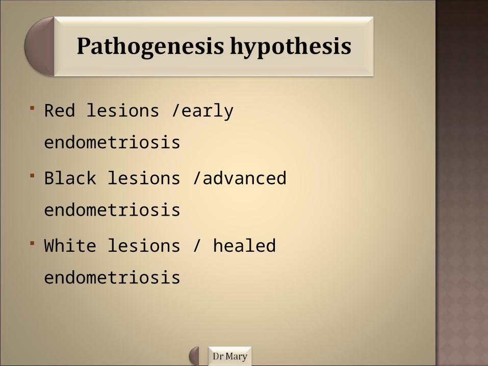
Red lesions /early endometriosis
Black lesions /advanced
endometriosis
White lesions / healed endometriosis

Genetics (positive family history)
Nulliparity
Early menarche
Hormones
Obesity
Uterine retroversion
Miscarriage.

Although a significant number of women with endometriosis remain asymptomatic, but symptomatic patients can be variable and reflects the depth and area of involvement.
Signs and symptoms includes:
1.Pelvic pain
2.Dysmenorrhoea
3.Dsyparinuria
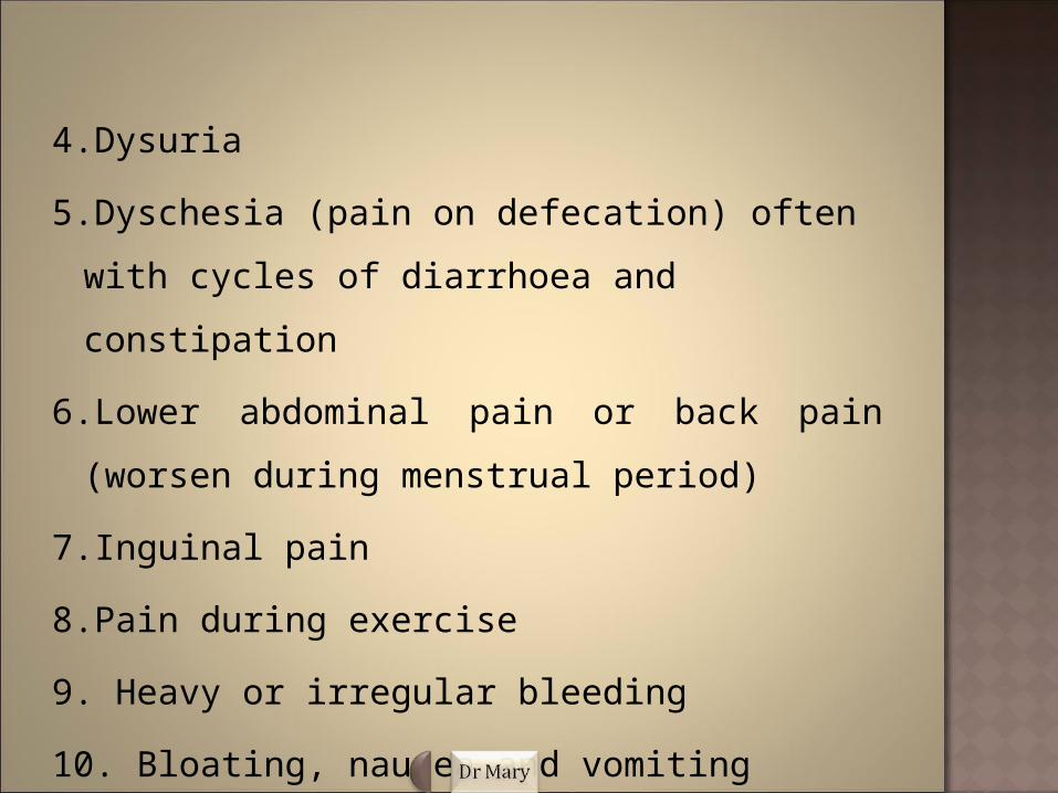
4.Dysuria
5.Dyschesia (pain on defecation) often with cycles of
diarrhoea and constipation
6.Lower abdominal pain or back pain (worsen during
menstrual period)
7.Inguinal pain
8.Pain during exercise
9. Heavy or irregular bleeding
10. Bloating, nausea and vomiting
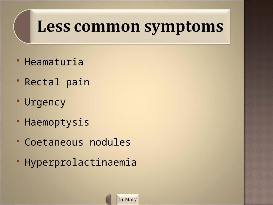
Heamaturia
Rectal pain
Urgency
Haemoptysis
Coetaneous nodules
Hyperprolactinaemia


History taking is an essential aspect in the evaluation of a patient with endometriosis, the following guidelines must be observed.
Having completed your bio data, the necessary important history based on the chief complaints of the patient with endometriosis must be asked.
Infertility/ pain is usually the chief complains of patients with endometriosis.
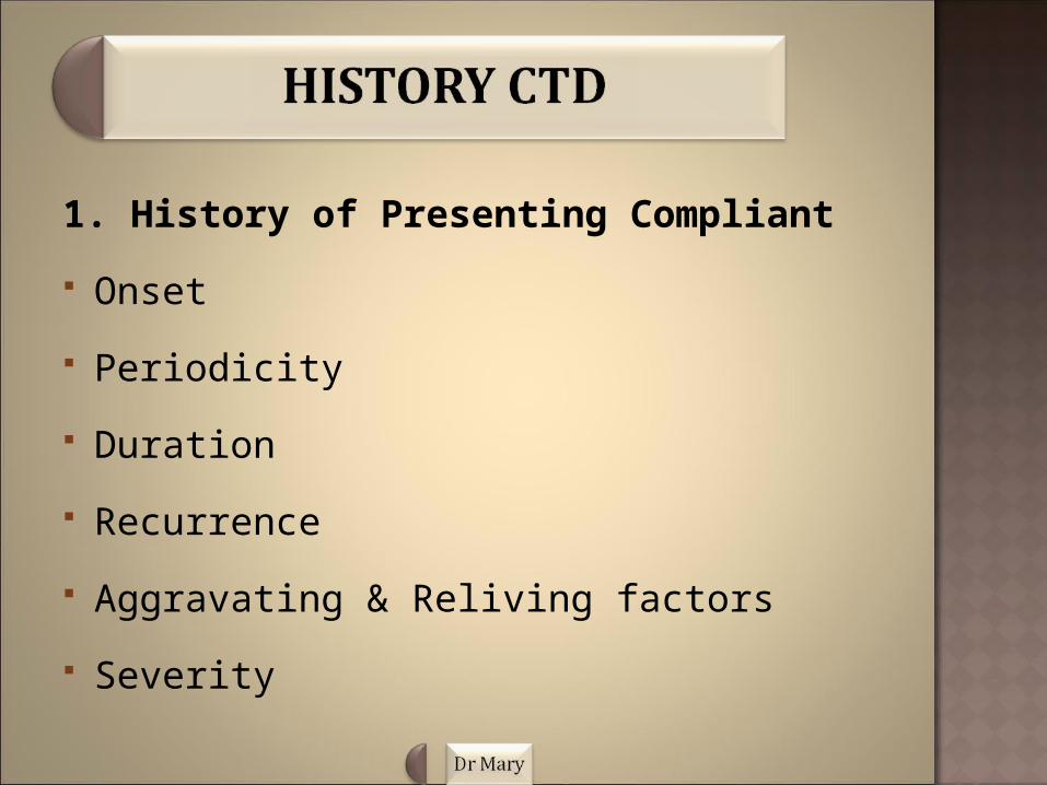
1. History of Presenting Compliant
Onset
Periodicity
Duration
Recurrence
Aggravating & Reliving factors
Severity
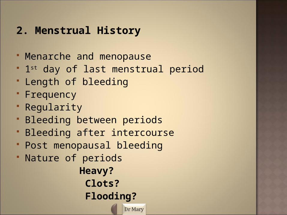
2. Menstrual History
Menarche and menopause 1st day of last menstrual period Length of bleeding Frequency Regularity Bleeding between periods Bleeding after intercourse Post menopausal bleeding Nature of periods Heavy? Clots? Flooding?
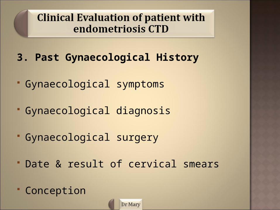
3. Past Gynaecological History
Gynaecological symptoms
Gynaecological diagnosis
Gynaecological surgery
Date & result of cervical smears
Conception
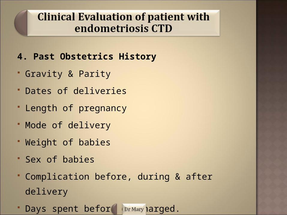
4. Past Obstetrics History
Gravity & Parity
Dates of deliveries
Length of pregnancy
Mode of delivery
Weight of babies
Sex of babies
Complication before, during & after delivery
Days spent before discharged.
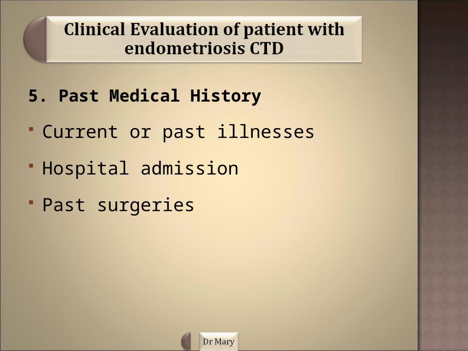
5. Past Medical History
Current or past illnesses
Hospital admission
Past surgeries

6. Drug History
Current medication
Prescribed/ over the counter medication
Herbal Remedies
Recreational drugs
Any known drug allergies.
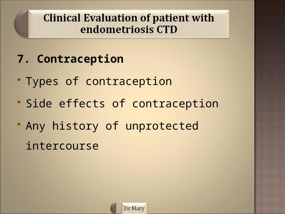
7. Contraception
Types of contraception
Side effects of contraception
Any history of unprotected intercourse

8. Family History History of endometriosis (occurs 10 times in
someone with positive family history)
Gynaecological condition
Malignancies
Consanguinity
History of demise, causes and age at demise.
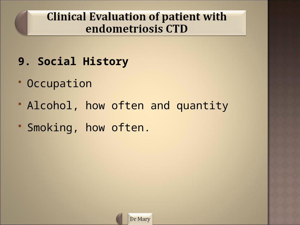
9. Social History
Occupation
Alcohol, how often and quantity
Smoking, how often.
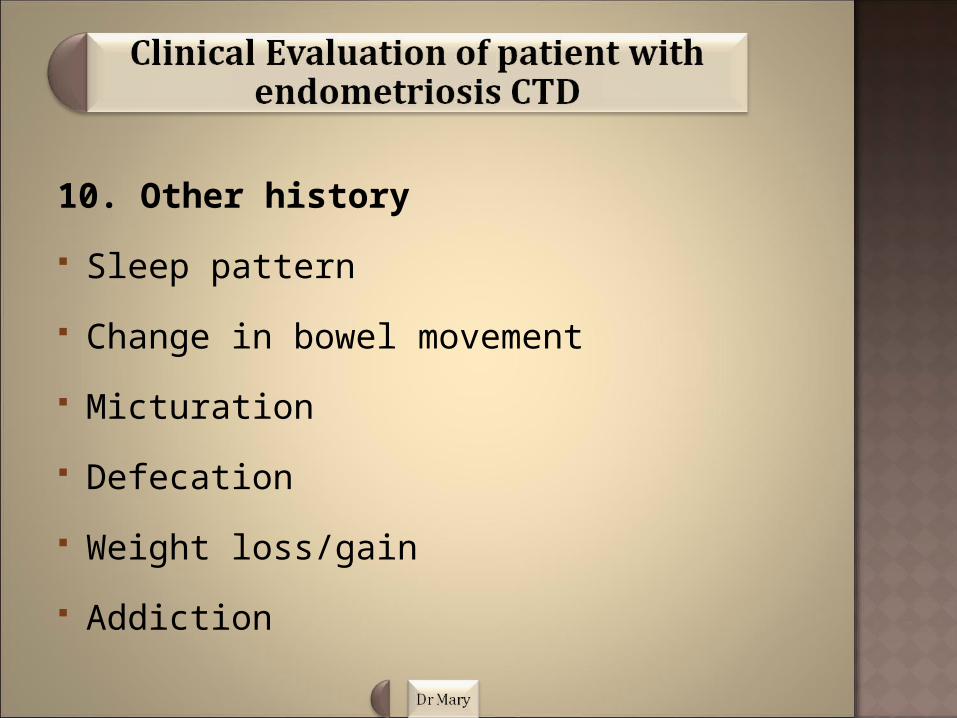
10. Other history
Sleep pattern
Change in bowel movement
Micturation
Defecation
Weight loss/gain
Addiction
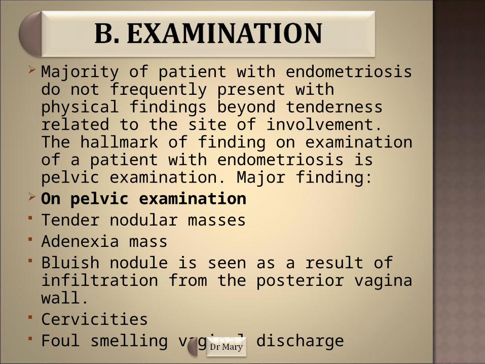
Majority of patient with endometriosis do not frequently present with physical findings beyond tenderness related to the site of involvement. The hallmark of finding on examination of a patient with endometriosis is pelvic examination. Major finding:
On pelvic examination Tender nodular masses Adenexia mass Bluish nodule is seen as a result of infiltration
from the posterior vagina wall. Cervicities Foul smelling vaginal discharge

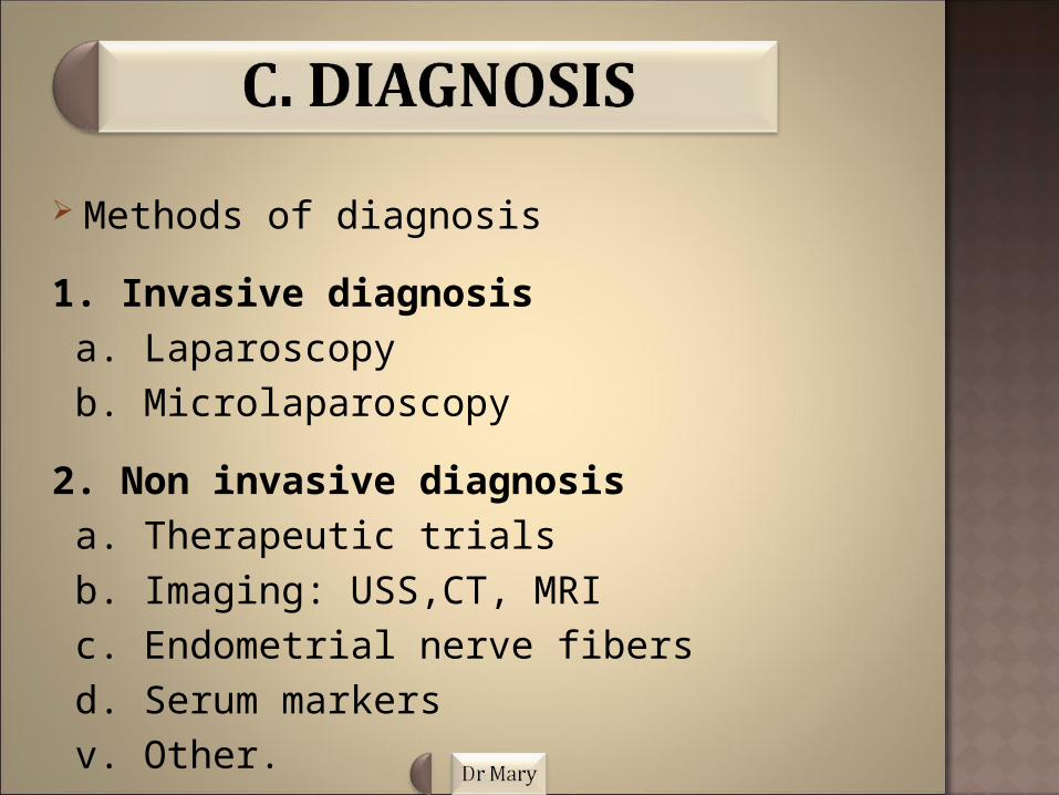
Methods of diagnosis
1. Invasive diagnosis a. Laparoscopy b. Microlaparoscopy
2. Non invasive diagnosis a. Therapeutic trials b. Imaging: USS,CT, MRI c. Endometrial nerve fibers d. Serum markers v. Other.

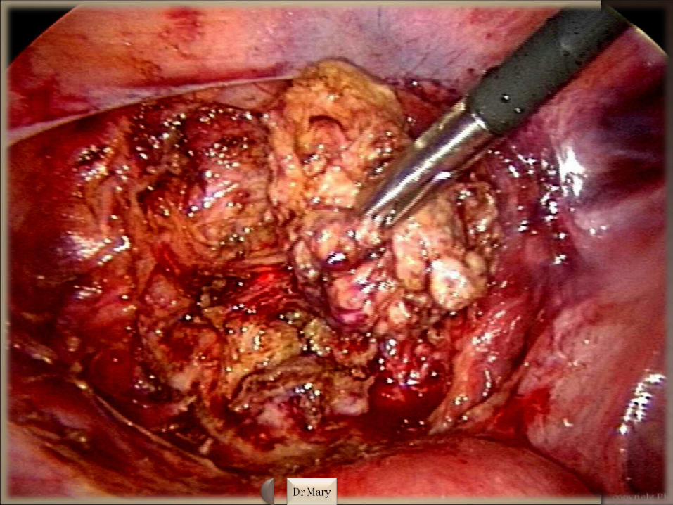
Invasive DiagnosisLaparoscopy: is the gold standard diagnostic
test.Advantages 1. Excludes other condition e.g. ovarian cancer 2. Treatment of endometriosis Disadvantages 1. requirement for surgery and anaesthesia 2. risk of major complications (bowel perforation) 3. visible inspection doesn't detect deep
endometriosis.
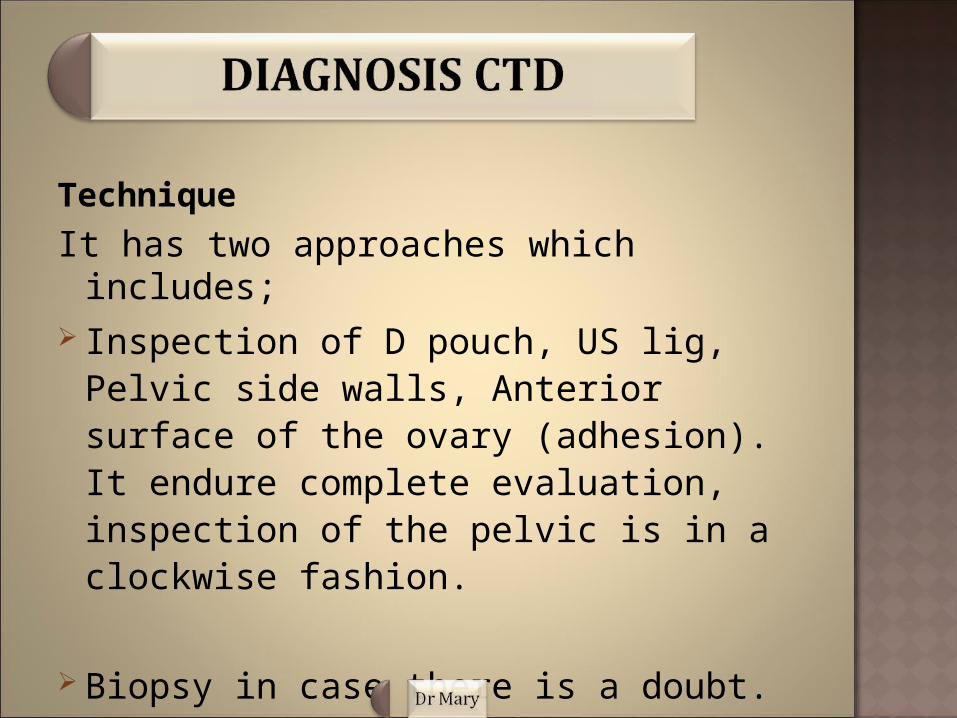
Technique
It has two approaches which includes; Inspection of D pouch, US lig, Pelvic side
walls, Anterior surface of the ovary (adhesion). It endure complete evaluation, inspection of the pelvic is in a clockwise fashion.
Biopsy in case there is a doubt.

Findings:A. Peritoneal i. Typical endometriosis : Black-blue, powder-burn
appearance, and doesn't require any biopsy.
ii. Atypical endometriosis: Lesion that lacks the typical black-blue, powder-burn appearance but however diagnosis may be difficult with standard laparoscopy so biopsy is necessary for confirmation of diagnosis.
B. Endometrioma
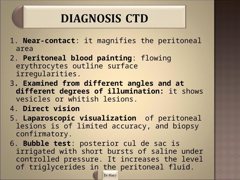
1. Near-contact: it magnifies the peritoneal area2. Peritoneal blood painting: flowing erythrocytes
outline surface irregularities.3. Examined from different angles and at
different degrees of illumination: it shows vesicles or whitish lesions.
4. Direct vision5. Laparoscopic visualization of peritoneal lesions
is of limited accuracy, and biopsy confirmatory. 6. Bubble test: posterior cul de sac is irrigated with
short bursts of saline under controlled pressure. It increases the level of triglycerides in the peritoneal fluid.
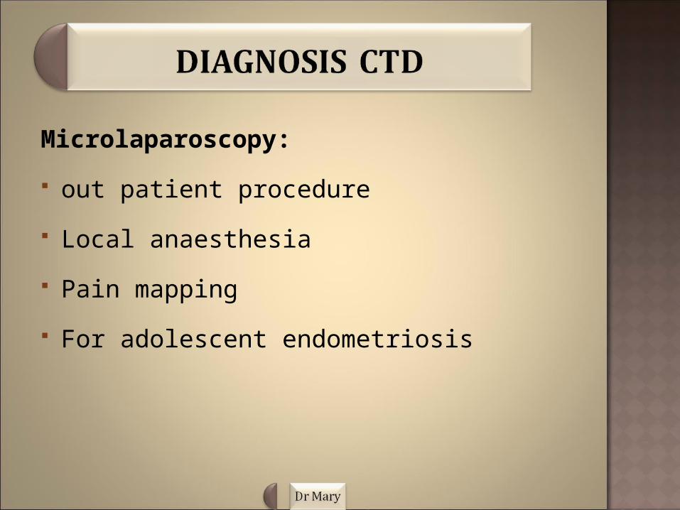
Microlaparoscopy:
out patient procedure
Local anaesthesia
Pain mapping
For adolescent endometriosis
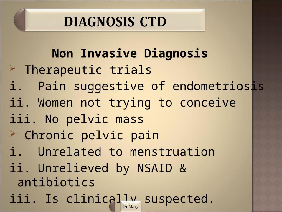
Non Invasive Diagnosis Therapeutic trialsi. Pain suggestive of endometriosisii. Women not trying to conceiveiii. No pelvic mass Chronic pelvic pain i. Unrelated to menstruationii. Unrelieved by NSAID & antibioticsiii. Is clinically suspected.

Imaging 1. Transvaginal ultrasound First line investigational tool for the suspecting
endometriosis Visualization of deep nodules (retrovaginal
septum) Results : Anechoic to echogenic cysts Masses containing multiple septations & solid
tissue Cysts with low-level echoes ( this is the
commonest finding 0f about 95%).

2. Transrectal ultrasound: it detect Rectal involvement Depth of infiltration Lesions on the posterior bladder wall. 3. CT : it has an important role in detecting an
unrelated involvement and possible renal insufficiency.
It has been replaced by MRI due to, poor specificity High radiation

4. MRI: it helps to detect pigmented hgic lesion and inadequately localized lesions.
Posses greater sensitivity which detects about 75% of mild disease
Evaluation of deep lesions Is also superior to ultrasound in diagnosis
rectosigmoid lesions and bladder of the endometriosis
Disadvantages It is expensive And not readily available
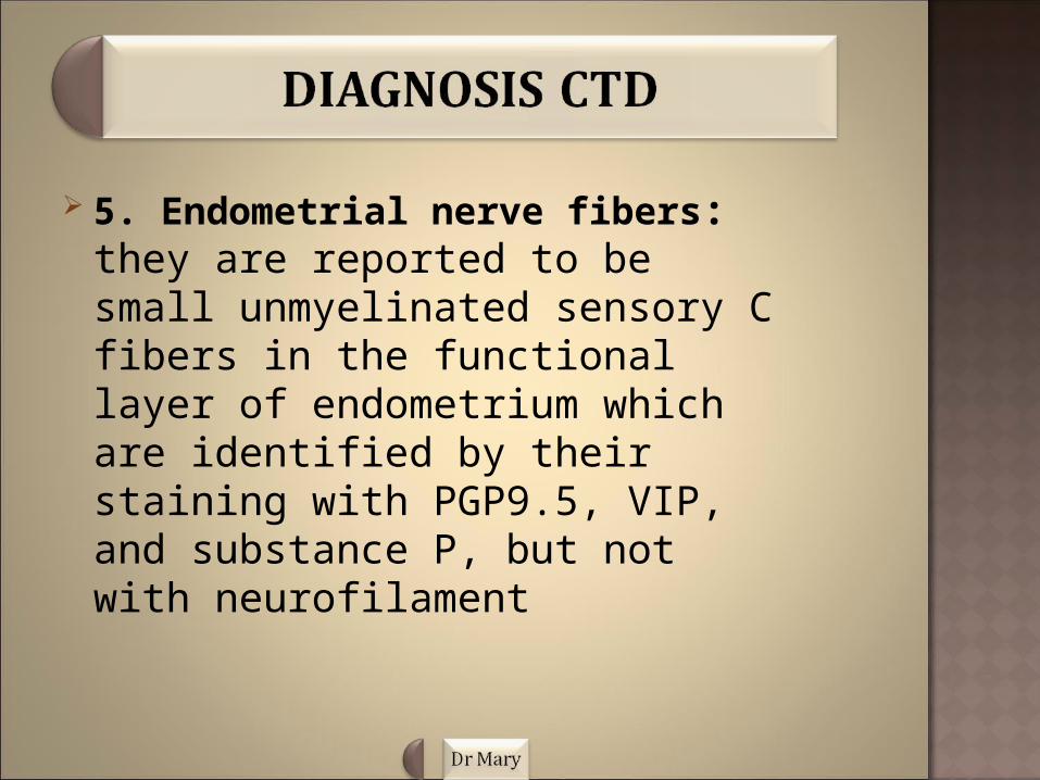
5. Endometrial nerve fibers: they are reported to be small unmyelinated sensory C fibers in the functional layer of endometrium which are identified by their staining with PGP9.5, VIP, and substance P, but not with neurofilament
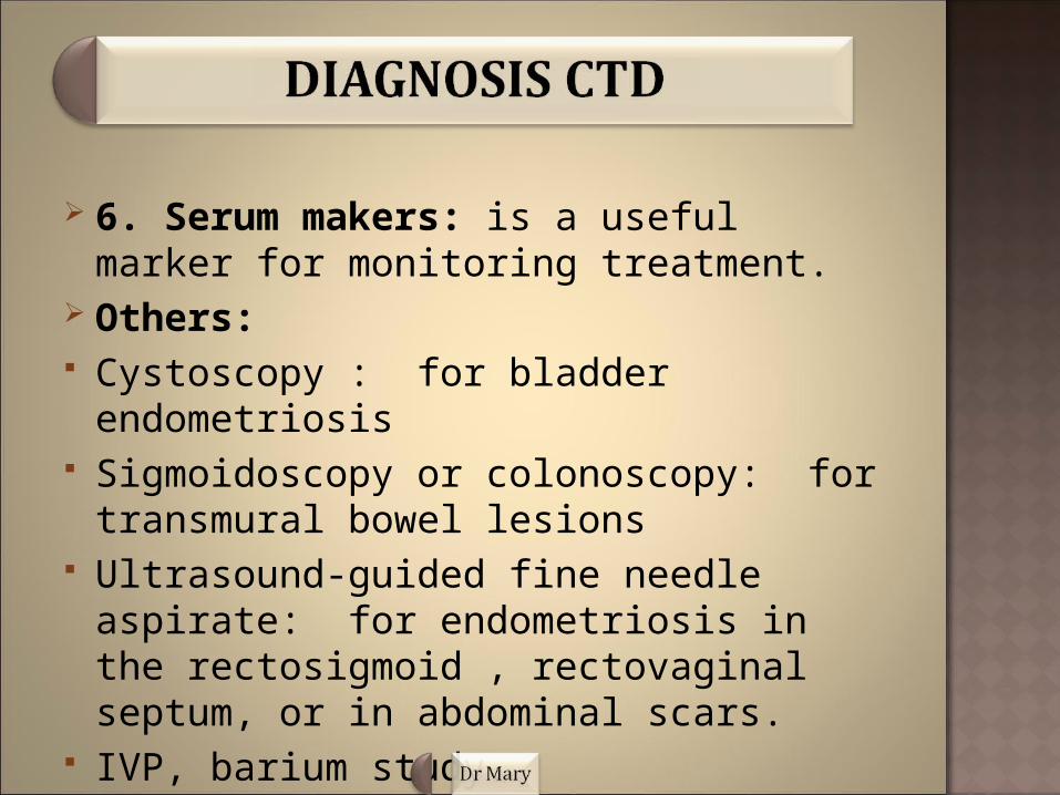
6. Serum makers: is a useful marker for monitoring treatment.
Others: Cystoscopy : for bladder endometriosis Sigmoidoscopy or colonoscopy: for transmural
bowel lesions Ultrasound-guided fine needle aspirate: for
endometriosis in the rectosigmoid , rectovaginal septum, or in abdominal scars.
IVP, barium study.

Treatment for endometriosis can be expectant, medical, or surgical depending on location, depth, severity of symptoms and as well as the desire of the patient to maintain or restore fertility.
Medical treatment: Is used in patients with pelvic pain or
dyspareunia and the aim of treatment is to focus on hormonal manipulation of the menstrual cycle to create the state of pseudopregnancy, pseudomenopause, or chronic anovulation..

Medication includes
1. Danazol
2. Gonadotropin-releasing hormone agonists
3. Oral contraceptive pills and other
4. Progestational agents
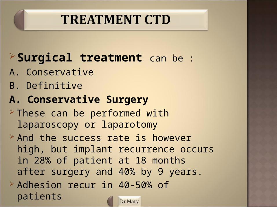
Surgical treatment can be :
A. Conservative B. Definitive
A. Conservative Surgery These can be performed with laparoscopy or
laparotomy And the success rate is however high, but
implant recurrence occurs in 28% of patient at 18 months after surgery and 40% by 9 years.
Adhesion recur in 40-50% of patients

B. Definitive surgery These include Hysterectomy and oophorectomy Is usually reserved for women with
intractable pain. And in one severe cases, one ovary may be retained.
Endometriosis may recur with exogenous estrogens replacement therapy, even in patient who has undergone oophorectomy.
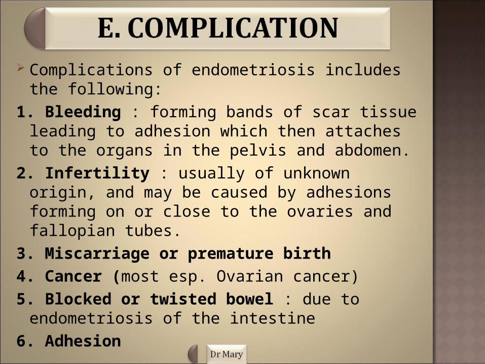
Complications of endometriosis includes the following:
1. Bleeding : forming bands of scar tissue leading to adhesion which then attaches to the organs in the pelvis and abdomen.
2. Infertility : usually of unknown origin, and may be caused by adhesions forming on or close to the ovaries and fallopian tubes.
3. Miscarriage or premature birth4. Cancer (most esp. Ovarian cancer)5. Blocked or twisted bowel : due to endometriosis
of the intestine6. Adhesion
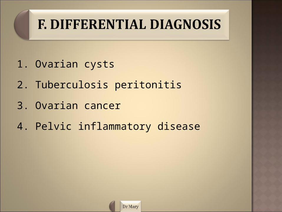
1. Ovarian cysts
2. Tuberculosis peritonitis
3. Ovarian cancer
4. Pelvic inflammatory disease
Prevention of endometriosis includes a wide range of activities known as interventions
Its aim is to reduce risks of threats to health. These leads us to the three categories of prevention of endometriosis;
1. Primary prevention2. Secondary prevention 3. Tertiary prevention

Primary prevention: Its aim at preventing disease or injury of
endometriosis before it ever occurs. This is done by;
i. Preventing exposures to hazards that can cause disease or injury which alters unhealthy behaviours that can lead to endometriosis.
ii. Enforcement to ban or control the use of hazardous substances.
iii. Education about healthy and safe habits.
2. Secondary prevention: Aims to reduce the impact of endometriosis or
injury that has already occur. This is done by;
i. Regular examination and screening to detect endometriosis in its earliest stage.
ii. To treat endometriosis as soon as possible to slow its progression.
ii. Encouraging personal strategies to prevent re-injury or recurrence, and implementing programs to return people to their original health.
iv. To prevent long-time complications of endometriosis.

3. Tertiary prevention: Aim to reduce the impact of an ongoing illness or
injury that has lasting effects, and is done by;
i. Helping people to manage long-term, often-complex health problems and injuries, in order to improve as much as possible their ability to function, their quality of life and life expectancy.
ii. Support groups that allow people to share strategies for living well
iii. Vocational rehabilitation programs to recover as early as possible.

The recurrence rate five years following surgery is between 20% and 40%, providing menopause has not been reached and hysterectomy has not been performed
Women who have undergone treatment for endometriosis needs to attend periodic examinations so they can be monitored using sonography
Note that endometriosis may recur after surgery or medical intervention if the underlying p causes is not probably treated.
Endometriosis is often a chronic disease, and
thorough discussions to ensure a good level of
patient understanding is essential.
It’s important to assess your level of symptoms,
your desire to have children in the future, as well as
your social and occupational needs for better health.
Comprehensive follow-up will aid in the assistance
of total rehabilitation.

Majority of patients with endometriosis will have increasing fertility problems. Fortunately, the results of assisted reproduction (such as IVF) after treatment for endometriosis are very good.
While some of these patients , even if they have an initial problem with their fertility, end up becoming pregnant after adequate and carefully monitored treatment.
In the same vain, some patients will require a higher level of technology to achieve a pregnancy, such as IVF or GIFT.
Note that, pregnancy is not a complete and definitive cure for endometriosis, the combination of pregnancy and breastfeeding significantly slows down the course of the disease and may even get rid of it entirely.
