magnetic resonance angiography shows increased arterial...
TRANSCRIPT

Research ArticleMagnetic Resonance Angiography Shows Increased ArterialBlood Supply Associated with Murine Mammary Cancer
Devkumar Mustafi ,1 Abby Leinroth,1 Xiaobing Fan,1 EricaMarkiewicz,1 Marta Zamora,1
Jeffrey Mueller,2 Suzanne D. Conzen,3 and Gregory S. Karczmar 1
1Department of Radiology, The University of Chicago, Chicago, Illinois 60637, USA2Department of Pathology, The University of Chicago, Chicago, Illinois 60637, USA3Department of Medicine, Section of Hematology and Oncology, The University of Chicago, Chicago, Illinois 60637, USA
Correspondence should be addressed to Gregory S. Karczmar; [email protected]
Received 11 October 2018; Accepted 6 January 2019; Published 17 January 2019
Academic Editor: Jie Tian
Copyright © 2019 Devkumar Mustafi et al. This is an open access article distributed under the Creative Commons AttributionLicense, which permits unrestricted use, distribution, and reproduction in any medium, provided the original work is properlycited.
Breast cancer is a major cause of morbidity and mortality in Western women. Tumor neoangiogenesis, the formation of new bloodvessels from pre-existing ones, may be used as a prognostic marker for cancer progression. Clinical practice uses dynamic contrastenhanced magnetic resonance imaging (DCE-MRI) to detect cancers based on increased blood flow and capillary permeability.However, DCE-MRI requires repeated injections of contrast media. Therefore we explored the use of noninvasive time-of-flight(TOF) MR angiography for serial studies of mouse mammary glands to measure the number and size of arteries feeding mammaryglands with and without cancer. Virgin female C3(1) SV40 TAg mice (n=9), aged 18-20 weeks, were imaged on a 9.4 Tesla smallanimal scanner. Multislice T
2-weighted (T2W) images and TOF-MRI angiograms were acquired over inguinal mouse mammary
glands. The data were analyzed to determine tumor burden in each mammary gland and the volume of arteries feeding eachmammary gland. After in vivoMRI, inguinal mammary glands were excised and fixed in formalin for histology. TOF angiographydetected arteries with a diameter as small as 0.1mm feeding the mammary glands. A significant correlation (r=0.79; p< 0.0001)was found between tumor volume and the arterial blood volume measured in mammary glands. Mammary arterial blood volumesranging from 0.08mm3 to 3.81mm3 weremeasured. Tumors and blood vessels found on in vivo T2W and TOF images, respectively,were confirmed with ex vivo histological images. These results demonstrate increased recruitment of arteries to mammary glandswith cancer, likely associated with neoangiogenesis. Neoangiogenesis may be detected by TOF angiography without injectionof contrast agents. This would be very useful in mouse models where repeat placement of I.V. lines is challenging. In addition,analogous methods could be tested in humans to evaluate the vasculature of suspicious lesions without using contrast agents.
1. Introduction
Breast cancer in humans is associated with increased bloodsupply and capillary permeability [1]. Typically, mammaryvasculature is detected and evaluated using dynamic contrastenhanced (DCE) MRI [2]. This method requires rapid imag-ing following an I.V. injection of contrast media. Increasedblood flow in cancers results in a greater rate of uptakeof contrast media and more rapid enhancement in MRimages. Changes in signal intensity following contrast mediainjection can be analyzed to determine contrast media uptakeand to calculate physiological parameters relating to bloodflow and permeability [2, 3].
While DCE-MRI is the preferred method for clinicaldetection of human breast cancer [4, 5], the use of DCE-MRI for serial measurements in mouse models of breastcancer is challenging. DCE-MRI requires the repeated use ofan I.V., which may lead to complications. The accumulationof contrast agents in certain tissues is also problematic asrepeated doses of contrast media can cause adverse reactions,such as kidney damage, in subjects [6]. Furthermore, DCE-MRI requires rapid imaging with time resolution of 1–2seconds; this means that signal-to-noise ratio and spatialresolution is limited [3].
Time-of-flight MR angiography is a noninvasive meansof assessing mammary gland vasculature. Time-of-flight
HindawiInternational Journal of Biomedical ImagingVolume 2019, Article ID 5987425, 6 pageshttps://doi.org/10.1155/2019/5987425

2 International Journal of Biomedical Imaging
angiography (TOFA) detects blood flow perpendicular to theimage slice based on decreases in the apparent T1 signal dueto in-flow [7]. TOFA primarily detects relatively rapid flowthrough arteries. Larger arteries are detected based on theirhigher signal-to-noise ratio (SNR) [8]. TOFA is thought to beless informative than DCE-MRI because there is concern thatTOFA cannot depict small vessels with low blood velocities[9, 10]. However, unlike DCE-MRI, TOFA does not usecontrast agents and is not limited by temporal resolution.Thus TOFA can be acquired over a longer period of time,which allows high spatial resolution. Images in all planes withhigh spatial resolution can be acquired to detect blood flow insmaller arteries [11, 12].
Previous studies in this lab have utilized serial imagingstudies of the SV40 TAg mouse model of human breastcancer to examine the initiation and progression of mam-mary cancers [13]. The sensitivity and specificity of detectingthe disease can be further improved by focusing on theconsequences of tumor angiogenesis: increased micro-vesseldensity with altered vascular characteristics [11, 12]. Wepropose that TOFA is a useful alternative to DCE-MRI forevaluating vasculature, but with less negative repercussions.Furthermore, these two types of MRI acquisition proto-cols should complement each other and could provide animproved understanding of the angiogenesis associated withcancer aggressiveness and growth.
Thegoal of the present researchwas to evaluate the qualityof TOFA in mouse mammary glands, and to determinethe range of sizes of arteries that could be detected. Inaddition, we tested whether TOFA detects changes in arterialblood supply associated with neoangiogenesis and growth ofmammary cancer.
2. Materials and Methods
2.1. Animal. Nine FVB/N mice homozygous for the SV40TAg transgene (originally provided as hemizygous TAg miceby Dr. Jeffrey E. Green of the National Cancer Institute’sMouse Models of Cancer Consortium) were weaned at 3weeks of age as previously described [14]. These mice weresacrificed after in vivoMRI studies at 18-20weeks of age.Micewere anesthetized prior to in vivo MR imaging, and anes-thesia was maintained during imaging with 1-2% isoflurane.Temperature, heart rate, and respiration were monitored bySA instruments (Stony Brook, NY, USA), and the respirationrate was used to gate imaging. After in vivo imaging, micewere sacrificed by an overdose of isoflurane and cervicaldislocation.
2.2. MRI Experiments. In vivo imaging: MR images wereacquired on a 9.4 Tesla small animal scanner (Bruker,Ettlingen, Germany) with 11.6 cm inner diameter activelyshielded gradient coils (maximum constant gradient strengthfor all axes: 230mT/m). The mouse was placed supineon an animal holder and inserted into a 30mm diameterquadrature volume coil (Rapid MR International, Colum-bus, OH, USA). Multislice RARE (Rapid Acquisition withRelaxation Enhancement) T
2-weighted (T2W) images with
fat suppression were acquired with the following parameters:
TR/TEeffective = 4000/20ms, field-of-view (FOV) = 25.6mm× 19.2mm, matrix size = 256 × 192, slice thickness = 0.5mm,RARE factor = 4, and number of excitations (NEX) = 2. Forthe inguinal glands (left and right) two interleaved sets ofimages were acquired to cover the slice gaps of 1mm and thencombined together for a total of 62 slices.
For TOFA, a flow compensated gradient echo sequencewith a short TR and thin slices was used to maximize infloweffects and depict flowing blood as a bright signal. Parametersfor TOF with the same in-plane resolution as T2W imageswere TR/TE = 10/3ms, flip angle = 60∘, FOV = 25.6mm ×19.2mm, matrix size = 256 × 192, slice thickness = 0.5mm, 61slices, and NEX = 4. The acquisition time for each slice was10.2 s and the total acquisition time for this TOF sequencewas10.4 minutes.
2.3. Histology. After in vivo MRI studies, the mammaryglands were excised and placed in 10% formalin for tissuefixation for two weeks, then put in a histology cassettein ethanol for immediate histological processing and H&Estaining of slices. An experienced breast pathologist (JM)evaluated the histological slides and tissue was classifiedas normal gland, in situ, or invasive cancers. H&E slideswere then scanned using a fully automated Leica microscope(DM-6000B, Leica Microsystems, Weltzar, Germany) forvisualization and H&E images were stored in TIFF format.
2.4. Data Analysis. The MRI data sets were processedand analyzed quantitatively using software written in IDL(Exelis VIS, Inc., Boulder, CO, USA). Amira 3D visual-ization and analysis software (FEI Visualization SciencesGroup, Burlington, MA, USA) was used to create three-dimensional displays of the tumors and blood vessels. Basedon criteria defined and published previously, we identifiedinvasive tumors in the inguinal mammary glands fromnine SV40 mice on T2W images [15]. Then regions-of-interest (ROIs) were manually traced around mammarycancers. Total tumor volume in each mammary glandwas determined by combining the volumes of all cancerROIs.
To determine the volumes of arteries feeding mammaryglands, the left and right inguinal mammary gland weremanually traced on T2W images first and the resulting ROIswere superimposed to theTOF images.Then, thresholdswereset to select only for those pixels representing blood vessels.Using these pixels, the total volumes of arteries in the left andright mammary gland were calculated.
We calculated the volumes of the mammary cancers orblood vessels on each slice by multiplying the total crosssectional area by the slice thickness of 0.5mm in both T2Wand TOF images. All of the slice volumes were added togetherto estimate the final tumor volume and vessel volumes oneach side of the mammary gland. The Pearson correlationtest was performed to examine whether there is a linearrelationship between mammary cancer volumes and bloodvolumes. The t-test was performed to determine whetherthere was a statistically significant difference for calculatedparameters. A p-value of less than 0.05 was consideredsignificant.

International Journal of Biomedical Imaging 3
(a)
(b)
(c)
Figure 1: In vivo MR images of a SV40 TAg mouse of 19 weeks of age. The top panel (a) shows T2W images of the three central 0.5 mmmthick slices through inguinal mouse mammary glands. In all images lymph nodes (LN) are labeled and tumors are indicated by red arrows.The middle panel (b) shows the TOF images of the corresponding slices as seen in the top panel. The bottom panel (c) shows blood vessels,shown in red as in TOF images in the middle panel, and overlaid on T2W images. Scale bars in all images are shown.
3. Results
Using TOFA, the smallest detectable artery on volume ren-dered images was 0.005mm3. In this case, the slice thicknesswas the limiting factor in determining size. The average (±standard deviation) SNRofmuscle in T2W images, measuredover ROIs (n = 27), was 17.3±6.4. The average SNR of muscleinTOF images,measured over ROIs (n = 27), was 3.6±1.9.Theaverage SNR of blood vessels in TOF, measured over ROIs(n = 27), was 61.1±22.0. The blood vessels had significantlyhigher SNR than muscle (p < 0.0004). This allowed for thedetection of blood flow in smaller arteries.
Using TOFA and T2W images, we were able to measurearterial blood volumes. Figure 1(a) shows three contiguousT2W images (left to right) of an SV40 TAg female mousewith a tumor (red arrow) developing beneath the lymphnode (denoted as LN). Figure 1(b) shows three TOF imagesfrom approximately the same slices as shown in Figure 1(a).Figure 1(c) shows blood vessels, in red, overlaid on theT2W images. These images demonstrate that blood volumeincreases with cancer burden, as blood vessels are moreabundant in the right gland, particularly beneath the tumor.In the left gland no cancer is evident and no blood vessels aredetected.
This qualitative relationship was visualized more easilywith three-dimensional rendering, (using Amira 3D soft-ware). Figure 2 shows a 2-dimensional depiction of the 3Dvolume-rendering of a SV40 TAg femalemouse at 18 weeks ofage. Greater arterial blood supply (in red) can be seen flowingtowards the tumors (labeled “Tu”) in the right gland (blue-to-green). In comparison, the left inguinal gland shows neithercancer nor arteries. Visual inspection of volume-renderedimages in 3D suggested differences in the organization ofarteries associated with mammary cancer. Arteries leadingto mammary glands with low or no tumor burden are wellorganized in a tree-like structure with alternating branches.Arteries leading to glands with higher tumor burdens donot follow this regular branching pattern. Instead the bloodvessels grow in a clump directed towards the tumor andsurrounding tissue.
A strong positive correlation (r = 0.79, p < 0.0001) wasfound between increasing tumor volume and blood volume,as seen in Figure 3. The best-fit straight line through the plotof tumor volume vs. blood volume is given by Blood-volume= 0.085∗Tumor-volume + 0.12.
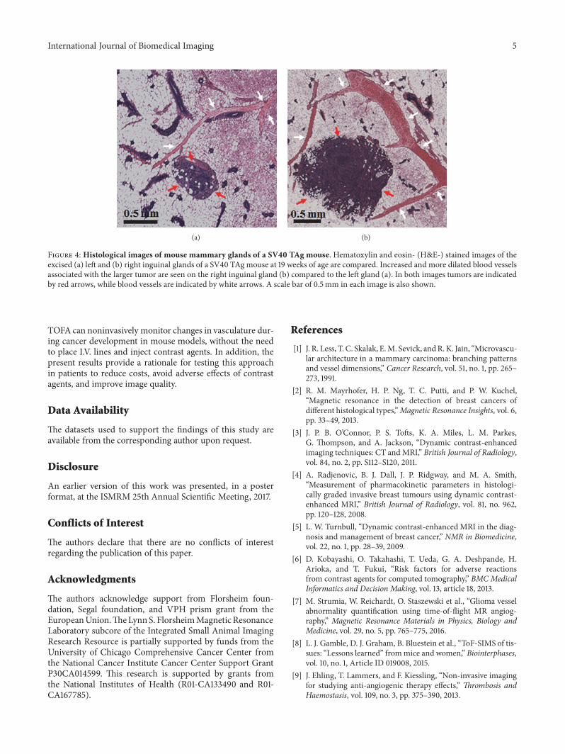
This result is consistent with histology. Figure 4 depictsthe histology of the mouse shown in Figure 2. This histologyshows that the right gland (Figure 4(b)), which has a larger

4 International Journal of Biomedical Imaging
Figure 2: Three dimensional volume rendered image showsmouse mammary glands and vessel densities of a SV40 TAgfemale mouse at 18 weeks of age. Only the ROIs of both inguinalmammary glands are shown in blue-to-green color—images wereacquired with a T2W RARE sequence. Blood vessels are shown inred—images of blood vessels were constructed from TOF datasets.Blood vessels in and near the right inguinal gland, compared to theleft gland, grew significantly as invasive cancers developed. Lymphnodes (LN) and tumors (Tu) are labeled. A scale bar of 2mm is alsoshown.
Blood volume = 0.0851(Tumor volume)+0.1167R = 0.79, p<0.0001
4
3
2
1
0
0 10 20 30 40
Tumor volume (mG3)
Bloo
d vo
lum
e (mG
3)
Figure 3: Plot of blood volume as a function of tumor volumein SV40 TAgmice. The scatter plot shows the relationship betweenthe tumor and blood volumes for a total of nine SV40 TAg miceof 18-20 weeks of age. There was a strong positive correlation (r =0.79, p < 0.0001) between the tumor volume and the blood volumeas indicated in the plot.
tumor and not only has more vessels but also more vascularcomplexity, as compared to the left gland (Figure 4(a)). Anexperienced breast pathologist (JM) identified cancers andblood vessels fromH&E-stained slices. Previous studies in thelab correlated MRI-TOFA results with both H&E-stained andCD31-stained slices, demonstrating the usefulness of MRI-TOFA in the use of H&E stained and CD31 stained histology[16].
4. Discussion
This study found a strong positive correlation betweenarterial blood volume in the mammary gland and mammary
cancer volume using TOFA. The correlation between vascu-lature and tumor aggressiveness has been previously studiedand established, and antiangiogenic therapies have becomean important option for anti-cancer therapies [7, 12, 17–20]. The results demonstrate the effectiveness of TOFA forqualitative and quantitative measurements of changes invasculature as cancer develops.
High-resolutionmagnetic resonance angiography (MRA)offers a macroscopic view of the entire arterial supply ofcancer, as documented in this study. MRA-based parametersmay have diagnostic utility. In a previous blinded study ofhuman brain cancer patients, a statistical analysis of theshapes of MRA-extracted blood vessels proved successfulin separating benign from malignant disease in all caseson the basis of image analysis before lesion resection [21].In contrast to perfusion imaging (e.g., DCE-MRI), high-resolution MRA shows the 3D anatomy of the arterial supplyto cancers [22]. The major advantage of TOFA of mousemodels lies in the ability to conduct repeated studies com-pletely noninvasively, avoiding use of intravenous cathetersand contrast agent injection. This makes TOFA a valuabletool for directly monitoring cancer development and theeffectiveness of antiangiogenic therapies without injuring orsacrificing animals. The method used here does not readilyprovide quantitative measurements of blood flow, but morequantitative TOFA methods are available [23].
A major concern with TOF imaging has been that itcannot detect small arteries; this could result in poor sensi-tivity to neoangiogenesis associated with mammary cancer.However, in this study at 9.4 T, TOA reliably measuredblood volumes as low as 0.08mm3 and detected individualarteries as small as 5x10⁻⁴mm3 in volume. The accuracy ofarterial blood volumemeasurements in this study was limitedprimarily by the slice thickness of 0.5mm, since in-planeresolution was 0.1mm × 0.1mm. In contrast to DCE-MRI,the TOF sequence does not require high temporal resolution.Therefore, more signal averaging can be used to acquire TOFimages with higher spatial resolution, to detect even smallerarteries. Furthermore, TOF images can be acquired in bothsagittal and coronal planes, in addition to the axial plane seenin the present study. This would allow evaluation of arterialblood flow in three-dimensions.
This study demonstrated a positive linear relationship(Figure 3) between the tumor growth and the local bloodvolume. The results suggest that blood volume in the mam-mary gland is approximately 8.5%of tumor volume.However,not all of the cancers identified in this study conformed tothe approximately linear relationship between blood volumeand tumor burden—there were several outliers. We arenot able to determine yet whether this reflects biologicalvariability ormeasurement error. Previous work suggests thatneoangiogenesis is biologically variable in cancers [9].
Previous studies have correlated the number of vesselswith aggressiveness in brain cancer [7, 17, 24]. To the best ofour knowledge the present study is the first to demonstratea strong correlation between tumor volume and local bloodvolume in mammary cancer, without use of contrast agents,noninvasively and in-vivo [2, 3]. The results demonstrate that
International Journal of Biomedical Imaging 5
(a) (b)
Figure 4: Histological images of mouse mammary glands of a SV40 TAg mouse. Hematoxylin and eosin- (H&E-) stained images of theexcised (a) left and (b) right inguinal glands of a SV40 TAgmouse at 19 weeks of age are compared. Increased and more dilated blood vesselsassociated with the larger tumor are seen on the right inguinal gland (b) compared to the left gland (a). In both images tumors are indicatedby red arrows, while blood vessels are indicated by white arrows. A scale bar of 0.5mm in each image is also shown.
TOFA can noninvasively monitor changes in vasculature dur-ing cancer development in mouse models, without the needto place I.V. lines and inject contrast agents. In addition, thepresent results provide a rationale for testing this approachin patients to reduce costs, avoid adverse effects of contrastagents, and improve image quality.
Data Availability
The datasets used to support the findings of this study areavailable from the corresponding author upon request.
Disclosure
An earlier version of this work was presented, in a posterformat, at the ISMRM 25th Annual Scientific Meeting, 2017.
Conflicts of Interest
The authors declare that there are no conflicts of interestregarding the publication of this paper.
Acknowledgments
The authors acknowledge support from Florsheim foun-dation, Segal foundation, and VPH prism grant from theEuropeanUnion.TheLynnS. FlorsheimMagnetic ResonanceLaboratory subcore of the Integrated Small Animal ImagingResearch Resource is partially supported by funds from theUniversity of Chicago Comprehensive Cancer Center fromthe National Cancer Institute Cancer Center Support GrantP30CA014599. This research is supported by grants fromthe National Institutes of Health (R01-CA133490 and R01-CA167785).
References
[1] J. R. Less, T. C. Skalak, E.M. Sevick, andR. K. Jain, “Microvascu-lar architecture in a mammary carcinoma: branching patternsand vessel dimensions,” Cancer Research, vol. 51, no. 1, pp. 265–273, 1991.
[2] R. M. Mayrhofer, H. P. Ng, T. C. Putti, and P. W. Kuchel,“Magnetic resonance in the detection of breast cancers ofdifferent histological types,”Magnetic Resonance Insights, vol. 6,pp. 33–49, 2013.
[3] J. P. B. O’Connor, P. S. Tofts, K. A. Miles, L. M. Parkes,G. Thompson, and A. Jackson, “Dynamic contrast-enhancedimaging techniques: CT andMRI,” British Journal of Radiology,vol. 84, no. 2, pp. S112–S120, 2011.
[4] A. Radjenovic, B. J. Dall, J. P. Ridgway, and M. A. Smith,“Measurement of pharmacokinetic parameters in histologi-cally graded invasive breast tumours using dynamic contrast-enhanced MRI,” British Journal of Radiology, vol. 81, no. 962,pp. 120–128, 2008.
[5] L. W. Turnbull, “Dynamic contrast-enhanced MRI in the diag-nosis and management of breast cancer,” NMR in Biomedicine,vol. 22, no. 1, pp. 28–39, 2009.
[6] D. Kobayashi, O. Takahashi, T. Ueda, G. A. Deshpande, H.Arioka, and T. Fukui, “Risk factors for adverse reactionsfrom contrast agents for computed tomography,” BMC MedicalInformatics and Decision Making, vol. 13, article 18, 2013.
[7] M. Strumia, W. Reichardt, O. Staszewski et al., “Glioma vesselabnormality quantification using time-of-flight MR angiog-raphy,” Magnetic Resonance Materials in Physics, Biology andMedicine, vol. 29, no. 5, pp. 765–775, 2016.
[8] L. J. Gamble, D. J. Graham, B. Bluestein et al., “ToF-SIMS of tis-sues: “Lessons learned” frommice and women,” Biointerphases,vol. 10, no. 1, Article ID 019008, 2015.
[9] J. Ehling, T. Lammers, and F. Kiessling, “Non-invasive imagingfor studying anti-angiogenic therapy effects,” Thrombosis andHaemostasis, vol. 109, no. 3, pp. 375–390, 2013.

6 International Journal of Biomedical Imaging
[10] J. Ehling, B. Theek, F. Gremse et al., “Micro–CT imaging oftumor angiogenesis: quantitative measures describing micro-morphology and vascularization,” The American Journal ofPathology, vol. 184, no. 2, pp. 431–441, 2014.
[11] M. O. Leach, “Application of magnetic resonance imaging toangiogenesis in breast cancer,” Breast Cancer Research, vol. 3,no. 1, pp. 22–27, 2001.
[12] M. Heijblom, J. M. Klaase, F. M. van den Engh, T. G. vanLeeuwen, W. Steenbergen, and S. Manohar, “Imaging tumorvascularization for detection and diagnosis of breast cancer,”Technology in Cancer Research & Treatment, vol. 10, no. 6, pp.607–623, 2011.
[13] X. Fan, D. Mustafi, E. Markiewicz et al., “Mammary cancerinitiation and progression studied with magnetic resonanceimaging,” Breast Cancer Research, vol. 16, no. 6, p. 495, 2014.
[14] P. A. Volden, E. L. Wonder, M. N. Skor et al., “Chronicsocial isolation is associated with metabolic gene expressionchanges specific tomammary adipose tissue,”Cancer PreventionResearch, vol. 6, no. 7, pp. 634–645, 2013.
[15] D. Mustafi, M. Zamora, X. Fan et al., “MRI accurately identifiesearly murine mammary cancers and reliably differentiatesbetween in situ and invasive cancer: Correlation of MRI withhistology,” NMR in Biomedicine, vol. 28, no. 9, pp. 1078–1086,2015.
[16] E. M.McAuley,D.Mustafi, B.W. Simons et al., “Magnetic Reso-nance Imaging and Molecular Characterization of a Hormone-Mediated Murine Model of Prostate Enlargement and BladderOutletObstruction,”TheAmerican Journal of Pathology, vol. 187,no. 11, pp. 2378–2387, 2017.
[17] C. Ishikawa, D. Ito, M. Kitagawa, and T. Watari, “Comparisonof conventional magnetic resonance imaging and nonenhancedthree dimensional time-of-flight magnetic resonance angiog-raphy findings between dogs with meningioma and dogs withintracranial histiocytic sarcoma: 19 cases (2010-2014),” Journalof the American Veterinary Medical Association, vol. 248, no. 10,pp. 1139–1147, 2016.
[18] R. M. Berman, A. M. Brown, S. D. Chang et al., “DCE MRI ofprostate cancer,” Abdominal Radiology, vol. 41, no. 5, pp. 844–853, 2016.
[19] J.-D. Yan, Y. Liu, Z.-Y. Zhang et al., “Expression and prognosticsignificance of VEGFR-2 in breast cancer,” Pathology - Researchand Practice, vol. 211, no. 7, pp. 539–543, 2015.
[20] L. Li, K. Wang, X. Sun et al., “Parameters of dynamic contrast-enhanced mri as imaging markers for angiogenesis and prolif-eration in human breast cancer,” Medical Science Monitor, vol.21, pp. 376–382, 2015.
[21] E. Bullitt, D. Zeng, G. Gerig et al., “Vessel tortuosity and braintumor malignancy: A blinded study,” Academic Radiology, vol.12, no. 10, pp. 1232–1240, 2005.
[22] L. M. Brubaker, E. Bullitt, C. Yin, T. Van Dyke, and W. Lin,“Magnetic resonance angiography visualization of abnormaltumor vasculature in genetically engineered mice,” CancerResearch, vol. 65, no. 18, pp. 8218–8223, 2005.
[23] S. Wagner, A. Helisch, G. Bachmann, and W. Schaper, “Time-of-Flight Quantitative Measurements of Blood Flow in MouseHindlimbs,” Journal of Magnetic Resonance Imaging, vol. 19, no.4, pp. 468–474, 2004.
[24] D. M. McDonald and P. L. Choyke, “Imaging of angiogenesis:from microscope to clinic,” Nature Medicine, vol. 9, no. 6, pp.713–725, 2003.

International Journal of
AerospaceEngineeringHindawiwww.hindawi.com Volume 2018
RoboticsJournal of
Hindawiwww.hindawi.com Volume 2018
Hindawiwww.hindawi.com Volume 2018
Active and Passive Electronic Components
VLSI Design
Hindawiwww.hindawi.com Volume 2018
Hindawiwww.hindawi.com Volume 2018
Shock and Vibration
Hindawiwww.hindawi.com Volume 2018
Civil EngineeringAdvances in
Acoustics and VibrationAdvances in
Hindawiwww.hindawi.com Volume 2018
Hindawiwww.hindawi.com Volume 2018
Electrical and Computer Engineering
Journal of
Advances inOptoElectronics
Hindawiwww.hindawi.com
Volume 2018
Hindawi Publishing Corporation http://www.hindawi.com Volume 2013Hindawiwww.hindawi.com
The Scientific World Journal
Volume 2018
Control Scienceand Engineering
Journal of
Hindawiwww.hindawi.com Volume 2018
Hindawiwww.hindawi.com
Journal ofEngineeringVolume 2018
SensorsJournal of
Hindawiwww.hindawi.com Volume 2018
International Journal of
RotatingMachinery
Hindawiwww.hindawi.com Volume 2018
Modelling &Simulationin EngineeringHindawiwww.hindawi.com Volume 2018
Hindawiwww.hindawi.com Volume 2018
Chemical EngineeringInternational Journal of Antennas and
Propagation
International Journal of
Hindawiwww.hindawi.com Volume 2018
Hindawiwww.hindawi.com Volume 2018
Navigation and Observation
International Journal of
Hindawi
www.hindawi.com Volume 2018
Advances in
Multimedia
Submit your manuscripts atwww.hindawi.com