lymphocyte activation and hepatic cellular infiltration in
TRANSCRIPT

Journal of Medical Virology 73:419–431 (2004)
Lymphocyte Activation and Hepatic CellularInfiltration in Immunocompetent MiceInfected by Dengue Virus
Hsuen-Chin Chen,1 Show-Yun Lai,1,7 Jui-Min Sung,1 Shu-Hwae Lee,8 Yu-Chin Lin,1 Wei-Kung Wang,2
Yee-Chun Chen,3,5 Chuan-Liang Kao,4 Chwan-Chuen King,6 and Betty A. Wu-Hsieh1*1Graduate Institute of Immunology, College of Medicine, National Taiwan University, Taiwan2Graduate Institute of Microbiology, College of Medicine, National Taiwan University, Taiwan3Department of Medicine, College of Medicine, National Taiwan University, Taiwan4Graduate Institute of Medical Technology, College of Medicine, National Taiwan University, Taiwan5Department of Internal Medicine, Division of Infectious Diseases, National Taiwan University Hospital, Taiwan6Institute of Epidemiology, College of Public Health, National Taiwan University, Taiwan7Bureau of Food and Drug Analysis, Department of Health, Taiwan8Animal Health Research Institute, Council of Agriculture, Executive Yuan, Taiwan
Activation and expansion of dengue virus-speci-fic T cells and abnormal liver functions in denguepatients have been documented. However, itremains to be determined whether T cells areinvolved in thepathogenicmechanismofdenguevirus infection. In this study, immunocompetentC57BL/6 mice were employed to study denguevirus-induced T cell activation. Mice were inocu-lated with 108 PFU dengue virus serotype 2 strain16681by the intravenous route. Dengueviral coreRNA was detected by RT-PCR in mouse serum,liver, spleen, and brain at different time pointsafter infection. Splenic T cells were activated asevidenced by their expression of CD69 and O-glycosylated CD43 at as early as day 3 afterinfection. Splenic T cell expression of O-glycosy-lated CD43 and IFN-g production coordinatelypeaked at day 5. Coincided with the peak ofsplenic T cell activation was hepatic lymphocyteinfiltration and elevation of liver enzymes. Flowcytometric analysis revealed the infiltrating CD8þ
T cell to CD4þ T cell ratio was 5/3. After a secondinoculation of dengue virus, hepatic T cell infiltra-tion and liver enzyme levels increased sharply.The infiltrating hepatic CD8þ T cell to CD4þ T cellratio increased to 5.8/1. A strong correlation wasfound between T cell activation and hepaticcellular infiltration in immunocompetent miceinfected with dengue virus. The kinetics of liverenzyme elevation also correlated with that of Tcell activation. These data suggest a relationshipbetween T cell infiltration and elevation of liverenzymes. J. Med. Virol. 73:419–431, 2004.� 2004 Wiley-Liss, Inc.
KEY WORDS: dengue virus; immunocompe-tent mice; T cell activation;hepatic cellular infiltration
INTRODUCTION
Dengue virus is a single positive-stranded RNA virusthat belongs to the family Flaviviridae. Four antigenti-cally related dengue viral serotypes cause a spectrum ofclinical illness and significant mortality and morbiditythroughout the tropics and subtropics [Monath, 1994].In the year 1998, dengue fever became the most impor-tant tropical infectious disease next to malaria. Thereare an estimated 50–100 million dengue infections,250,000–500,000 cases of dengue hemorrhagic feverand 25,000 deaths annually [Gubler, 1998; Guzman andKouri, 2002].
It has been reported that soluble CD4 (sCD4), solubleCD8 (sCD8), and soluble IL-2 receptor (sIL-2R) arefound in the sera of children with dengue hemorrhagicfever [Kurane et al., 1991]. The acute sera of patients
Grant sponsor: National Health and Research Institute (ROC);Grant number: NHRI-CN-CL8903P; Grant sponsor: NationalTaiwan University Hospital; Grant numbers: NTUH 90A12,91A24.
*Correspondence to: Betty A. Wu-Hsieh, Graduate Institute ofImmunology, National Taiwan University, College of Medicine,No. 1 Jen-Ai Road, Section 1, Taipei, Taiwan.E-mail: [email protected]
Accepted 15 March 2004
DOI 10.1002/jmv.20108
Published online in Wiley InterScience(www.interscience.wiley.com)
� 2004 WILEY-LISS, INC.

with dengue fever contained higher than normal levelsof sCD4 and sIL-2R but not sCD8. The levels of sCD8,sCD4, and sIL-2R in dengue hemorrhagic fever patientsare significantly higher than in dengue fever patients ondays 3–4 after onset of fever. Recently, Zivna et al.[2002] compared the frequency of peptide-specific T cellsin subjects who experienced dengue hemorrhagic feveras a result of secondary infection and those who haddengue fever as a secondary infection, and found thatthe frequency of specificT cellswerehigher in the formerthan the latter. These findings suggest that the activa-tion and expansion of dengue virus-specific T cells,especially that of CD8þT cells is greater in subjectswithmore severe clinical syndrome and support the hypoth-esis that activation of dengue virus-specific T cells playsan important role in the pathogenesis of denguehemorrhagic fever [Zivna et al., 2002]. Therefore, it iscritical to understand the kinetics of T cell activation inrelation to the pathogenesis of dengue. Such a study isbest carried out in experimental animals.Several small animalmodels have been established to
study dengue and to test potential dengue vaccines.Early attempts by Falgout et al. [1990] showed thatintracerebral inoculation of LD100 and LD50 denguevirus into BALB/c mice produced encephalitis. SCIDmice engrafted with human peripheral blood lympho-cytes (hu-PBL-SCID), humanK562 orHepG2 cells werealso used to study dengue virus infection [Wu et al.,1995; Lin et al., 1998; An et al., 1999]. In the former, hu-PBL-SCID model dengue virus serotype-1 WesternPacific strain 74 was used and the virus was recoveredat low rates from different organ systems. In the latter,HepG2-SCIDmodel, animals showed neurological signsafter injection with dengue virus serotype-2 strainTr1751 and died at 2 weeks post-infection. A/J miceinfected intravenously with dengue virus serotype-2strain PL-046 developed transient thrombocytopeniaand produced anti-platelet antibody after primary aswell as secondary infection [Huang et al., 2000]. AG129mice with defects in IFN-a, b, g receptor genes infectedwith mouse-adapted dengue virus serotype-2 of NewGuinea C strain experienced transient viremia and diedwithin 12 days of infection [Johnson andRoehrig, 1999].Each of these animal models has its merit, but none ofthemhasbeenused to evaluateT cell response to denguevirus.In this study, immunocompetent C57BL/6 mice were
employed to investigate T cell response to dengue virusinfection and to study the role of T cells in immuno-pathogenesis of dengue disease.Micewere infectedwithhigh titers of dengue virus serotype-2 strain 16681,originally isolated from a dengue hemorrhagic feverpatient from Thailand [Russell et al., 1967]. RT-PCRand real-timeRT-PCR revealed dengue viral RNA in thesera and various tissues of the infected mice. T cells inthe infectedmicewere activated and functionally active,as evidenced by production of IFN-g. CD8 T cellsconstituted the majority of activated T cells. Coincidedwith the kinetics of T cell activation was liver enzymeelevation and hepatic T cell infiltration. Taken together,
the results of this study point to the possibility of T cell-mediated liver damage in dengue disease.
MATERIALS AND METHODS
Virus
Dengue virus serotype-2 strain #16681 was usedthroughout this study. Dengue virus serotype-2#16681 was originally isolated from a Thai patient whosuffered denguehemorrhagic fever [Russell et al., 1967].The virus was propagated in insect cell line C6/36cultured inDMEM (Gibco-BRL,Gaithersburg,MD) andM&M(Sigma-Aldrich, St.Louis,MO) (1:1 ratio) contain-ing 2% heat-inactivated fetal calf serum (BiologicalIndustries, Kibbutz Beit Haemek, Israel) at 288C. Thevirus titer was at �2�108 PFU per ml. C6/36 culturesupernatant (Mock) and UV-inactivated virus (UV-DV)were used as controls. Virus stocks were inactivated byUV treatment at 50 mJ/cm2 in 30 sec by SpectrolinkerXL-1000 crosslinker (Spectronics Corporation, West-bury, NY). Loss of infectivity was confirmed by plaqueassay.
Infection
C57BL/6 mice were obtained from the JacksonLaboratory (Bar Harbor, ME) and bred at NationalTaiwan University College of Medicine LaboratoryAnimal Center. Mice at 4–5 weeks of age were injectedintravenously with 1� 108 PFU in 0.5 ml volume.Tissueswere removed frommice at different time pointsafter infection as indicated in the legend of each figure.Mice were bled from the orbital vein and sera were usedforRT-PCR, real-rimeRT-PCRassays, and liver enzymedeterminations. In some experiments,micewere givenasecond inoculation of virus (1� 108 PFU) at day 7 afterprimary infection. Mice injected with 0.5 ml of UV-inactivated virus, mock C6/36 culture supernatant, orPBS were used as controls.
Reagents
Immunological reagents including: conjugated anti-CD4 (clone GK1.5), anti-CD8 (clone 53-6.7), anti-CD69(cloneH1.2F3), anti-CD44 (clone IM7), and IFN-g (cloneXMG 1.2) antibodies and IFN-g ELISA kits wereobtained from eBioscience (La Jolla, CA). Anti-O-glycosylated CD43 (clone 1B11) was obtained from BDPharMingen (San Diego, CA). RPMI 1640, HEPESbuffer, L-glutamine, sodium pyruvate, non-essentialamino acids, penicillin/streptomycin were purchasedfrom Gibco-BRL. 2-Mercaptoethanol was purchasedfrom Sigma-Aldrich.
Real-Time RT-PCR Assays
Real-time RT-PCR assay was employed to quantifydengue virus in sera [Wang et al., 2002]. Briefly, dengueviral RNA was isolated from serum using the QIAampviral RNA mini kit (Qiagen, Hilden, Germany). Analiquot (2.5 ml) of purified RNA and known amounts ofpositive-sense RNA (2.5, 25, 250, 2,500, 25,000, and
420 Chen et al.

2,500,000 copies) were subjected to real-time RT-PCRusing the designed primers (d2C16A, 50-GCTGAAACG-CGAGAGAAACC-30 and d2C46B, 30-TCCCTGCTCCT-GGTIATTTTGAC-50) and probe (VICd2C38B, 30-TGT-CGACTGTTTCTCTAAGAGTGAACCTTACGA-50), andthe Taqman one-step RT-PCR master mix reagent kit(PE Biosystems, Foster City, CA). The amplificationcondition was 488C for 30 min and 958C for 10 min,followedby40cycles of958Cfor15secand608Cfor1min,as recommended by the manufacturer. The ABI Prism7700 sequence detector was used to analyze the emittedfluorescence during amplification. A positive result isdefinedby the cycle number (CTvalue) required to reachthe threshold, which is ten times the standard deviationof the mean baseline emission calculated for PCR cycles3–15. Since 2.5 ml of the 50-ml RNA elutes, which werederived from 100 ml of serum, was used in each reaction,thenumber of dengue virusRNAcopies per reactionwasdivided by 5 ml (100 ml�2.5 ml/50 ml) and multiplied by1,000 to determine the number of RNA copies per ml ofserum. The sensitivity of the assay is 2.5 copies of RNAper reaction, corresponding to 357 copies per ml ofserum.
Detection of Dengue Virus in Tissuesby RT-PCR
Spleen, liver, brain, and serum were collected fromcontrol aswell as infectedmice at days 1, 3, 5, and 7 afterinfection. RNeasy mini kit (Qiagen) was used to extractRNA from 30 mg of tissue. Tissue was homogenized in600 ml of RLT buffer with 0.1% of b-ME (2-mercap-toethanol) and RNA was extracted with 70% ethanol.RNA extract was filtered through RNeasy mini column(Qiagen) into collection tube by centrifugation for 15 secat 10,000 rpm. The RNA was washed with buffer RPEand DEPC water and stored at –808C. The RNA wasreverse transcribed and cDNA amplified in one reactiontube with primer set C14A and C69B and RT/Tag mix(Gibco-BRL). The sequences of C14A and C69B are 50-AATATGCTGAAACGCGAGAGAAACCGCG-30 (corre-sponding to genome positions 136–163 of the DEN-2Jamaica strain) and 50-CCCATCTCTTCAGTATCCC-TGCTGTTGG-30 (corresponding to genome positions278–305 of the dengue virus serotype-2 Jamaica strain),respectively [Deubel et al., 1986; Wang et al., 2000].Theywere designed to amplify a 170-bp product in the Cregion of dengue viral RNA. The RT-PCR reaction tookplace in the following condition: 40min at 558C, 2min at948C followed by 35 cycles of 45 sec at 948C, 45 sec at628C, and 45 sec of 688C, then 5 min at 688C. The PCRproductswere diluted 1:500 andamplified by 40 cycles ofPCRreaction (45 sec at 948C,45 sec at 628C,and45 secat688C, followed by 5 min at 688C) with C22A (50-GCGTGTCGACTGTACAACAG-30) and C63B (50-GAG-TGTTAGGGTGGTTGTCGT-30) primer set and TaqDNA polymerase (Klen Tag, St. Louis, MO). The nestedRT-PCR products were loaded onto 2% agarose gel forseparation. The size of nested RT-PCR products was125 bp. HPRT gene was amplified as a control. The
primer set for HPRT amplification was 50-GTTGGATA-CAGGCCAGACTTTGTTG-30 and 50-GAGGGTAGGCT-GGCCTATGGCT-30. The molecular size for PCRproduct of HPRT was 352 bp.
Immunostaining and Flow CytometricAnalysis of Cell Surface Marker
One million of spleen cells were placed in the wells ofV-bottomed 96-well plate. Cells were spun down at 48C,supernatants removed, and washed two times in stain-ing buffer. Phycoerythrin (PE)-conjugated anti-CD4 orPE-conjugated anti-CD8 and fluorescein isothiocyanate(FITC)-conjugated anti-CD69, FITC-conjugated anti-CD43 activation-associated glycoform, or FITC-conju-gated anti-CD44 antibodies at final concentration of1 mg/ml were added to the wells. Thirty minutes afterincubation at 48C, the cellswere spundownandwashed.The cell pellets were resuspended in staining buffercontaining 1% paraformaldehyde. Cells were acquiredby FACSCalibur (BD Biosciences, San Jose, CA). Datawere analyzed by CellQuest.
IFN-g Enzyme-Linked Immunoabsorbent Assay
Spleen cells were harvested from uninfected controlmice and mice inoculated with mock supernatant, UV-inactivated virus or live dengue virus at different timepoints after inoculation. Single cell suspension wasprepared and the red cells were lysed by treatment in0.85%Tris-NH4Clbuffer.Splenocytesat5� 105 in100mlof RPMI medium containing 20 mM of HEPES, L-glutamine, sodiumpyruvate, non-essential amino acids,2-mercaptoethanol (5� 10�2 mM), streptomycin/peni-cillin, 10% heat-inactivated fetal calf serum, and rhIL-2(0.04 ng/ml) were added to round-bottomed wells in the96-well plates. Virus at 1.5� 108 PFU/ml was inacti-vated by UV as described above. One tenth of 1 ml ofinactivated virus was added to each well. The cultureplate was left in 378C incubator for 48 hr before culturesupernatantswereharvested fordeterminationof IFN-gconcentration by ELISA assay. Coating and blockingantibody set for IFN-gwas purchased from eBioscience.The avidin-horseradish peroxidase and TMB substratewere used for color development. Known concentrationsof recombinant murine IFN-g were used as standards.
Intracytoplasmic IFN-g Staining
Single cell suspension was prepared from freshlyharvested spleens at different time points after infec-tion. Spleen cells at 5�105 cells were suspended in100 ml of RPMI 1640medium containing rhIL-2 (0.04 ng/ml). Cells were added to flat-bottomed 96-well platesthatwerecoatedwith100ml ofanti-CD28antibody10mg/ml (clone 37.51, BD PharMingen). Dengue virus of1.5� 107 PFU was added to the wells. Six hours beforeharvest, monesin (2 mM, Sigma-Aldrich) was added tothe culture. Cells were stained with allophycocyanin(APC)-conjugated anti-CD4 or anti-CD8 antibody,and FITC-conjugated 1B11 antibody at 1 mg/ml finalconcentration for 30min. Cells were thenwashed, fixed,
Hepatic Lymphocyte Infiltration in Dengue 421

and permabilized by Cytofix (4% paraformaldehyde indPBS) for 20min.After twowashes,PE-conjugatedanti-IFN-g antibody in the Perm/Wash buffer was added for30 min. After incubation on ice, cell were washed andfixed in FACS staining buffer/2% paraformaldehyde.FACSCalibur flow cytometer was used for cell acquisi-tion. Data were analyzed by CellQuest.
Immunohistochemical Staining
Liver tissues removed from control as well as denguevirus-infected mice were embedded in O.C.T. in dry icebath. Frozen tissues were then cryosectioned (Shandon,CRYOTOME1 SME, Pittsburgh, PA) at 5 mmthickness,fixed in acetone for 5 min and air-dried. The sectionswere washed with PBS and blocked in medium contain-ing 5% goat serum for 20 min. Rat anti mouse-CD4(clone GK 1.5 at 1:100 dilution) or anti-CD8 (clone 53-6.7, at 1:100 dilution) antibodywas added to the sectionsand left in wet chamber at 48C overnight. Sections werewashed twice in PBS before peroxidase-conjugated goatanti-rat IgG antibody (Jackson ImmunoResearchLaboratories, Inc., West Grove, PA) was added. After5 hr incubation at 378C, the sections were washed andDAB substrate (Vector Lab., Burlingam, CA) wasapplied. One to three minute(s) was allowed for colordevelopment. Hematoxylin was used as a counterstain.
H & E Staining
Liver tissues were fixed in 4% neutral formalinsolution for at least 24hr and then embedded inparaffin.The tissues were sectioned at 3 mm in thickness. Afterdeparaffinization and rehydration, the sections werestained in hematoxylin for 15 min, washed, and stainedin eosin for 3 min. The sections were then dehydratedbefore mounting.
Isolation and Identificationof Lymphocytes in the Liver
Liver tissues were collected frommice with one or twoinoculations of virus or from mice receiving intrave-nously injectionofConA (20mg/kg,Sigma-Aldrich). Thetissues were pressed between two ground glass slides.Cell debris was removed by passing the suspensionthrough the cotton column packed in Pasteur pipette.The single cell suspension was centrifuged and resus-pended in HBSS. The cell suspension in 4 ml wasoverlaid on 3 ml of Ficoll-Pague (Amersham PharmaciaBiotech, Uppsala, Sweden). After centrifugation at 400gat room temperature for 30min, the cells at the interfacewere collected andwashed.Cellswere spundownat 48C,and washed two times in staining buffer. The cells werestained by PE-conjugated anti-CD4 or anti-CD8 andFITC-conjugated anti-CD44 antibodies at a final con-centration of 1 mg/ml.
Determination of Liver Enzyme Levels
Mouse blood was collected from the orbital vein.Serum obtained from clotted blood was frozen at �708C
before assay. ALT and AST levels were determined inseparate vials by a colometrical method. Coenzymepyridoxal-5-phosphate was added to facilitate thedetermination of ALT activity. ALT catalyzes and pro-duces pyruvate and glutamate. The pyruvate producedis then reduced to lactate by lactate dehydrogenas (LD)with the concomitant oxidation of NADH to NADþ. Therate of NADH oxidation is proportional to the ALTactivity of the sample. AST catalyzes and producesoxaloacetate and is reduced to malate dehydrogenease(MDH) with the concomitant oxidation of NADH toNADþ. Likewise, the rate of NADH oxidation is propor-tional to theASTactivity of the sample.NADHoxidationwasmeasured at 340 nm and read by automated AbbottAlcyon 300i (Abbott Park, IL).
Detection and Isotyping DengueVirus Specific Antibodies
Mice were bled from the orbital vein. Sera werecollected after the clotted blood was centrifuged. Serawere kept in �708C before assay. EIA plates (NalgeNunc, Dermark) were used for antibody detection.Dengue virus at 5� 107 PFU/ml was sonicated for totalof 15 min on ice and diluted tenfold in coating buffer.Supernatants from uninfected C6/36 culture werediluted tenfold and used as mock control. The wellswere coated with 50 ml of the virus preparation and themock supernatant overnight at 48C.Thewellswere thenwashed with PBS/0.5% Tween-20 for three times andblocked with blocking buffer at 48C overnight and thenwashed. Duplicate samples of sera and control super-natants were diluted 1:200 before addition to wells. Theplates were left at room temperature for 1 hr. Afterwashing, detection antibody biotin-conjugated goatanti-mouse IgG in 1:5,000 or biotin-conjugated goatanti-mouse IgM in 1:5,000 was added. The plates wereagain left at room temperature for 1 hr beforewashwithbuffer. After the addition of avidin-conjugated horse-radish peroxidase (1:250) the plates were left at roomtemperature for 30min.Thewellswere thenwashedfivetimes with buffer and let stand for 1–2 min beforeaddition of TMB substrate (eBioscience). The plateswere left at room temperature and protected from lightfor 15 min. The color development was stopped by 2 NH2SO4. The absorbance was read at 450 nm.
In Situ Detection of DNA Fragmentation
Liver tissues were fixed in 4% paraformadehyde.Fixed sections were treated with 0.1% Triton1 X-100 in0.1% sodium citrate for 2 min on ice. The sections werethen blocked with 0.1 M Tris-HCl (pH 7.5) containing20% FCS. DNA fragmentation was detected on sectionsby using the In Situ Cell Death Detection Kit AP(Boehringer Mannheim, Mannheim, Germany). Thestaining was performed according to manufacturer’sinstructions. In brief, TUNEL reaction mixture (TdT-mediated dUTPnick end labeling) at enzyme solution tolabel solution 1:9 was added. Converter-POD (perox-idase-conjugated anti-fluorescein antibody) as added
422 Chen et al.

after wash. DAB was used as substrate for colordevelopment and methyl green as a counterstain.
RESULTS
Dengue Virus Disseminates inImmunocompetent Mice
C57BL/6micewere inoculated intravenouslywith 108
PFU of dengue virus serotype-2, strain 16681. RT-PCRresults showed that dengue viral RNAwas detectable inmouse spleen, liver, brain, and serumat days 1, 3, 5, and7 after infection (Fig. 1A,B), demonstrating that denguevirus was disseminated in the mouse. Real-time RT-PCR revealed that viral RNA peaked at day 3 whenthere was 2.7�106–3.6� 106 copies of viral RNA permlof serum in the mouse. The level fell to 5.1�104–4.6�105 copies perml of serumat day 5. Together, theseresults show that dengue virus serotype-2 strain 16681inoculated intravenously established viremia and dis-seminated to various tissues in immunocompetentmice.
T Cells Are Activated AfterDengue Virus Infection
The expression ofCD69 is commonly used as amarkerto assess early lymphocyte activation [Testi et al., 1989].In the course of dengue virus infection, the numbers ofCD4þ and CD8þ T cells expressing CD69 at days 3, 5,and 7 after infection were significantly higher than the
control mice (Table I). During this time, the number of Tcells in infected mice that expressed CD69 was 2.0–3.5times of that in the control mice. These data show thatdengue virus infection induced T cell activation.
To understand further the kinetics of T cell activationand function, another activation marker was studied. Ithas been reported in the LCMV model that increasedexpression of O-glycosylated CD43, as recognized by1B11 monoclonal antibody, on CD8þ T cells, correlateswith their effector function [Harrigton et al., 2000]. Theexpression of O-glycan by CD4þ T cells and its correla-tion with activation state has also been reported inmiceafter infection with Moloney murine sarcoma andleukemia virus complex (MoMSV) [Schepers et al.,2002]. Therefore, 1B11 monoclonal antibody was usedto identify and enumerate activated and functional Tcells. Data inFigure 2 show that at day 3 of infection, thenumbers of CD8þ and CD4þ T cells expressing O-glycosylatedCD43indenguevirus-infectedmicebecamesignificantly higher than the control mice. The meannumbers of O-glycan-expressing CD8þ andCD4þT cellspeaked at day 5 of infection and declined thereafter. Atthepeak, theywere7.0-and1.2-fold,respectively,higherthanthecontrolmice.Incontrast,O-glycosylatedCD43Tcells in mice inoculated with mock or UV-inactivatedvirus were not significantly different from control mice.The results suggest that T cells were functionallyactivated after dengue virus infection.
Fig. 1. Dengue virus disseminated in immunocompetentmice. (A) Viral core genes were detected byRT-PCR in the spleen (Sp), liver (L), brain (Br), and (B) serum at different time points after infection. RT-PCRproduct of viral core gene is 125 bp. Control mice were injected with PBS. þ, Culture supernatant fromdengue virus-infected C6/36 cells; Live-DV, tissues or serum RNA from mice inoculated with live denguevirusmice; UV-DV, tissues or serumRNA frommice inoculated withUV-inactivated dengue virus; HPRT,house-keeping gene. The molecular size for HPRT is 352 bp.
Hepatic Lymphocyte Infiltration in Dengue 423

Activated T Cells Produce IFN-g
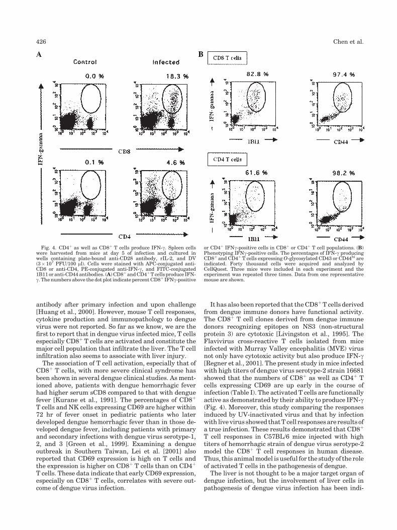
To investigate the effector function of activated Tcells, spleen cells collected at different time points ofinfection were cultured and the supernatants wereassayed for IFN-g. Not only spleen cells from infectedmice produced IFN-g (Fig. 3) but the kinetics of IFN-gproduction also followed that of T cell activation (Fig. 2).IFN-g production (19.3� 6.9 ng/ml) peaked at day 5 ofinfection,when18.3%of splenicCD8þTcells and4.6%ofCD4þ T cells were IFN-g producers (Fig. 4A). Incontrast, spleen cells from mice inoculated with UV-inactivated dengue virus and those with mock super-natants produced <1/10 levels of IFN-g at 5 days afterinoculation (Fig. 3). Interestingly, of the total IFN-gproducing cells, about 76% were CD8þ T cells and 24%CD4þ T cells, demonstrating that the majority offunctionally active cells wereCD8þT cells. Phenotypingshowed 82.8% of CD8þ-IFN-gþ T cells and 61.6% ofCD4þ-IFN-gþ T cells expressed O-glycosylated CD43and >97% of all IFN-gþ T cells were CD44hi (Fig. 4B).These results demonstrate that most activated CD8þ
T cells expressing O-glygan and CD44hi were function-ally active in mice infected by dengue virus.
Hepatic Infiltration in Mice InfectedWith Dengue Virus
In examining liver sections, cellular infiltration wasobserved inmice at day 5 after infection (Fig. 5B). H&Estain revealed that the infiltrating cells were mostlylymphoid cells. Immunohistochemical staining reveal-ed that the infiltrating cells constituted of both CD8þ
and CD4þ T cells (Fig. 6B,E). The distribution of CD4þ
andCD8þTcells followeddifferent patterns.Whilemostof theCD4þT cells clustered around the portal veinwithsome scattered in the parenchyma, most of CD8þ T cellswere scattered in the hepatic acinus (personal observa-tion). Flow cytometric analysis of hepatic infiltratingT cells showed that of total infiltrating T cells isolated,about 62.5% were CD8þ T cells and 37.5% CD4þ T cells(Fig. 7B), contrast to acute hepatitis induced by Con Awhere themajority of infiltrating cellswereCD4þTcells(71.7%) (Fig. 7A). Moreover, 69.9% of infiltrating CD8þ
Tcellswere ofCD44hi phenotype (Fig. 7D). These resultsare consistent with the observation in Figure 4A that
most of the functionally active T cells in the spleen wereCD8þ T cells.
Liver Injury
Serum ALT and AST levels were then assayed todetermine whether T cell infiltration was associatedwith liver damage. The results in Table II show thatliver enzyme levels in dengue virus-infected mice weresignificantly higher than control mice (days 3 and 5 forALT and days 3, 5, 7 for AST), although UV-inactivateddengue virus also induced a transient elevation of ALTand AST at day 3 after inoculation. In situ detection ofDNA fragmentation by TUNEL assay shows that livercells are apoptotic (Fig. 8), similar to what is reported ininfected humans [Couvelard et al., 1999]. Interestingly,the kinetics and peak of liver enzyme levels correlatedwith that of activated T cells (Fig. 2) suggesting thatactivated T cells were associated with liver damage.Moreover, the liver enzyme levels also correlated withthe peak of IFN-g production (Fig. 3). The degree ofcellular infiltration also positively correlated with thelevel of IFN-g produced (Table III).
Second Inoculation of Dengue Virus InducesGreater Liver Damage
In a separate experiment, mice were given a secondinoculum of dengue virus at the time when activatedT cell numbers and specific IgM levels declined and be-fore specific IgG rose. The AST levels in these miceincreased rapidly. At the third day after second inocula-tion,AST levelswere1.7 times that ofmice givenasingledose of the virus. Interestingly, a second virus inocula-tion resulted in more pronounced cellular infiltration(Figs. 5C and 6C,F). The infiltrating CD8þ/CD4þ T cellratio was greater in mice receiving a second dose ofvirus (5.8/1) than in those given one inoculation (3/2)(Fig. 7B,C). Moreover, although in mice receiving onlyone dengue virus inoculation only 69.9% of CD8þ T cellsexpressedCD44hi phenotype,>95%of infiltratingCD8þ
Tcells inmice receiving seconddengue virus inoculationexpressed CD44hi phenotype (Fig. 7E), illustratingfurther the association of activated CD8þ T cells andliver injury in dengue.
TABLE I. T Cells Express CD69 After Infection
Cell typea
Cell number (�106)
Control
Days after infection
3 5 7
CD8þ T cells 0.4� 0.1 1.4� 0.3** (3.5)b 1.1� 0.8** (2.8) 1.0� 0.4** (2.5)CD4þ T cells 0.8� 0.4 2.7� 0.7** (3.4) 2.8� 1.7** (3.4) 1.6� 0.9* (2.0)
aSpleen cells were collected from control mice andmice infected with dengue virus serotype-2 virus at days3, 5, and7after infection.Thenumber of lymphocytes expressingCD69 (�106)was calculated fromthe totalnumber ofCD4þ orCD8þTcells and thepercent of cells expressingCD69. Six toninemicewere included foreach time point.bThe mean number of cells expressing CD69 in infected mice divided by that in control mice.*P< 0.05, comparing the number of CD69þ cells in infected mice with that in uninfected mice.**P< 0.005, comparing the number of CD69þ cells in infected mice with that in uninfected mice.
424 Chen et al.

DISCUSSION
Both immune enhancement and viral virulencehypotheses recognize the importance of achieving highviral titer in the cause of severe dengue disease[Halstead, 1970, 1980, 1988; Rosen, 1977a; Gubleret al., 1978; Gubler, 1988]. The antibody enhancementhypothesis attempts to explain the observation ofindividuals experiencing secondary infection with aheterologous dengue serotype had a significantly higher
risk of developing dengue hemorrhagic fever/dengueshock syndrome [Halstead, 1970, 1980, 1988; Sangka-wibha et al., 1984; Burke et al., 1988; Kliks et al., 1989].It was shown in vitro that in the presence of low titers ofheterologous antibody, dengue virus replicates tohighertiters in human monocytes [Kliks et al., 1989]. Thishypothesis emphasizes the importance of heterologousantibody in enhancing the replication of the secondvirusin antigen-presenting cells. The enhancement results inprofound activation of preexisting cross-reactive cyto-toxic T cells and release of cytokines and cellular medi-ators, thus leading to subsequent immunopathologicalprocesses and dengue hemorrhagic fever/dengue shocksyndrome [Innis, 1995; Gubler, 1998; Rothman andEnnis, 1999]. However, the link between T cell activa-tion and pathology hasnever been established. The viralvirulence hypothesis was based on the observation thatsome dengue viruses have greater epidemic potentialthan others [Rosen, 1977b; Gubler et al., 1978]. These‘virulent’ virus strains have the advantage to replicatefaster and to higher levels, and as a result cause higherlevels of viremia. To study immunopathogenesis ofdengue in immunocompetent mice, virus virulence andtiter were taken into considerations. We establishedsuccessfully this mouse model by intravenous injectionof high titers of the hemorrhagic strain dengue virusserotype-2 16681 [Russell et al., 1967]. The infectedmicebecame viremic and the virus disseminated. Moreover,the infection induced T cell activation.
Immunocompetent mice have not been used widely tostudy T cell responses to dengue virus. A/J miceinoculated intravenousely with dengue virus developedtransient thrombocytopenia and produced anti-platelet
Fig. 3. IFN-gproduction peaks at day 5 of infection. Spleen cellswereharvested from same groups of mice at the same time points asdescribed in legendofFigure 2.ViruswereUV-inactivatedandadded tothe wells containing 5� 105 spleen cells. Culture supernatants werecollected after 48 hr of incubation. IFN-g concentration in the super-natants was determined by ELISA. Six to ten mice were used at eachtime point. *, ** indicate P< 0.05 and P< 0.005, respectively. TheP value was calculated from comparing the IFN-g concentration in thespleen cell culture supernatants from mock supernatant, UV-inacti-vated virus, or live virus infected mice with that in supernatants fromuninfected control mice.
Fig. 2. T cell expression ofO-glycosylated CD43 peaks at day 5 afterdengue virus infection. Splenocytes were harvested fromdengue virus-infectedmice at days 1, 3, 5, and 7after infection. Spleen cells frommicereceiving PBC (control), C6/36 culture supernatant (mock), and UV-inactivated dengue virus (UV-DV) were harvested at day 5 afterinoculation. Cells were stained with PE-conjugated anti-CD8 or PE-conjugated anti-CD4 and FITC-conjugated 1B11 antibodies. Thenumber of CD8þ (A) and CD4þ (B) T cells expressing O-glycan in thecourse of infection was calculated from the total number of T cells andthe percentage of cells stained positive by 1B11 monoclonal antibody.Each symbol represents one mouse. The horizontal lines indicate themean value. *,** indicate P< 0.05 and P< 0.005, respectively,comparing the mean number of cells from infected mice at each timepoint after infection with that from control mice without infection.
Hepatic Lymphocyte Infiltration in Dengue 425
antibody after primary infection and upon challenge[Huang et al., 2000]. However, mouse T cell responses,cytokine production and immunopathology to denguevirus were not reported. So far as we know, we are thefirst to report that in dengue virus infected mice, T cellsespecially CD8þ T cells are activated and constitute themajor cell population that infiltrate the liver. The T cellinfiltration also seems to associate with liver injury.The association of T cell activation, especially that of
CD8þ T cells, with more severe clinical syndrome hasbeen shown in several dengue clinical studies. As ment-ioned above, patients with dengue hemorrhagic feverhad higher serum sCD8 compared to that with denguefever [Kurane et al., 1991]. The percentages of CD8þ
T cells and NK cells expressing CD69 are higher within72 hr of fever onset in pediatric patients who laterdeveloped dengue hemorrhagic fever than in those de-veloped dengue fever, including patients with primaryand secondary infections with dengue virus serotype-1,2, and 3 [Green et al., 1999]. Examining a dengueoutbreak in Southern Taiwan, Lei et al. [2001] alsoreported that CD69 expression is high on T cells andthe expression is higher on CD8þ T cells than on CD4þ
T cells. These data indicate that early CD69 expression,especially on CD8þ T cells, correlates with severe out-come of dengue virus infection.
It has also been reported that theCD8þT cells derivedfrom dengue immune donors have functional activity.The CD8þ T cell clones derived from dengue immunedonors recognizing epitopes on NS3 (non-structuralprotein 3) are cytotoxic [Livingston et al., 1995]. TheFlavivirus cross-reactive T cells isolated from miceinfected with Murray Valley encephalitis (MVE) virusnot only have cytotoxic activity but also produce IFN-g[Regner et al., 2001]. The present study in mice infectedwith high titers of dengue virus serotype-2 strain 16681showed that the numbers of CD8þ as well as CD4þ Tcells expressing CD69 are up early in the course ofinfection (Table I). The activated T cells are functionallyactive as demonstrated by their ability to produce IFN-g(Fig. 4). Moreover, this study comparing the responsesinduced by UV-inactivated virus and that by infectionwith livevirus showed thatT cell responsesare results ofa true infection. These results demonstrated that CD8þ
T cell responses in C57BL/6 mice injected with hightiters of hemorrhagic strain of dengue virus serotype-2model the CD8þ T cell responses in human disease.Thus, this animalmodel is useful for the study of the roleof activated T cells in the pathogenesis of dengue.
The liver is not thought to be a major target organ ofdengue infection, but the involvement of liver cells inpathogenesis of dengue virus infection has been indi-
Fig. 4. CD4þ as well as CD8þ T cells produce IFN-g. Spleen cellswere harvested from mice at day 5 of infection and cultured inwells containing plate-bound anti-CD28 antibody, rIL-2, and DV(3� 107 PFU/100 ml). Cells were stained with APC-conjugated anti-CD8 or anti-CD4, PE-conjugated anti-IFN-g, and FITC-conjugated1B11 or anti-CD44 antibodies. (A) CD8þ andCD4þT cells produce IFN-g. The numbers above the dot plot indicate percent CD8þ IFNg-positive
or CD4þ IFNg-positive cells in CD8þ or CD4þ T cell populations. (B)Phenotyping IFNg-positive cells. The percentages of IFN-g producingCD8þ and CD4þ T cells expressingO-glycosylated CD43 or CD44hi areindicated. Forty thousand cells were acquired and analyzed byCellQuest. Three mice were included in each experiment and theexperiment was repeated three times. Data from one representativemouse are shown.
426 Chen et al.

cated by clinical signs of hepatomegaly, abnormal liverfunction, pathological findings, and detection of viralantigen [Bhamarapravati et al., 1967; Rosen et al., 1989;Hall et al., 1991; Lanciotti et al., 1992; Kangwanponget al., 1995; Couvelard et al., 1999; Huerre et al., 2001].In examining fatal dengue hemorrhagic fever cases,liver is the tissue most often the virus is isolated [Rosenet al., 1989; Innis, 1995; Bhamarapravati, 1997; Huerreet al., 2001]. Dengue patients were reported to haveelevatedAST levels andASTelevation isassociatedwithhemorrhage [Kalayanarooj et al., 1997; Monath et al.,2000]. In dengue virus induced hepatitis, the level ofAST is higher than ALT with a ratio of around 1–1.5 while other types of virally induced hepatitis havehigher ALT levels than AST [Kuo et al., 1992; Nguyenet al., 1997; Lei et al., 2001]. Serum liver enzyme ASTlevel is higher in dengue hemorrhagic fever patientsthan in dengue fever patients [Kalayanarooj et al., 1997;Monath et al., 2000;Wahid et al., 2000]. It has also beenreported that dengue virus induces HepG2 apoptosisand production of RANTES [Lin et al., 2000a,b].Together, these reports indicate that dengue virusinfection can cause liver pathology. The mechanism ofliver damage has never been examined.
Examining liver pathology of 100 fatal pediatric (age5 months to 14 years) dengue hemorrhagic fever cases,Bhamarapravati et al. [1967] reported that cellular
infiltration was noted in 64 cases. Megakaryocytes,lymphocytoid cells, and rarely neutrophils wereobserved in sinusoids. Cellular infiltration in the portalareas was composed of young lymphocytes, plasmacy-toid cells and some histiocytes [Bhamarapravati et al.,1967]. A recent liver histopathological report of five fatalpediatric (age 10–72 months) cases revealed that liverhistopathology was detected in four out of five post-mortem examinations [Huerre et al., 2001], but little orno inflammatory response was found in four cases andmoderate infiltration, mainly in the periportal area inonly one case [Huerre et al., 2001]. Therefore, it appearsthat liverhistopathology is common inpatientswhodiedof dengue hemorrhagic fever but it remains to bedetermined virus infecting liver cells, lymphocyte infil-tration or both is the cause of liver pathology.
In this study, mice infected with dengue virus hadsignificantly higher liver enzyme levels (days 3, 5, and 7,Table II) when the numbers of activated T cells werehigh (days 3, 5, and 7, Fig. 2). Analysis of infiltratingcells showed that they are activated lymphocytes andthe majority are CD8þ T cells (Fig. 7B,D). In situdetection of DNA fragmentation by TUNEL assayshowed that liver cells were apoptotic (Fig. 8), similartowhat is reported in infected humans [Couvelard et al.,1999]. These data strongly indicate that liver injury indengue virus infection is associated with activated
Fig. 5. Dengue virus infection results in hepatic cellular infiltration. Liver tissues were removed fromcontrol mouse (A), mouse at 5 days after receiving one inoculation of virus (B) and mouse at 3 days afterreceiving a second inoculation of virus (C). Tissues were fixed in 10% buffered formaldehyde and stainedwith hematoxylin and eosin. The magnification was 200�.
Hepatic Lymphocyte Infiltration in Dengue 427

lymphocyte, especially CD8þ T cell infiltration. How-ever, these results do not rule out the contribution ofdirect viral damage to the liver.Dengue virus infection induced specific antibody
response. Specific IgM became significantly higher atday 3 and peaked at day 5 of infection while specific IgGwas detectable at day 5–7 and peaked at day 14(unpublished data). The infiltration in liver was morepronounced in mice receiving a second dose of denguevirus (Figs. 5C and 6C,F) at day 7 when T cells andspecific IgM declined and before specific IgG elevated.The liver enzyme AST levels in these mice are muchhigher than in those receiving only one injection. Theratio of infiltrating CD8þ to CD4þ T cells was 3/2 in
mice receiving single inoculation and became 5.8/1 inmice receiving two viral inoculations (Fig. 7B,C), show-ing that CD8þ T cells greatly expanded upon secondinoculation. This correlation strengthened further theassociation between liver injury and CD8þ T cell infil-tration. It is worth noting that same observation wasrepeated in mice receiving lower dose of inoculum(1.5�107 PFU), indicating the phenomenon is notcaused by high viral titer per se. Although it has notbeen documented that patients who later developdengue hemorrhagic fever have been inoculated withdengue virus by multiple mosquito bites at differenttimes within a short time frame, the chances of a personliving in an endemic area being bitten by infected
Fig. 6. The cellular infiltrates in the liver consist of CD8þ T and CD4þ T cells. Liver tissues were fromcontrol mouse (A, D), mouse at 5 days after receiving one inoculation of virus (B, E) and mouse at 3 daysafter receiving a second inoculation of virus (C,F). Cryosectioned tissues were stainedwith rat anti-mouseCD8 (A,B,C) or anti-CD4 antibody (D,E,F) followed by peroxidase-conjugated goat anti-rat IgGantibody.DAB was used as substrate for color development. Arrows point at positive cells. The magnification was200�.
428 Chen et al.

mosquitoes more than once are high. This model predi-cts that upon receiving a second dose of virus, the rapidexpansion of CD8þ T cells can very well result ininfiltration to the liver. Experiments are underway toinvestigate the direct causal relationship betweenCD8þ
T cell infiltration and liver injury.
Clinical studies have shown that serum IFN-g levelsare high in dengue hemorrhagic fever patients [Kuraneet al., 1989, 1991]. This present study showed that IFN-glevels are highat thepeak of T cell activation (Fig. 3) andthe extent of cellular infiltration correlates with theamounts of IFN-g secreted (Table III). However, it
TABLE II. Liver Enzyme Levels Increase in Mouse Sera After Infection
Liver enzymea (U/L) Inoculumb Controlc
Days after 1st inoculationdDays after 2ndinoculation
3 5 7 3
ALT Live DV 25.7� 10.4 38.3� 16.2* 36.7� 15.6** 30.5� 5.5 33.0� 4.9*UV-DV 37.2� 4.1* 36.0� 18.3 27.0� 7.1Mock 30.4� 1.7 29.3� 9.9 32.0� 2.8
AST Live DV 43.3� 5.4 67.1� 28.0* 94.8� 32.3** 82.2� 17.1** 164.3� 75.2**UV-DV 64.8� 5.2** 58.0� 10.0 63.0� 7.1Mock 48.4� 9.2 35.3� 19.7 42.0� 5.7
aALT and AST levels were determined by Abbott Alcyon 300i. The enzyme levels are presented as units per liter (U/L).bMice were inoculated with live dengue virus (Live DV), UV-inactivated virus (UV-DV), or mock supernatant (mock).cControls were normal mice without inoculation.dSera were collected frommice at days 3, 5, and 7 after one inoculation and day 3 after second inoculation. Five to six mice were included in eachexperiment.*P< 0.05, comparing the level of ALT orAST of live dengue virus,UV-inactivated dengue virus, ormock culture supernatant inoculatedmicewiththat of control uninfected mice.**P< 0.005, comparing the level of ALT or AST of live dengue virus, UV-inactivated dengue virus, or mock culture supernatant inoculated micewith that of control uninfected mice.
Fig. 7. Activated CD8þ T cells constitute the major T cell populationinfiltrating the liver in DV-infected mice. Liver tissues were removedfrom mouse at 16 hr after Con A injection (A), mouse at 5 days afterreceiving one inoculation of virus (B, D) and mouse at 3 days afterreceiving a second inoculation of virus (C, E). Single cell suspensionwas prepared and mononuclear cells were separated on Ficoll-Paque
gradient. Themononuclear cellswere stainedwithPE-conjugated anti-CD8 and APC-conjugated anti-CD4 antibodies (A, B, C) or PE-conjugated anti-CD8 and FITC-conjugated anti-CD44 antibodies (D,E). The numbers above the dot plots indicate the percentage of CD4þ orCD8þT cells in the total mononuclear cells isolated (A,B,C) or CD8þTcells expressing CD44hi phenotype (D, E).
Hepatic Lymphocyte Infiltration in Dengue 429

remains to be investigated whether high IFN-g level isan indication of T cell activation or directly related toliver injury.In summary, by injecting high titers of dengue virus
serotype-2 hemorrhagic strain to immunocompetentC57BL/6 mice, a mouse model to study T cell activationis developed. As Rothman et al. pointed out that variousimmune mechanism(s) is important to different stagesof dengue infection [Rothman and Ennis, 1999], theresults of this study revealed the possibility that liver
injury in dengue is related to CD8þ T cell activation andinfiltration.
ACKNOWLEDGMENTS
H.-C. Chen and S.-Y. Lai contributed equally to thismanuscript. We are grateful to the excellent technicalassistance provided by Miss M.-Y. Liao.
REFERENCES
An J, Kimura-Kuroda J, Hirabayashi Y, Yasui K. 1999. Developmentof a novel mouse model for dengue virus infection. Virology 263:70–77.
Bhamarapravati N. 1997. Pathology of dengue infections. Oxon, UK:Cab International, Inc. p 115.
Bhamarapravati N, Tuchinda P, Boonypaknavik V. 1967. Pathology ofThailand haemorrhagic fever: A study of 100 autopsy cases. AnnTrop Med Parasitol 61:500–510.
BurkeDS,NisalakA,JohnsonD,ScottRM.1988.Aprospective studyofdengue infections in Bangkok. Am J Trop Med Hyg 38:172–180.
Couvelard A, Marianneau P, Bedel C, Drouet MT, Vachon F, Henin D,Deubel V. 1999. Report of a fatal case of dengue infection withhepatitis: Demonstration of dengue antigens in hepatocytes andliver apoptosis. Human Pathol 30:1106–1110.
Deubel V, Kinney RM, Trent DW. 1986. Nucleotide sequence anddeduced amino sequence of the structural proteins of dengue type 2virus, Jamaica genotype. Virology 155:365–377.
Falgout B, BrayM, Schlesinger JJ, Lai CJ. 1990. Immunization ofmicewith recombinant vaccine virus expressing authentic dengue virusnonstructural protein NS1 protects against lethal dengue virusencephalitis. J Virol 64:4356–4363.
Green S, Pichyanghul S, Vaughn DW, Kalayanarooj S, NimmannityaS, Nisalak A, Kurane I, Rothman AL, Ennis FA. 1999. Early CD69expression on peripheral blood lymphocytes from children withdengue hemorrhagic fever. J Infect Dis 180:1429–1435.
Gubler DJ. 1988. Dengue. In: Monath TP, editor. Epidemiology ofarthropod-borne viral disease. Boca Raton, FL: CRP Press, Inc.pp 233–260.
Gubler DJ. 1998. Dengue and dengue hemorrhagic fever. ClinMicrobiol Rev 11:480–496.
GublerDJ,ReedD,RosenL,HitchockJC. 1978.Epidemiologic, clinical,andvirologic observations ondengue in theKingdomofTonga.AmJTrop Med Hyg 27:581–589.
Guzman MG, Kouri G. 2002. Dengue: And update. Lancet Infect Dis1:33–42.
Hall WC, Crowell TP, Watts DM, Barros VL, Kruger H, Pinheiro F,Peters CJ. 1991. Demonstration of yellow fever and dengueantigens in formalin-fixed paraffin-embedded human liver byimmunohistochemical analysis. Am J Trop Med Hyg 45:408–417.
TABLE III. Correlation Between IFN-g Productionand Cellular Infiltration in the Liver
Experiment Infiltration scoresaIFN-g produced byspleen cells (ng/ml)b
I þþ 21.0þ 12.4� 2.8c
II þþ 19.6þ 7.2� 3.2c
aScoring of cellular infiltration.þþ, 10–30 cells per focus;þ, lower than10 cells per focus; �, no significant infiltration.bSpleen cellswere harvested frommice at 5 days after virus inoculationand stimulatedwith inactivated virus in vitro as described inmaterialsand methods. Supernatants were collected at 24 hr of incubation andIFN-g concentration was determined by ELISA.cSpleen cells were collected from mice inoculated with inactivatedvirus.
Fig. 8. Liver cell undergo apoptosis after dengue virus infection.DNA fragmentation was detected by in situ TUNEL staining. DNase Itreated tissue was used as positive control (A). Uninfected normal (B)and dengue virus-infected (C) mouse liver sections were treated withFITC-conjugated TdT-mediated dUTP nick end labeling mixture andperoxidase-conjugated anti-FITCantibody.DABwasused as substrateand methyl green as counterstain.
430 Chen et al.

Halstead SB. 1970. Observations related to pathogenesis of denguehemorrhagic fever. VI. Hypotheses and discussion. Yale J Biol Med42:350–362.
Halstead SB. 1980. Dengue hemorrhagic fever-public health problemand a field for research. Bull WHO 58:1–21.
Halstead SB. 1988. Pathogenesis of dengue: Challenges to molecularbiology. Science 239:476–481.
Harrigton LE, Galvan M, Baurn LG, Altman JD, Ahmed R. 2000.Differentiating betweenmemory and effectorCD8T cells by alteredexpression of cell surface O-glycan. J Exp Med 191:1241–1246.
Huang KJ, Chen SC, Liu HS, Lin YS, Yeh TM, Liu CC, Lei HY. 2000.Manifestation of thrombocytopenia in dengue-2-virus-infectedmice. J Gen Virol 8:2177–2182.
Huerre MR, Lan NT, Marianneau P, Hue NB, Khun H, Hung NT,Khen NT, Drouet MT, Huong VTQ, Ha DQ, Buisson Y, Deubel V.2001. Liver histopathology and biological correlates in five casesof fatal dengue fever in Vietnamese children. Virchows Arch 438:107–115.
Innis BL. 1995. Dengue and dengue hemorrhagic fever. London, UK:Champman Hall, Inc. p 103.
Johnson AK, Roehrig JT. 1999. New mouse model for dengue virusvaccine testing. J Virol 73:783–786.
Kalayanarooj S, Vaughn DW, Nimmannitya S, Green S, SuntayakornS, Kunentrasai N, Vitamitrachai W, Ratanachueke S, Kiapoloj S,Innis BL, Rothman AL, Nisalak A, Ennis FA. 1997. Early clinicaland laboratory indicators of acute dengue illness. J Infect Dis 176:313–321.
Kangwanpong D, Bhamarapravati N, Lucia HL. 1995. Diagnosingdengue virus infection in archived autopsy tissues by means of thein situ PCR method: A case report. Clin Diagn Virol 3:165–172.
Kliks SC, Nisalak A, Bradndt WE, Wahl L, Burke DS. 1989. Antibody-dependent enhancement of dengue virus growth in human mono-cytes as a risk for dengue hemorrhagic fever. Am J Trop Med Hyg40:444–451.
Kuo CH, Tai DI, Chang-Chien CS, Lan CK, Chiou SS, Liaw YF. 1992.Liver biochemical tests and dengue fever. Am J Trop Med Hyg47:265–270.
Kurane I, Innis BL, Nisalak A, Hoke C, Nimmeannitya S, Meager A,Ennis FA. 1989. Human T cell responses to dengue virus antigens.proliferative responses and interferon gamma production. J ClinInvest 83:506–513.
Kurane I, Innis BL, Nimmeannitya S, Nisalak A, Meager A, Janus J,Ennis FA. 1991. Activation of T lymphocytes in dengue virusinfections. High levels of soluble interleukin 2 receptor, solubleCD4, solubleCD8, interleukin 2, and interferon-g in sera of childrenwith dengue. J Clin Invest 88:1473–1480.
Lanciotti RS, Calisher CH,GublerDJ, ChanfmGJ,VorndamAV. 1992.Rapid detection and typing of dengue viruses from clinical samplesby using reverse transcriptase-polymerase chain reaction. J ClinMicrobiol 30:545–551.
Lei HY, Yeh TM, Liu HS, Lin YS, Chen SH, Liu CC. 2001.Immunopathogenesis of dengue virus infection. J Biomed Sci 8:377–388.
Lin YL, Liao CL, Chen LK, Yeh CT, Liu CI, Ma SH, Huang YY, HuangYL,KaoCL,KingCC. 1998. Study of dengue virus infection inSCIDmice engrafted with human K562 cells. J Virol 72:9729–9737.
Lin YL, Liu CC, Lei HY, Yeh TM, Lin YS, Chen RMY, Liu HS. 2000a.Infection of five human liver cell lines by dengue-2 virus. J MedVirol 60:425–431.
Lin YL, Liu CC, Chung JI, Lei HY, Yeh TM, Lin YS, Hung YH, Liu HS.2000a. Involvement of oxidative stress, NF-IL-6 and RANTES
expression in dengue-2 virus infected human liver cells. Virology276:114–126.
Livingston PG,Kurane I,Dai LC,Okamato Y, Lai CJ,MenR,Karaki S,Takiguchi M, Ennis FA. 1995. Dengue virus-specific, HLA-B35-restricted, human CD8þ cytotoxic T lymphocyte (CTL) clones.Recognition of NS3 amino acids 500 to 508 by CTL clones of twodifferent serotype specificities. J Immunol 154:1287–1295.
Monath TP. 1994. Dengue: The risk to developed and developingcountries. Proc Natl Acad Sci USA 91:2395–2400.
Monath B, Patwari AK, Anand VK. 2000. Hepatic dysfunction inchildhood dengue infection. J Trop Pediatr 46:40–43.
Nguyen TL, Nguyen TH, Tieu NT. 1997. The impact of denguehaemorrhagic fever on liver function. Res Virol 148:273–277.
Regner M, Lobigs M, Blanden R, Milburn P, Mullbacher A. 2001.Antiviral cytotoxic T cells cross-reactively recognize disparatepeptide determinants from related viruses but ignore more similarself- and foreign-determinants. J Immunol 166:3820–3828.
Rosen L. 1977a. The Emperor’s new clothes revisited, or reflections onthe pathogenesis of dengue hemorrhagic fever. Am JTropMedHyg26:337–343.
Rosen L. 1977b. Dengue: An overview. Sydney, Australia: AcademicPress, Inc. pp 484–493.
Rosen L, Khin MM, Tin U. 1989. Recovery of virus from the liver ofchildren with fatal dengue. Reflections on the pathogenesis of thedisease and its possible analogy with that of yellow fever. Res Virol140:351–360.
Rothman AL, Ennis FA. 1999. Immunopathogenesis of denguehemorrhagic fever. Virology 257:1–6.
Russell PK, Udomsakdi S, Halstead SB. 1967. Antibody response indengue and dengue hemorrhagic fever. Jpn JMed Sci Biol 20:103–108.
Sangkawibha N, Rojanasuphot S, Ahandrink S, Viriyapongse S,Jatanasen S, Salitul V, Phanthumachinda B, Halstead SB. 1984.Risk factors indengue shock syndrome:Aprospective epidemiologicstudy in Rayong, Thailand. Am J Epidemiol 120:653–669.
Schepers K, Toebes M, Sotthewes G, Vyth-Dreese FA, Dellemijn TAM,Melief CJM, Ossendorp F, Schumacher TNM. 2002. Differentialkinetics of antigen-specific CD4þ and CD8þ T cell response in theregression of retrovirus-induced sarcomas. J Immunol 169:3191–3199.
Testi R, Phillips JH, Lanier LL. 1989. T cell activation via Leu-23(CD69). J Immunol 143:1123–1128.
Wahid SF, Sanusi S, Zawawi MM, Ali RA. 2000. A comparison of thepattern of liver involvement in dengue hemorrhagic fever withclassic dengue fever. South Asian J Trop Med Public Health 31:259–263.
Wang WK, Lee CN, Kao CL, Lin YL, King CC. 2000. Quantitativecompetitive reverse transcription-PCR for quantification of denguevirus RNA. J Clin Microbiol 38:3306–3310.
Wang WK, Shung TL, Tsai YC, Kao CL, Chang SM, King CC. 2002.Detection of dengue virus replication in peripheral blood mono-nuclear cells fromdenguevirus type 2-infected patients bya reversetranscription-real-time PCR assay. J ClinMicrobiol 40:4472–4478.
WuSJL,HayesCG,DuboisDR,WindheuserMG,KangYH,WattsDM,Sieckmann DG. 1995. Evaluation of the severe combined immuno-deficient (SCID) mice as a animal model for dengue viral infection.Am J Trop Med Hyg 52:468–476.
Zivna I, Green S, Vaughn DW, Kalayanarooj S, Stephens HAF,Chandananyingyong D, Nisalak A, Ennis FA, Rothman AL. 2002.T cell responses to an HLA-B*07-restricted epitope on the dengueNS3 protein correlate with disease severity. J Immunol 168:5959–5965.
Hepatic Lymphocyte Infiltration in Dengue 431