lumbar radiculopathy post lumbar discectomy: a case study
TRANSCRIPT
University of North DakotaUND Scholarly Commons
Physical Therapy Scholarly Projects Department of Physical Therapy
2016
Lumbar Radiculopathy Post Lumbar Discectomy:A Case StudyAmy HarmonUniversity of North Dakota
Follow this and additional works at: https://commons.und.edu/pt-grad
Part of the Physical Therapy Commons
This Scholarly Project is brought to you for free and open access by the Department of Physical Therapy at UND Scholarly Commons. It has beenaccepted for inclusion in Physical Therapy Scholarly Projects by an authorized administrator of UND Scholarly Commons. For more information,please contact [email protected].
Recommended CitationHarmon, Amy, "Lumbar Radiculopathy Post Lumbar Discectomy: A Case Study" (2016). Physical Therapy Scholarly Projects. 556.https://commons.und.edu/pt-grad/556
Lumbar Radiculopathy Post Lumbar Discectomy: A Case Study
by
Amy Harmon Bachelor of Science in Kinesiology and Health Science
University of Wyoming, 2012
A Scholarly Project Submitted to the Graduate Faculty of the
Department of Physical Therapy
School of Medicine and Health Sciences
University of North Dakota
in partial fulfillment of the requirements for the degree of
Doctor of Physical Therapy
Grand Forks, North Dakota May, 2016
This Scholarly Project, submitted by Amy Harmon in partial fulfillment of the requirements for the Degree of Doctor of Physical Therapy from the University of North Dakota, has been read by the Advisor and Chairperson of Physical Therapy under whom the work has been done and is hereby approved,
£~4~ (Chairperson, Physical erapy)
ii
Title
Department
Degree
PERMISSION
Lumbar Radiculopalhy Post Lumbar Discectomy: A Case Study
Physical Therapy
Doctor of Physical Therapy
In presenting this Scholarly Project in partial fulfillment of the requirements for a graduate degree from the University of North Dakota, I agree that the Department of Physical Therapy shall make it freely available for inspection. I further agree that permission for extensive copying for scholarly purposes may be granted by the professor who supervised my work or, in her absence, by the Chairperson of the department. It is understood that any copying or publication or other use of this Scholarly Project or part thereof for financial gain shall not be allowed without my written permission. II is also understood that due recognition shall be given to me and the University of North Dakota in any scholarly use which may be made of any material in this Scholarly Project.
Signature
Date
iii
TABLE OF CONTENTS
LIST OF FIGURES ......... "" ........... "" .......... "" ......... "" ........................... v
LIST OF TABLES ......................................................................... vi
ACKNOWLEDGEMENTS ............................................................... vii
ABSTRACT .............................................................................. viii-ix
CHAPTER I.
II.
BACKGROUND AND PURPOS ..................... 10-11
CASE DESCRiPTION .................................. 12-13
Examination, Evaluation and Diagnosis ............ 13-21
Prognosis and Plan of Care ............................. 21-22
Intervention ................................................ 23-27
Outcomes ....................................................... 28
III. DISCUSSION ............................................... 29-32
Reflective Practice ............................................ 32
REFERENCES ........................................................................ 33-36
iv
LIST OF FIGURES 1. SlR Initial Test Position ........................................................... 16
2. SlR Ending Position of Positive Test... ........................................ 16
3. Initial Slump Test Position .......................................................... 17
4. Final Slump Test Position .......................................................... 18
5. Negative Trendelenburg Test Position .......................................... 19
6. Positive Trendelenburg Test Position .......................................... 19
7. Negative Thomas Hip Test Position ............................................ 20
8. Positive Thomas Test Position .................................................... 21
v
LIST OF TABLES
1. Special Tests to Rule In Or Out Lumbar Nerve Roots ...................... 15
2. Plan of Care ....................................................................... 23-24
vi
ACKNOWLEDGEMENTS
I would like to thank my clinical instructor for all of his support and
assistance though my clinical rotation and for teaching me new therapy skills to
use in this case study.
VII
ABSTRACT
Background and Purpose: Low back pain (LBP) is one of the most
common and costly musculoskeletal diagnosis in America. Approximately 50% of
individuals with LBP have recurrent episodes by 1 year, 60% by 2 years, and
70% by 5 years.1 Micro-lumbar discectomies are often performed to relieve the
radiculopathy and pain associated with nerve root entrapment. The purpose of
this case study was to describe the clinical decision process implemented for the
post-operative assessment and intervention of a patient with a L4-5, L5-S1
discectomy and to outline the associated outcomes.
Case Description: The patient, a 26 year-old male, underwent a L4-5 and
L5-S1 micro-discectomy 10 months prior to presenting to physical therapy with a
diagnosis of right L4-5, S1 radiculopathy. Co-morbidities included alcohol and
tobacco addiction. Prior to this episode of care, the patient attempted a multitude
of care options, with no systemic relief.
Plan of Care: Multiple interventions were utilized throughout the treatment
sessions due to low patient tolerance. The patient reported a decrease in pain,
an increase in time for onset of pain, increased work tolerance, and a
centralization of radicular symptoms.
Clinical Decision Making: Clinical decisions were based on current
evidence and the International Classification of Functioning, Health and Disease
viii
(ICF) model. Outcomes were evaluated through the use of visual analog pain
scale, location of radicular symptoms, and the Oswestry Disability Index.
Reflection on Practice: Using a combination of intervention resulted in
the best functional outcomes for this patient. The patient's substance abuse
contributed to the lack of program adherence.
ix
CHAPTER I
BACKGROUND AND PURPOSE
Low back pain (LBP) is one of the most common and costly
musculoskeletal diagnoses in the United States and the single leading cause of
disability worldwide. Back pain is one of the most frequent reasons that young
adults miss work and are prevented from engaging in recreational activities.3
Avoidance behavior is significantly motivated by anticipation of pain and fear
avoidance beliefs4 Approximately SO% of individuals with LBP have recurrent
episodes by 1 year, 60% by 2 years and 70% by S years.1
The patient in this case study underwent a L4-S and LS-S1 discectomy 10
months prior to presenting to physical therapy with a diagnosis of right L 1-3
radiculopathy. Micro lumbar discectomies are often performed to relieve the
radiculopathy and pain associated with nerve root entrapment.
The patient's co-morbidities included alcohol and tobacco addiction. Moon, Choi
and Kim5 found limiting supportive evidence in the causal relationship of smoking
and low back pain. Abstinence from smoking may be a positive primary
preventative measure, but the authors concluded that there was limited evidence
that smoking cessation is beneficial for previously established musculoskeletal
problemss
Despite the frequency with which lumbar disc herniation occurs, there is
considerable controversy regarding its pathophysiology and treatment. Currently,
10
multiple physical therapy strategies are utilized for the treatment and
management of chronic low back pain, including manipulation, postural
correction, core strengthening and stabilization, motor control exercises, aerobic
activities, strain counterstrain, muscle energy, electrotherapy, stretching, aquatic
therapy, and aerobic activity2,6,7,8,9,10,11,12 Each technique provides benefits and
desired outcomes, but the literature is unable to agree on the optimal and most
effective strategy for the management of chronic low back pain. However,
exercise is the most agreed upon recommendation for reducing chronic low back
pain. 2,6,7,8,8,10,11
The purpose of this case study was to describe the clinical decision
process implemented for post-operative assessment and intervention for a
patient with an L4-5 and L5-S1 discectomy and to outline the associated
outcomes.
11
CHAPTER II
CASE DESCRIPTION
The patient was a 26 year-old male with a long history of chronic back
pain. The initial injury occurred, four year ago, when he crashed on his bike. He
saw a chiropractor for 4 months, which slightly improved his radicular symptoms.
The patient, a full-time server, re-injured his back at work 6 months after initial
injury; The injury resulted in his inability to work an entire shift in the dining room
due to an increase in low back pain and radicular symptoms. He had stopped
participating in recreational activities, which include playing basketball, golf and
snowmobiling. The patient reported difficulty sleeping due to pain. Prior to this
episode of care, the patient had received chiropractic, acupuncture, massage
treatment, physical therapy, and cortisone injections with no systematic relief.
Subsequently, the patient underwent an L4-5 and L5-S1 micro discectomy.
At the onset of physical therapy, the patient's complaints were right lower
extremity (LE) radiculopathy in the L4-S1 nerve roots, hip pain, and tightness in
his gluteus and calf muscles. The patient lived at home with his grandmother and
must descend a flight of stairs with one railing to enter his bedroom. He had
never ambulated with an assistive device. He was not on any physician
prescribed medication but did report taking Aleve, as recommended, when pain
increased or in anticipation of pain with certain physical activities. The patient did
not report past abuse of physician-prescribed medications. He has a past history
12
of alcohol abuse and smoking (10 years). Before the initial injury occurred, the
patient did not report any restrictions in mobility, ADLs or recreational activities.
At time of initial evaluation the patient was unable to participate in any sort of
physical activity without pain and was unable to work a full shift without severe
pain. His goals were to return to his recreational activities without pain as well as
gain the ability to work a full shift. The patient discharged himself after 8 physical
therapy visits. Prior to his self-discharge, the patient attended 8 physical therapy
visits over a 5-week period; He missed 2 appointments.
Examination, Evaluation and Diagnosis
Examination: Observing the patient at the initial examination revealed
increased kyphosis, forward head, and decreased lordosis. The patient did not
exhibit a normal gait pattern but ambulated with a shuffle, including bilateral
decreased step length, no apparent heel strike or toe off on either foot, and
moved at a decreased speed. There was no rhythm in the arm swing with his
stride. A positive Trendelenburg sign was found bilaterally, indicating weakened
gluteus medius muscles. He had a decreased mass of his right gluteus max
compared to his left, which coincided with the radicular symptoms and the
positive Trendelenburg sign. The patient's gait also appeared 'stiff' with apparent
muscle guarding.
The patient demonstrated decreased range of motion (ROM) in bilateral
LEs. Strength levels were tested with manual muscle tests. A manual muscle
test (MMT) is applied to a limb or other body part after it has completed its full
ROM. Manual resistance (concentric force) is applied by the therapist in
13
opposition to the contracting muscle and is graded on a 0-5 scale with 0 being no
muscle activity and 5 described as normal or best strength. 13 This test examines
the group of muscles involved in the motion rather than individual muscles. 13
MMT revealed limitations on the right: 4/5 for right hip flexion secondary to pain,
and 4/5 for right knee extension and dorsiflexion (OF).
The patient's lumbar flexion and extension was measured at 25% of
normal active range of motion (AROM). The McKenzie measurement technique
was utilized to assess for lumbar AROM (40-60 degrees is normal).14 Passive
range of motion (PROM) was limited by about half of normal due to pain.
Patient's hip flexion was measured at 112 degrees of AROM and PROM was
within normal limits but was accompanied by pain. The patient appeared guarded
with PROM.
The following tests were utilized to rule in reported radiculopathy
stemming from the spinal nerve roots. Regarding sensory status, the sensory
distribution of each nerve root is called a dermatome. 15 A positive dermatome
occurs when the patient does not feel, or has diminished feeling in the area of
skin.15 A myotome is a group of muscles supplied by a single nerve root and are
assessed using an isometric muscle hold of at least 5 seconds. 15 A positive
myotome is when weakness is detected during the isometric hold. Positive
myotomes on the right with OF, great toe extension, PF and positive dermatomes
on the right medial ankle, dorsum of foot, lateral boarder of the foot were found.
These results match the L4-5, L5-S1 radicular symptoms supporting nerve root
involvement. The positive special tests were Straight Leg Raise (SLR), Slump,
14
Thomas and Trendelenburg were discovered upon initial evaluation. A positive
Thomas Test was later found on the second visit. The sensitivity and specificity
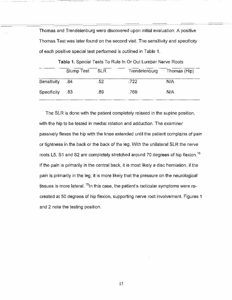
of each positive special test performed is outlined in Table 1.
Table 1. Special Tests To Rule In Or Out Lumbar Nerve Roots
Slump Test SLR Trendelenburg Thomas (Hip)
Sensitivity .84 .52 .722 N/A
Specificity .83 .89 .769 N/A
The SLR is done with the patient completely relaxed in the supine position,
with the hip to be tested in medial rotation and adduction. The examiner
passively flexes the hip with the knee extended until the patient complains of pain
or tightness in the back or the back of the leg. With the unilateral SLR the nerve
roots L5, S1 and S2 are completely stretched around 70 degrees of hip flexion. 15
If the pain is primarily in the central back, it is most likely a disc herniation, if the
pain is primarily in the leg, it is more likely that the pressure on the neurological
tissues is more lateral. 151n this case, the patient's radicular symptoms were re
created at 50 degrees of hip flexion, supporting nerve root involvement. Figures 1
and 2 note the testing position.
15
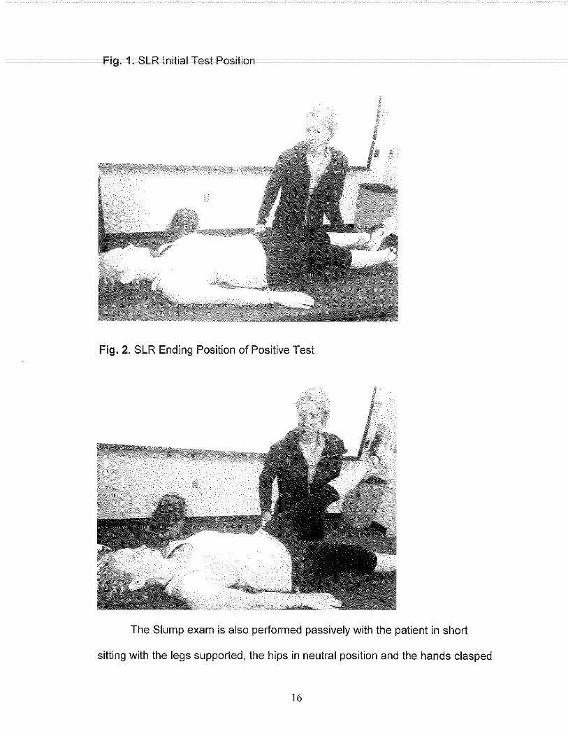
Fig. 2. SLR Ending Position of Positive Test
The Slump exam is also performed passively with the patient in short
sitting with the legs supported, the hips in neutral position and the hands clasped
16
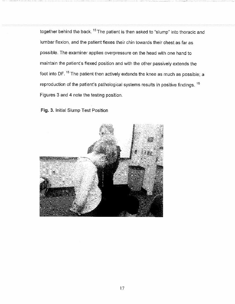
together behind the back. 15 The patient is then asked to "slump" into thoracic and
lumbar flexion, and the patient flexes their chin towards their chest as far as
possible. The examiner applies overpressure on the head with one hand to
maintain the patient's flexed position and with the other passively extends the
foot into OF. 15 The patient then actively extends the knee as much as possible; a
reproduction of the patient's pathological systems results in positive findings. 15
Figures 3 and 4 note the testing position.
Fig. 3. Initial Slump Test Position
17
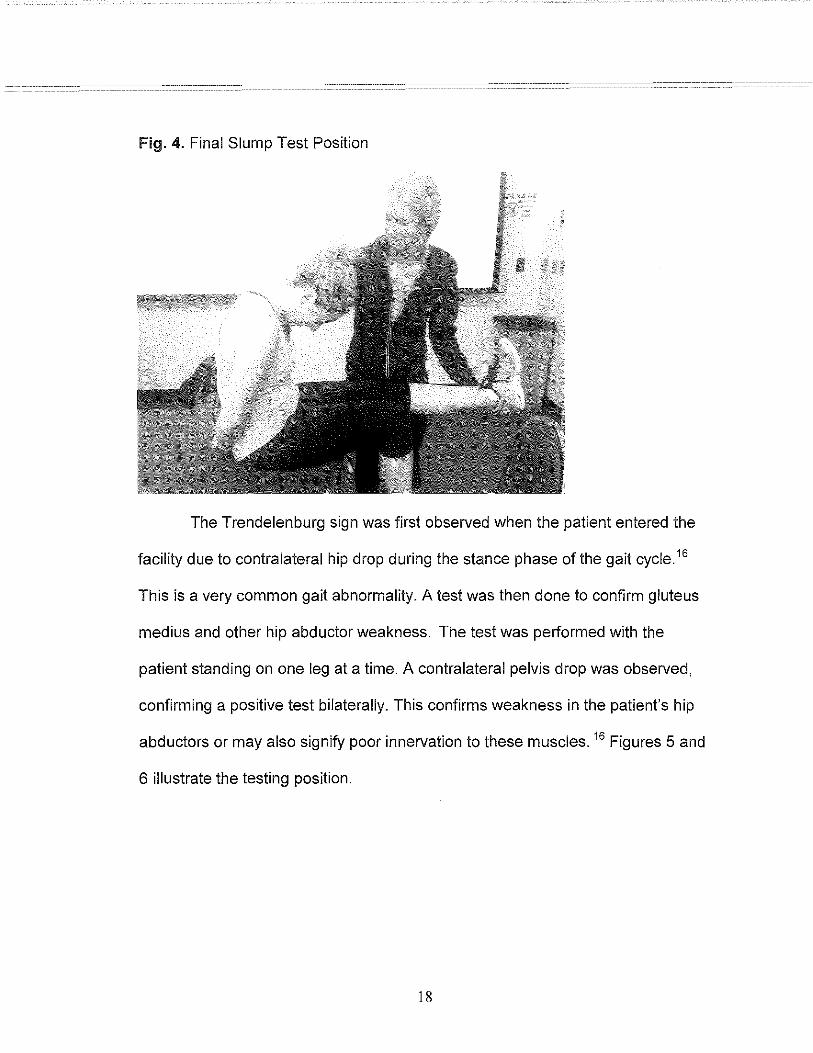
Fig. 4. Final Slump Test Position
The Trendelenburg sign was first observed when the patient entered the
facility due to contralateral hip drop during the stance phase of the gait cycle. 16
This is a very common gait abnormality. A test was then done to confirm gluteus
medius and other hip abductor weakness. The test was performed with the
patient standing on one leg at a time. A contralateral pelvis drop was observed,
confirming a positive test bilaterally. This confirms weakness in the patient's hip
abductors or may also signify poor innervation to these muscles. 16 Figures 5 and
6 illustrate the testing position.
18
~--~~---~~-----cc-.c ccc.c:cc.c.ccc.cc---~----------~---~--
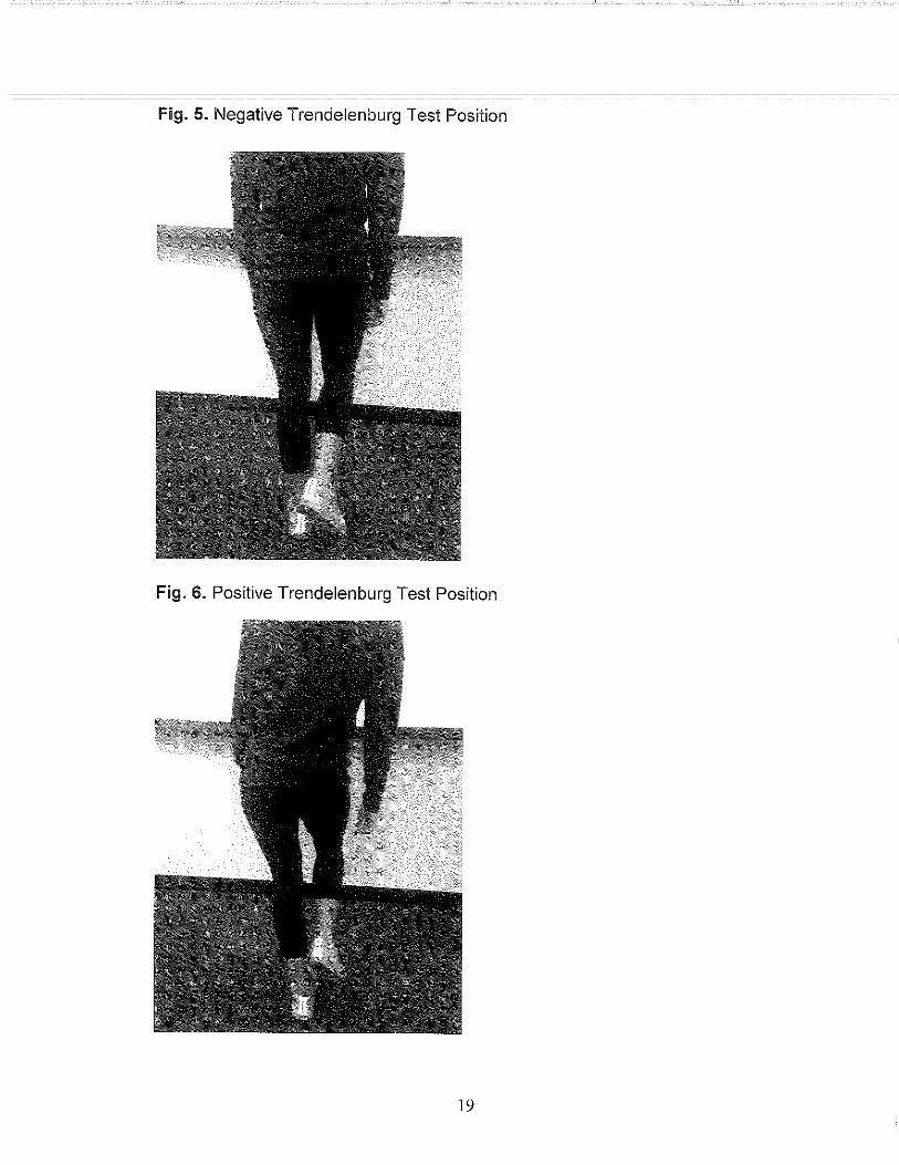
Fig. 5. Negative Trendelenburg Test Position
Fig. 6. Positive Trendelenburg Test Position
19
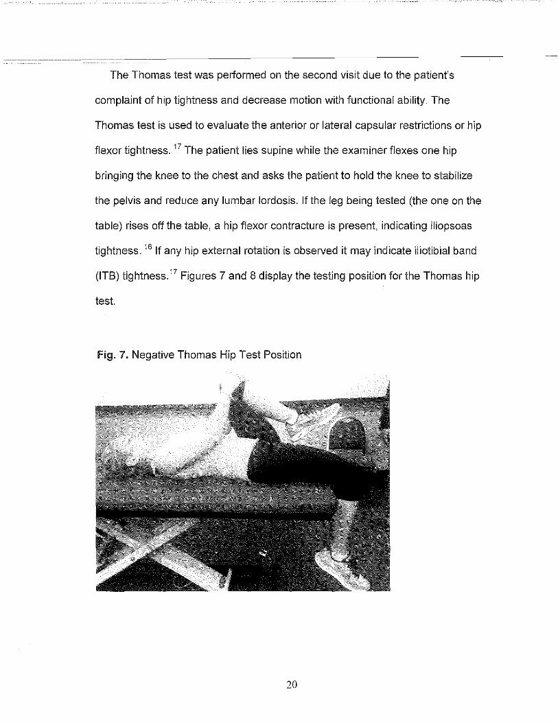
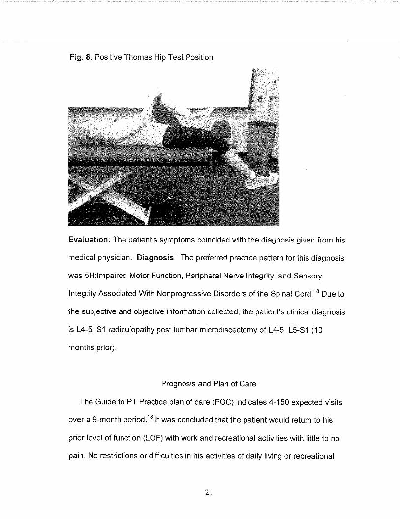
The Thomas test was performed on the second visit due to the patient's
complaint of hip tightness and decrease motion with functional ability. The
Thomas test is used to evaluate the anterior or lateral capsular restrictions or hip
flexor tightness. 17 The patient lies supine while the examiner flexes one hip
bringing the knee to the chest and asks the patient to hold the knee to stabilize
the pelvis and reduce any lumbar lordosis. If the leg being tested (the one on the
table) rises off the table, a hip flexor contracture is present, indicating iliopsoas
tightness. 16 If any hip external rotation is observed it may indicate iliotibial band
(ITS) tightness. 17 Figures 7 and 8 display the testing position for the Thomas hip
test.
Fig. 7. Negative Thomas Hip Test Position
20
~~--------------------------------------------
Fig. 8. Positive Thomas Hip Test Position
Evaluation: The patient's symptoms coincided with the diagnosis given from his
medical physician. Diagnosis: The preferred practice pattern for this diagnosis
was 5H:lmpaired Motor Function, Peripheral Nerve Integrity, and Sensory
Integrity Associated With Nonprogressive Disorders of the Spinal Cord. i8 Due to
the subjective and objective information collected, the patient's clinical diagnosis
is L4-5, S1 radiculopathy post lumbar microdiscectomy of L4-5, L5-S1 (10
months prior).
Prognosis and Plan of Care
The Guide to PT Practice plan of care (POC) indicates 4-150 expected visits
over a 9-month period. i8 It was concluded that the patient would return to his
prior level of function (LOF) with work and recreational activities with little to no
pain. No restrictions or difficulties in his activities of daily living or recreational
21
activities were anticipated. The patient was scheduled twice a week for 12
weeks. The patient's insurance provider allotted more physical therapy visits than
anticipated.
The patient's short term goals were to: 1) be able to demonstrate correct lifting
techniques 2) centralization of radicular symptoms, and 3) increase flexion and
extension to 50% of normal to improve functional mobility. Long term goals
included to: 1) return to full work duties without pain 2) work a full shift without
pain, and 3) return to recreational activities with little or no pain.
The discharge criteria for this patient included being able to sleep without
issues, perform normal tasks at work with no pain or radicular symptoms, and
develop normal gait mechanics. The plan at the initial treatment was to re
evaluate and examine the patient at the 4th and 8th week mark.
22
Intervention
Patient attended therapy for 8 sessions over the course of 5 weeks.
Multiple interventions were utilized due to patient reports of pain, discomfort or
re-creation of radicular symptoms. A detailed description of the POC is in Table
2. The patient's home exercise program (HEP) consisted of exercises first
performed in the clinic to ensure correct form and body mechanics as well as to
clarify patient understanding.
Day 1
Day2
Day 3
Table 2. Plan of Care
Treatment
Initial Evaluation Patient Education: Correct Lifting Techniques Harmful effects Substance Abuse Substance Abuse Referral IFC E-Stim and Hot Pack: 20 Hz 20 minutes
SCS: Posterior Innominate L4, L5, S1 Trunk Stabilization: Posterior Pelvic Tilts 3x20 Bridges 3x20 Bridges with Marching 3x20 Bridges with Heel Slides 3x20 IFC E-Stim and Hot Pack: 22 Hz 20 minutes Muscle Energy Technique: Pelvis SCS: Posterior Innominate L4, L5,S1 Nerve Flossing: R Sciatic N. 8 min Stretching: 30 sec x2 Lumbar Extension and Flexion Hip Flexion Hamstrings IFC E-Stim and Hot Pack: 25 Hz 20 minutes
23
HEP
Static Stretch Hamstrings with Strap Ice LB With Pain
Trunk Stabilization Exercises
Trunk Stabilization Exercises Static Stretching Hamstrings with Strap and Lumbar Flexion
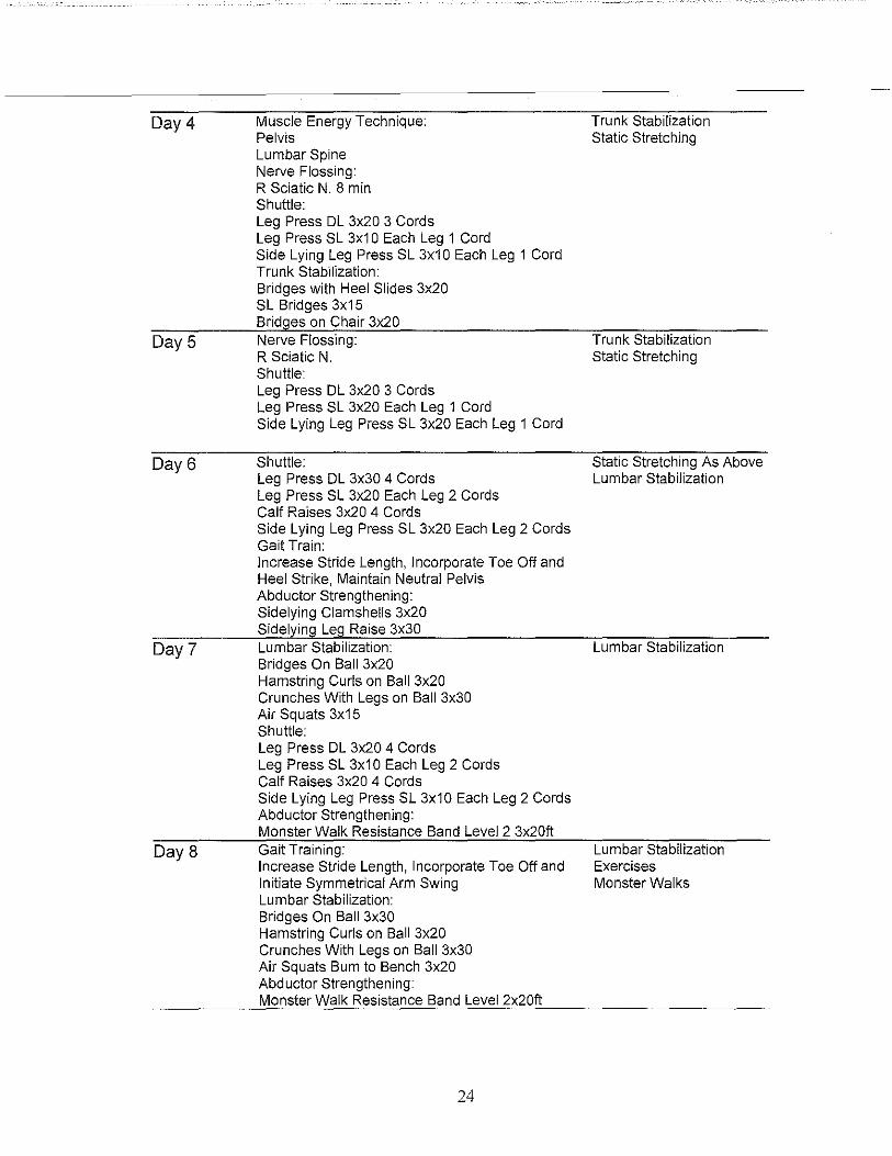
Day4
Day 5
Day6
Day 7
Day 8
Muscle Energy Technique: Pelvis Lumbar Spine Nerve Flossing: R Sciatic N. 8 min Shuttle: Leg Press DL 3x20 3 Cords Leg Press SL 3x1 0 Each Leg 1 Cord Side Lying Leg Press SL 3x10 Each Leg 1 Cord Trunk Stabilization: Bridges with Heel Slides 3x20 SL Bridges 3x15 Bridges on Chair 3x20 Nerve Flossing: R Sciatic N. Shuttle: Leg Press DL 3x20 3 Cords Leg Press SL 3x20 Each Leg 1 Cord Side Lying Leg Press SL 3x20 Each Leg 1 Cord
Shuttle: Leg Press DL 3x30 4 Cords Leg Press SL 3x20 Each Leg 2 Cords Calf Raises 3x20 4 Cords Side Lying Leg Press SL 3x20 Each Leg 2 Cords Gait Train: Increase Stride Length, Incorporate Toe Off and Heel Strike, Maintain Neutral Pelvis Abductor Strengthening: Sidelying Clamshells 3x20 Sidelying Leg Raise 3x30 Lumbar Stabilization: Bridges On Ball 3x20 Hamstring Curls on Ball 3x20 Crunches With Legs on Ball 3x30 Air Squats 3x15 Shuttle: Leg Press DL 3x20 4 Cords Leg Press SL 3x1 0 Each Leg 2 Cords Calf Raises 3x20 4 Cords Side Lying Leg Press SL 3x10 Each Leg 2 Cords Abductor Strengthening: Monster Walk Resistance Band Level 2 3x20ft Gait Training: Increase Stride Length, Incorporate Toe Off and Initiate Symmetrical Arm Swing Lumbar Stabilization: Bridges On Ball 3x30 Hamstring Curls on Ball 3x20 Crunches With Legs on Ball 3x30 Air Squats Bum to Bench 3x20 Abductor Strengthening: Monster Walk Resistance Band Level 2x20ft
24
Trunk Stabilization Static Stretching
Trunk Stabilization Static Stretching
Static Stretching As Above Lumbar Stabilization
Lumbar Stabilization
Lumbar Stabilization Exercises Monster Walks
On the first day, after the examination and evaluation was completed, the
patient was educated on proper body mechanic techniques for lifting objects to
help prevent future agitation of his spine. Then the patient was provided with a
referral for his reported substance use. The negative affects of alcohol and
tobacco on the body were discussed, including how the substances can slow the
healing process through reduced oxygen and blood supply, increase the risk of
fractures, and alter psychological well-being.
On the second visit the treatment started with SCS to the patient's right
posterior innominate as well as his L4, L5 and S1 vertebrae to address the
complaints of radicular pain. The patient was then reassessed for pain level and
location of radicular symptoms. The patient reported a centralization of his
symptoms (moved from lower calf to crease of knee) after each 60sec hold for
each position of the SCS. Each area was only treated once. SCS was selected
based on evidence indicating patients who do not respond to interventions
including pharmacology, physical therapy, biofeedback, acupuncture and
therapeutic exercise (TE) have a decrease or complete resolution in pain after
SCS treatment12
Lumbar stabilization exercises included performing a posterior pelvic tilt in
supine to help engage his abdominals, bridges, bridges with marching and
bridges with heel slides. The patient was instructed to exhale to a count of 5 and
hold the exercise throughout exhalation. The marches and heel slides were
implemented to challenge core strength by introducing mobility and creating a
decreased base of support. The patient had some difficulty maintaining level hips
25
and not rotating with this exercise. Extra time was spent educating the patient on
correct body mechanics and cues to pay attention to such as placing a wooden
dowel or broom across the hips for easier observation.
The patient missed the next scheduled appointment, so he only came in
once this week. On the third treatment day, the patient reported moving furniture
at work, which resulted in an increase of pain intensity. He was instructed to
avoid such activities if possible until recovery had improved. The patient's pelvis
was slightly anteriorly rotated on the left. A muscle energy technique was
performed and the pelvis was then reassessed for symmetry. ses was
performed, with centralization to the middle of the thigh. Sciatic nerve flossing
was used to desensitize the nerve so that the referring symptoms are not as
easily reproduced. Following nerve flossing, the patient was instructed on lumbar
flexion and extension exercises to help relieve some pain while at work. The
patient was not given any exercises on this day due to his inability to mobilize
without an increase in pain.
On day 4, the patient was re-evaluated for progress and the previous poe
was continued. A centralization of radicular symptoms was reported since the
initial visit and a decrease in overall pain on the visual analog scale, 8 to 7. The
shuttle press was introduced to help increase gluteus strength as well as engage
the core and implemented closed chain mobility activities. Due to the patient's
progress in his trunk stabilization exercises, the patient was advanced to
performing bridging on a chair to increase the difficulty of the exercise.
26
Day 5 consisted of nerve flossing to continue to desensitize his pain and
the shuttle press were continued. The patient was late arriving on day 5 and thus
a shorter session resulted. The repetitions were increased on the shuttle press
since the patient reported feeling "stronger in his leg muscles."
The patient ambulated with decreased speed, lack of arm movement, and
great difficulty implementing a toe off. Consequently gait training was introduced
on the 6th visit. The focus was to increase stride length, develop a heel strike and
toe off. The session ended with abductor strengthening.
The patient did not appear to display motivation to improve on this day; he
had to repeatedly be cued with his exercises and took long rest breaks. The
patient was absent for the following appointment.
On day 7, the patient progressed his trunk stabilization exercises from a
chair to a ball. This change was designed to reduce the stability under his feet
and require him to further engage his core stabilizers.
On the last day of treatment, the patient was re-evaluated and reported his
radicular pain moved upward from the mid-thigh to just below his gluteal fold and
pain levels decreased from 7 to 5. He was able to demonstrate a moderate heel
strike and toe off, as well as maintain an increased step length. The patient was
also able to incorporate a reciprocal arm swing during gait. The patient's gait
appeared to be quicker but still slower than a normal speed for his age. The
patient discharged himself after this treatment.
27
Outcomes
The patient discharged himself from therapy, The therapy plan was not
completed and all of patient's goals were not achieved, Throughout treatment the
patient irregularly attended PT, missing 2 visits out of the total of 10 visits during
the 5 weeks, This pattern of action coincides with the previous behaviors towards
therapy and other treatment alternatives that he explained,
The patient reported improved quality of sleep, a centralization of his
radicular symptoms (moved from calf to just inferior of the gluteal fold), an overall
decrease in the severity of pain according to the visual analog scale (8-5), and an
ability to work longer hours before his symptoms were created, The pain
remained in the anterior hip but declined from 6 to 3, The patient's hip flexion
also increased from 107 to 119 degrees, The patient reported an improvement in
his functional abilities, including being able to work an entire shift The onset of
pain was delayed and the pain was reported as a desire to sit down after two
hours, later extending this urge to 5 hours to help relieve pain,
There was a noticeable change in the patient's gait pattern, The step
length increased and an observable heel strike and toe off was also present The
patient still demonstrated difficulty with his arm swing symmetry, The patient was
still unable to engage in his desired recreational activities and never sought help
for the substance use,
28
CHAPTER III
DISCUSSION
LBP is widespread and debilitating condition; however, there is insufficient
evidence in support of a set protocol. Researchers and therapists are able to
agree on the need for physical activity, but the exercise regimens vary and a
consensus has not been reached on which exercises are most effective. 8
Oosterhuis et el8 concluded that exercise therapy does not seem to be effective
for acute LBP but is helpful to patients with chronic pain.
Common advice to reduce back pain includes engaging in regular activity,
consuming a balanced diet, maintaining proper posture, performing proper lifting
techniques and smoking cessation. 1 The patient was educated on all of these
topics; however, this education did not alter his behaviors. The inability to comply
with the physical therapy plan could have resulted from a lack of motivation or
from difficulty changing behavior pattems.
Resistive exercise was implemented based on evidence that trunk
strengthening appears effective compared to no exercise, and that an increase in
intensity and motivation would increase the treatment effect, resulting in a faster
return to work2. 5,6 As the exercises became more difficult, the patient continued
to improve and move closer to his goals. However, periodically, it was difficult to
motivate the patient and his internal motivation varied between therapy sessions.
29
SCS appeared to be the most immediate form of relief of all of the
interventions chosen. According to the theory of SCS, there is a constant state of
hypertonicity due to aberrant neuromuscular activity between muscle agonist and
antagonist. 20 SCS is proposed to correct aberrant proprioceptive input, reset
gamma bias, and interrupt the reflex pathway resulting in a more relaxed
muscle. 11•2o The patient presented with neuromuscular complications and the
radicular pain led to muscle guarding. This technique was ideal for this patient
because it required the patient to relax as the therapist passively moved the
specific area targeted into position. This was one of the only techniques utilized
where the patient did not complain of discomfort and both therapist and patient
saw immediate results.
Nerve flossing did not seem to provide additional benefits for reducing
radicular pain on top of SCS and therapeutic exercise. Although the patient had a
decline in the onset of radicular symptoms, this may have been attributable to the
lumbar stabilization exercises as well as the SCS techniques. No high or
intermediate level research was found on nerve flossing for lumbar radicular
pain. More research needs to be done involving the use of nerve flossing and
stretching for LBP associated with radicular symptoms.
The patient's substance abuse could have led to his decreased healing
process. Lee et al18 found that smokers tended to have decreased mental and
physical health scores, more musculoskeletal disorders, and chronic pain (nearly
two times the risk) than nonsmokers. Smoking has different effects on human
tissues and may cause LBP through multiple mechanisms. 4, 18 Nicotine, the
30
addictive ingredient in cigarettes, has been shown to affect the immune system,
alter bone mass density, stimulate the sympathetic nervous system (SNS),
disrupt the vascular system by reducing blood supply, place a strain on the liver,
and cause cell death, 4 Decreased calcium absorption in smokers may also be a
contributing factor to decreased bone mineral density along with the depletion of
bone marrow B-Iymphocytes, which are vital for bone homeostasis, 4 Cigarette
smoking has been shown to lead to increased fracture rates affecting the hip,
spine, and distal radius, and other osteoporosis-associated fractures, 4 The
patient's 1 O-year history of smoking may have played a prominent role in his
delayed healing process and contributed to the development of his chronic pain
and pathology, The patient was provided education on the damaging affects of
alcohol including cirrhosis of the liver, permanent damage to the brain, jaundice,
nerve damage, and malnutrition 23, The patient did not report altering either of
these behaviors during therapy,
Psychological factors including distress and depression, may also
influence patients with chronic LBP, Early identification of these risk factors
through a screening questionnaire may lead to more effective treatment A
health-related outcome measure, such as the SF36, could be used to determine
the psycho-social behavioral factors influencing patients, These screens were not
included in the initial evaluation and POC, The data from these screening tools
may have also provided some insight into his substance use and motivation,
31
Reflective Practice
There are a few things that I would have done differently with this patient.
Pincus, Burton and Vogel22 found that psychological factors such as distress,
depression, and somatization have a unique contribution in the development of
chronic low back pain. It would have been beneficial to utilize a psychological
questionnaire to assess the severity of his substance use, abuse, or addiction.
Psychological screening would have helped provide insight regarding
inconsistent and unmotivated behaviors that affect compliance and if further
psychological assessment would have identified contributing factors to the
original pathology. Coping strategies and fear avoidance techniques can
minimize the role of these psychological factors and may have been helpful to
the patient. 22
A variety of additional interventions could have been implemented. A
massage instead of a hot pack may have provided quicker relaxation. Manual
therapy of grade I or II distraction could have been used to reduce hip pain and
grade III distraction or a posterior glide to increase ROM. The Mackenzie15
methods may have increased lumbar ROM and decreased pain. These
alternative interventions may have produced different results.
32
REFERENCES
1. Available at: http://www.acatoday.org/leveI2_css.cfm?T1ID=13&T2ID=68.
Accessed May 17, 2015.
2. Hauggaard A, Persson AL. Specific spinal stabilisation exercises in patients
with low back pain - a systematic review. Physical Therapy Reviews.
2007; 12(3):233-248.
3. Pfingsten M, Leibing E, Harter W, et al. Fear-avoidance behavior and
anticipation of pain in patients with chronic low back pain: a randomized
controlled study. Pain Med. 2001 ;2(4):259-66.
4. Leboeuf-Yde C, Yashin A, Lauritzen T. Does smoking cause low back pain?
Results from a population-based study. Journal Of Manipulative &
Physiological Therapeutics [serial online]. February 1996; 19(2):99-1 08.
Available from: CINAHL with Full Text, Ipswich, MA. Accessed May 17,
2015.
5. Moon HJ, Choi KH, Kim DH, et al. Effect of Lumbar Stabilization and
Dynamic Lumbar Strengthening Exercises in Patients With Chronic Low
Back Pain. Annals of Rehabilitation Medicine. 2013;37(1):110-117.
doi: 1 0.5535/arm.2013.37. 1.110.
6. Hahne, AJ.; Ford, JJ.; McMeeken, JM. Conservative management of lumbar
disc hemiation with associated radiculopathy: a systematic review. Spine,
2010, 35, 11, 488-504
33
7. Bakhtiary AH, Safavi-Farokhi Z, Rezasoltani A. lumbar stabilizing exercises
improve activities of daily living in patients with lumbar disc herniation. J
Back Musculoskeletal Rehabil2005; 18:55-60.
8. Oosterhuis T, Costa lO, Maher CG, De vet HC, Van tulder MW, Ostelo RW.
Rehabilitation after lumbar disc surgery. Cochrane Database Syst Rev.
2014;3:CD003007.
9. Slade, S.C., Ther, M.M., & Keating, J.L. (2006). Trunk Strengthening
exercises for chronic low back pain: A systematic review techniques
[Electronic Version]. Journal of Manual & Manipulative Therapy, 29, 163-
173.
10.Filiz, S., Sibel, E., Hale, K., Kazim, C., & Yesim, K. (2009). Comparison of
isokinetic exercise versus standard exercise training in patients with chronic
low back pain: a randomized controlled study [Electronic version]. Clinical
rehabilitation, 23, 238-247.
11. Krantz DB. Strain Counterstrain vs. Therapeutic Exercise for Low Back Pain.
Jones Institute. 2011
12. Dardzinski, JA, Ostrov, B.E., & Hamann, L.S. (2000). Myofascial pain
unresponsive to standard treatment: successful use of a strain and
counterstrain technique with physical therapy [PDF]. Journal of clinical
rheumatology, 4,169-174.
13. Hislop HJ, Avers D, Brown M. Daniels and Worthingham's Muscle Testing,
Techniques of Manual Examination and Performance Testing. Saunders;
2014.
34
14. Donelson, R, Aprill, C, Medcalf, R, Grand, W. A Prospective Study of
Centralization of Lumbar and Referred Pain: A Predictor of Symptomatic
Discs and Annual Competence. Diagnostics and Therapeutics.
1997;22(10): 1115-1112.
15. Magee DJ. Orthopedic Physical Assessment. Elsevier Health Sciences;
2013.
16. Available at: http://www.thestudentphysicaltherapist.com/trendelenbu rg
test.htm!. Accessed June 1, 2015.
17 .Available at: http://www.pthaven.com/page/show/157779-thomas-test.
Accessed June 1, 2015.
18.AP. Guide to Physical Therapist Practice. Amer Physical Therapy Assn;
2001.
19.1ee JJ, Patel R, Biermann JS, Dougherty PJ. The musculoskeletal effects of
cigarette smoking. J Bone Joint Surg Am. 2013;95(9):850-9.
20. Wong, C.K., & Schauer, C. (2004). Reliability, validity and effectiveness of
strain counterstrain techniques [PDF]. Journal of Manual & Manipulative
Therapy, 12, 107-112.
21. Lewis, C., & Flynn, T.w. (2001).The use of strain-counterstrain in the
treatment of patients with low back pain [Electronic Version]. Journal of
Manual and Manipulative Therapy, 2, 92- 98.
35
22. Pincus T, Burton AK, Vogel S, Field AP. A systematic review of
psychological factors as predictors of chronicity/disability in prospective
cohorts of low back pain. Spine. 2002;27(5):E1 09-20.
23. Available at: http://www .d rugfreeworld .org/d rugfacts/alcohollshort -term-Iong
term-effects.html. Accessed June 13, 2015.
36