percutaneous endoscopic lumbar discectomy for … · percutaneous endoscopic lumbar discectomy for...
TRANSCRIPT
Percutaneous Endoscopic Lumbar discectomy for Foraminal to Superior Migrated Disc at L5-
S1 level using the Contralateral Interlaminar Approach: A Technical Case Report
Keun Lee, M.D.1, Hyeun-Sung Kim, M.D. Ph.D.
2, Jee-Soo Jang, M.D. Ph.D.
2, Yong-Hun Pee, M.D.
1,
Jin-Uk Kim, M.D. 3, Jun-Ho Lee, M.D.
3, Il-Tae Jang, M.D. Ph.D.
4
1Department of Neurosurgery, Nanoori Jooan Hospital, Incheon, Republic of Korea
2Department of Neurosurgery, Nanoori Suwon Hospital, Suwon, Gyeonggi-do, Republic of Korea
3Department of Neurosurgery, Nanoori Incheon Hospital, Incheon, Republic of Korea
4Department of Neurosurgery, Nanoori Hospital, Seoul, Republic of Korea
Running head: Contralateral Interlaminar Approach to foraminal - superior disc at L5-S1
Corresponding author:
Hyeun-Sung Kim, M.D. Ph.D.
Department of Neurosurgery, Nanoori Suwon Hospital, Suwon, Gyeonggi-do, Republic of Korea.
e-mail:[email protected]
Tel: 02-1588-9797
Fax: 032-721-9618
Conflicts of Interest: N/A
Abstract
Objective: In cases of foraminal superior migration of L5-S1 HNP, it is very difficult to access
by rigid endoscopic procedure. Thus, we attempted a contralateral interlaminar approach to
expose the exiting nerve root and contralateral foramen to remove the symptomatic disc and
preserve the functional structures.
Methods: Between January 2013 and January 2014, five patients who received the percutaneous
endoscopic lumbar discectomy for foraminal superior migration of L5-S1 lumbar HNP via a
contralateral interlaminar approach were included in this study. Through a contralateral
interlaminar approach, we could expose the exiting nerve root only, to remove the symptomatic
disc without structural damage. We confirmed the radiologic result with an immediate
postoperative MRI, and the clinical result was examined using a VAS.
Results: In all cases, the superior migrated disc from the L5-S1 foraminal space was removed
completely and demonstrated in the immediate postoperative MRI. The mean preoperative
Visual Analogue Score was decreased at the postoperative state, from 7.8 ± 0.84 to 1.4 ± 0.55.
Despite the small number of cases, outcomes were satisfactory.
Conclusion: We obtained excellent clinical results in treating foraminal to superior migrated
disc herniation at L5-S1 using percutaneous endoscopic lumbar discectomy via a contralateral
interlaminar approach.
Keywords: contralateral, disc herniation, foraminal, lumbar, interlaminar

Introduction
In the treatment of radiating pain in the lower limbs related to the lumbar disc herniation,
lumbar discectomy following laminectomy has been considered the gold standard1)
. However,
due to the development of relevant equipment, such as the drill, forceps, laser, radiofrequency
(RF) probe, and high-resolution optic endoscope, it has become possible to treat most lumbar
disc herniations with percutaneous endoscopic lumbar discectomy (PELD)4, 5, 10, 13, 14)
.
However, in the case of the L5-S1 level, approaches to foraminal lesions are limited due to
several anatomical barriers. The high iliac crest, the enlarged L5 transverse process, the
hypertrophied facet joint, or the narrow neural foramen, all of which can make the
transforaminal approach difficult. Thus, the endoscopic approach, using an interlaminar
approach or transiliac approach, has been considered3, 5, 13)
.
In the case of lumbar disc herniation with foraminal to superior migration from the L5-S1 level,
both transforaminal and ipsilateral interlaminar approaches have technical difficulties, while an
appropriate approach with endoscope has not been discussed. In order to approach the
contralateral foramen, which has been considered as the most difficult part to be approached
with the endoscope, the authors of this study attempted an approach that passed the interlaminar
window from the contralateral side to the lesion. The authors effectively removed the
symptomatic ruptured disc that was compressing the exiting nerve root, by approaching through
the contralateral foramen to the legion. This study reports the contralateral interlaminar
approach for the first time, as we believe there is no literature showing a method of approaching
the disc fragment in superior migration towards the foramen at the L5-S1 level, using a rigid
endoscope.
Materials and Methods
This study examined patients with a superior migrated disc from the L5-S1 foraminal space who
visited the hospital between January 2013 and January 2014 for radiating pain in a single lower
limb. Among these, five patients refractory to conservative treatment for more than six weeks
underwent percutaneous endoscopic lumbar discectomy via a contralateral interlaminar
approach. The operation was followed by a measurement of pain improvement using a visual
analog scale (VAS). Nerve decompression was evaluated with an immediately postoperative
MRI.
Operative techniques
Prior to the operation, patients received preventative antibiotic treatment and were prepared in a
prone position on a radiolucent table. The operation was undertaken with conscious sedation
using midazolam and fentanyl. In order to avoid any injuries to exiting nerve roots and
traversing nerve roots during the approach, the operation proceeded with continuous feedback
from the patient. C-arm guidance was used to identify the L5-S1 disc space. The skin was
marked at the level of the disc space in the midline. Discography was conducted in advance for
a transforaminal approach; 0.8% indigo carmine (Carmine, Korea United Pharmaceutical,
Yoenki, Korea) mixed with contrast (Iobrix injection, Taejoon Pharm , Korea) was injected.
Using an 18-gauge spinal needle, from a point 2–3 cm away on the contralateral side of the
midline, the needle was inserted at an approximately 45-degree angle passing the middle of the
interspinous ligament. The 18-gauge spinal needle was docked targeting the medial border of
the inferior facet. Following the needle, a guide wire was inserted, which was followed by the
insertion of an obturator, working channel, and an endoscope, just beyond the ligamentum
flavum (Fig.1). With maximum preservation of surrounding structures, the ligamentum flavum
was divided with a probe, and then the bevel of the working channel penetrated through the gap
of the ligamentum flavum and was rotated.
When the ligamentum flavum was divided, the contralateral foraminal area was exposed (Fig.2).
The divided ligamentum flavum was pushed down, protecting the traversing nerve root and
limiting additional injuries due to the approach. Having reached the foraminal area of the lesion,
the epidural fat was dissected with an RF electrode (Ellman International, Hewlett, NY). The
blue-stained ruptured disc, which had been intensely compressing the exiting nerve root, was
removed with endoscopic forceps (Fig.3). Having confirmed the decompression of the exiting
nerve root, the scope was removed after bleeding control.
Immediately after the operation, the decompression of exiting nerve root was confirmed with
postoperative MRI. On the operation day, the patient had reduced pain and no problem in
walking.
Results
The average age of the five patients was 55.8 ± 10.04. Three of them were male, two female. All
cases were confirmed through MRI inspection immediately after the operations; they showed
that the ruptured discs had been successfully removed. The average VAS was reduced from 7.8
± 0.84 preoperatively to 1.4 ± 0.55 postoperatively (Table 1). As this was a technical case
report, the number of cases was small, but the results were satisfying in all of cases. There were
no complications such as dysthesia, hematoma, or infection. None of the cases needed any
additional open surgery.

Illustrative case
A 43-year-old man presented with an intense left lower limb radiation pain, which had not
responded to more than 6 weeks of conservative treatment (Fig. 4A). On the MR images, a
ruptured disc, which had migrated superiorly from the left foramen at the L5-S1 level, was
compressing the left L5 nerve root (Fig. 4B). Employing the contralateral interlaminar
approach, a working channel and endoscope were inserted at a spot 2.5 cm to the right of the
midline, towards the medial border of the inferior articular process of the lesion. The
ligamentum flavum was divided, which was followed by removal of the ruptured disc that had
been blue-stained (Fig. 4C). The MRI scan, which immediately followed the operation, showed
that the ruptured disc had been successfully removed and that the left L5 nerve root was well
decompressed (Fig. 4D).
Discussion
Since Kambin et al. first introduced lumbar disc decompression via a posterolateral approach,
there have been remarkable advances in PELD7)
. With the development of various instruments
related to spinal endoscopy, now nearly all symptomatic lumbar HNP can be operated on via
endoscope. Among them, as L5-S1 level has characteristic anatomical features, such as the iliac
crest, a large facet joint, a narrow foramen, and a wide interlaminar space; therefore, several
approaches to ruptured discs at the L5-S1 level have been studied, such as transforaminal,
ipsilateral interlaminar, and transiliac appoaches3, 6, 13)
. Choi et al. compared the transforaminal
approach to the interlaminar approach in detail depending on the location of the lesion, but
limited the lesion to intracanal disc herniation and did not examine foraminal discs6)
. In
particular, it is difficult to approach ruptured discs that have migrated from the foraminal to the
superior in the L5-S1 level with rigid endoscopes, and such approaches have not been reported
yet.
Some researchers have studied the use of the contralateral approach to remove inferior-migrated
ruptured disc fragments. Kim et al. succeeded in removing down-migrated ruptured discs at the
L4-5 level via a contralateral transforaminal approach8)
. Yeom et al. also succeeded in removing
ruptured disc fragments that were distally migrated from L3-4 and L4-5 to below the midpedicle
level via a contralateral transforaminal approach without excessive pivoting of the rigid
endoscope to the cranial side15)
.
Although the traditional ipsilateral transforaminal approach can be taken for nerve root
compression due to superior migration, the same approach is difficult for cases of superior
migration in the foramen at the L5-S1 level due to anatomical barriers, such as the high iliac
crest, large L5 transverse process, or hypertrophy of the facet joint. Using an excessive cranial
to caudal skin entry point due to the high iliac crest increases the possibility of encountering an
existing nerve root injury, further hindering the approach to the superior migration disc
fragment. It is also difficult to approach superior migrated disc fragments in the foramen via the
ipsilateral interlaminar approach due to the inferior facet and laminar. Thus, traditionally,
microscopic lumbar discectomy via the paramedian transmuscular approach has been used for
this type of lesion. However, microscopic approaches require partial facetectomy; if more than
40–50% is removed, instability may arise, inducing and maintaining postoperative pain2, 9)
.
The discs, facet joints, supraspinous ligament, and paraspinal muscles of the lumbar spine are
important structures that maintain the stability of segmental motion, and instability can be
increased when these structures and surrounding muscles and ligaments are damaged11, 12)
. For
ruptured disc fragments that were foraminal to superior migrated at L5-S1, percutaneous
endoscopic lumbar discectomy via a contralateral interlaminar approach can maximally preserve
these structures and minimize muscle, laminar, and facet injury, while enabling the surgeon to
approach the lesion-side foramen, remove the migrated disc fragments, and decompress with a
direct view of the exiting nerve root.
If the interlaminar space is relatively narrow, it might render an approach to superior migration
difficult, as the contralateral laminar would block the path. In such a case, an approach
trajectory can be acquired by undercutting the inferior margin of the lesion-side facet using an
endoscopic drill, dilator, or reamer. Fortunately, in the present cases, we were able to approach
the contralateral foramen and remove the ruptured disc fragments without using an endoscopic
drill. If the ligamentum flavum is split after docking at the medial border of the contralateral
inferior facet, the contralateral foramen and lateral recess are exposed. When the bevel of the
working channel is rotated 180 degrees, the split ligamentum flavum is inferiorly displaced,
naturally protecting the traversing nerve root and preventing additional approach-induced injury.
Since directly approaching the working channel at a sharp angle may cause injury to the
thecal sac, it is important to safely perform docking of the working channel to the medial border
of the inferior facet of lesions at an approximately 45-degree angle in the beginning. In addition,
when dissection is carried out in order to split the ligamentum flavum without clarifying the
boundaries of the bone and ligamentum, this may cause exiting nerve root injury, thereby
requiring attention.
Conclusion
This study achieved a good result through a contralateral interlaminar approach for the
foraminal superior migrated L5-S1 Lumbar HNP. This can be a considerable option for the
treatment of ruptured discs that had previously been difficult to approach with a rigid
endoscope.
References
1. Andrews DW, Lavyne MH: Retrospective analysis of microsurgical and standard
lumbar discectomy. Spine (Phila Pa 1976) 15: 329-335, 1990
2. Bae JS, Kang KH, Park JH, Lim JH, Jang IT: Postoperative Clinical Outcome and Risk
Factors for Poor Outcome of Foraminal and Extraforaminal Lumbar Disc Herniation. J
Korean Neurosurg Soc 59: 143-148, 2016
3. Choi G, Kim JS, Lokhande P, Lee SH: Percutaneous endoscopic lumbar discectomy by
transiliac approach: a case report. Spine (Phila Pa 1976) 34: E443-446, 2009
4. Choi G, Lee SH, Bhanot A, Raiturker PP, Chae YS: Percutaneous endoscopic
discectomy for extraforaminal lumbar disc herniations: extraforaminal targeted
fragmentectomy technique using working channel endoscope. Spine (Phila Pa 1976)
32: E93-99, 2007
5. Choi G, Lee SH, Raiturker PP, Lee S, Chae YS: Percutaneous endoscopic interlaminar
discectomy for intracanalicular disc herniations at L5-S1 using a rigid working channel
endoscope. Neurosurgery 58: ONS59-68; discussion ONS59-68, 2006
6. Choi KC, Kim JS, Ryu KS, Kang BU, Ahn Y, Lee SH: Percutaneous endoscopic lumbar
discectomy for L5-S1 disc herniation: transforaminal versus interlaminar approach.
Pain Physician 16: 547-556, 2013
7. Kambin P, Sampson S: Posterolateral percutaneous suction-excision of herniated lumbar
intervertebral discs. Report of interim results. Clin Orthop Relat Res: 37-43, 1986
8. Kim JS, Choi G, Lee SH: Percutaneous endoscopic lumbar discectomy via contralateral
approach: a technical case report. Spine (Phila Pa 1976) 36: E1173-1178, 2011
9. Kotil K, Akcetin M, Bilge T: A minimally invasive transmuscular approach to far-lateral
L5-S1 level disc herniations: a prospective study. J Spinal Disord Tech 20: 132-138,
2007
10. Mayer HM, Brock M: Percutaneous endoscopic discectomy: surgical technique and
preliminary results compared to microsurgical discectomy. J Neurosurg 78: 216-225,
1993
11. Panjabi MM: The stabilizing system of the spine. Part I. Function, dysfunction,
adaptation, and enhancement. J Spinal Disord 5: 383-389; discussion 397, 1992
12. Panjabi MM: The stabilizing system of the spine. Part II. Neutral zone and instability
hypothesis. J Spinal Disord 5: 390-396; discussion 397, 1992
13. Ruetten S, Komp M, Godolias G: A New full-endoscopic technique for the interlaminar
operation of lumbar disc herniations using 6-mm endoscopes: prospective 2-year results
of 331 patients. Minim Invasive Neurosurg 49: 80-87, 2006
14. Ruetten S, Komp M, Merk H, Godolias G: Full-endoscopic interlaminar and
transforaminal lumbar discectomy versus conventional microsurgical technique: a
prospective, randomized, controlled study. Spine (Phila Pa 1976) 33: 931-939, 2008
15. Yeom KS, Choi YS: Full endoscopic contralateral transforaminal discectomy for
distally migrated lumbar disc herniation. J Orthop Sci 16: 263-269, 2011

Figure legends
Fig. 1. Docking of working channel at the medial border of inferior facet
Fig. 2. Endoscopic view of contralateral interlaminar approach (LF: splitted ligamentum flavum, RD:
ruptured disc, FT: fatty tissue, EXR: exiting nerve root)
Fig. 3. Removal of foraminal to superior migration disc with endoscopic forcep
Fig. 4. Illustrative case (A: Preoperative X-ray, B: Preoperative MRI, C: Removed rupture disc, D:
Postoperative MRI)
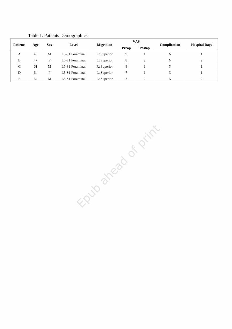
Table 1. Patients Demographics
Patients Age Sex Level Migration VAS
Complication Hospital Days Preop Postop
A 43 M L5-S1 Foraminal Lt Superior 9 1 N 1
B 47 F L5-S1 Foraminal Lt Superior 8 2 N 2
C 61 M L5-S1 Foraminal Rt Superior 8 1 N 1
D 64 F L5-S1 Foraminal Lt Superior 7 1 N 1
E 64 M L5-S1 Foraminal Lt Superior 7 2 N 2
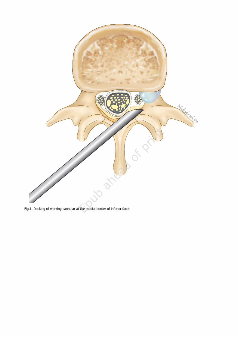
Fig.1. Docking of working cannular at the medial border of inferior facet
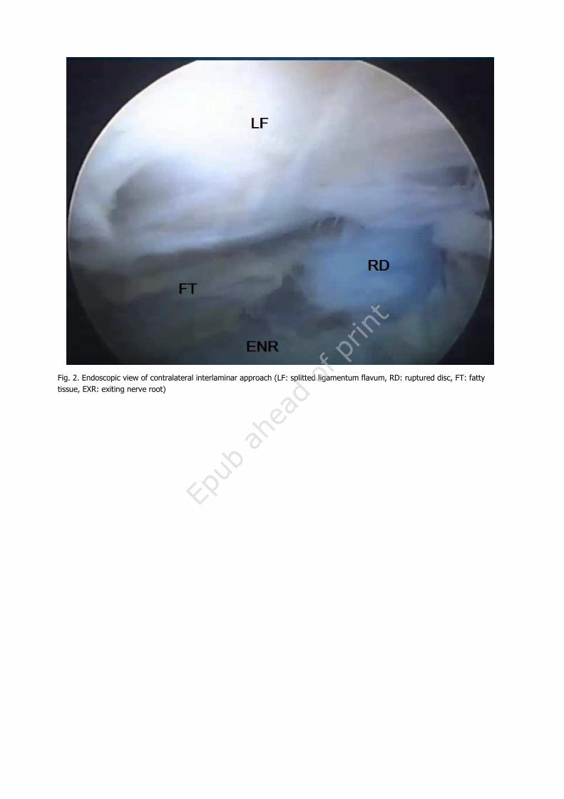
Fig. 2. Endoscopic view of contralateral interlaminar approach (LF: splitted ligamentum flavum, RD: ruptured disc, FT: fatty
tissue, EXR: exiting nerve root)
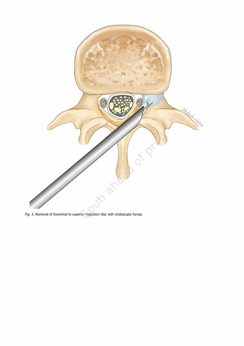
Fig. 3. Removal of foraminal to superior migration disc with endoscopic forcep
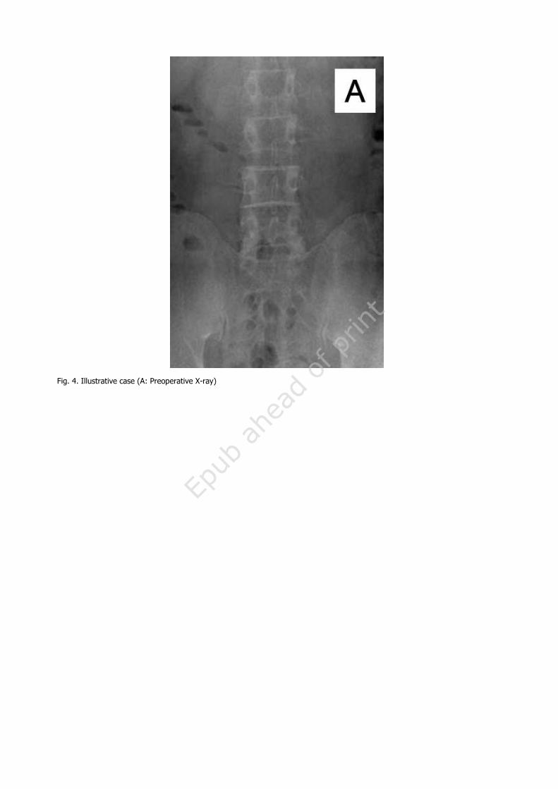
Fig. 4. Illustrative case (A: Preoperative X-ray)
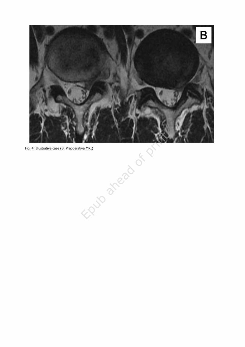
Fig. 4. Illustrative case (B: Preoperative MRI)
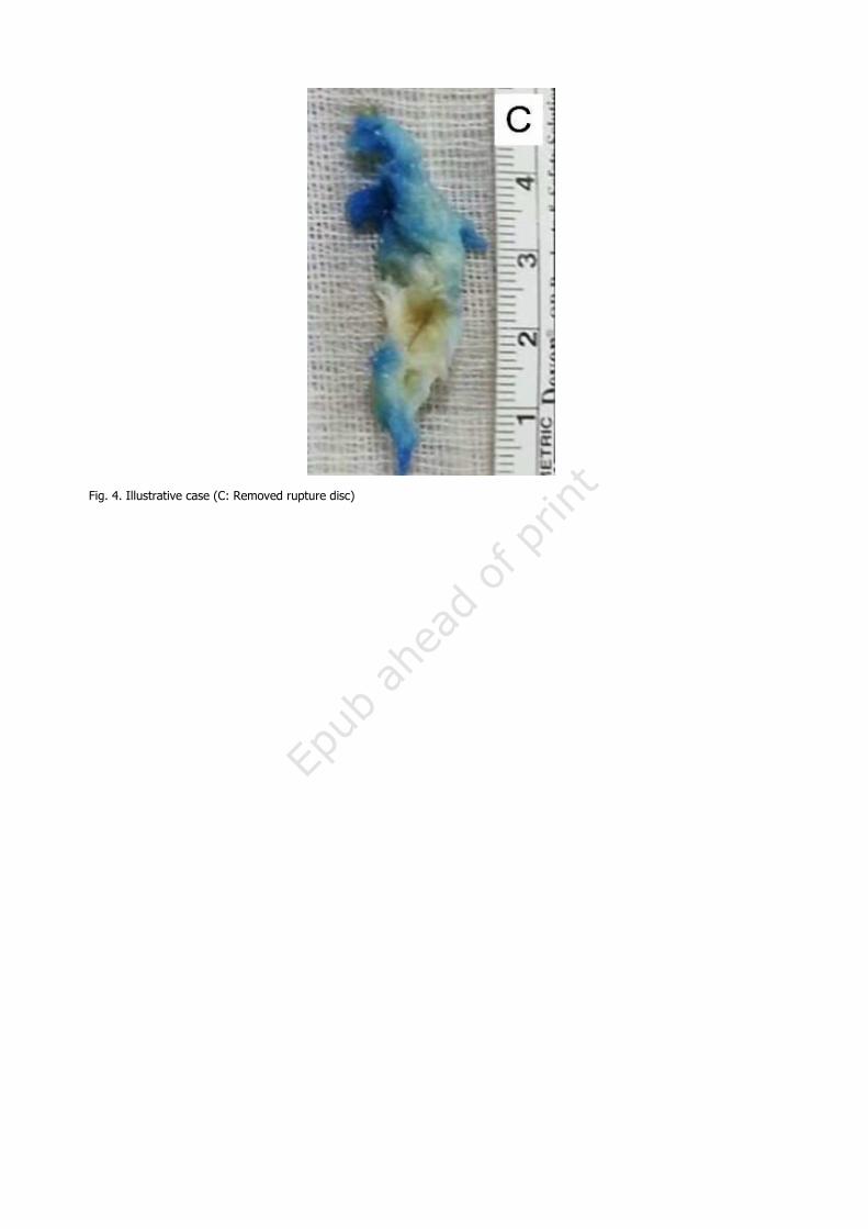
Fig. 4. Illustrative case (C: Removed rupture disc)
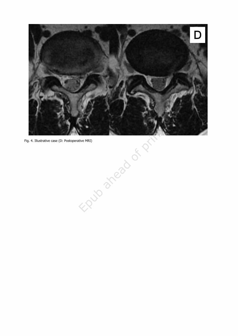
Fig. 4. Illustrative case (D: Postoperative MRI)