low level quantification of c &wyl ester transfer protein in plas,ma
TRANSCRIPT

Low level quantification of c &wyl ester transfer protein in plas,ma subfractions and cell culture media by monoclonal antibody-based immunoassay
Ronald W. Clark, 1 James B. Moberly,' and Mark J. Bamberger
Department of Cardiovascular and Metabolic Diseases, Central Research Division, Pfizer, Inc., Groton, CT 06340
Abstract Sensitive immunoradiometric (IRMA) and ELISA Supplementary key words sandwich blotting immunoblotting assays for cholesteryl ester transfer protein (CETP) have been native gel electrophoresis prebeta migrating Superose Superdex developed using two different monoclonal antibodies (MAbs). HepG2 cells The MAbs were prepared against human plasma CETP and demonstrated specificity by their inhibition of cholesteryl ester transfer activity and by immunoblots of crude plasma fractions and whole media from transfected CHO cells. For these sandwich-type assays, one MAb, 2F8, is used for capture, and the second MAb, 2E7, is iodinated (IRMA) or conjugated with alkaline phosphatase (ELISA) and used for detection. Both as- says are linear and provide sensitivities much greater than previ- ously reported. The IRMA allows for the accurate quantifica- tion of CETP in the range of 0.5-20 ng/assay (5-200 ng/ml), the ELISA 0.05-5 ng/assay (0.5-50 ng/ml). Using the IRMA, the mean plasma CETP concentration in 44 normolipidemic in- dividuals was determined to be 2.10 k 0.36 pglml; 2.05 k 0.33 for males (n = 25) and 2.16 + 0.40 for females (n = 19). Values ranged from 1.28 to 2.97 pg/ml and CETP mass correlated well with cholesteryl ester transfer activity ( r = 0.913, n = 23). The distribution of CETP in human plasma was examined both by gel permeation fast protein liquid chromatography (FPLC) and by native gel electrophoresis. For FPLC using agarose resins, a low ionic strength, isotonic buffer system resulted in near total recoveries of CETP, and demonstrated a peak for CETP mass centered at molecular masses of 150 to 180 kilodaltons, larger than that for free monomeric CETP. Native acrylamide gel elec- trophoresis of plasma from six individuals, followed by 2F8/2E7 sandwich immunoblotting, showed CETP migrating within a size range of 170-220 kilodaltons. This result is consistent with suggestions that plasma CETP is associated with small-sized HDL. Agarose gel electrophoresis showed plasma CETP, as well as purified recombinant CETP, to be prebeta migrating. For de- termining the concentration of CETP in the media of cultured HepG2 cells, advantage was taken of the high sensitivity of the ELISA. CETP levels were found to increase linearly over the 100-h culture period, reaching 8.0 k 0.4 ng/ml (18.0 k 1.3 ng/mg cell protein). 811 These sensitive, direct immunoassays for CETP mass should be valuable aids for examining the be- havior of CETP in plasma and other complex systems, as well as for studying the synthesis and secretion of CETP by different cells and tissues.-Clark, R. W., J. B. Moberly, and M. J. Bamberger. Low level quantification of cholesteryl ester transfer protein of plasma subfractions and cell culture media by monoclonal antibody-based immunoassay. J. Lipid Res. 1995. 36: 876-889.
Cholesteryl ester transfer protein (CETP) catalyzes the transfer of cholesteryl esters (CE), triglyceride, and phos- pholipid between lipoproteins in the plasma of many animal species, including humans (1). Most plasma CE is generated on high density lipoproteins (HDL) through the action of 1ecithin:cholesterol acyltransferase (LCAT). CETP acts to redistribute a portion of this CE to triglyceride-rich lipoproteins, which are transformed via lipolysis to remnants and low density lipoprotein (LDL), or to LDL directly. Because this exchange of CE for triglyceride causes a net loss of CE from HDL, CETP has been proposed to play an important role in determining the balance between LDL- and HDL-cholesterol levels. That CETP can play such a role in vivo is suggested by the very high HDL levels observed for subjects genetically defective for CETP expression (2), and by the reduced HDL levels displayed by CETP transgenic mice (3). These contrasting states represent potentially antiathero- genic and atherogenic lipoprotein profiles, respectively, and call for more detailed studies on the role of CETP in intravascular lipoprotein metabolism, as well as its func- tion in different cells and tissues.
Abbreviations: CETP, cholesteryl ester transfer protein; CE, cholesteryl ester; E, total cholesterol; CO, cholesteryl oleate; CMC, critical micelle concentration; VLDL/IDL, very low density lipoprotein/intermediate density lipoprotein; LDL, low density lipoprotein; HDL, high density lipoprotein; IRMA, immunoradiometric assay; MAb, monoclonal anti- body; FPLC, fast protein liquid chromatography.
*To whom correspondence should be addressed. 2Present address: Baxter Healthcare Corporation, Renal Division
Research MPR-Dl, McGaw Park, IL 60085-6730.
876 Journal of Lipid Research Volume 36, 1995
by guest, on Novem
ber 23, 2018w
ww
.jlr.orgD
ownloaded from

For CETP to transfer neutral lipid it must associate with the lipoproteins involved and bind the lipid to be transferred. Much effort has been expended to determine the degree to which CETP is associated with lipoprotein subclasses in the plasma and how factors such as surface charge, apolipoprotein content, plasma proteins, and ionic strength may be involved in such interactions. CETP interactions in whole plasma or in defined assays with isolated lipoproteins or synthetic vesicles have fre- quently been examined using gel filtration techniques to separate components followed by assessment of CETP content by CE-transfer activity (4-6). Other studies have examined CETP interaction with lipoproteins immobi- lized on Sepharose (7) or used both gel filtration and im- mobilized lipoproteins in separate experiments (8). In one investigation, plasma lipoprotein subclasses were specifically removed by apolipoprotein A-I and A-I1 immunoaffinity and the amount of CETP activity remaining in the non- binding plasma fraction was determined (9). Single dimension native polyacrylamide gel electrophoresis (PAGE) (10, 11) and two-dimensional agarose/PAGE (12) have also been used to characterize the CETP-associated species in human plasma with regard to size classes and surface charge. In none of these studies, however, was an attempt made to determine CETP mass. In studying the effects of anti-CETP antibodies on CETP binding to lipo- proteins or vesicles in artificial systems, Swenson et al. (13) determined relative mass levels in chromatography frac- tions by sodium dodecyl sulfate (SDS)/PAGE followed by scanning of immunoblots, but did not determine actual concentrations. Likewise, demonstrations of the secretion of CETP by cells in culture have either relied solely on CE- transfer activity and inhibition of such by specific antibodies (14-16) or combined this evidence with that derived by im- munoprecipitation and immunoblotting (17).
The lack of quantitative information on CETP mass in the above studies appears due in large part to lack of a sensitive and reliable assay for measuring CETP at the low levels found in chromatographic fractions and cell culture media. We have developed a series of monoclonal antibodies (MAb) to human CETP. Two MAbs, identified as 2F8 and 2E7, have been found to function well in double-MAb sandwich immunoassays. Immunoradio- metric (IRMA) and ELISA assays have been stan- dardized using 2F8 as the capture antibody and either io- dinated or alkaline phosphatase-conjugated 2E7 as the detection MAb. Conditions for optimizing the detection of CETP under different conditions, including the selec- tion and concentration of appropriate detergents, are described, as well as the use of these assays to measure CETP in gel filtration fractions and cell conditioned media.
The IRMA and ELISA for CETP described in this report are direct noncompetitive assays that take advan- tage of the high specificity and low background of monoclonal antibodies, and are unaffected by widely
varying lipid and lipoprotein levels. For quantification of CETP at low levels, the ELISA provides a sensitivity 40-50 times greater than that previously reported for both indirect competitive (10) and immunoradiometric (18, 19) assays.
MATERIALS AND METHODS
Materials Non-labeled lipids, alkaline phosphatase and, routine
chemicals were purchased from Sigma. [SH]cholesteryl oleate was from New England Nuclear, 1251-iodine and 125I-labeled streptavidin were from Amersham. All chro- matography resins, columns, and standards were ob- tained from Pharmacia, except butyl Toyopearl 650M which was from TOSOHAAS. Reagents for SDS-PAGE were from Bio-Rad, those for agarose electrophoresis were from Beckman (Paragon system), and precast acrylamide minigels for native protein electrophoresis were from Daiichi. For the enzymatic determinations of cholesterol and triglyceride, enzymes were purchased from Boehringer Mannheim. The microBCA protein assay kit, lodo-gen and all detergents were from Pierce. Costar High Binding flat-bottom, stripwell plates were used for the IRMA and ELISA. Cell culture media and other rea- gents were from GIBCO, except supplements for hybri- doma culture which were from Sigma.
Human hepatoma HepG2 cells were obtained from the American Type Culture Collection, Rockville, MD. Chinese hamster ovary (CHO) cells stably transfected with the human CETP cDNA were acquired from Columbia University and the laboratory of Dr. Alan Tall.
Purification of CETP For the initial immunization of mice, CETP was par-
tially purified from a d > 1.21 g/ml fraction obtained from human plasma after sequential ultracentrifugation. This plasma fraction was processed by FPLC using hydrophobic interaction chromatography followed by ca- tion and then anion exchange. The plasma subfraction was loaded onto a butyl Toyopearl 650M column in 10 mM Tris, 500 mM NaCl, pH 7.4, and CETP was eluted with a linear gradient beginning with 50 mM Tris and ending with H 2 0 (20). CETP activity was determined by measuring [SHICE transfer from LDL to HDL. CETP- containing fractions were pooled, dialyzed against 50 mM acetate, pH 4.5, and loaded onto a CM-Sepharose column (21). CETP was then eluted using a 0-1.0 M NaCl gradient in acetate buffer. The pooled CETP frac- tion was then dialyzed against 10 mM Tris, pH 7.4, ap- plied to a Mono-Q column, and eluted with 0-1.0 M NaCl. The three purification steps increased CETP specific activity by 175 x , 10 x, and 12 x , respectively,
Clark, MobcrrY, and Bamberger Immunoassay of CETP using monoclonal antibodies 877
by guest, on Novem
ber 23, 2018w
ww
.jlr.orgD
ownloaded from

resulting in a specific activity approximately 21000 times that of the starting material and a purity of 60-70s.
For calibration of the IRMA and ELISA and for other analytical studies, CETP was purified from cell culture media conditioned by CHO cells expressing full-length human CETP. Purification using hydrophobic interaction and anion exchange chromatography, as described above, was sufficient for obtaining CETP that appeared pure by SDS-PAGE/silver staining and by amino acid analysis (see Analytical Methods). A d > 1.063 g/ml pooled hu- man plasma fraction, frozen at -70°C in 10% sucrose, was used as a secondary standard. This secondary stan- dard was compared to fresh lots of purified recombinant CETP every 3-4 months and found to be stable over a 1-year period.
Preparation of lipoprotein substrates
Labeled and unlabeled lipoprotein substrates were pre- pared according to the method described by Morton and Zilversmit (22). Blood was obtained from fasted donors using EDTA as anticoagulant. After centrifugation the plasma was adjusted to 3.5 mM N-ethylmaleimide to in- hibit 1ecithin:cholesterol acyltransferase. To produce la- beled lipoproteins, 3.0 ml of unilamellar liposomes, la- beled with [3H]cholesteryl oleate (CO), was added to 150 ml plasma. The liposome solution consisted of egg phos- phatidylcholine, CO, triolein, and BHT at a mole % composition of 82:12:5.6:0.4, respectively, and contained 4.0 mCi[W]CO (250 nmol). The label containing plasma was incubated for 18 h at 37OC to allow the endogenous CETP activity to incorporate [3H]CO into all the lipoprotein classes. Lipoprotein subfractions were ob- tained from both labeled and unlabeled plasma by se- quential ultracentrifugation using potassium bromide to adjust density. VLDL/IDL, LDL, and HDL were iso- lated from the d < 1.019 g/ml, the 1.019-1.063 g/ml, and the 1.10-1.21 g/ml ranges, respectively. Isolated lipoproteins were dialyzed extensively against 100 mM sodium phos- phate, pH 7.4, containing 1 mM EDTA and 0.02% NaN3.
Assay of cholesteryl ester transfer
For determining CETP activity of fractions isolated during purification of CETP, and for the screening of hybridoma supernatants for anti-CETP activity, an assay measuring [SHJCE transfer from LDL (4 nmol CE) to HDL (2.5-5.0 nmol CE) was performed (170 pl assay volume). The source of CETP activity in assays of hybri- doma supernatants was the d > 1.21 g/ml human plasma fraction (150 p g protein). Samples were incubated at 37OC for 1.0-18 h (20-30% transfer of label) and LDL was precipitated with Na,H,P04/MnC12 (100 mM/10 m M final concentrations). Radiolabel present in the HDL- containing supernatant, obtained by centrifugation, was determined by liquid scintillation.
To determine the relative CETP activity of different human plasma samples, an HDL to LDL transfer assay was set up in which the variation due to the lipoprotein content of the sample (2.5 ,u1 plasma, 8-14 nmol total cholesterol) was minimized by using much larger amounts of exogenous donor [3H]HDL (30 nmol 'E) and acceptor LDL (125 nmol E). For assay of transfer ac- tivity in HepG2 conditioned media the donor was [3H]HDL (5 nmol 'IC) and the acceptor was a mix of VLDL/IDL (50 nmol E) and LDL (25 nmol TC). For both assays acceptor lipoprotein was precipitated at the end of the incubation period by adding P043-/MnC12 as described above.
The inhibition of CE-transfer by anti-CETP monoclonal antibodies in whole human plasma was deter- mined by measuring [3H]C0 transfer from exogenous HDL (5 nmol CE) to the non-HDL lipoproteins of the sample. Non-HDL lipoproteins were precipitated at the end of the assay by addition of an equal volume of 20% (wtlvol) PEG 8000 (23).
Preparation of anti-CETP monoclonal antibodies
For the initial immunization, 10 pg of partially purified CETP in complete Freund's adjuvant was injected in- traperitoneally into Balb/c mice. After 1 month the animals were boosted with 10 ,ug in incomplete adjuvant. Spleen lymphocytes were fused with mouse myeloma cells (SP2/0) using PEG 1000 (24). Media conditioned by the resulting hybridomas were screened for CETP inhibition in an assay measuring CE transfer from labeled LDL to unlabeled HDL (see above). Many media were screened simultaneously in an ELISA using immobilized partially purified CETP. Positive hybridomas were cloned by limit- ing dilution and were injected intraperitoneally into pristane-primed Balb/c mice for ascites production. After delipidation the ascites fluid was processed by Protein A Superose (Pharmacia) chromatography for isolation of purified monoclonal antibodies. For MAbs of the IgG, subclass, such as 2E7 and 2F8, a high salt buffer was used for binding to Protein A.
IRMA and ELISA for determination of CETP mass
A series of monoclonal antibodies to CETP were exa- mined for their ability to function as capture and detec- tion antibodies in double-MAb sandwich assays. TWO an- tibodies, 2F8 and 2E7,3 served these functions well and allowed for the development of sensitive and linear assays. For the data shown in this paper, 2F8 functioned as the capture MAb and iodinated or alkaline phosphatase-
3Requests for samples of the 2F8 and 2E7 MAbs should be directed to R. W. Clark.
878 Journal of Lipid Research Volume 36, 1995
by guest, on Novem
ber 23, 2018w
ww
.jlr.orgD
ownloaded from
conjugated 2E7 as the detection MAb, although similar performance was seen with the antibodies in reverse roles. Use of the same MAb as both the capture and detection antibody resulted in complete elimination of CETP detec- tion. This result, for both the 2E7 and 2F8 MAb, suggests that the determinants recognized by these MAbs are monovalent.
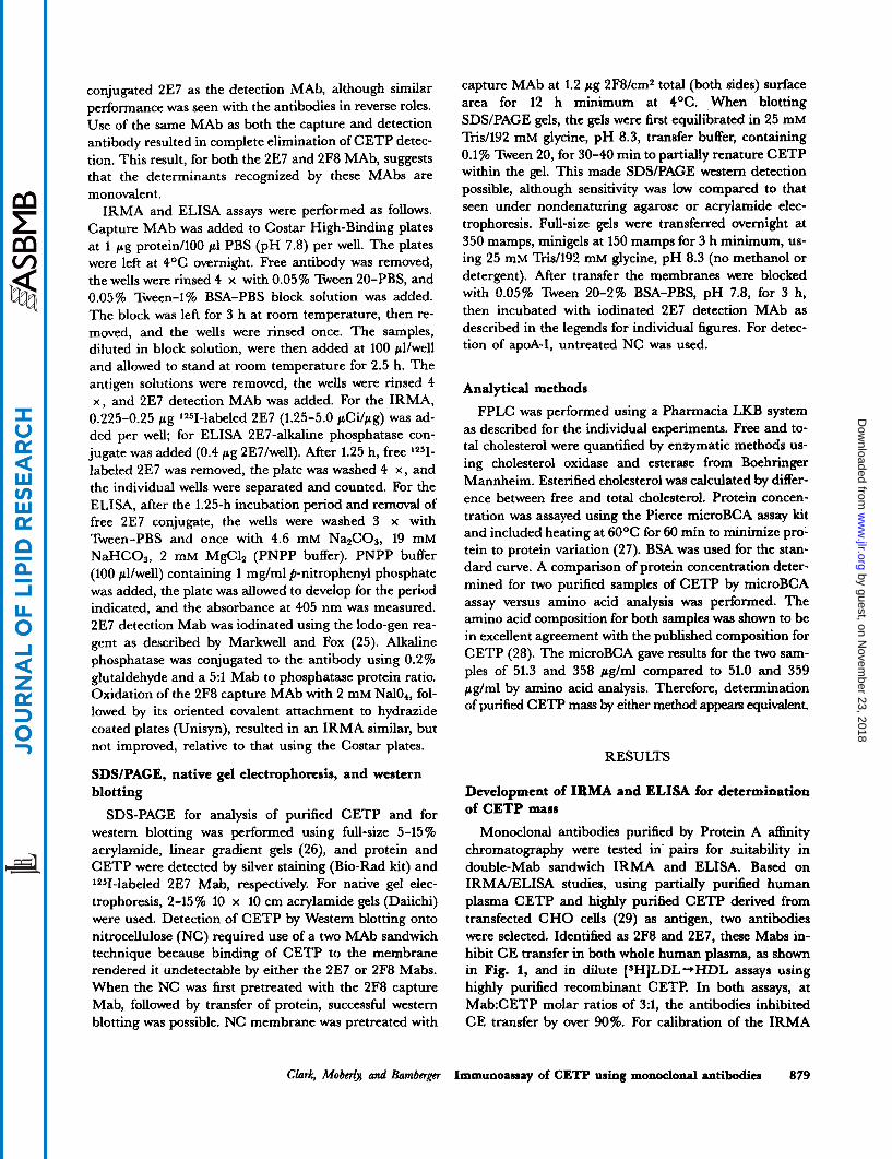
IRMA and ELISA assays were performed as follows. Capture MAb was added to Costar High-Binding plates at 1 pg protein/100 p1 PBS (pH 7.8) per well. The plates were left at 4OC overnight. Free antibody was removed, the wells were rinsed 4 x with 0.05% Tween 20-PBS, and 0.05% Tween-1% BSA-PBS block solution was added. The block was left for 3 h at room temperature, then re- moved, and the wells were rinsed once. The samples, diluted in block solution, were then added at 100 pllwell and allowed to stand at room temperature for 2.5 h. The antigen solutions were removed, the wells were rinsed 4 x , and 2E7 detection MAb was added. For the IRMA, 0.225-0.25 pg 1251-labeled 2E7 (1.25-5.0 pCilpg) was ad- ded per well; for ELISA 2E7-alkaline phosphatase con- jugate was added (0.4 pg 2E7Iwell). After 1.25 h, free 1251- labeled 2E7 was removed, the plate was washed 4 x , and the individual wells were separated and counted. For the ELISA, after the 1.25-h incubation period and removal of free 2E7 conjugate, the wells were washed 3 x with Tween-PBS and once with 4.6 mM Na&03, 19 mM NaHC03, 2 mM MgClz (PNPP buffer). PNPP buffer (100 pllwell) containing 1 mg/ml p-nitrophenyl phosphate was added, the plate was allowed to develop for the period indicated, and the absorbance at 405 nm was measured. 2E7 detection Mab was iodinated using the lodo-gen rea- gent as described by Markwell and Fox (25). Alkaline phosphatase was conjugated to the antibody using 0.2% glutaldehyde and a 5:l Mab to phosphatase protein ratio. Oxidation of the 2F8 capture MAb with 2 mM Na104, fol- lowed by its oriented covalent attachment to hydrazide coated plates (Unisyn), resulted in an IRMA similar, but not improved, relative to that using the Costar plates.
SDSIPAGE, native gel electrophoresis, and western blotting
SDS-PAGE for analysis of purified CETP and for western blotting was performed using full-size 5-15 % acrylamide, linear gradient gels (26), and protein and CETP were detected by silver staining (Bio-Rad kit) and ‘2JI-labeled 2E7 Mab, respectively. For native gel elec- trophoresis, 2-15% 10 x 10 cm acrylamide gels (Daiichi) were used. Detection of CETP by Western blotting onto nitrocellulose (NC) required use of a two MAb sandwich technique because binding of CETP to the membrane rendered it undetectable by either the 2E7 or 2F8 Mabs. When the NC was first pretreated with the 2F8 capture Mab, followed by transfer of protein, successful western blotting was possible. NC membrane was pretreated with
capture MAb at 1.2 pg 2F8/cm2 total (both sides) surface area for 12 h minimum at 4OC. When blotting SDSlPAGE gels, the gels were first equilibrated in 25 mM Tris/192 mM glycine, pH 8.3, transfer buffer, containing 0.1% Tween 20, for 30-40 min to partially renature CETP within the gel. This made SDWPAGE western detection possible, although sensitivity was low compared to that seen under nondenaturing agarose or acrylamide elec- trophoresis. Full-size gels were transferred wemight at 350 mamps, minigels at 150 mamps for 3 h minimum, us- ing 25 mM TrisA92 mM glycine, pH 8.3 (no methanol or detergent). After transfer the membranes were blocked with 0.05% Tween 20-276 BSA-PBS, pH 7.8, for 3 h, then incubated with iodinated 2E7 detection MAb as described in the legends for individual figures. For detec- tion of apoA-I, untreated NC was used.
Analytical methods
FPLC was performed using a Pharmacia LKB system as described for the individual experiments. Free and to- tal cholesterol were quantified by enzymatic methods us- ing cholesterol oxidase and esterase from Boehringer Mannheim. Esterified cholesterol was calculated by differ- ence between free and total cholesterol. Protein concen- tration was assayed using the Pierce microBCA assay kit and included heating at 6OoC for 60 min to minimize pro- tein to protein variation (27). BSA was used for the stan- dard curve. A comparison of protein concentration deter- mined for two purified samples of CETP by microBCA assay versus amino acid analysis was performed. The amino acid composition for both samples was shown to be in excellent agreement with the published composition for CETP (28). The microBCA gave results for the two sam- ples of 51.3 and 358 F g / d compared to 51.0 and 359 p g / d by amino acid analysis. Therefore, determination of purified CETP mass by either method appears equivalent.
RESULTS
Development of IRMA and ELISA for determination of CETP mass
Monoclonal antibodies purified by Protein A affinity chromatography were tested in pairs for suitability in double-Mab sandwich IRMA and ELISA. Based on IRMNELISA studies, using partially purified human plasma CETP and highly purified CETP derived from transfected CHO cells (29) as antigen, two antibodies were selected. Identified as 2F8 and 2E7, these Mabs in- hibit CE transfer in both whole human plasma, as shown in Fig. 1, and in dilute [SH]LDL-*HDL assays using highly purified recombinant CETP. In both assays, at Mab:CETP molar ratios of 3:1, the antibodies inhibited CE transfer by over 90%. For calibration of the IRMA
Clark, Mobcrh and Bambcrgcr Immunoassay of CETP using monoclonal antibodies 879
by guest, on Novem
ber 23, 2018w
ww
.jlr.orgD
ownloaded from

40 ! , 1
1 10 20 Mab (ug/ml)
Fig. 1. Inhibition by anti-CETP monoclonal antibodies of cholesteryl ester (CE) transfer in whole human plasma. CE transfer from HDL to nonHDL lipoproteins was measured by adding [SH]cholesteryl oleate- labeled HDL in tracer amounts (5 nmol total cholesterol, 60,000 dpm) to an aliquot of whole plasma (1,000 nmol cholesterol) with or without anti-CETP Mabs in a total volume of 200 pI (1.9 pg CETF'/ml). Samples were incubated at 37OC for 4.5 h. An equal volume of 20% (wt/vol) poly- ethylene glycol 8000 in 200 mM glycine (pH 10) was then added to precipitate nonHDL lipoproteins. The samples were centrifuged and radioactivity in an aliquot of the HDL-containing supernatant was measured by liquid scintillation counting. The degree of HDL --*
nonHDL CE transfer was calculated from the decrease in HDL dpm over the incubation period relative to the zero timepoint. For control samples, approximately 50% of labeled CE was transferred to nonHDL during the incubation period. The 2F8 (-O-) and 2E7 (-R-) MAbs were tested at 1.25, 2.5, 5.0, 10, and 20 pglml.
and ELISA, CETP was purified from culture media con- ditioned by Chinese hamster ovary cells stably transfected with human CETP cDNA (29). Purified CETP, processed by SDS-PAGE and visualized by silver staining (Fig. 2A), showed a 71/66 kilodalton doublet characteris- tic for CETP expressed by this cell line, with no other bands apparent. The purity of the CETP preparation was verified by amino acid analysis (see Material and Methods). In Fig. 2B-C, SDS/PAGE immunoblots show the specific detection of 73/68 and 71/66 kilodalton doublets for plasma and recombinant CETP, respectively, consistent with earlier reports (20, 29, 30).
In the experiment shown in Fig. 3 the IRMA and ELISA are compared. A serial dilution of CETP was made in Tween/BSA block solution and used to create standard curves of 0.5-20 ng CETP/well for the IRMA and 0.05-5 ng CETP/well for the ELISA. As shown in Fig. 3A, the IRMA is linear over the entire range and is suitable for the accurate determination of CETP mass down to levels of 0.5 ng CETP per IRMA well (5 ng/ml). For accurate quantification, this represents a several-fold increase in sensitivity compared to previously described immunocompetitive (10) and immunoradiometric (18, 19)
assays for CETP. The ELISA is also linear (Fig. 3B), es- pecially in the lower range of the standard curve after ex- tended development. Using the ELISA, CETP levels as low as 0.05 ng CETP/well(O.5 ng/ml) can be quantified. In the same experiment, the level of CETP in a pooled human plasma sample was determined by the two assays and compared. The sample was serially diluted in ten steps to form a sample set of 0 . 1 ~ to 0.00025~ whole plasma concentration. The IRMA was then used to meas- ure CETP levels for the seven samples at the high end of the dilution series (ca. 0.5-20 ng CETP/well) and the ELISA for the seven samples at the low end of the series (0.05-5 ng CETP/well). The mean CETP concentration (pg/ml rt SD) for the sample was determined to be 2.05 * 0.06 by IRMA and 1.98 * 0.10 by ELISA, demonstrating that the two assays yield equivalent results.
Because these assays were developed for measuring CETP from a variety of lipid-containing sources, the effects of lipids and lipoproteins on the IRMA were evalu- ated. Preincubation of purified CETP with liposomes containing phospholipid, cholesteryl ester, and triglycer- ide was found to greatly inhibit CETP detection, while the same treatment with various plasma lipoprotein sub- fractions had little or no effect (Fig. 4). Detergents, in- cluding Tween 20 and 80, Triton X-100 and X-114, Brij 56, CHAPS, octyl glucoside, and sodium cholate were tested, at concentrations above and below the CMC, for their ability to restore IRMA CETP detection in the presence of liposomes. CHAPS, when used at 0.75% in preparing the standard curves, was found to completely restore the IRMA signal and Tween 20 at 0.05% was also effective to a large extent (Fig. 4). Triton X-100 and X-114 at 0.025% performed similarly to 0.05% Tween 20 (data not shown), while other detergents proved less effective. We found the concentrations of Triton X-100 (0.5-LO%), reported by Marcel et al. (10) to improve the specificity of a competi- tive RIA using the TP2 anti-CETP Mab and utilized in three other mass assays for CETP (18, 19, 31), not only to be unnecessary but caused a large reduction in detection of CETP by the 2F8/2E7 IRMA. This suggests that the antigenic sites for the 2E7 and 2F8 MAbs are such that apoA-I does not compete with their binding to CETP as has been reported to be the case for the TP2 MAb (10). Furthermore, when standard curves (25-100 ng CETP/ml) were prepared using 0.05% Tween 20 and either purified recombinant CETP or the d > 1.063 g/ml plasma fraction (secondary standard), identical curves were obtained whether the solutions were supplemented with purified apoA-I (50 pg/ml) in all steps or not (data not shown). This demonstrates a lack of interference by apoA-I under standard assay conditions.
As lipoprotein levels can vary several-fold among in- dividual plasma samples, the effects of lipoproteins on CETP determination by IRMA were studied further. An
880 Journal of Lipid Research Volume 36, 1995
by guest, on Novem
ber 23, 2018w
ww
.jlr.orgD
ownloaded from

1 A
- 200 K -
1 2 C
116K - 97.4 K - 66.2 K -
- 97.4 K - - 66.2 K - riiL 0
- 45K - - 31K -
45 K - Fig. 2. Purification of CETP and detection in conditioned media by western blotting. CETP was purified from culture media conditioned by transfected C H O cells expressing full length CETP in a two-step procedure using hydrophobic interaction and anion exchange chromatography. To assess purity, 1.0- to 1.5-pg aliquots were separated by SDS-PAGE using 5-15% gels and protein bands were visualized by silver staining (A). Lane 1 shows molecular weight standards, lanes 2-4 pooled CETP-containing fractions eluted from Q-Sepharose at mean NaCl concentra- tions of 170, 155, and 130 mM, respectively. The purified CETP doublet has an apparent molecular mass of 71/66 kDa (albumin ran at a higher molecular weight than expected relative to the other standards). For western blot detec- tion of CETP in whole conditioned media (B), nitrocellulose (NC) sections were pretreated with the 2F8 capture Mab and blotting was conducted as described in Materials and Methods. After blocking, the blot was incubated with 75,000 and 3,400 cpm/cm2 1*5I-labeled 2E7 detection Mab and 125I-labeled streptavidin, respectively, for 1.5 h. For the experiment shown, 350 ng CETP (650 pg total media protein) was added to lane 1, and 2 pg biotinylated SDSlPAGE standards (Bio-Rad) to lane 2. To obtain similar intensities lane 1 was photographed after an 11-day film exposure, and lane 2 after a 3-day exposure. C. For comparison of recombinant CETP to human plasma CETP, aliquots of media and plasma were electrophoresed in adjacent lanes (C). Plasma CETP was partially purified by butyl toyopearl chromatography, and an aliquot containing 225 ng CETP and 27 pg total protein was applied. Whole C H O media (350 ng CETP) was applied to lane 2. Electrophoresis and immunoblotting were conducted as for B.
experiment was performed in which a plasma pool was supplemented with VLDLIIDL, LDL, or HDL plasma subfractions. The plasma was first diluted to 0 . 2 5 ~ whole plasma concentration with 1% BSA/PBS block solution, containing either 0.05% Tween 20 or 0.75% CHAPS, supplemented with lipoprotein such that the added VLDL/IDL, LDL, and HDL concentrations were 100 and 50, 200 and 100, and 80 and 40 mg cholesterol/dl, as shown in Table 1. The samples were then diluted further, with block solution only, to 0.04, 0.02, and 0 .01~ whole plasma concentrations, and CETP levels were determined by IRMA. Values shown are the mean for the final three dilutions and indicate no significant effect of added lipoprotein on CETP determination by the IRMA. The results also demonstrate that use of 0.05% Tween 20 or 0.75% CHAPS gave equal results. Therefore, although there may be an advantage in using CHAPS as detergent when performing experiments with synthetic lipid vesicles or in other specialized studies, Tween 20 appears sufficient for routine assay of CETP in plasma. Omission of all de- tergent from the assay results in plasma CETP values 10-40% less than those obtained with Tween or CHAPS,
Clark, Moberly, and Bamberger
indicating that detergent is required to fully expose anti- genic determinants otherwise masked by interaction with lipid or other plasma proteins.
Use of the IRMA for determination of CETP concentration in plasma
Having calibrated the IRMA, CETP concentrations were determined in human plasma samples obtained from 44 normolipidemic individuals. Total and HDL- cholesterol levels for these fasted donors were 178 k 32 and 46 ~f: 11 mg/dl, respectively. Mean plasma CETP levels for these subjects were determined by IRMA to be 2.10 -+ 0.36 pg/ml. Males had an average value of 2.05 0.33 pg/ml (n = 25), females 2.16 + 0.40 (n = 19). Values ranged from 1.28 to 2.97 pg/ml and mass values correlated well with CETP activity (Fig. 5, r = 0.914). This mean value of 2.1 pg CETP/ml is similar to the 2.1 to 2.7 pglml reported by Fukasawa, Arai, and Inoue (31) for 20 normolipemic Japanese adults. It is also within 20-2576 of the 1.7 pg/ml (n = 50) and 1.8 pg/ml (n = 79) determined by Marcel et al. (10) and McPherson et al. (32), respectively, using the same competitive RIA.
Immunoassay of CETP using monoclonal antibodies 881
by guest, on Novem
ber 23, 2018w
ww
.jlr.orgD
ownloaded from

250
1 3 4 / 2.0
-m- 3.0 hr development ]
0 1 2 3 4 5
CETP ( q h e l l )
0 5 10 15 20
CETP (nghvell) A
7 1.4
1 2 -
1.0-
0.8 - 0.6 - 0.4 -
0 0 0 1 0 1 0 3 0 4 O S 0.2 -
CETP (-11) 0.0 - - J
=I I I . I 1 ’ I
0 1 2 3 4 5
CETP (nghell) B
Fig. 3. Double monoclonal antibody sandwich IRMA and ELISA for determination of CETP mass. Both the IRMA and ELISA were performed as described in Materials and Methods. Costar high-binding strip-well plates were used. Standard curves were formed by serial ddution of purified CETP in block solution. The insets in A and B show the lower ranges of the standard curves, 0.5-5 ng CETP for the IRMA and 0.05-0.5 ng CETP for the ELISA. For the IRMA the 2F8 anti-CETP Mab served as capture antibody and 1251-labeled 2E7 as the detection antibody (A). Values shown are the means of duplicate wells. The specific activities (SA) for the 2E7 MAb were 0.76 pCilpg (-e-), 2.6 pCilpg (-A-), and 4.9 pCi/pg (-m-), which followed iodination by 4 days for the medium and high SA 2E7, and 12 weeks for the low SA 2E7. Control wells, receiving block solution only as the antigen sample, were processed on the same plate as for the standard curves. The mean background of four control wells each for the low, medium, and high SA standard curves was 330, 170, and 360 cpmlwell, respectively, and the cpmlwell at 0.5 ng CETP/well for the corresponding SAs was 1390, 3430, and 7040 cpm. This translates to a CETP specific signal to background ratio of 3.3 at the low SA and 19 for both the medium and high SA curves. The ELISA for CETP (B) was performed similarly to the IRMA, except the detection 2E7 antibody was conjugated to alkaline phosphatase. Mean absorbance for six background control wells (block only, no antigen) was 0.064 after 3 h development and 0.084 after 16 h. After 16 h, at 0.05 ng CETWwell, the absorbance was 0.224 and the CETP specific signal to background ratio was 1.8. Values shown for the IRMA and ELISA are the means for duplicate and triplicate wells, respectively, after subtraction of the background signal.
Examination of plasma CETP distribution using sandwich immunoassays
We were interested in using these immunoassays not merely to determine CETP concentrations, but to study the protein’s interaction with lipoproteins in native plasma. Initially, plasma CETP distribution was exa- mined by FPLC gel permeation chromatography. Su- perose 6 (Pharmacia) is an agarose-based medium that is often used for generating plasma lipoprotein profiles. It allows for the separation of VLDL, LDL, and HDL into distinct peaks (33), and has been used to study the loca- tion of cholesteryl ester transfer protein activity in human plasma (6). When human plasma was fractionated by Su- perose 6 chromatography using a typical 150 m M NaCl buffer system (Fig. 6A), the total recovery of CETP, de- termined by IRMA, was found to be incomplete (73%). A trailing of the CETP peak towards a size range much smaller than expected was also observed, suggesting that hydrophobic interactions of CETP with the agarose resin
were occurring. Such interactions may explain the partial recovery of CETP activity (76%) reported previously (6) for Superose 6 chromatography. To minimize such inter- actions, a low ionic strength buffer consisting of 65 m M sucrose, 225 mM mannitol, and 2.5 mM Tris, pH 8.1 (SMT buffer) was tested. Using SMT buffer the recovery of CETP for the same plasma sample was 93% (Fig. 6B). Also, the peak for recovered CETP mass was centered at a molecular mass of approximately 150 kilodaltons, sug- gesting a complex larger than monomeric CETP alone. CETP has the potential to exist in the plasma as free monomeric protein or as HDL-bound or self-associated dimeric or oligomeric protein. To better resolve these potential forms, plasma was fractionated using Superdex 200. Superdex 200 is a dextradagarose composite medium with a molecular weight size exclusion limit of 1.3 x lo6 versus 40 x lo6 for Superose 6. Although this resin is only part agarose, plasma fractionated using NaCl buffer yielded only 54% recovery of CETP mass (data not
882 Journal of Lipid Research Volume 36, 1995
by guest, on Novem
ber 23, 2018w
ww
.jlr.orgD
ownloaded from

35 i
0 2 4 6 8 10
35
15 10
5 4 e- o , . I . l . I = l ’ l ’
0 2 4 6 8 10
:IF//, 10
5 0
d i 4 6 8 10
35 30 25 20 15 10 5 0
0 2 4 6 8 10
CETP (ng) Fig. 4. Effects of liposomes and lipoproteins on detection of CETP by IRMA. A mixed multi- and unilamellar liposome preparation was formed by partial clarification of a lipid vesicle suspension by sonication. The liposomes consisted of egg phosphatidylcholine-cholesteryl ole- ate-triolein-BHT 81.7:12.06.00.4. Human VLDL/IDL, LDL, and HDL fractions were isolated by sequential centrifugation as described in Materials and Methods. For the experiment shown, purified CETP (100 ng/100 p1) was preincubated in 0.25% BSA/PBS containing liposomes, VLDL/IDL, LDL, or HDL at concentrations of 10, 30, 150, and 50 mg total cholesterol/dl. CETP in unsupplemented BSAlPBS served as the control. After 60 min at room temperature the samples were serially diluted to 10, 5, 2.5, and 1.0 ng CETP/lOO pl using liposome containing (2 mg cholesterol/dl) BSA/PBS for liposome-preincubated samples and BSA/PBS only for the lipoprotein-preincubated samples. Diluent solu- tions contained no detergent or Tween 20 or CHAPS, such that all sam- ples forming the standard curves contained either no detergent or 0.05% Tween 20 or 0.75% CHAPS. The IRMA was performed as described in Fig. 3, and the standard curves for samples supplemented with liposomes or lipoproteins (with or without detergent) were compared to those for samples lacking both supplements and detergent. No supplement, no de- tergent (-U-); (+) supplement, no detergent (-.-); (+) supplement (+) Tween 20 (-A-); (+) supplement (+) 0.75% CHAPS (-V-).
shown), less than that seen with Superose 6. This lowered recovery is probably due to the smaller pore size and greater surface area of the resin. As was the case for Su- perose, use of the low ionic strength SMT buffer resulted in near complete recovery of CETP off the column (Fig. 6C). The plasma CETP profile shown in Fig. 6C is for the
same high-HDL individual as for Superose study, but for plasma collected during a different month. The main CETP peak is centered at 183 kilodaltons. This is a some- what greater size class than that seen for the Superose ex- periment and may be related to actual differences in CETP distribution for the samples collected at different times, and/or to differences in the degree to which CETP inacts with the Superose and Superdex resins at low ionic strength. It has been shown previously that the ability of purified CETP to associate with isolated lipoproteins (8) or with lipid vesicles (13) is greatly reduced in low ionic strength/hypotonic buffers. In contrast, the above results suggest that once CETP is already associated with lipoprotein, presumedly HDL, the complex may in large part resist dissociation in a low ionic strength/isotonic buffer.
The distribution of CETP-containing particles in plasma was next studied by native agarose and acryla- mide electrophoresis followed by western blotting. Double MAb sandwich blotting was performed as described in Materials and Methods. For agarose gel electrophoresis, approximately 5-7.5 ng CETP w a s loaded per lane (Fig. 7, top gel). The autoradiogram shows band intensity after a 2-day exposure. CETP present in whole plasma, a d > 1.21 g/ml plasma subfraction or purified and applied with albumin or apoA-I as carrier, migrates with prebeta mo- bility. An apoA-I western for the same samples (Fig. 7, bottom gel) shows both alpha- and prebeta-migrating bands. This prebeta mobility of plasma CETP differs
TABLE 1 . Lack of effect of added lipoprotein on IRMA determination of CETP in plasma
Plasma CETP Levels
Addition to 25 x olasma
~~ ~ ~
0.05% Tween 20 0.75% CHAPS
pf/ml SD
Control, no addition 2.36 i 0.09 2.36 f 0.04 VLDLIIDL, 100 mg/dl 2.45 i 0.10 2.51 f 0.06 VLDLIIDL, 50 mg/dl 2.43 i 0.10 2.42 f 0.03
2.35 f 0.07 LDL, 200 mg/dl LDL, 100 mg/dl 2.46 f 0.04 2.29 f 0.01
2.53 i 0.07 HDL, 80 mg/d HDL, 40 mgldl 2.52 5 0.05 2.43 * 0.15
2.47 * 0.08
2.48 * 0.04
Typically, for the IRMA determination of plasma CETP, the samples are diluted with block solution to 0.025-0.020~ whole plasma concentra- tion, In this experiment the potential effects of high concentration of lipoprotein were evaluated by supplementing a human plasma pool (180 mg total cholesterol/dl) with additional lipoprotein subfractions. Lipoprotein i isotonic salt were added such that the initial 0 . 2 5 ~ dilut- ed plasma contained additional lipoprotein at the levels indicated. The samples were incubated at 37°C for 30 min, then serial-diluted with 1 I BSA/PBS * detergent to 0.04, 0.02, and 0 . 0 1 ~ . IRMA results are the mean for the 0.04, 0.02, and 0 . 0 1 ~ dilutions (assayed in duplicate) f standard deviation. CETP present in the added VLDLlIDL and LDL was negligible, but was significant for the HDL-supplemented samples (18% of total for 80 mg/mI samples) and IRMA results are corrected for this supplemented CETP.
Clark. Maberlv. and Bambermr /- Immunoassay of CETP using monoclonal antibodies 883
by guest, on Novem
ber 23, 2018w
ww
.jlr.orgD
ownloaded from

2 6 4 Humanplasma
m- 0 20
il
U 12
i r = 0.9996 4 1 1 . 1 . 1 . 1 . 1 ' 1 . 1
0.1 0.2 0.3 0.4 0.5 0.6 0.7
CETP (ng/assay)
Fig. 5. CETP mass correlates with activity. CETP mass in human plasma and HepG2 condition media was determined by IRMA and ELISA, respectively. For determining CETP activity of human plasma samples, 2.5 pl test plasma (source of CETP, 10-15 nmol total cholesterol) was incubated for 6 h with an excess of donor 3H-labeled HDL (30 nmol 'E) and acceptor LDL (125 nmol E) to minimize the effects of endogenous lipoprotein. For HepG2 conditioned media, equal aliquots from the six wells for each time point (24, 48, 72, and 100 h) were pooled and then realiquoted at 75 pl per assay. A lipoprotein mix (75 $1) containing 3H-labeled HDL (5 nmol E) and VLDWIDL and LDL (50 and 25 nmol TC, respectively) was added, and the samples were incubated for 30 h at 37OC. Both plasma and conditioned media assays were terminated by the addition of PO43-/MnCl2 (100/20 mM). After centrifugation the transfer of (3H]cholesteryl oleate was deter- mined from the loss in supernatant (HDL) counts. The data for human plasma is for a subset of 23 individuals assayed together for both CETP mass and activity within the same 24-h period.
somewhat from the alpha-2 mobility described by Marcel et al. (10). The presence of apoA-I did not alter the pre- beta mobility of CETP but did greatly enhance detection by the 2E7 MAb. It appears that cotransfer of CETP and apoA-I to nitrocellulose preserves the availability of the epitope for 2E7 recognition, that is otherwise lost when CETP alone is transferred. As both plasma-associated and purified CETP appear as prebeta species, such single dimension agarose gels are uninformative in distinguish- ing between lipoprotein bound and free CETP. Native acrylamide gel electrophoresis, however, does demon- strate that CETP in whole plasma exists within a rela-
tively narrow size class (Fig. 8A) compared to that of apoA-I (Fig. 8B). Based on the molecular weight stan- dards, the bulk of CETP in the plasma from the six in- dividuals tested in this experiment migrated at apparent molecular masses of 170-220 kilodaltons.
Use of the ELISA to quantify low levels of CETP secreted by HepGZ cells
Advantage was taken of the sensitivity of the ELISA to quantify low levels of CETP found in media conditioned by HepGP cells (Fig. 9). The accumulation of CETP in the media of the CETP expressing CHO cells was also measured for comparison. The cells were allowed to con- dition the media for the periods indicated, after which the media was removed for CETP determination by IRMA (CHO cells) or ELISA (HepG2 cells). There was a near linear accumulation of CETP into the media of both cul- tures during the course of the experiment. Values are ex- pressed as pg CETP/mg cell protein for CHO cell condi- tioned media, ng/ml for HepG2 media. As was seen earlier for human plasma samples, the mass of CETP in HepGP conditioned media, determined by ELISA, closely reflected the ability of these samples to promote [3H]CE transfer (Fig. 5).
DISCUSSION
Sensitive IRMA and ELISA for CETP using monoclonal antibodies
Immunoradiometric (IRMA) and ELISA assays for measuring CETP mass have been developed using two different MAbs for antigen capture and detection. These specific anti-CETP MAbs inhibit cholesteryl ester trans- fer in assays using both human plasma and HepGP cell conditioned media as the source of CETP, as well as purified CETP derived from transfected CHO cells. The specificity of the monoclonal antibodies is also demon- strated by western blot analysis using a crude human plasma fraction and whole media conditioned by trans- fected cells. In both cases detection is limited to a doublet typical for CETP from these two sources (20, 29, 30).
At 5 ng CETP/ml the IRMA achieves a CETP-specific signal nearly 20-fold greater than background, ensuring accurate measurements at this level or below. The ELISA shows excellent linearity between 0.5 and 5 ng CETP/ml and due to the low and stable background allows for ac- curate determination of CETP down to 0.5 ng/ml. Previ- ously published mass assays for CETP have reported limits for accurate quantification of 20 ng/ml or greater (10, 18, 19). Therefore, the IRMA and ELISA described in this report offer an increased sensitivity of 4- to $@fold, and should be of advantage for measuring CETP at the low levels typical for analytical fractions and cell culture media.
884 Journal of Lipid Research Volume 36, 1995
by guest, on Novem
ber 23, 2018w
ww
.jlr.orgD
ownloaded from

10 20 30 40 50 60 70 80 500
mua m r w w = 0 E
500
8 E e - 400
.. P
Li - h 300
0 .. g 200 X
Y F .g 100
: c
0
10 20 30 40 50 60 70 80
A 10 20 30 40 50 60 70 80 Fraction number (0.5 ml fractions)
10 20 30 40 50 60
c - 400 e
tw
.. CD r
300
0 s ..
200 X
- F .c 100 E 2 0.
0 10 20 30 40 50 60
Fraction number (0.4 ml fractions) C
Measurement of human plasma CETP concentrations
We have used the IRMA to determine CETP concen- trations in 44 normolipidemic adult subjects. The mean CETP level was 2.10 * 0.36 p g / d and ranged from 1.28 to 2.97 pg/ml. This is similar to that determined by Fukasawa et al. (31) (2.1-2.7 pg/ml), and close to the 1.7-1.8 p g / d reported by Marcel et al. (10) and McPher- son et al. (32). It is, however, nearly twice the 1.1 pglml value found by Ritsch et al. (18). The mass of purified CETP preparations was determined by amino acid analy- sis in the studies of Marcel et al. (lo), Ritsch et al. (18), and ourselves. Therefore, it is unlikely that these differ- ences in mean plasma concentrations are related to the method of protein estimation. Whereas our assay and
E
n 300
0 Li .. s? 7 200
Y F X
c 100 9) c
0
10 20 30 40 50 60 70 80
Fraction number (0.5 ml fractions) B
Fig. 6. Examination of CETP distribution in human plasma by gel filtration chromatography. FPLC chromatography was performed using either two HR10130 Pharmacia Superose 6 (A and B) or Superdex 200 (C) columns connected in series. The buffers used were isotonic, but one (A) was isoionic 150 mM NaCI, 2.5 mM Tris, 1 mM EDTA, 0.02% NaNs, pH 7.8 (NaCI buffer), the other (B and C) was low ionic strength 65 mM sucrose, 225 mM mannitol, 10 mM Tris, 1 mM EDTA, 0.02% NaN3, pH 8.1 (SMT buffer). The flow rate was 0.1 mumin throughout the runs. Chromatography was initiated and, after 0.5 ml of flow, injec- tion of 0.5 ml whole plasma was started. For Superose chromatography, collection of 0.5-ml fractions began at 7.5 ml and ended at 47.5 ml; for Superdex 0.4-ml fractions were collected between 10.0 and 42.0 ml. Pro- tein (-El-), cholesterol (-0-), and CETP (-A-) levels were determined by microBCA (Pierce), enzymatic, and IRMA assays, respectively. The plasma for all runs was from the same high-HDL individual, but col- lected during a different month for C (81 mg/d HDL-C, 194 mg/dl TPC) than for A and B (76 mg/d HDL-C, 202 mg/dl TPC). Recovery ofCETP for A, B, and C was 73%,93%, and 105% respectively. Arrows at the upper a x i s indicate the elution point for native molecular weight standards. From left to right these are thyroglobulin (669K), ferritin (440K), catalase (232K), and aldolase (158K). HA indicates the peak for human albumin contained in the plasma sample itself.
those of Fukasawa et al. (31) and Marcel et al. (10) use MAbs, this latter assay uses a polyclonal antibody prepa- ration for both capture and detection of CETP. To obtain the highly purified CETP necessary for polyclonal gener- ation, an approximate 30,000-fold enrichment from hu- man plasma was required. If this lengthy purification process generated a partially denatured or altered prepa- ration of CETP, then a portion of the polyclonal prepara- tion could be directed against antigenic determinants not present in native plasma. This would be one explanation for an underestimation of plasma CETP using purified CETP as standard. Use of monoclonal antibodies avoids these possibilities, first by requiring only partially purified CETP for immunization and, second by the fact that once a monoclonal preparation is shown to interact with native
Clark, Moberh and Bamberp Immunoassay of CETP using monoclonal antibodies 885
by guest, on Novem
ber 23, 2018w
ww
.jlr.orgD
ownloaded from

1 2 3 4 5 6 7 8 determinants for two different MAbs. If this were the case one would expect an underestimation of plasma CETP
a - pre-p -
P - origin -
a - pre-p -
P - origin - Fig. 7. Identification of CETP by western blotting after agarose gel electrophoresis. For electrophoresis 4.0 pl of each sample was applied to 0.5% agarose gels (Beckman Paragon) and allowed to absorb into the gel over 5 min (roughly 2.5 pl absorbed). Electrophoresis was carried out for 30 min at 100 volts. Lanes 1-2 (whole plasma) were cut from the gel and alpha, beta, and prebeta lipoprotein bands were visualized by Sudan
concentration. However, the mean value we have deter- mined in human plasma is as high or higher than that reported using different assays. Of course, if the reactivity of the purified recombinant CETP for the MAbs was less than that of plasma CETP, then plasma CETP concentra- tion would be overestimated. For calibration of the IRMA and ELISA we chose to use highly purified CETP derived from the transfected CHO cells (29). These cells have been stably transfected with full-length wild-type cDNA, therefore, the expressed CETP is identical in amino acid sequence and total protein molecular weight to human CETP. The advantage in using the conditioned media as the source standard CETP is that CETP comprises about 0.5-1.0% of the total protein in the media versus approxi- mately 0.003% in human plasma (2.1 pg CETP/70 mg protein per ml plasma). Thus highly purified CETP can be obtained using an abbreviated two-step purification protocol that increases the chances of obtaining un- altered, nondenaturated protein. The CETP secreted by
black staining. The remainder-of the gel was blotted by absorption onto nitrocellulose. For the CETP western (top gel) nitrocellulose (NC) was
TweenlBSAIPBS, and CETP was detected with 1*SI-labeled 2E7 as described in Fig. 2. ApoA-I was identified in the same samples (bottom gel) using untreated NC and a mix of iodinated Chemicon and Pierce anti-A-I monoclonal antibodies were used for detection (60000 cpmkm2). Lanes 3-4 are duplicates of lanes 1-2 and show results for whole plasma. The sample for lanes 1 and 3 is from a high HDL subject (HDL-C = 59 mg/dl), that for lanes 2 and 4 from a low HDL subject (HDL-C = 36 mgldl), both with plasma CETP levels of about 2 pg/ml (5 ngllane). The composition and approximate amounts of samples ab- sorbed onto lanes 5-8 are: purified CETP + BSA (7.5 ng + 22.5 &, purified CETP + apoA-I (7.5 ng + 1.25 pg), apoA-I (1.25 pg) and a d > 1.21 glml plasma fraction (4 ng CETP, 160 pg protein).
the transfected CHO ceil line does differ slightly from
ting indicated an apparent molecular mass Of 71/66 kilod&ons for the cell derived CETP doublet versus 73/68 kilodaltons for plasma (Fig. 2c), a difference similar to that observed by Inazu et al. (29). We feel that such minor differences are unlikely to unequal responses in the 2E7/2F8 sandwich assays. For example, when highly purified cell derived CETP was used to con- struct standard curves for the IRMA, identical curves resulted when different Q-Sepharose fractions were used, even though these fractions had different ratios of the 71
pretreated with 2F8 capture Mab, blocked after transfer with plasma in the extent Of glYcosylation* Western
plasma CETP, clonal specificity makes it unlikely that any other antibody in the preparation reacts otherwise. Re- cently an IRMA for CETP, using a monoclonal and oligoclonal antibodies was described by Mezdour et al. (19). The TP4 MAb, homologous to TP2 (10, 19), was used for antigen capture, and peptide antibodies against residues 290-306 of CETP for detection. Mean plasma CETP concentration in 40 subjects was determined to be 1.1 5 0.4 pglml by this assay. As the calibration curve was constructed using a CETP-rich fraction that was itself calibrated by the previously described competitive RIA (lo), this value is a relative one (35% less than the RIA). Until this IRMA is calibrated independently with a pure preparation of CETP it remains unclear how its results compare to those derived by other assays.
When we began development of these MAb sandwich assays we had considered the possibility that the high specificity of monoclonals could itself be a problem be- cause all forms of CETP might not contain the specific
and 66 kilodalton forms of the protein. This suggests that the 5 kilodalton difference in glycosylation state did not affect the reactivity of CETP to the two antibodies.
One line of evidence consistent with our determination of a mean plasma CETP concentration of 2.1 pg/ml is the fold-purification reported by those preparing highly purified CETP from human plasma. Based on the enrich- ment in CE transfer activity of the purified CETP, Fukasawa et al. (31), Kat0 et al. (34), and Ritsch et al. (18) obtained 26000-, 28000-, and 30000-fold purifications, respectively. Assuming a mean plasma protein concentra- tion of 70 mg/ml this would indicate CETP plasma con- centration of 2.3-2.7 pg/ml which is close to the value we have obtained. Ohnishi, Yokoyama, and Yamamoto (20) and Hesler, Swenson, and Tall (35) obtained CETP preparations showing 43000- and 55000-fold purifica- tions, corresponding to CETP concentrations of 1.6 and 1.3 pg/ml. Although these extrapolated values are lower than those discussed above, they are still higher than the 1.1 pg/ml value determined by Ritsch et al. (18). Of course such values will be overestimated if CETP activity is lost
886 Journal of Lipid Research Volume 36, 1995
by guest, on Novem
ber 23, 2018w
ww
.jlr.orgD
ownloaded from

- 669K Y . . - 440K - 232K
4 5 6 7 8 9 - Origin
- 669K - 440K - 232K - 158K - 158K - 67K - 67K
B 4 5 6 7 8 9
-Origin
- 669K - 440K
- 67K
Fig. 8. Detection of CETP in human plasma by native gel electrophoresis and western blotting. A: Samples and standards were prepared in Tris/glycine buffer and remained on ice until electrophoresis. Electrophoresis was per- formed for 4 h at 100 volts (room temp) at which point the tracking dye reached the bottom of the gel. Electrophore- sis was then continued for an additional 1 h and 45 min. Transfer of proteins to 2F8 Mab pretreated nitrocellulose, blocking, and detection with 125I-labeled 2E7 was as described in Fig. 2. Lanes 1-2 show the Coomassie blue staining profile for 1.0 and 0.2 p1, respectively, of a whole plasma pool from six individuals; lane 3 shows 12.5 p g of native molecular weight standards (Pharmacia). The autoradiogram for lanes 4-9 from the same gel shows CETP detection for 1.0 pl of plasma from each of the six individuals. Individuals for the first three lanes had total cholesterollHDL-C ratios < 4.0; those for the last three lanes had ratios > 4.5. CETP values for the six subjects, determined by IRMA, ranged from 1.24 to 2.42 pg CETP/ml with the lowest and highest CETP plasma run on lanes 6 and 7, respectively. Based on the molecular mass standards, the bulk of CETP in the plasma of these individuals migrated at apparent molecular masses of 220-170 kilodaltons. B. ApoA-I was detected in 1.0-pl plasma samples from the same six in- dividuals using a gel run in parallel to that for CETP. Proteins were transferred to untreated nitrocellulose and apoA-I was detected as described above.
during the purification, and underestimated if an inhibi- tor of CETP activity (36, 37) were removed during the process. The extent to which differences in reported CETP concentration can be explained by the use of differ- ent assays versus actual variations in CETP levels for different populations remains to be determined.
Use of the IRMA and ELISA for measuring CETP levels in analytical fractions and cell culture media
We have examined the distribution of CETP in human plasma using both gel permeation FPLC and native gel electrophoresis. Chromatography of human plasma on Superose 6, followed by determination of CETP concen- tration by IRMA, demonstrated that use of a typical iso- tonic salt buffer led to incomplete recovery and trailing of CETP from the column. This result appears due to hydrophobic interactions between CETP and the agarose matrix of the column, and may explain the partial recov- ery of CETP activity previously reported for Superose 6 (6). Both partial recoveries and trailing were also noted using Superdex 200. Use of an isotonic, low ionic strength buffer system, however, achieved near total recovery of CETP using both Superose and Superdex chromatogra- phy. Plasma from a high-HDL individual, fractionated by chromatography on Superdex 200, showed a main CETP
peak centered at 183 kilodaltons (Fig. 6C). The distribu- tion of CETP in plasma was also studied by native agarose and acrylamide electrophoresis followed by western blotting. Plasma fractionated by agarose elec- trophoresis demonstrated that CETP was present as a prebeta migrating species (Fig. 7). However, both purified CETP and CETP present in a d > 1.21 g/ml plasma frac- tion exhibited prebeta mobility. Therefore, single dimen- sion agarose electrophoresis cannot discriminate between lipoprotein bound and free CETP. Native acrylamide gel electrophoresis, however, did show that plasma CETP from six individuals migrated within a relatively narrow apparent size range of 170-220 kilodaltons (Fig. 8A). Considering that the molecular mass of plasma CETP it- self is about 70 kilodaltons, this result is consistent with the association of CETP with small-sized HDL as has been suggested previously (10). This size range also en- compasses that observed for peak CETP fractions sepa- rated on Superdex 200 (Fig. 6C). The FPLC plasma CETP profiles differ from those seen by native gel elec- trophoresis, however, in that a significant portion of CETP is eluted at a size range extending into that for hu- man albumin. One explanation may be that in spite of the near total recovery of CETP, sufficient interaction of CETP with the column resin is occurring to retard its elu- tion. Also, displacement of a portion of lipoprotein-
Clark, Moberh and Bamberpr Immunoassay of CETP using monoclonal antibodies 887
by guest, on Novem
ber 23, 2018w
ww
.jlr.orgD
ownloaded from

.. I ' I ' I ' 1 . 1 - 50
V 10
- 0
50 -
z 20
0
0- 4 , . 1 . 1 . , . , . 1 1
Conditioning Period (hrs) 0 20 40 60 80 100 A
" J / T T T J
0 20 40 60 80 100
Conditioning Period (hrs) B
Fig. 9. Secretion of CETP by HepG2 cells and by transfected C H O cells expressing human C E T P C E T P levels in HepG2 (-m-) and C H O (-O-) cell conditioned media were determined by ELISA and IRMA, respectively (A). Both cell types were grown in Costar 6-well culture plates using RPMI1640/10% FBS. When the cells were nearly confluent, the media was removed; the cells were washed once with media and then allowed to condition 1.5 ml fresh media: 1.0, 24, 48, 72, and 100 h for HepG2, 1.25, 18, 24, 48, and 72 h for C H O cells. One 6-well plate was used for each time point, and conditioned media from each well were as- sayed separately for CETP in duplicate. Values shown represent the mean for each 6-well set * SD, and are expressed as ng CETP/mg cell protein for HepG2 cells and @g/mg for C H O cells. Cell protein was de- termined by microBCA assay (Pierce) after solubilizing the cells in 1.5% CHAP/PBS. HepC2 conditioned media was found to promote C E trans- fer and this activity was inhibited by the 2F8 MAb (B). For assay of C E transfer, 75 p1 HepC2 conditioned media was added to a mix of VLDL/IDL (50 nmol total cholesterol), LDL (25 nmol TC) and a trace amount of 3H-labeled HDL ( 5 nmol TC, 68000 dpm cholesteryl oleate) in a total volume of 300 &I. After 30 h incubation at 37OC, VLDL/IDL/LDL was precipitated with phosphatehanganese and the amount of radiolabel transferred was calculated from the decrease in su- pernatant (HDL) counts. For testing the effects of an anti-CETP anti- body, assays were incubated in the presence (-A-) or absence (-v-) of 3 kg/ml 2F8 MAb.
associated CETP, causing it to elute as a smaller particle, could be caused by this same interaction or by use of the low ionic strength buffer itself. Our examination of plasma CETP distribution by FPLC chromatography us- ing Superose and Superdex suggests that for elution of CETP using these and other agarose-containing resins,
such as Sepharose, a low ionic strength buffer may be of use. The alternative is to use Sephadex as several groups have done in the past (4, 8, 13, 38). Sephadex has the dis- advantage of not being able to withstand the pressures typical for FPLC, necessitating flow rates of 0.05 mllmin or less. The development of sensitive assays for CETP mass, such as the IRMA and ELISA described in this report, should make it easier to test the suitability of the non-agarose resins that have recently become commer- cially available.
We have taken advantage of the high sensitiyity of the CETP ELISA to determine CETP concentrations in the media of cultured HepG2 cells. The amount of CETP secreted, expressed as ng/mg cell protein, increased linearly over the 100-h culture period. At 100 h CETP levels had reached 18.0 * 1.3 nglmg cell protein (8.0 f 0.4 nglml). This is 2700-fold less than that seen for CHO cell line (48 pglmg). The ability of this ELISA to quantify CETP concentrations at levels of 0.5 ng/ml should be a valuable aid in studies concerning the synthe- sis and secretion of CETP by different cells and tissues.
I
The authors wish to thank Tom Sand for culture of the trans- fected CHO cells used for obtaining purified CETP and An- thony Lanzetti for performing the amino acid analysis.
Manuscript recezucd 29 August 1994 and in rvkedfonn 8 Nwember 1994.
REFERENCES
1.
2.
3.
4.
5.
6.
7.
Tall, A. R. 1993. Plasma cholesteryl ester transfer protein. J. Lipid Res. 34: 1255-1274. Inazu, A., M. L. Brown, C. B. Hesler, L. B. Agellon, J. Koizumi, K. Takata, Y. Maruhama, H. Mabuchi, and A. R. Tall. 1990. Increased high-density lipoprotein levels caused by a common cholesteryl-ester transfer protein gene mutation. N. Engl. J- Med. 323: 1234-1238. Agellon, L. B., A. Walsh, T. Hayek, P. Moulin, X. C. Jiang, S. A. Shelanski, J. L. Breslow, and A. R. Tall. 1991. Reduced high density lipoprotein cholesterol in human cholesteryl ester transfer protein transgenic mice. J Bzol. Chm. 266 10796-10801. Pattnaik, N. M., and D. B. Zilversmit. 1979. Interaction of cholesteryl ester transfer protein with human plasma lipoproteins and phospholipid vesicles. J Biol. Chem. 254: 2782-2786. Swenson, T. L., R. W. Brocia, and A. R. Tall. 1988. Plasma cholesteryl ester transfer protein has binding sites for neu- tral lipids and phospho1ipids.J Biol. Chem. 263: 5150-5157. Speijer, H., J. E. M. Groener, E. van Ramshorst, and A. van Tol. 1991. Different locations of cholesteryl ester trans- fer protein and phospholipid transfer protein activities in plasma. Atherosclerosis. 90: 159-168. Morton, R. E. 1985. Binding of plasma-derived lipid trans- fer protein to lipoprotein substrates. J. Biol. Chem. 260: 12593-12599.
888 Journal of Lipid Research Volume 36, 1995
by guest, on Novem
ber 23, 2018w
ww
.jlr.orgD
ownloaded from

8. Nishida, H. I., H. Arai, and T. Nishida. 1993. Cholesteryl ester transfer mediated by lipid transfer protein as influenced by changes in the charge characteristics of plasma lipoproteins. J. Biol. Chcm. 268: 16352-16360.
9. Cheung, M. C., A. C. Wolf, K. D. Lum, J. H. Tollefson, and J. J. Albers. 1986. Distribution and localization of leci- thin:cholesterol acyltransferase and cholesteryl ester trans- fer activity in A-I-containing lipoproteins. J. Lipid Res. 27:
10. Marcel, Y. L., R, McPherson, M. Hogue, H. Czarnecka, Z. Zawadzki, P. K. Weech, M. E. Whitlock, A. R. Tall, and R. W. Milne. 1990. Distribution and concentration of cholesteryl ester transfer protein in plasma of normolipemic subjects. J. Clin. Invest. 85: 10-17. Moulin, P., M. C. Cheung, C. Bruce, S. Zhong, T. Cocke, H. Richardson, and A. R. Tall. 1994. Gender effects on the distribution of the cholesteryl ester transfer protein in apolipoprotein A-I-defined lipoprotein subpopulations. J. Lipid Res. 35: 793-802.
12. Francone, 0. L., A. Gurakar, and C. Fielding. 1989. Distri- bution and functions of 1ecithin:cholesteryl acyltransferase and cholesteryl ester transfer protein in plasma lipoproteins. J. Biol. Chcm. 264: 7066-7072.
13. Swenson, T. L., C. B. Hesler, M. L. Brown, E. Quinet, P. P. Trotta, M. F. Haslanger, F. C. A. Gaeta, Y. L. Marcel, R. W. Milne, and A. R. Td. 1989. Mechanism of cholesteryl ester transfer protein inhibition by a neutralizing monoclonal antibody and mapping of the monoclonal anti- body epitope. J. Biol. Chem. 264: 14318-14326.
14. Faust, R. A., and J. Albers. 1987. Synthesis and secretion of plasma cholesteryl ester transfer protein by human hepatocarcinoma cell, HepG2. Artniosclmis. 7: 267-275.
15. Swenson, T. L., J. S. Simmons, C. B. Hesler, C. Bisgaier, and A. R. Tall. 1987. Cholesteryl ester transfer protein is secreted by HepG2 cells and contains asparagine-linked carbohydrate and sialic acid.J. Biol. C h . 262: 16271-16274.
16. Faust, R. A,, and J, Albers. 1988. Regulated vectorial secre- tion of cholesteryl ester transfer protein (LTP-I) by the CaCo-2 model of human enterocyte epithelium. J. Biol.
17. Albers, J., J. H. Tollefson, G. Wolfbauer, and R. E. Al- bright, J . 1992. Cholesteryl ester transfer protein in human brain. Int. J . Clin. Lab. RW. 21: 264-266.
18. Ritsch, A., B. Auer, B. Foger, S. Schwartz, and J. Patsch. 1993. Polyclonal antibody-based immunoradiometric assay for quantification of cholesteryl ester transfer protein. J Lipid RES. 34: 673-679.
19. Mezdour, H., I. Kora, H. J. Parra, A. Tartar, Y. L. Marcel, and J. Fruchart. 1994. Two-site enzyme immunoassay of cholesteryl ester transfer protein with monoclonal and oligoclonal antibodies. Clin. C h . 40: 593-597.
20. Ohnishi, T., S. Yokoyama, and A. Yamamoto. 1990. Rapid purification of human plasma lipid transfer proteins. J.
21. Gambert, P., C. Bouzerand-Gambert, A. Athias, M. Far- nier, and C. Lallemant. 1990. Human low density lipoprotein subfractions separated by gradient gel elec- trophoresis: composition, distribution, and alterations in- duced by cholesteryl ester transfer protein. J. Lipid Res. 31:
22. Morton, R. E., and D. B. Zilversmit. 1981. A plasma inhibi- tor of triglyceride and cholesteryl ester transfer activities. J.
1135-1144.
11.
Chm. 263: 8786-8789.
Lipid Res. 31: 397-406.
1199-1210.
23.
24.
25.
26.
27.
28.
29.
30.
31.
32.
33.
34.
35.
36.
37.
38.
Biol. C h . 256: 11992-11995. Dias, V. C., H. G. Parsons, N. D. Boyd, and P. Keane. 1988. Dual-precipitation method for determination of high- density lipoprotein (HDL), HDLP, and HDL, cholesterol concentrations. Clin. C h . 34: 2322-2327. Kohler, G., and C. Milstein. 1975. Continuous culture of fused cells secreting antibody of predefined specificity. Na-
Markwell, M. K., and E Fox. 1980. Protein-protein inter- actions within Paramyxoviruses identified by native disulfide bonding or reversible chemical cross-linking. J. Viml. 33: 152-166. Laemmli, U. K. 1970. Cleavage of structural proteins dur- ing the assembly of the head of bacteriophage T4. Nature.
Wiechelman, K. J., R. D. Braun, and J. D. Fitzpatrick. 1988. Investigation of the bicinchonic protein assay: identification of the groups responsible for color formation. Anal. Biochem. 175: 231-237. Drayna, D., A. S. Jarnagin, McLean, W. Henzel, W. Kohr, C. Fielding, and R. Lawn. 1987. Cloning and sequencing of human cholesteryl ester transfer protein cDNA. Nature.
Inazu, A., E. M. Quinet, S. Wang, M. L. Brown, S. Stevenson, M. L. Barr, P. Moulin, and A. R. Tall. 1992. Al- ternative splicing of the mRNA encoding the human cholesteryl ester transfer protein. B k h b t y . 31: 2352-2358. Stevenson, S. C., S. Wang, L. Deng, and A. R. Tall. 1993. Human plasma cholesteryl ester transfer protein consists of a mixture of two forms reflecting variable variable glycosy- lation at asparagine 341. Biochcmktty. 32: 5121-5126. Fukasawa, M., H. Arai, and K. Inoue. 1992. Establishment of anti-human cholesteryl ester transfer protein monoclonal antibodies and radioimmunoassaying of the level of cholesteryl ester transfer protein in human plasma. J. Bio-
McPherson, R., C. J. Mann, A. R. Tall, M. Hogue, L. Martin, R. W. Milne, and Y. L. Marcel. 1991. Plasma con- centrations of cholesteryl ester transfer protein in hyper- lipoprotenemia. Arfcrwscln: Thmmb. 11: 797-804. Gerdes, L. U., C. Gerdes, I. B. Klausen, and 0. Faerge- man. 1992. Generation of analytical plasma lipoprotein profiles with two prepacked Superose 6B columns. Clin. Chim. Acta. 205: 1-9. Kato, H., T. Nakanishi, H. Arai, H. I. Nishida, and T. Nishida. 1989. Purification, microheterogeneity, and stabil- ity of human lipid transfer p.0tein.J Biol Chm 264: 4082-4087. Hesler, C. B., T. L. Swenson, and A. R. Tall. 1987. Purification and characterization of a human plasma chdesteryl ester transfer pr0tein.J. BWL Chmr 262 2215-2282. Son, Y. C., and D. Zilversmit. 1984. Purification and charac- terization of human plasma proteins that inhibit lipid trans- fer activities. Biochim. Biophys. Acta. 795: 473-480. Nishide, T., J. H. Tollefson, and J. J. Albers. 1989. Inhibi- tion of lipid transfer by a unique high density lipoprotein subclass containing an inhibitor protein. J. Lipid Res. 30
Kinoshita, M., H. Arai, M. Fukasawa, T. Wantanabe, K. Tsukamoto, Y. Hashimoto, K. Inoue, K. Kurukawa, and T. Teramoto. 1993. Apolipoprotein E enhances lipid exchange between lipoproteins mediated by cholesteryl ester transfer protein. J. Lipid Res. 34: 261-268.
fun. 256: 495-497.
227: 680-685.
327: 632-634.
c h . 111: 696-698.
149-158.
Clark, Moberly, and Bamberger Immunoassay of CETP using monoclonal antibodies 889
by guest, on Novem
ber 23, 2018w
ww
.jlr.orgD
ownloaded from