low cost negative pressure wound therapy for treatment … · low cost negative pressure wound...
TRANSCRIPT

220International Journal of Scientific Study | July 2016 | Vol 4 | Issue 4
Low Cost Negative Pressure Wound Therapy for Treatment of Diabetic Foot UlcersN Arun Kumar1, S Surya Nihar2, K Chetan3
1Head, Department of General Surgery, St Martha’s Hospital, Bengaluru, Karnataka, India, 2Post-graduate Student, Department of General Surgery, St Martha’s Hospital, Bengaluru, Karnataka, India, 3Senior Consultant, St Martha’s Hospital, Bengaluru, Karnataka, India
a limb is lost somewhere in the world as a consequence of diabetes.1
In 1993, Fleischmann et al.2 described a new technique called negative pressure wound therapy (NPWT) where sub-atmospheric pressure was applied over the surface of the wound to promote wound healing.
The exact mechanism of action of NPWT is still debated. Negative pressure applies mechanical forces to the wound to promote wound healing; these are known as micro- and macro-strain.3-5 The visible changes that occur after the application of negative pressure is called as macro-strain, and the changes at the cellular level are called as micro-strain. Macro-strain includes drawing the wound edges together, removal of exudates, and even distribution of negative pressure. Micro-strain includes microdeformational changes occurring at the cellular level due to cell stretch resulting in a reduction of edema, enhancement of perfusion, and promotion of granulation tissue formation by facilitating cell migration.
INTRODUCTION
Many techniques have been tried over the centuries to heal chronic foot ulcers. Although there exists no ideal wound dressing, the management of chronic wounds especially diabetic wounds has seen many new developments.
Diabetic ulcers are the most common cause of chronic wounds.
The lifetime risk of a person with diabetes developing a foot ulcer is as high as 25% and it is said that every 30 s
Original Article
AbstractBackground: Many techniques have been tried in the treatment of chronic diabetic ulcers. Recent studies have shown that application of negative pressure wound therapy (NPWT) has got an important role in healing of such wounds. However, this technology is commercially available and is expensive. Cost-effective alternatives can, therefore, be of great value to patients. Hence, we conducted a study to evaluate the effects of NPWT on wound healing in diabetic ulcers using hospital based devices.
Objective: To determine the effectiveness of “low cost” NPWT in the treatment of diabetic foot ulcers.
Materials and Methods: A prospective observational study was conducted on 35 patients with diabetic foot ulcers. After an initial debridement, NPWT was applied using the standard wall suction apparatus and gauze - freely available in all hospital set-ups.
Results: Mean ulcer size was 49.4 cm2 at the start of therapy. The mean ulcer size at the end of 1, 2, and 3 weeks was 41.5 cm2, 30.2 cm2 and 24 cm2, respectively (P value is significant <0.05). The mean duration of healing was 22.4 days. Out of 35 wounds, 26 underwent split skin grafting, 6 wounds healed by secondary intention, and 3 wounds required local flaps for coverage. None of them needed amputation.
Conclusion: “Low cost” negative pressure therapy using wall suction is an effective treatment for faster healing of diabetic wounds.
Key words: Diabetes mellitus, Foot ulcers, Negative pressure wound therapy
Access this article online
www.ijss-sn.com
Month of Submission : 05-2016 Month of Peer Review : 06-2016 Month of Acceptance : 07-2016 Month of Publishing : 07-2016
Corresponding Author: Dr. N Arun Kumar, #214, 11th Main, Lakkasandra Extension, Bengaluru, Karnataka, India. Phone: +91-9845017098. E-mail: [email protected]
Print ISSN: 2321-6379Online ISSN: 2321-595X
DOI: 10.17354/ijss/2016/410

Kumar, et al.: Economical NPWT for Diabetic Foot Ulcers
221 International Journal of Scientific Study | July 2016 | Vol 4 | Issue 4
Based on this concept, commercial form of NPWT system also known as vacuum-assisted closure (VAC)6 (KCI, San Antonio) has become available since many years. After its introduction, it has evolved into a widely accepted treatment modality of chronic wounds.
However due to its high cost, the commercially available forms of NPWT cannot be used for unaffordable patients and hospitals with limited facilities, especially in rural places. Shalom et al.7 devised a “homemade” and cost-effective NPWT and the results were comparable to commercial NPWT.
Based on the same principles, we have applied NPWT to the wound surface using simple and easily available equipment available in any hospital settings.
MATERIALS AND METHODS
This study was a prospective observational study carried out to evaluate the effects of NPWT on diabetic foot ulcers and conducted in the Department of General Surgery from November 2013 to November 2015 at St. Martha’s Hospital, Bengaluru. 35 patients presenting with diabetic foot ulcers and consenting for the procedure were studied after Ethical and Scientific Committee approval. All the wounds underwent prior debridement and wound swab was sent for culture and sensitivity testing. The patients were treated with broad spectrum antibiotics initially, later according to the sensitivity pattern.
NPWT was applied using sterile gauzes as wound bed cover over which a Ryle’s tube was placed for the suction purpose. This dressing was secured with the help of sterile Ioban (iodinated drape). The Ryle’s tube was connected to sterile tubing which in turn was attached to wall suction with a continuous pressure of −125 mmHg. The presence of negative pressure was confirmed by frequently checking the readings of suction apparatus and also by the collapse of dressing material after application of suction (Figure 1a-c).
The dressing was changed twice every week. At every dressing change, the wound was washed with saline and assessed for relevant parameters. The results were analyzed weekly till the wound was satisfactorily covered with healthy granulation tissue. In our study, we used paired t-test to check the level of statistical significance. A P < 0.05 was considered significant. Physician consultation was taken regularly for the management of diabetes and other comorbidities.
Inclusion Criteria• Foot ulcers in diabetic patients aged more than
16 years.
Exclusion Criteria• Malignant ulcers, ulcers with exposed bone or blood
vessels, underlying osteomyelitis• Critical limb ischemia (ankle-brachial index <0.4).
RESULTS AND OBSERVATIONS
A total of 35 patients were studied of which 22 were males (63%). Most of the patients belonged to age group 45-54 years (46%). 21 patients (60%) had a duration of diabetes mellitus (DM) <10 years. Most patients (82%) had glycosylated hemoglobin levels >7.5%. Hypertension was the most commonly associated comorbidity (31%). Chronic kidney disease was seen in 4 cases and peripheral vascular disease was seen in 4 cases (ankle-brachial index of <0.8).
The majority of the ulcers presented were spontaneous in onset (63%). Left foot was the most commonly involved (42%). The mean duration of ulcer was 7.02 weeks.
About 32 cases had purulent discharge at the time of presentation (91%). Pus culture and sensitivity was done in all the patients on admission (Graph 1). Escherichia coli was the most common isolate 12 cases followed by Staphylococcus aureus 6 cases and Pseudomonas 4 cases. 7 cases showed mixed growth (both Gram-positive and negative). Out of the 6 cases of S. aureus, 4 cases were methicillin-resistant S. aureus (MRSA).
Most patients had ulcer size in the range of 31-50 cm2 (57%). The mean ulcer size at admission was 49.4 cm2. The mean ulcer size at the end of 1, 2, and 3 weeks was 41.5 cm2, 30.2 cm2, and 24 cm2, respectively. P value, calculated using paired t-test is significant (P < 0.01).
Evaluation of the granulation tissue at the time of application of NPWT showed that only unhealthy granulation tissue was seen in all wounds. At the end of 4 weeks, 28 wounds had attained 100% granulation and were taken up for skin grafting (Figure 2a-c). Rest 7 wounds had attained granulation >75% of wound area.
Figure 1: (a-c) Method of application of negative pressure wound therapy with wall suction, (b and c) The Ryle’s tube
being connected to the wall suction apparatus
a b c

Kumar, et al.: Economical NPWT for Diabetic Foot Ulcers
222International Journal of Scientific Study | July 2016 | Vol 4 | Issue 4
In our study time taken from the start of therapy to the time, they were ready for coverage ranged from 11 to 46 days. The mean duration of healing was 22.4 days.
About 26 wounds underwent split skin grafting, 3 wounds developed only partial granulation cover and these required local flap cover for wound closure and healing. In 6 patients not willing for surgery, the wounds had healed by complete epithelialization. No patient required any form of amputation.
DISCUSSION
Most of our patients are males in working age group, engaged in occupations like farming where they are always prone to trivial trauma. In our study, 21 of the 35 patients had duration of diabetes <10 years (60%). 29 patients (82%) had a poor glycemic control with the HbA1c levels more than 7.5%. Our results are comparable to Bansal et al.8 where 51.46% had DM for <10 years and another study by Alva et al.9 where glycosylated hemoglobin levels are more than 7% in 74% of patients. The above facts substantiate that majority of the patients were ignorant regarding foot care during the early years of DM treatment.
The mean duration of ulcer was 7.02 weeks, with most of the patients having ulcer for the duration of <6 weeks
(51%). In a study by Jeffcoate et al.,10 the mean ulcer duration was 15 days. In another study by Margolis,11 the mean duration of diabetic foot ulcers was 5.39 months.
Pus culture and sensitivity was done for all the patients on admission (Graph 1). In our study, the most common isolates were Gram-negative organisms (18 cases). E. coli was the most common isolate (12 patients, 34%) followed by S. aureus (6 patients, 17%). Of the 6 cases of S. aureus, 4 cases were MRSA. Our results are comparable to a study by Alva et al.9 in which Gram-negative organisms (55.8%) were the major isolates. After the completion of therapy, once the wound culture was negative the wounds were subsequently taken up for grafting or for flap cover.
All the patients showed a significant decrease of wound discharge including the MRSA wounds. Of the 4 MRSA, 3 patients were taken up for Split-thickness skin grafts and one patient healed by complete epithelization (Figure 3a-f). The role of NPWT in reducing the bacterial load has been proven in a study by Nain et al.12 where they found that 40% of wounds were sterile after 3 weeks of VAC therapy as compared to only 20% of the wounds treated by conventional dressing turning sterile after 3 weeks of treatment.
The mean ulcer size at admission was 49.4 cm2. In our study, measurements of the wound were taken at the end of 1st, 2nd, and 3rd week. These findings are comparable to the mean ulcer size observed in the patients undergoing NPWT in a study conducted by Nather13 which ranged from 6.9 cm2 to 124 cm2 with a mean size of 54.6 cm2. As the duration of treatment with NPWT increases, larger wounds showed satisfactory decrease in size and
Graph 1: Culture-organism isolated
Figure 2: (a-c) Status of wound on admission, 2 weeks after negative pressure wound therapy and before grafting,
respectively
a b cFigure 3: (a-f) Wound culture showed methicillin-resistant
Staphylococcus aureus. Patient was started on appropriate antibiotics and negative pressure wound therapy. Patient
was not willing for surgery. Wound had healed by complete epithelialization
a
d e f
b c
Kumar, et al.: Economical NPWT for Diabetic Foot Ulcers
223 International Journal of Scientific Study | July 2016 | Vol 4 | Issue 4
better granulation tissue. The mean ulcer size at the end of 1, 2, and 3 weeks was 41.5 cm2, 30.2 cm2, and 24 cm2, respectively. P < 0.001 was calculated using paired t-test and is statistically significant.
After the commencement of NPWT, the percentage of granulation at the end of every week was analyzed. At the end of the 3rd week, most of the wounds - 19 wounds (54%) had attained granulation of 100% and were taken up for grafting. Rest 16 patients had >75% granulation tissue over the wound. Lone et al.14 observed granulation tissue deposition in 92.85% of wounds by 2nd week and 100% granulation by the 5th week of treatment in patients treated with NPWT.
32 wounds, 91% of cases developed healthy granulation after being treated by NPWT before closure of the wound. 3 wounds that did not attain satisfactory granulation were taken up for local flap cover. Nather,13 Armstrong et al.,15 also observed early deposition of granulation tissue in patients treated with NPWT.
The end point or the time taken for wound closure was taken as the day the wounds were treated by graft, flap or by complete epithelialization. In our study, at the end of 2 weeks, 6 patients attained complete closure and 19 patients attained complete wound closure at the end of 3rd week. Split skin grafting was the most common treatment modality used for wound closure following NPWT. Of the 35 patients, 26 (74%) underwent skin grafting. 6 patients not willing for grafting had healed by complete epithelialization of the wounding regular dressing. 3 patients were closed with a flap. Nain et al.12 had similar findings where split skin grafting was the most common mode of closure employed (65% of patients). Lone et al.14 also had similar results where grafting was used in 86.4% of patients treated with NPWT.
In our study, the mean duration of healing is 22.4 days. A study by McCallon et al.16 observed the mean time for complete healing was 22.8 days. Vuerstaek et al.17 had similar results where the average time to heal in the study group was 29 days. Nather13 observed that the mean time to heal was 23.3 days. In our study out of the 35 ulcers, 30 healed within a month of initiation of the therapy.
Based on the principles of negative pressure therapy by Morykwas et al.6 we have used easily available materials like wall suction and gauze to perform NPWT. The usage of foam mandates gas sterilization of foam – not readily available in all hospitals. Shalom et al.7 had treated 15 patients successfully using wall suction and gelatin sponge. Gill et al.18 performed similar studies using wall suction and foam to treat complex traumatic wounds.
After foam contamination was noted with the usage of VAC system, Kiyokawa et al.19 used saline irrigation with continuous aspiration and negative pressure for treatment of intractable ulcers. We have not noted any instance of wound infection or gauze contamination after the initiation of therapy.
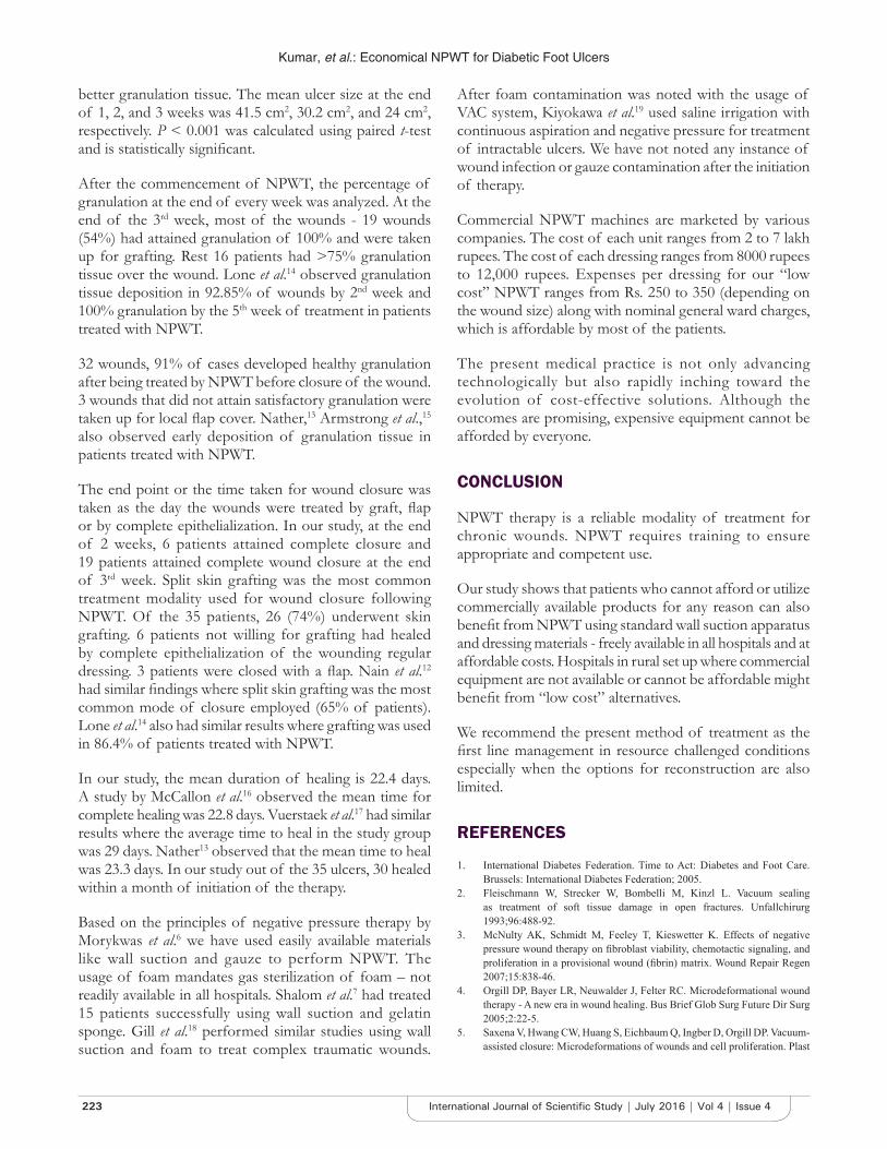
Commercial NPWT machines are marketed by various companies. The cost of each unit ranges from 2 to 7 lakh rupees. The cost of each dressing ranges from 8000 rupees to 12,000 rupees. Expenses per dressing for our “low cost” NPWT ranges from Rs. 250 to 350 (depending on the wound size) along with nominal general ward charges, which is affordable by most of the patients.
The present medical practice is not only advancing technologically but also rapidly inching toward the evolution of cost-effective solutions. Although the outcomes are promising, expensive equipment cannot be afforded by everyone.
CONCLUSION
NPWT therapy is a reliable modality of treatment for chronic wounds. NPWT requires training to ensure appropriate and competent use.
Our study shows that patients who cannot afford or utilize commercially available products for any reason can also benefit from NPWT using standard wall suction apparatus and dressing materials - freely available in all hospitals and at affordable costs. Hospitals in rural set up where commercial equipment are not available or cannot be affordable might benefit from “low cost” alternatives.
We recommend the present method of treatment as the first line management in resource challenged conditions especially when the options for reconstruction are also limited.
REFERENCES
1. International Diabetes Federation. Time to Act: Diabetes and Foot Care. Brussels: International Diabetes Federation; 2005.
2. Fleischmann W, Strecker W, Bombelli M, Kinzl L. Vacuum sealing as treatment of soft tissue damage in open fractures. Unfallchirurg 1993;96:488-92.
3. McNulty AK, Schmidt M, Feeley T, Kieswetter K. Effects of negative pressure wound therapy on fibroblast viability, chemotactic signaling, and proliferation in a provisional wound (fibrin) matrix. Wound Repair Regen 2007;15:838-46.
4. Orgill DP, Bayer LR, Neuwalder J, Felter RC. Microdeformational wound therapy - A new era in wound healing. Bus Brief Glob Surg Future Dir Surg 2005;2:22-5.
5. Saxena V, Hwang CW, Huang S, Eichbaum Q, Ingber D, Orgill DP. Vacuum-assisted closure: Microdeformations of wounds and cell proliferation. Plast

Kumar, et al.: Economical NPWT for Diabetic Foot Ulcers
224International Journal of Scientific Study | July 2016 | Vol 4 | Issue 4
Reconstr Surg 2004;114:1086-96.6. Morykwas MJ, Simpson J, Punger K, Argenta A, Kremers L, Argenta J.
Vacuum-assisted closure: State of basic research and physiologic foundation. Plast Reconstr Surg 2006;117 7 Suppl:121S-6.
7. Shalom A, Eran H, Westreich M, Friedman T. Our experience with a “homemade” vacuum-assisted closure system. Isr Med Assoc J 2008;10:613-6.
8. Bansal E, Garg A, Bhatia S, Attri AK, Chander J. Spectrum of microbial flora in diabetic foot ulcers. Indian J Pathol Microbiol 2008;51:204-8.
9. Alva KA, Aithala PS, Rai R, Rekha B. Clinical and microbiological profile of diabetic foot in patients admitted at a tertiary care center in Mangalore. Muller J Med Sci Res 2013;4:3.
10. Jeffcoate WJ, Chipchase SY, Ince P, Game FL. Assessing the outcome of the management of diabetic foot ulcers using ulcer-related and person-related measures. Diabetes Care 2006;29:1784-7.
11. Margolis DJ, Allen-Taylor L, Hoffstad O, Berlin JA. Diabetic neuropathic foot ulcers: The association of wound size, wound duration, and wound grade on healing. Diabetes Care 2002;25:1835-9.
12. Nain PS, Uppal SK, Garg R, Bajaj K, Garg S. Role of negative pressure wound therapy in healing of diabetic foot ulcers. J Surg Tech Case Rep 2011;3:17-22.
13. Nather A. Role of negative pressure wound therapy in healing of diabetic
foot ulcers. J Surg Tech Case Rep 2011;3:10-1.14. Lone AM, Zaroo MI, Laway BA, Pala NA, Bashir SA, Rasool A. Vacuum-
assisted closure versus conventional dressings in the management of diabetic foot ulcers: A prospective case-control study. Diabet Foot Ankle 2014;5.
15. Armstrong DG, Lavery LA; Diabetic foot study consortium. Negative pressure wound therapy after partial diabetic foot amputation: A multicentre, randomised controlled trial. Lancet 2005;366:1704-10.
16. McCallon SK, Knight CA, Valiulus JP, Cunningham MW, McCulloch JM, Farinas LP. Vacuum-assisted closure versus saline-moistened gauze in the healing of postoperative diabetic foot wounds. Ostomy Wound Manage 2000;46:28-32, 34.
17. Vuerstaek JD, Vainas T, Wuite J, Nelemans P, Neumann MH, Veraart JC. State-of-the-art treatment of chronic leg ulcers: A randomized controlled trial comparing vacuum-assisted closure (VAC) with modern wound dressings. J Vasc Surg 2006;44:1029-37.
18. Gill NA, Hameed A, Sajjad Y, Ahmad Z, Rafique Mirza MA. “Homemade” negative pressure wound therapy: Treatment of complex wounds under challenging conditions. Wounds 2011;23:84-92.
19. Kiyokawa K, Takahashi N, Rikimaru H, Yamauchi T, Inoue Y. New continuous negative-pressure and irrigation treatment for infected wounds and intractable ulcers. Plast Reconstr Surg 2007;120:1257-65.
How to cite this article: Kumar NA, Nihar SS, Chetan K. Low Cost Negative Pressure Wound Therapy for Treatment of Diabetic Foot Ulcers. Int J Sci Stud 2016;4(4):220-224.
Source of Support: Nil, Conflict of Interest: None declared.