long-term outcomes after curative resection for patients with macroscopically solitary...
TRANSCRIPT
ORIGINAL PAPER
Long-term outcomes after curative resection for patientswith macroscopically solitary hepatocellular carcinomawithout macrovascular invasion and an analysis of prognosticfactors
Shu-Hong Li • Wei Wei • Rong-Ping Guo •
Ming Shi • Zhi-Xing Guo • Zhi-Yuan Chen •
Cheng-Zuo Xiao • Mu-Yan Cai • Lie Zheng
Received: 25 June 2013 / Accepted: 9 August 2013 / Published online: 22 August 2013
� Springer Science+Business Media New York 2013
Abstract The long-term outcome and prognostic factors
after curative in patients with single hepatocellular carci-
noma (HCC) without macrovascular invasion are still
unclear. The objective of this study is to evaluate the effect
of curative resection on survival and analyze the prognostic
clinicopathologic factors, especially the presence of micro-
vascular invasion (MVI), in these patients. Two hundred and
sixty consecutive patients with single HCC without macro-
vascular invasion who underwent curative resection from
December 2004 to December 2007 were retrospectively
reviewed in this study. Survival rates were calculated by
using the Kaplan–Meier method. Univariate and multivari-
ate analyses of 14 clinicopathologic factors were performed
to determine the significant prognostic factors. No patient
died within 1 month after the operation. The 1-, 3-, and
5-year overall survival rates after curative resection were
96.54, 83.46, and 74.01 %, respectively. Multivariate anal-
ysis revealed that only the presence of MVI was an
independent negative prognostic factor affecting overall
survival. The 1-, 3-, and 5-year disease-free survival rates
were 79.62, 62.69, and 56.01 %, respectively. The presence
of MVI was the only independent unfavorable prognostic
factor for disease-free survival. According to our analysis,
patients with single HCC without macrovascular invasion
after curative resection can be expected to have considerable
long-term survival. The presence of MVI was an indepen-
dent negative prognostic factor for both overall survival and
disease-free survival. To improve the prognosis, these
patients should be followed up more carefully and might be
good candidates for adjuvant therapy.
Keywords Hepatocellular carcinoma �Hepatectomy � Prognostic factor � Survival �Tumor recurrence � Microvascular invasion
Shu-Hong Li and Wei Wei contributed to the manuscript equally.
S.-H. Li � W. Wei � R.-P. Guo (&) � M. Shi � Z.-Y. Chen �C.-Z. Xiao
Department of Hepatobiliary Oncology, Cancer Center,
Sun Yat-sen University, 651 Dongfeng Road East,
Guangzhou 510060, People’s Republic of China
e-mail: [email protected]
S.-H. Li � W. Wei � R.-P. Guo � M. Shi � Z.-X. Guo �Z.-Y. Chen � C.-Z. Xiao � M.-Y. Cai � L. Zheng
State Key Laboratory of Oncology in South China,
Cancer Center, Sun Yat-sen University, 651 Dongfeng Road
East, Guangzhou 510060, People’s Republic of China
Z.-X. Guo
Department of Ultrasound, Cancer Center, Sun Yat-sen
University, 651 Dongfeng Road East, Guangzhou 510060,
People’s Republic of China
C.-Z. Xiao
Department of General Surgery, Shenzhen Shajing Affiliated
Hospital of Guangzhou Medical University, 3 Shajing
Town Street, Shengzheng 518104, People’s Republic of China
M.-Y. Cai
Department of Pathology, Cancer Center, Sun Yat-sen
University, 651 Dongfeng Road East, Guangzhou 510060,
People’s Republic of China
L. Zheng
Department of Medical Imaging and Interventional Radiology,
Cancer Center, Sun Yat-sen University, 651 Dongfeng Road
East, Guangzhou 510060, People’s Republic of China
123
Med Oncol (2013) 30:696
DOI 10.1007/s12032-013-0696-3
Introduction
Hepatocellular carcinoma (HCC) is the fifth most common
malignancy worldwide [1]. Although it is common in Asia
and Africa, the incidence of HCC has increased rapidly in
the USA [2]. Because of technical improvements in hepatic
resection and earlier diagnosis, the overall and disease-free
survival rates after resection of HCC have improved sig-
nificantly in recent decades [3]. However, long-term sur-
vival remains unsatisfactory because of a high incidence of
recurrence. Even after curative resection, the postoperative
recurrence rate remains high, and most recurrences tend to
occur in remnant liver.
The tumor number [4–7] and macrovascular invasion
[4, 7] are poor predictive factors after resection in HCC
patients. The latest TNM classification for HCC from
UICC/AJCC also emphasizes that the number of nodes and
vascular invasion are the crucial factors for classification
and prognosis of HCC [8]. Although several previous
studies have reported the outcomes and prognostic factors
after hepatectomy in patients with single HCC [7, 9, 10],
they were performed in patients who had confounding
factors, such as noncurative resection [7, 9] and macro-
vascular invasion [7, 9, 10]. So far, no study has focused on
patients with single HCC without macrovascular invasion,
and the long-term outcome and prognostic factors of these
patients remain unknown.
Tumor vessel invasion can be divided into macroscopic
and microscopic or only microscopic. Macroscopic vessel
invasion is an important prognostic factor after surgical
resection of HCC [4, 7]. Although several studies have
suggested that the presence of MVI is an independent
factor predictive of poor survival after resection of HCC
[11–14], the prognostic significance of MVI after curative
resection of patients with solitary HCC without macro-
vascular invasion is still unclear.
We retrospectively reviewed 260 patients with single
HCC without macrovascular invasion. The purpose of this
study was to evaluate the prognostic value of clinical and
histopathologic variables, especially MVI, after curative
resection.
Materials and methods
Patients
From December 2004 to December 2007, 491 consecutive
patients with macroscopically single HCC underwent
hepatectomy in the Hepatobiliary Department of the Sun
Yat-sen University Cancer Center. The diagnosis of HCC
was confirmed by histopathologic examination. Patient
inclusion criteria for this study were as follows: (1)
macroscopically single HCC; (2) no evidence of macro-
vascular invasion as evaluated by two independent, expe-
rienced imaging physicians who examined the preoperative
imaging and resected specimens; (3) underwent curative
hepatic resection; (4) received no previous therapy. Cura-
tive resection of single HCC without macrovascular inva-
sion was performed as described. First, the tumor was
resected. Second, negative surgical margins were con-
firmed by histologic examination. Third, the absence of
extrahepatic metastasis was confirmed. Fourth, the absence
of a residual tumor was confirmed by dynamic contrast-
enhanced computerized tomography (CT) or ultrasonog-
raphy 3–5 weeks postsurgery. Of these 491 patients, 293
patients met these criteria. Thirty-three of the 293 patients
were excluded from the study because detailed records
regarding survival and/or cancer recurrence were not
available, and the remaining 260 patients were enrolled in
this study.
Clinicopathologic variables
We obtained the following clinicopathologic information
from chart review: gender, age, ALT, AST, albumin, total
bilirubin, prothrombin time, hepatitis B serology, HCV-Ab,
cirrhosis, AFP, tumor size, tumor capsule, Edmondson–
Steiner grade, microvascular invasion, resection margin,
blood loss, blood transfusion, and hepatectomy procedure.
Tumor size was based on the largest diameter of the tumor
specimen. The width of the surgical margin was measured
as the smallest distance from the tumor edge to the resec-
tion line.
The histologic grade (I–IV) was determined by using the
criteria of Edmondson and Steiner and was based on the
areas showing the highest grade. Microvascular invasion
was defined as the presence of clusters of cancer cells
floating in the vascular space lined by endothelial cells on
histopathologic examination of the resected specimens.
The presence of MVI was assessed by two independent
pathologists.
Patient follow-up
After discharge, ultrasonography or contrast-enhanced
dynamic CT was performed monthly in the first
2–3 months after surgery, then every 2–3 months in the
first year, and every 3–6 months thereafter. When tumor
recurrence or metastasis was suspected, further investiga-
tions, including magnetic resonance imaging (MRI),
hepatic angiography, and biopsies, were performed. Tele-
phone calls were performed when necessary. The follow-
up data were regularly updated in the database for each
patient. Follow-up data were obtained from the patient’s
database for this study. Follow-up ended on September 1,
Page 2 of 8 Med Oncol (2013) 30:696
123
2012 or the date of the patient’s death. By the end of fol-
low-up, none of the 260 patients were lost. The median
follow-up of the 260 patients was 60.90 months (range
5.40–93.87 months).
Statistical analysis
Overall survival (OS) was measured from the date of
hepatectomy to death or the most recent follow-up time.
Disease-free survival (DFS) was measured from the date of
hepatic resection to the date of the first diagnosis of tumor
recurrence or the most recent follow-up time. Survival rates
were calculated by using the Kaplan–Meier method. Sur-
vival rates were compared by the log-rank test for uni-
variate analysis. All of the variables that were significant in
univariate analysis were then entered into a Cox propor-
tional hazards model for multivariate analysis by using
stepwise selection to identify independent variables corre-
lated with disease-free survival and overall survival. Sta-
tistical analysis was performed by using SPSS 16.0 for
Windows. p values \ 0.05 were considered statistically
significant.
Results
Clinicopathologic characteristics
There were 35 women and 225 men. The median age was
49 years (range 15–76, mean 49.15 ± 11.68). All 260
patients in this study were stage I according to the 7th
NCCN TNM classification for HCC. Table 1 summarizes
the clinicopathologic characteristics of the 260 patients
with single HCC without macrovascular invasion.
Predictors of overall survival
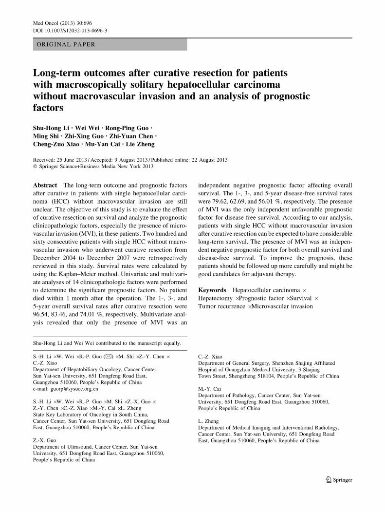
No patients died within 1 month after the operation. The
1-, 3-, and 5-year overall survival rates for this series of
patients were 96.54, 83.46, and 74.01 %, respectively
(Fig. 1).
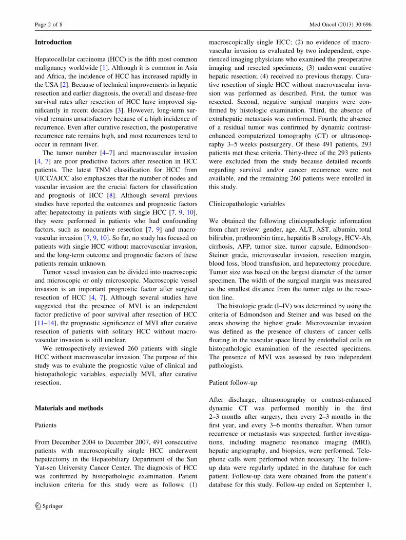
The results of univariate analysis for survival rate after
curative resection of single HCC without macrovascular
invasion are summarized in Table 1. According to univariate
analysis, the presence of microvascular invasion (p = 0.015)
was a significant prognostic factor associated with poor
prognosis (Fig. 2), while resection margin C 1 cm, tumor
size [ 5 cm, and AFP elevation were not significant
(Table 1). The overall survival rate tended to be higher in
patients who underwent curative resection with resection
margin C 1 cm (77.46 % at 5 years) than in those who with
resection margin \ 1 cm (66.94 % at 5 years), although this
difference did not reach statistical significance (p = 0.0504).
To further evaluate the relationship between tumor size, AFP
level and overall survival in patients with single HCC without
macrovascular invasion, we divided these patients into
groups according to the different cutoff values and compared
overall survival rates between these groups. We defined 3
groups according to tumor size: group I (n = 158), tumor
size B 5 cm; group II (n = 115), tumor size between 5 and
10 cm; group III (n = 47), tumor size [ 10 cm. We found no
difference in overall survival between these three groups
(p = 0.202). We defined low-AFP, intermediate-AFP, and
high-AFP groups by using 100, 400, and 1000 ng/ml as cutoff
values, respectively. We found that AFP level was not asso-
ciated with survival in this population (low AFP: p = 0.784;
intermediate AFP: p = 0.796; high AFP: p = 0.157).
The results of a multivariate analysis model including
microvascular invasion and resection margin indicated that
only microvascular invasion was a significant predictive
factor of survival after curative resection for single HCC
without macrovascular invasion (Table 2). The overall
survival rates at 1, 3, and 5 years for patients with and
without MVI were 88.46, 71.15, 63.30 and 98.56, 86.54,
76.70 %, respectively (Table 1).
Predictors of disease-free survival
During follow-up, 122 patients were diagnosed as having
HCC recurrence, and 138 patients had no evidence of
recurrence. The overall recurrence rate was 46.92 %. At
the date of the first diagnosis of tumor recurrence, 105
patients had developed intrahepatic recurrence only, 15
patients had developed extrahepatic recurrence only, and 2
patients had developed both intrahepatic and extrahepatic
recurrence. The disease-free survival rates for this series of
patients at 1, 3, and 5 years were 79.62, 62.69, and
56.01 %, respectively (Fig. 3).
Univariate analysis identified two significant prognostic
factors for disease-free survival: gender (p = 0.031) and
microvascular invasion (p = 0.031) (Figs. 4, 5). The
cumulative disease-free survival rate tended to be higher in
patients who underwent curative resection with resection
margin C 1 cm (59.68 % at 5 years) than in those with
resection margin \ 1 cm (48.51 % at 5 years), although this
difference did not reach statistical significance (p = 0.090).
Multivariate analysis including microvascular invasion,
gender, and resection margin indicated that only microvas-
cular invasion was a significant predictor of disease-free
survival for this series of patients (Fig. 4). The disease-free
survival rates at 1, 3, and 5 years for patients with and
without MVI were 65.38, 51.92, 45.93 and 83.17, 65.38,
58.53 %, respectively (Table 1).
Med Oncol (2013) 30:696 Page 3 of 8
123

Table 1 Clinicopathologic characteristics of the 320 curatively resected patients with single hepatocellular carcinoma (HCC) without macro-
vascular invasion and univariate analysis of survival after hepatectomy according to clinicopathologic factors
Variables No. of patients (%) Overall survival rate (%) p values
1-year 3-year 5-year
Gender 0.106
Male 225 (86.5) 94.29 71.43 65.71
Female 35 (13.5) 96.89 85.33 75.31
Age (years) 0.447
B 50 142 (54.6) 95.07 83.10 75.18
[ 50 118 (45.4) 98.31 83.90 72.65
ALT(U/L) 0.694
B 40 133 (51.2) 95.49 82.71 76.50
[ 40 127 (48.8) 97.64 84.25 71.45
AST(U/L) 0.258
B 45 188 (72.3) 97.34 84.57 76.42
[ 45 72 (27.7) 94.44 80.56 67.79
Child-Pugh A classification 260 (100.0) 96.54 83.46 74.01 –
Hepatitis B serologya 0.835
Positive 235 (90.4) 96.17 83.40 73.81
Negative 24 (9.2) 100.00 83.33 79.06
HCV-Aba 0.774
Positive 3 (1.2) 100.00 66.67 66.67
Negative 256 (98.5) 96.48 83.60 74.39
Cirrhosis 0.203
Yes 213 (81.9) 96.24 82.16 72.05
No 47 (18.1) 97.87 89.36 82.98
AFP elevationa 0.368
Yes 149 (57.3) 95.30 80.54 71.55
No 110(42.3) 98.18 87.27 77.12
Tumor size (cm) 0.166
B 5 129 (49.6) 98.45 87.60 77.17
[ 5 131 (50.4) 94.66 79.39 70.88
Tumor capsule 0.523
Absent 102 (39.2) 93.14 79.41 71.51
Incomplete 67 (25.8) 98.51 85.07 72.90
Complete 91 (35.0) 98.90 86.81 77.55
Edmondson Steiner grade 0.169
I 31 (11.9) 100.0 96.77 83.75
II 133 (51.2) 97.74 84.21 74.12
III ? IVb 96 (36.9) 93.75 78.13 70.72
Microvascular invasion 0.015
Yes 52 (20.0) 88.46 71.15 63.30
No 208 (80.0) 98.56 86.54 76.70
Hepatectomy procedure
Anatomic resection 174 (66.9) 96.51 81.40 69.50 0.259
Nonanatomic resection 86 (33.1) 96.55 84.48 76.25
Resection margin (cm) 0.050
\ 1 86 (33.1) 97.67 81.40 66.94
C 1 174 (66.9) 95.98 84.48 77.46
Blood loss (ml) 0.346
Page 4 of 8 Med Oncol (2013) 30:696
123

Discussion
Hepatocellular carcinoma has a tendency to invade the
intrahepatic vasculature, such as the portal veins (PVs) and
the hepatic veins (HVs). PV involvement is more common
than HV involvement [15]. In patients with HCC, tumor
vascular invasion can be divided into macroscopic and
microscopic. Macroscopic vascular invasion, such as a
tumor thrombus in the portal vein portal and/or hepatic
vein, has been identified as one of the most important
prognostic factors affecting survival or recurrence after
surgical resection of HCC [16, 17]. The extent of the tumor
thrombus in the portal vein and/or hepatic vein is correlated
with overall survival after treatment with partial hepatec-
tomy [18, 19].
Although some studies have suggested that the presence
of MVI is an independent predictor of poor survival after
resection of HCC, the clinical significance of microvascular
invasion for patients with single HCC without macrovascu-
lar invasion remains unclear. MVI occurrence rates are
between 23 and 29 % in single HCC [12, 20]. Shirabe et al.
[20] investigated 218 HCC patients without any extrahepatic
metastases and vascular invasion detected during preopera-
tive evaluation. Among the 218 patients, 146 patients had
single HCC. They found that 42 of the 146 single HCC
patients had MVI. Cho et al. [12] reported that among 230
Table 1 continued
Variables No. of patients (%) Overall survival rate (%) p values
1-year 3-year 5-year
B 500 224 (86.2) 95.98 84.38 75.27
[ 500 36 (13.8) 100.00 77.78 66.08
Blood transfusion 0.744
Yes 37 (14.2) 95.96 83.86 73.76
No 223 (85.8) 100.00 81.08 75.68
Ligation of the hilus 0.464
Yes 208 (80.0) 96.15 82.21 72.90
No 52 (20.0) 98.08 88.46 78.42
ALT alanine aminotransferase, AST aspartate aminotransferase, HCV-Ab hepatitis C virus antibody, AFP a-fetoproteina Not all data available for all patientsb Only 4 patients with undifferentiated HCC
Fig. 1 Survival curve for patients with single hepatocellular carci-
noma (HCC) without macrovascular invasion who underwent curative
resection between 2004 and 2007 (n = 260 patients)
Fig. 2 Overall survival curve for patients with and without micro-
vascular invasion (MVI). The overall survival rate of the MVI(-)
group was significantly better than that of the MVI(?) group
(p = 0.015)
Med Oncol (2013) 30:696 Page 5 of 8
123

HCC patients with single HCC less than 10 cm in diameter,
53 patients had MVI. Our results are similar, as we found that
22.8 % patients with single HCC without macrovascular
invasion had MVI, which suggests that even in early-stage
HCC patients, the MVI rate is relatively high. MVI is often
an independent factor for overall survival [11, 13, 14, 21] and
disease-free survival [12, 13, 21]. In a report of 408 HCC
patients within the Milan criteria who underwent partial
hepatectomy, Fan et al. [14] identified the absence of
microvascular invasion as a favorable predictor for overall
survival. Cho et al. [12] found that the presence of micro-
vascular invasion was a significant prognostic factor for
disease-free survival in both single large HCC ([ 5–10 cm
in diameter) patients and single small HCC (B 5 cm)
patients. Sumie [21] investigated 110 HCC patients without
macroscopic vascular invasion who underwent curative
resection. In their study, MVI was identified as an indepen-
dent risk factor for both recurrence-free survival and disease-
free survival. Similar findings have been reported by other
authors. We also identified the presence of MVI as an inde-
pendent predictor for both poor survival and disease-free
survival after curative resection of HCC. The overall survival
rate and disease-free survival rate were significantly worse
for patients with MVI than those without MVI. These find-
ings indicate that microscopic venous invasion may play an
important role in short-term survival and cancer cell
spreading in HCC patients after curative resection. The
presence of vascular invasion has been considered direct
Table 2 Independent predictors for overall survival identified by
multivariate analysis
Variables Coefficient SE p Hazard
ratio
95 % CI
Microvascular
invasion
0.632 0.265 0.017 1.881 1.119–3.160
Fig. 3 Disease-free survival curve for patients with single hepato-
cellular carcinoma (HCC) without macrovascular invasion who
underwent curative resection between 2004 and 2007 (n = 260
patients)
Fig. 4 Disease-free survival according to gender. The disease-free
survival rate of males was significantly better than that of females
(p = 0.031)
Fig. 5 Disease-free survival according to microvascular invasion
(MVI). The disease-free survival rate of the MVI(-) group was
significantly better than that of the MVI(?) group (p = 0.031)
Page 6 of 8 Med Oncol (2013) 30:696
123

evidence of intrahepatic metastasis. Therefore, it is impor-
tant to detect whether MVI is present in the resected speci-
men of patients with single HCC without macrovascular
invasion because of its importance in predicting both overall
survival and disease-free survival after curative resection.
MVI(?) HCC patients need careful follow-up, and these
patients may be good candidates for adjuvant chemotherapy.
Although some investigators have reported that postopera-
tive TACE could improve the prognosis of HCC patients
with macroscopic portal vein tumor thrombi, its efficacy in
MVI(?) HCC patients is still controversial. So far, no report
has focused on its efficacy in MVI(?) patients with single
HCC without macroscopic vascular invasion. In a future
study, we will further analyze this tissue to improve the
prognosis of these HCC patients (Table 3).
The prognostic significance of resection margin in post-
operative survival and disease-free survival remains contro-
versial in HCC. Some investigators have found that the extent
of the liver resection margin was an independent prognostic
factor for overall survival [22, 23] and disease-free survival
[22–24]. However, other investigators have found that the
extent of liver resection margin did not influence postopera-
tive overall survival [25, 26] or disease-free survival [25–28].
In the present study, we found that the resection margin did
not influence postoperative overall survival or disease-free
survival in patients with single HCC without macrovascular
invasion who underwent curative resection. Although both
the cumulative overall survival rate and disease-free survival
rate tended to be higher in patients who underwent curative
resection with resection margin C 1 cm than those with
resection margin \ 1 cm, this difference did not reach sta-
tistical significance (p = 0.0504; p = 0.090, respectively).
These results imply that resection margin C 1 cm has little
beneficial effect in prolonging both overall survival rate and
disease-free survival rate for patients with single HCC with-
out macrovascular invasion who undergo curative resection.
This observation might have 2 explanations. First, tumor
recurrence could be classified as intrahepatic metastasis and
multicentric occurrence, according to its origin. Multicentric
recurrence could occur anywhere in the liver remnant and
might not be prevented by a wide resection margin. Among
the 15 paired samples from 15 HCC patients who underwent
re-resection, Poon et al. [29] demonstrated that 8 of 9 (89 %)
resected early recurrent (B 1 year) tumors were intrahepatic
metastases, whereas all 6 (100 %) resected late recurrent
([ 1 year) tumors were classified as multicentric occur-
rences. In our study, 57 of the 122 patients with recurrent
HCC developed late intrahepatic recurrence ([ 1 year) that
may have mainly originated from multicentric occurrences.
Second, HCC has a propensity to disseminate by means of
vascular invasion, and intrahepatic metastasis is likely to be
present beyond 1 cm in most patients before operation. Lai
et al. [30] investigated 23 resected liver specimens by serial
sectioning followed by histologic examination, and they
found that 20 of the 23 specimens had either microsatellites or
histologic venous permeation. Among these 20 specimens,
either microsatellites or histologic venous permeation
extended beyond 1 cm from the resection margin in 17
specimens. In their study, 9 specimens had single HCC
without a gross tumor thrombus. Six of the 9 specimens had
either venous permeation or microsatellites, and either
microsatellites or histologic venous permeation was beyond
1 cm from the resection margin in 4 of these 6 specimens.
However, a 2-cm margin improved the potential for a cure in
macroscopically solitary HCC without vascular invasion. Our
previous study showed that micrometastases extended
beyond the 1-cm margin in 28 (24.8 %) patients but beyond
the 2-cm margin in only nine (8.0 %) patients with macro-
scopically solitary tumors without vascular invasion [15].
Thus, a wide resection margin (2 cm) should be recom-
mended for the patient to survive the operation.
Tumor size is not an indicator of poor prognosis in HCC
patients with single tumors after hepatic resection [7, 9, 10].
Yang et al. [9] reported that solitary large HCC patients had
similar overall survival and disease-free survival to small
HCC patients. The latest American Joint Commission on
Cancer (AJCC) staging classification for HCC also classified
single HCC without vascular invasion as T1, irrespective of
its size. The present study is consistent with the previous
studies showing that tumor size did not affect the postoper-
ative outcomes in patients with single HCC. Although AFP
level is the most effective marker for HCC diagnosis and
recurrent surveillance, the prognostic value of AFP has been
controversial. While some studies suggested that AFP was
associated with survival after resection [10, 31], other studies
found that AFP was not a predictor of survival. In the present
study, AFP level did not have prognostic value for overall
survival or disease-free survival.
There are some limitations of this study. First, it was a
retrospective study. Second, we examined only the resected
specimens for MVI. Whether MVI was present in the liver
remnant is not known. Lastly, sampling may have affected
the pathologists’ diagnosis of MVI, and inadequate sam-
pling may have caused false-negative detection.
In conclusion, in patients with single HCC without mac-
rovascular invasion, we found that the presence of MVI was a
poor prognostic factor for both overall survival and disease-
free survival after curative resection. These findings suggest
Table 3 Independent predictors for disease-free survival identified by
multivariate analysis
Variables Coefficient SE p Hazard
ratio
95 % CI
Microvascular
invasion
0.451 0.210 0.032 1.569 1.039–2.370
Med Oncol (2013) 30:696 Page 7 of 8
123

that MVI can be employed to estimate both overall survival
and disease-free survival after curative resection in patients
with single HCC without macrovascular invasion. Patients
with MVI may be good candidates for adjuvant chemotherapy.
Further studies to analyze whether adjuvant chemotherapy can
improve the survival of these patients are necessary.
Acknowledgments We thank Yu Zhang and Yun-Xian Mo for their
contribution to this study. This work was supported by grants from the
National Natural Science Foundation of China (No. 81172037/
H1606) and Guangzhou municipal science and technology project of
China (No.2012J4100078).
Conflict of interest None.
References
1. Bosch FX, Ribes J, Borras J. Epidemiology of primary liver
cancer. Semin Liver Dis. 1999;19(3):271–85.
2. El-Serag HB, Mason AC. Rising incidence of hepatocellular
carcinoma in the United States. N Engl J Med. 1999;340(10):
745–50.
3. Poon RT, Fan ST, Lo CM, Ng IO, Liu CL, Lam CM, et al.
Improving survival results after resection of hepatocellular car-
cinoma: a prospective study of 377 patients over 10 years. Ann
Surg. 2001;234(1):63–70.
4. Vauthey JN, Lauwers GY, Esnaola NF, Do KA, Belghiti J, Mirza
N, et al. Simplified staging for hepatocellular carcinoma. J Clin
Oncol. 2002;20(6):1527–36.
5. Sun HC, Zhang W, Qin LX, Zhang BH, Ye QH, Wang L, et al.
Positive serum hepatitis B e antigen is associated with higher risk
of early recurrence and poorer survival in patients after curative
resection of hepatitis B-related hepatocellular carcinoma. J Hep-
atol. 2007;47(5):684–90.
6. Kiriyama S, Uchiyama K, Ueno M, Ozawa S, Hayami S, Tani M,
et al. Triple positive tumor markers for hepatocellular carcinoma
are useful predictors of poor survival. Ann Surg. 2011;254(6):
984–91.
7. Ariizumi SI, Kotera Y, Takahashi Y, Katagiri S, Yamamoto M.
Impact of hepatectomy for huge solitary hepatocellular carci-
noma. J Surg Oncol. 2012;107(4):408–13.
8. Edge SB BD, Carducci MA et al, eds., editor. American Joint
Committee on Cancer (AJCC) Cancer Staging Manual. 7th ed.
New York: Springer; 2009.
9. Yang LY, Fang F, Ou DP, Wu W, Zeng ZJ, Wu F. Solitary large
hepatocellular carcinoma: a specific subtype of hepatocellular
carcinoma with good outcome after hepatic resection. Ann Surg.
2009;249(1):118–23.
10. Zhou L, Rui JA, Wang SB, Chen SG, Qu Q. Prognostic factors of
solitary large hepatocellular carcinoma: the importance of dif-
ferentiation grade. Eur J Surg Oncol. 2011;37(6):521–5.
11. Ng KK, Vauthey JN, Pawlik TM, Lauwers GY, Regimbeau JM,
Belghiti J, et al. Is hepatic resection for large or multinodular
hepatocellular carcinoma justified? Results from a multi-institu-
tional database. Ann Surg Oncol. 2005;12(5):364–73.
12. Cho YB, Lee KU, Lee HW, Cho EH, Yang SH, Cho JY, et al.
Outcomes of hepatic resection for a single large hepatocellular
carcinoma. World J Surg. 2007;31(4):795–801.
13. Wang CC, Iyer SG, Low JK, Lin CY, Wang SH, Lu SN, et al.
Perioperative factors affecting long-term outcomes of 473 con-
secutive patients undergoing hepatectomy for hepatocellular
carcinoma. Ann Surg Oncol. 2009;16(7):1832–42.
14. Fan ST, Poon RT, Yeung C, Lam CM, Lo CM, Yuen WK, et al.
Outcome after partial hepatectomy for hepatocellular cancer
within the Milan criteria. Br J Surg. 2011;98(9):1292–300.
15. Shi M, Zhang CQ, Zhang YQ, Liang XM, Li JQ. Micrometas-
tases of solitary hepatocellular carcinoma and appropriate
resection margin. World J Surg. 2004;28(4):376–81.
16. Ikai I, Arii S, Kojiro M, Ichida T, Makuuchi M, Matsuyama Y,
et al. Reevaluation of prognostic factors for survival after liver
resection in patients with hepatocellular carcinoma in a Japanese
nationwide survey. Cancer. 2004;101(4):796–802.
17. Shah SA, Greig PD, Gallinger S, Cattral MS, Dixon E, Kim RD,
et al. Factors associated with early recurrence after resection for
hepatocellular carcinoma and outcomes. J Am Coll Surg. 2006;
202(2):275–83.
18. Chen XP, Qiu FZ, Wu ZD, Zhang ZW, Huang ZY, Chen YF,
et al. Effects of location and extension of portal vein tumor
thrombus on long-term outcomes of surgical treatment for
hepatocellular carcinoma. Ann Surg Oncol. 2006;13(7):940–6.
19. Shi J, Lai EC, Li N, Guo WX, Xue J, Lau WY, et al. Surgical
treatment of hepatocellular carcinoma with portal vein tumor
thrombus. Ann Surg Oncol. 2010;17(8):2073–80.
20. Shirabe K, Itoh S, Yoshizumi T, Soejima Y, Taketomi A,
Aishima S, et al. The predictors of microvascular invasion in
candidates for liver transplantation with hepatocellular carci-
noma-with special reference to the serum levels of des-gamma-
carboxy prothrombin. J Surg Oncol. 2007;95(3):235–40.
21. Sumie S, Kuromatsu R, Okuda K, Ando E, Takata A, Fukushima
N, et al. Microvascular invasion in patients with hepatocellular
carcinoma and its predictable clinicopathological factors. Ann
Surg Oncol. 2008;15(5):1375–82.
22. Shi M, Guo RP, Lin XJ, Zhang YQ, Chen MS, Zhang CQ, et al.
Partial hepatectomy with wide versus narrow resection margin for
solitary hepatocellular carcinoma: a prospective randomized trial.
Ann Surg. 2007;245(1):36–43.
23. Wang J, Xu LB, Liu C, Pang HW, Chen YJ, Ou QJ. Prognostic
factors and outcome of 438 Chinese patients with hepatocellular
carcinoma underwent partial hepatectomy in a single center.
World J Surg. 2010;34(10):2434–41.
24. Shinkawa H, Uenishi T, Takemura S, Ohba K, Ogawa M, Ichi-
kawa T, et al. Risk factors for postoperative recurrence of non-B
non-C hepatocellular carcinoma. J Hepatobiliary Pancreat Sci.
2010;17(3):291–5.
25. Liu L, Miao R, Yang H, Lu X, Zhao Y, Mao Y, et al. Prognostic
factors after liver resection for hepatocellular carcinoma: a single-
center experience from China. Am J Surg. 2012;203(6):741–50.
26. Giuliante F, Ardito F, Pinna AD, Sarno G, Giulini SM, Ercolani
G, et al. Liver resection for hepatocellular carcinoma \/=3 cm:
results of an Italian multicenter study on 588 patients. J Am Coll
Surg. 2012;215(2):244–54.
27. Poon RT, Fan ST, Ng IO, Wong J. Significance of resection
margin in hepatectomy for hepatocellular carcinoma: a critical
reappraisal. Ann Surg. 2000;231(4):544–51.
28. Chun JM, Kwon HJ, Sohn J, Kim SG, Park JY, Bae HI, et al.
Prognostic factors after early recurrence in patients who under-
went curative resection for hepatocellular carcinoma. J Surg
Oncol. 2011;103(2):148–51.
29. Poon RT, Fan ST, Ng IO, Lo CM, Liu CL, Wong J. Different risk
factors and prognosis for early and late intrahepatic recurrence after
resection of hepatocellular carcinoma. Cancer. 2000;89(3):500–7.
30. Lai EC, You KT, Ng IO, Shek TW. The pathological basis of
resection margin for hepatocellular carcinoma. World J Surg.
1993;17(6):786–90; discussion 91.
31. Santambrogio R, Opocher E, Costa M, Barabino M, Zuin M,
Bertolini E, et al. Hepatic resection for ‘‘BCLC stage A’’ hepa-
tocellular carcinoma. The prognostic role of alpha-fetoprotein.
Ann Surg Oncol. 2012;19(2):426–34.
Page 8 of 8 Med Oncol (2013) 30:696
123