locking plate for proximal humeral fracture in the elderly
TRANSCRIPT
Locking Plate for Proximal Humeral Fracture in the Elderly Population: Serial Change of Neck Shaft Angle
Aditya C Pawaskar, MD, Kee-Won Lee, MD, Jong-Min Kim, MD, Jin-Woong Park, MD, Iman W Aminata, MD, Hong-Jun Jung, MD, Jae-Myeung Chun, MD, In-Ho Jeon, MD
Department of Orthopedic Surgery, Asan Medical Center, University of Ulsan College of Medicine, Seoul, Korea
Original Article Clinics in Orthopedic Surgery 2012;4:209-215 • http://dx.doi.org/10.4055/cios.2012.4.3.209
Received February 21, 2012; Accepted March 29, 2012Correspondence to: In-Ho Jeon, MDDepartment of Orthopedic Surgery, Asan Medical Center, University of Ulsan College of Medicine, 86 Asanbyeongwon-gil, Songpa-gu, Seoul 138-736, KoreaTel: +82-2-3010-3896, Fax: +82-2-488-7877E-mail: [email protected]
Elderly patients depend on an intact musculoskeletal system in order to maintain function and independence in activities of daily living. Proximal humeral fractures account for around 5% of all fractures and there is an in-creasing rate in elderly patients.1) There is a lack of agree-
Background: We conducted this radiographic study in the elderly population with proximal humeral fracture aiming to evaluate 1) the serial changes of neck-shaft angle after locking plate fixation and 2) find relationship between change in neck shaft angle and various factors such as age, fracture pattern, severity of osteoporosis, medial support and initial reduction angle.Methods: Twenty-five patients who underwent surgical treatment for proximal humeral fracture with locking plate between Sep-tember 2008 and August 2010 are included. True anteroposterior and axillary lateral radiographs were made postoperatively and at each follow-up visit. Measurement of neck shaft angle was done at immediate postoperative, 3 months postoperative and a final follow-up (average, 11 months; range, 8 to 17 months). Severity of osteoporosis was assessed using cortical thickness sug-gested by Tingart et al.Results: The mean neck shaft angles were 133.6o (range, 100o to 116o) at immediate postoperative, 129.8o (range, 99o to 150o) at 3 months postoperative and 128.4o (range, 97o to 145o) at final follow-up. The mean loss in the neck-shaft angle in the first 3 months was 3.8o as compared to 1.3o in the period between 3 months and final follow-up. This was statistically significant (p = 0.002), indicating that most of the fall in neck shaft angle occurs in the first three months after surgery. Relationship between neck shaft angle change and age (p = 0.29), fracture pattern (p = 0.41), cortical thickness (p = 0.21), medial support (p = 0.63) and initial reduc-tion accuracy (p = 0.65) are not statistically significant.Conclusions: The proximal humerus locking plate maintains reliable radiographic results even in the elderly population with proximal humerus fracture.Keywords: Proximal humeral fracture, Neck shaft angle, Elderly population, Locking plate
ment on the best treatment of these fractures, particularly in older patients. Traditional internal fixation stabilization methods for proximal humeral fractures include percuta-neous K-wiring, screw fixation, tension band wiring and conventional plates and screws, intramedullary nailing.2-4) Surgical treatment either by open reduction and internal fixation or prosthetic replacement in these population has been reported being successful, however, there have been concerns with regard to poor bone quality in geriatric pa-tients.5)
In order to overcome the problems associated with osteoporotic unstable proximal humeral fractures, several implants have been developed during the last decades.
Copyright © 2012 by The Korean Orthopaedic AssociationThis is an Open Access article distributed under the terms of the Creative Commons Attribution Non-Commercial License (http://creativecommons.org/licenses/by-nc/3.0)
which permits unrestricted non-commercial use, distribution, and reproduction in any medium, provided the original work is properly cited.Clinics in Orthopedic Surgery • pISSN 2005-291X eISSN 2005-4408
210
Pawaskar et al. Locking Plate for Proximal Humeral Fracture in the Elderly PopulationClinics in Orthopedic Surgery • Vol. 4, No. 3, 2012 • www.ecios.org
One of the most recent developments is the locking com-pression plate (LCP). The LCP allows controlled fracture compression and has been widely used as angular stability offers several advantages in osteoporotic bone.6)
Several studies have yielded promising clinical re-sults with the LCP.7,8) Although outcomes of the LCP for proximal humeral fracture have been well documented and the knowledge of the LCP is continuously increasing, there are few studies which follow the serial postoperative changes in the neck shaft angle after use of the LCP in the elderly.
We conducted this radiographic study in the elderly population with proximal humeral fractures aiming to evaluate 1) the serial changes of neck-shaft angle after LCP fixation and 2) find the relationship between the change in neck shaft angle and various factors such as age, fracture pattern, severity of osteoporosis, medial support and ini-tial reduction angle.
METHODS
This study was conducted in a tertiary referral hospital. All patients who underwent surgical treatment for a proximal humeral fracture with a locking plate during the study pe-riod were collected from the hospital trauma registration and reviewed for the study.
The criteria for inclusion in the present study were a proximal humeral fracture that had been treated surgically up to three weeks after the initial injury. The cut off value of age at the time of surgery was 65 years or older. Be-tween September 2008 and August 2010, 41 patients met the criteria. We excluded patients with isolated fractures of the greater tuberosity or lesser tuberosity, a pathologi-cal fracture, patients with polytrauma, and patients who were not able to comprehend the given informed consent reliably. In addition, those patients with four part fracture dislocation who required prosthetic replacement were also excluded. Individuals who were managed non-operatively (as a consequence of being deemed unfit for surgery) were excluded from the analysis. We also excluded patients with incomplete data and lost follow-up. Twenty-five patients were available for the final analysis. Senior authors con-ducted all the operations during the study period.
Operative TechniqueSurgery was performed with the patient in the beach-chair position on a radiolucent table. The standard deltopectoral approach was used. The fracture was exposed and special attention was paid to minimize surgical trauma to the ad-jacent soft tissue. We did not expose the medial soft tissue
hinge in order to reduce the fracture. The reduction was confirmed with the use of image intensification. The LCP was positioned 2 cm posterior to the bicipital groove, with care being taken to ensure that a sufficient gap was main-tained between the plate and the long head of the biceps. Medial hinge was considered an important radiographic indicator when assessing initial reduction accuracy. When fracture reduction and subsequent screw positioning was considered adequate, the plate was fixed definitively with the insertion of locking screws into the humeral head. Tension band type sutures were added over the greater tu-berosity to provide additional stability.
Postoperatively, the arm was immobilized in a sling and controlled passive mobilization exercise with abduc-tion and flexion beyond 90o was started within three weeks postoperatively, depending on the stability of the osteosyn-thesis and the bone quality.
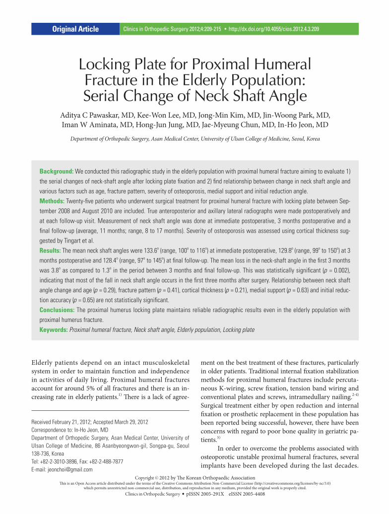
Radiographic EvaluationTrue anteroposterior and axillary lateral radiographs were made postoperatively and at each follow-up visit. The assessment of radiographs was done at three points in the postoperative period, immediate postoperative, at 3 months postoperative, and a final follow-up (average, 11 months; range, 8 to 17 months). All radiographs were tak-en by the same two technicians in the same setting as stan-dard protocol in order to minimize potential errors. Also, the radiographs were analyzed by one author. The neck-shaft angle was measured as described by Agudelo et al.9) (Fig. 1). Radiographs were assessed to find the presence of bridging osseous trabeculae, which is suggestive of heal-
Fig. 1. Measurement of neck shaft angle.
211
Pawaskar et al. Locking Plate for Proximal Humeral Fracture in the Elderly PopulationClinics in Orthopedic Surgery • Vol. 4, No. 3, 2012 • www.ecios.org
ing, as well as failure of the fixation. A fracture pattern was determined and confirmed by the senior surgeons based on the Neer classification.10) Severity of osteoporosis in the proximal humerus was assessed using cortical thickness suggested by Tingart et al.11) On the anteroposterior view, we measured the lateral and medial cortical thickness at two different levels and calculated the mean of the four measurements. Level 1 was the most proximal level of the humeral diaphysis and level 2 was 20 mm distal to level 1. The presence or absence of medial support, as described by Gardner et al.12) was assessed. We divided the patients into two groups; those with medial support and those without medial support. On the anteroposterior view, we determined that the fracture has medial support when 1) the medial cortex was intact without comminution, and reduced anatomically, and/or 2) the shaft was impacted into the head fragment, and/or 3) a locking screw was placed in the inferomedial quadrant of the humeral head within 5 mm of the subchondral bone.
Statistical AnalysisChanges in neck-shaft angle (difference between the im-mediate postoperative angle and the last follow-up angle) were compared with fracture pattern and presence or ab-sence of medial support using t-test. We also used t-test to see whether the fall in the angle during the first 3 months was higher than the fall in the angle between 3 months and the final follow-up period. We fitted univariate re-gression for age and combined cortical thickness (CCT) to see if there is any association between the neck-shaft angle changes and continuous variables of age and CCT. In addition, we fitted a linear mixed model to see whether changing pattern over time is statistically significant. The linear mixed model includes additional random-effect terms, so it is often appropriate for clustered data or data gathered over time regarding the same individuals.13) All reported p-values were 2-sided, and p-values < 0.05 were considered statistically significant. A package developed by Pinheiro and Bates13) and R software were used for all statistical analyses.
RESULTS
The average age of the patients was 70 years (range, 65 to 82 years). Twenty-one (84%) patients were women and four (16%) were men. Twenty-two (88%) patients sus-tained the fracture due to a trivial fall such as falling from a standing position, two were involved in a motor-vehicle accident and one was physically assaulted. There were ten (40%) Neer type 2 fractures and fifteen (60%) Neer type 3 fractures.
All fractures were united within three months of the index operation. Complications included postoperative delirium and superficial stitch abscess in one patient each. There were no deep infections, nerve or vascular injuries, complications related to hardware such as loosening or screw cutout, nonunion or osteonecrosis of the humeral head.
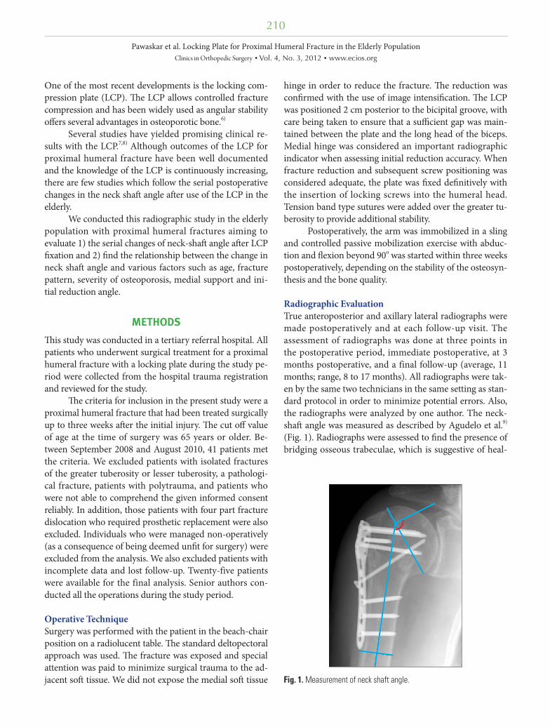
Neck Shaft AngleThe mean neck shaft angles at different points in time are represented in Table 1 and Fig. 2. The mean loss of neck shaft angle between the immediate postoperative and the final follow-up period was 5.6o (range, 0o to 14o) (Fig. 2). The mean loss in the neck-shaft angle in the first 3 months was 3.8o as compared to 1.3o in the period between 3 months and the final follow-up. This was statistically sig-nificant (p = 0.002), indicating that most of the decrease in neck shaft angle occurs in the first three months after sur-gery. Only three patients showed a total loss of reduction of greater than 10o.
We considered an immediate postoperative neck shaft angle of less than 120o to be varus malalignment.9) There were three cases with varus malalignment in our se-ries. The mean fall in the neck shaft angle in these was 5.3o.
Table 1. Neck Shaft Angle Change in the Serial Radiographs
Time after surgery Mean neck shaft angle (o) (SD, range)
Immediate 133.6 (11.6, 100-150)
3 mo 129.8 (12.1, 99-150)
Final follow-up 128.4 (12.1, 97-145) Fig. 2. Changes in the neck shaft (NS) angle over various time periods.
212
Pawaskar et al. Locking Plate for Proximal Humeral Fracture in the Elderly PopulationClinics in Orthopedic Surgery • Vol. 4, No. 3, 2012 • www.ecios.org
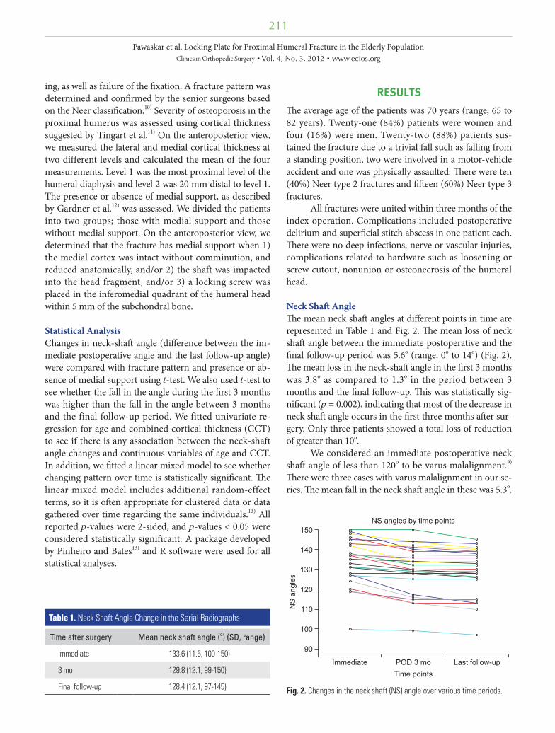
Relationship between the Change in Neck Shaft Angle and Various Clinical FactorsAgeThere was a trend that with increasing patient age, the fall in neck shaft angle decreased, but this was not statistically significant (p = 0.29) (Fig. 3).
Fracture patternThe mean decrease in 2-part fractures was 5.9o and in 3-part fractures it was 4.7o. There was no statistically sig-nificant relationship between the change in neck shaft angle and fracture pattern (p = 0.41) (Fig. 4).
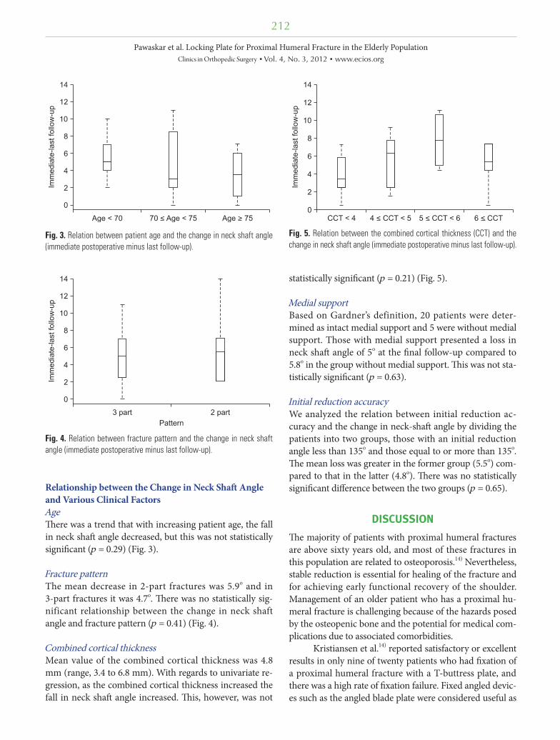
Combined cortical thicknessMean value of the combined cortical thickness was 4.8 mm (range, 3.4 to 6.8 mm). With regards to univariate re-gression, as the combined cortical thickness increased the fall in neck shaft angle increased. This, however, was not
statistically significant (p = 0.21) (Fig. 5).
Medial supportBased on Gardner’s definition, 20 patients were deter-mined as intact medial support and 5 were without medial support. Those with medial support presented a loss in neck shaft angle of 5o at the final follow-up compared to 5.8o in the group without medial support. This was not sta-tistically significant (p = 0.63).
Initial reduction accuracyWe analyzed the relation between initial reduction ac-curacy and the change in neck-shaft angle by dividing the patients into two groups, those with an initial reduction angle less than 135o and those equal to or more than 135o. The mean loss was greater in the former group (5.5o) com-pared to that in the latter (4.8o). There was no statistically significant difference between the two groups (p = 0.65).
DISCUSSION
The majority of patients with proximal humeral fractures are above sixty years old, and most of these fractures in this population are related to osteoporosis.14) Nevertheless, stable reduction is essential for healing of the fracture and for achieving early functional recovery of the shoulder. Management of an older patient who has a proximal hu-meral fracture is challenging because of the hazards posed by the osteopenic bone and the potential for medical com-plications due to associated comorbidities.
Kristiansen et al.14) reported satisfactory or excellent results in only nine of twenty patients who had fixation of a proximal humeral fracture with a T-buttress plate, and there was a high rate of fixation failure. Fixed angled devic-es such as the angled blade plate were considered useful as
Fig. 3. Relation between patient age and the change in neck shaft angle (immediate postoperative minus last follow-up).
Fig. 4. Relation between fracture pattern and the change in neck shaft angle (immediate postoperative minus last follow-up).
Fig. 5. Relation between the combined cortical thickness (CCT) and the change in neck shaft angle (immediate postoperative minus last follow-up).
213
Pawaskar et al. Locking Plate for Proximal Humeral Fracture in the Elderly PopulationClinics in Orthopedic Surgery • Vol. 4, No. 3, 2012 • www.ecios.org
they resisted angular deformation and torsion.15) However, Meier et al.16) did not recommend internal fixation with angled blade plate in unstable proximal humerus fractures due to the high rate of complications (33%) including protrusion of the blade into the glenohumeral joint (22%). Disadvantages of the percutaneous pinning technique include the potential for pin migration, loss of reduction, and pin-site infection. Injury to important anatomic struc-tures concerning the shoulder is also a potential concern with this technique.17) Moreover, fixation using intramed-ullary nailing has also shown variable results.18,19)
The LCP is a relatively new modality in the arma-mentarium of the surgeon in tackling this category of fractures.20) Several advantages have been outlined in re-cent literature. According to Bjorkenheim et al.,8) the main advantage of this plate was apparent in elderly patients, since there were no failures of the internal fixation in this group, and they were able to attain an activity level, which was sufficient to satisfy their needs regarding independent daily living. They are site-specific, low-profile plates. The plate is precontoured for the proximal humerus, and the insertion of locking screws obviates the need for a plate-to-bone compression, preserving the blood supply to the bones.20) The insertion of multiple polyaxial locking screws through the specific targeting device into the fragments of the humeral head provides fixed-angle support in multiple planes, which in theory should maintain the reduction achieved, while allowing for early mobilization.21) The key to this technology is the fixed-angle relationship between the screws and plate. The threaded screw heads are locked into the threaded plate holes in order to prevent screw toggle, slide, and pull-out, thus diminishing the possibility of a primary or secondary loss of reduction.6) The load is transmitted from the bone to the plate via the screw-plate threaded connection. Since the plate and screws form a single stable system, the stability of the fracture depends on the stiffness of the entire construct.6)
The mean age of the patients in our study group was 70 years. Also, 84% of our sample population was women. By the Neer classification, there were 10 two part fractures and 15 three part fractures. The demographic and frac-ture-related characteristics in our study group were thus comparable to those in previous studies.7,15,16)
We observed two complications in 25 patients dur-ing the one-year follow-up. There was one case with a superficial infection, which was effectively treated with an-tibiotics. This is similar to the 5% superficial infection rate reported by Koukakis et al.22) The other was postopera-tive delirium from which the patient recovered within 24 hours. While complications such as subacromial impinge-
ment, initial malreduction, nonunion, and osteonecrosis have been reported in literature, the overall complication rate in our study was encouraging.2,7) This may be due to our exclusion of Neer type 4 fractures, which have report-ed a prevalence of 21% to 75% regarding osteonecrosis.23) We did have three cases with initial varus malalignment, but the mean fall in neck shaft angle in these cases (5.3o) matched that of the entire sample (5.6o).
In our series, the mean loss of neck shaft angle was 5.9o in two part fractures and 4.7o in three part fracture pa-tients. This loss occurred maximally within the first three months of surgery. Zhang et al.24) reported an early loss of fixation in the initial 12 weeks after surgery. They reported a mean loss of 1.6o ± 1.3o in patients with medial support and 1.3o ± 1.2o in patients without medial support in two part fractures, and a mean loss of 3.5o ± 2.4o in patients with medial support and 6.1o ± 3.1o in patients without medial support in three part fractures.
We observed that the patient age did not signifi-cantly affect the loss in neck shaft angle. Moonot et al.25) demonstrated no significant difference in the functional outcome between patients under and over 65 years old at a mean follow-up of 11 months post PHILOS plate fixation. The LCP may thus provide an acceptable surgical option in proximal humerus fractures irrespective of patient age.
We used the combined cortical thickness as de-scribed by Tingart et al.11) as an indicator of bone quality. The combined cortical index did not significantly affect the change in neck shaft angle. Siwach et al.26) concluded in their study that proximal humerus fractures with grade IV osteoporosis by Singh’s index did worse as compared to those with grade II osteoporosis. Bjorkenheim et al.8) used an LCP in 72 patients and concluded that this method was safe and recommended its use in treating proximal hu-merus fractures in elderly patients with poor bone stock. In this study, we found that Tingart’s cortical index pre-sented minimal effect on the decrease of neck shaft angle. This is in accordance to a recent biomechanical study on locking plates in the proximal humerus where the authors concluded that the mechanical strength of the plate was unaffected based on a threshold combined proximal hu-merus cortical thickness of 4 mm.27)
Evaluation of fractures on the basis of medial sup-port as described by Gardner et al.12) revealed that the loss of neck shaft angle was less when there was medial support, although this was not statistically significant. We could not replicate the statistical results obtained by Gard-ner et al.12) but two part fractures showed a slightly better result than three part fractures. Gardner et al.12) stressed the importance of recreating continuity of the medial col-

214
Pawaskar et al. Locking Plate for Proximal Humeral Fracture in the Elderly PopulationClinics in Orthopedic Surgery • Vol. 4, No. 3, 2012 • www.ecios.org
umn or placement of inferomedial screws in the humeral head. Analysis of the histomorphometry and microstruc-tural architecture of the humeral head bone stock also demonstrates that trabecular thickness and density are the greatest in the medial region.28) Liew et al.29) also found screw purchase to be significantly greater in screws placed into the medial subchondral bone.
Yang et al.30) in a recent study concluded that medial support and fracture pattern were the only two factors significantly related to the functional outcome, with the two part and three part fractures performing better than the four part fractures. Analysis of the fracture pattern with changes in the neck shaft angle in our study did not reveal any significant correlation. The mean loss of neck shaft angle was 5.9o for 2 part fractures and 4.7o for 3 part fractures. In two part and three part fractures, the fracture pattern thus did not significantly affect the radiological outcome of the treatment.
We also analyzed the effect of immediate postop-erative neck shaft angle on the final outcome. Division of patients into two groups, those with a neck shaft angle less than 135o and those with a neck shaft angle equal to or more than 135o, revealed a slightly higher fall in the former group, though not statistically significant. Agudelo et al.9) found a statistically significant association between varus malreduction immediately following surgery and early loss of fixation. Leonard et al.20) reported a better outcome in patients with intraoperative restoration of the humeral neck shaft angle to greater than 90o compared to those who were fixed with an angle less than 90o, though it was not statistically significant. They advocate optimal restora-tion of the neck shaft angle in order to avoid the potential
complication of varus collapse.The limitation of our study is in regards to the
relatively small sample size. Also, we have not clinically assessed the patients nor applied functional scoring and hence could not correlate the functional outcome with the radiological findings. However, the results from our radio-logical analysis demonstrate that the locking plate with its ease of use, respect for biology, polyaxial fixation and low complication rates, is a useful tool in treating Neer type 2 and type 3 proximal humerus fractures in patients over the age of 65 years.
Although age and the deterioration of bone quality with age may be areas of concern, the proximal humerus locking plate offers a reliable option to the surgeon in order to treat these types of fractures in the elderly. Our findings emphasize the fact that a meticulous surgical technique, concerning the initial reduction and correct placement of the locking plate and screws, can provide a better outcome and fewer complications.
CONFLICT OF INTEREST
No potential conflict of interest relevant to this article was reported.
ACKNOWLEDGEMENTS
This work was supported by the Global Frontier R&D Pro-gram on “Human-centered Interaction for Coexistence” funded by the National Research Foundation of Korea grant funded by the Korean Government (MEST; NRF-M1AXA003-2011-0028358).
REFERENCES1. Helmy N, Hintermann B. New trends in the treatment
of proximal humerus fractures. Clin Orthop Relat Res. 2006;442:100-8.
2. Charalambous CP, Siddique I, Valluripalli K, et al. Proximal humeral internal locking system (PHILOS) for the treat-ment of proximal humeral fractures. Arch Orthop Trauma Surg. 2007;127(3):205-10.
3. Wijgman AJ, Roolker W, Patt TW, Raaymakers EL, Marti RK. Open reduction and internal fixation of three and four-part fractures of the proximal part of the humerus. J Bone Joint Surg Am. 2002;84(11):1919-25.
4. Mittlmeier TW, Stedtfeld HW, Ewert A, Beck M, Frosch B, Gradl G. Stabilization of proximal humeral fractures with an angular and sliding stable antegrade locking nail (Targon
PH). J Bone Joint Surg Am. 2003;85 Suppl 4:136-46.
5. Bastian JD, Hertel R. Osteosynthesis and hemiarthroplasty of fractures of the proximal humerus: outcomes in a con-secutive case series. J Shoulder Elbow Surg. 2009;18(2):216-9.
6. Wagner M. General principles for the clinical use of the LCP. Injury. 2003;34 Suppl 2:B31-42.
7. Fankhauser F, Boldin C, Schippinger G, Haunschmid C, Szyszkowitz R. A new locking plate for unstable frac-tures of the proximal humerus. Clin Orthop Relat Res. 2005;(430):176-81.
8. Bjorkenheim JM, Pajarinen J, Savolainen V. Internal fixation of proximal humeral fractures with a locking compression plate: a retrospective evaluation of 72 patients followed for a minimum of 1 year. Acta Orthop Scand. 2004;75(6):741-5.
215
Pawaskar et al. Locking Plate for Proximal Humeral Fracture in the Elderly PopulationClinics in Orthopedic Surgery • Vol. 4, No. 3, 2012 • www.ecios.org
9. Agudelo J, Schurmann M, Stahel P, et al. Analysis of efficacy and failure in proximal humerus fractures treated with lock-ing plates. J Orthop Trauma. 2007;21(10):676-81.
10. Neer CS 2nd. Displaced proximal humeral fractures. I. Classification and evaluation. J Bone Joint Surg Am. 1970;52(6):1077-89.
11. Tingart MJ, Apreleva M, von Stechow D, Zurakowski D, Warner JJ. The cortical thickness of the proximal humeral diaphysis predicts bone mineral density of the proximal hu-merus. J Bone Joint Surg Br. 2003;85(4):611-7.
12. Gardner MJ, Weil Y, Barker JU, Kelly BT, Helfet DL, Lo-rich DG. The importance of medial support in locked plating of proximal humerus fractures. J Orthop Trauma. 2007;21(3):185-91.
13. Pinheiro J, Bates D. Mixed-effects models in S and S-plus. New York: Springer; 2000.
14. Kristiansen B, Barfod G, Bredesen J, et al. Epidemiol-ogy of proximal humeral fractures. Acta Orthop Scand. 1987;58(1):75-7.
15. Hintermann B, Trouillier HH, Schafer D. Rigid internal fixa-tion of fractures of the proximal humerus in older patients. J Bone Joint Surg Br. 2000;82(8):1107-12.
16. Meier RA, Messmer P, Regazzoni P, Rothfischer W, Gross T. Unexpected high complication rate following internal fixa-tion of unstable proximal humerus fractures with an angled blade plate. J Orthop Trauma. 2006;20(4):253-60.
17. Rowles DJ, McGrory JE. Percutaneous pinning of the proxi-mal part of the humerus: an anatomic study. J Bone Joint Surg Am. 2001;83(11):1695-9.
18. Rajasekhar C, Ray PS, Bhamra MS. Fixation of proximal humeral fractures with the Polarus nail. J Shoulder Elbow Surg. 2001;10(1):7-10.
19. Bernard J, Charalambides C, Aderinto J, Mok D. Early failure of intramedullary nailing for proximal humeral frac-tures. Injury. 2000;31(10):789-92.
20. Leonard M, Mokotedi L, Alao U, Glynn A, Dolan M, Flem-ing P. The use of locking plates in proximal humeral frac-tures: comparison of outcome by patient age and fracture
pattern. Int J Shoulder Surg. 2009;3(4):85-9.
21. Badman BL, Mighell M. Fixed-angle locked plating of two-, three-, and four-part proximal humerus fractures. J Am Acad Orthop Surg. 2008;16(5):294-302.
22. Koukakis A, Apostolou CD, Taneja T, Korres DS, Amini A. Fixation of proximal humerus fractures using the PHILOS plate: early experience. Clin Orthop Relat Res. 2006;442:115-20.
23. Leyshon RL. Closed treatment of fractures of the proximal humerus. Acta Orthop Scand. 1984;55(1):48-51.
24. Zhang L, Zheng J, Wang W, et al. The clinical benefit of me-dial support screws in locking plating of proximal humerus fractures: a prospective randomized study. Int Orthop. 2011;35(11):1655-61.
25. Moonot P, Ashwood N, Hamlet M. Early results for treat-ment of three- and four-part fractures of the proximal hu-merus using the PHILOS plate system. J Bone Joint Surg Br. 2007;89(9):1206-9.
26. Siwach R, Singh R, Rohilla RK, Kadian VS, Sangwan SS, Dhanda M. Internal fixation of proximal humeral fractures with locking proximal humeral plate (LPHP) in elderly pa-tients with osteoporosis. J Orthop Traumatol. 2008;9(3):149-53.
27. Wallace MJ, Bledsoe G, Moed BR, Israel HA, Kaar SG. Re-lationship of cortical thickness of the proximal humerus and pullout strength of a locked plate and screw construct. J Orthop Trauma. 2012;26(4):222-5.
28. Hepp P, Lill H, Bail H, et al. Where should implants be an-chored in the humeral head? Clin Orthop Relat Res. 2003; (415):139-47.
29. Liew AS, Johnson JA, Patterson SD, King GJ, Chess DG. Ef-fect of screw placement on fixation in the humeral head. J Shoulder Elbow Surg. 2000;9(5):423-6.
30. Yang H, Li Z, Zhou F, Wang D, Zhong B. A prospective clinical study of proximal humerus fractures treated with a locking proximal humerus plate. J Orthop Trauma. 2011;25(1):11-7.