local abdominal examination
TRANSCRIPT


Tropical medicine department
• Gastroentrology and hepatology unit• Faculty of medicine• Zagazig university• Egypt


Also, The abdomen is divided into 9 regions by:
2 lateral vertical planes; passing from the mid-clavicular lines, continued downwards, to the mid-point between the anterior superior iliac spine and the pubic symphysis (right and a left lateral line drawn vertically through points halfway between the anterior superior iliac spines and the middle line).2 horizontal planes; the subcostal (passing across the abdomen to connect the lowest points on the costal margin); and the interiliac (passing across the abdomen to connect the tubercles of the iliac crests)

subcostal
interiliac


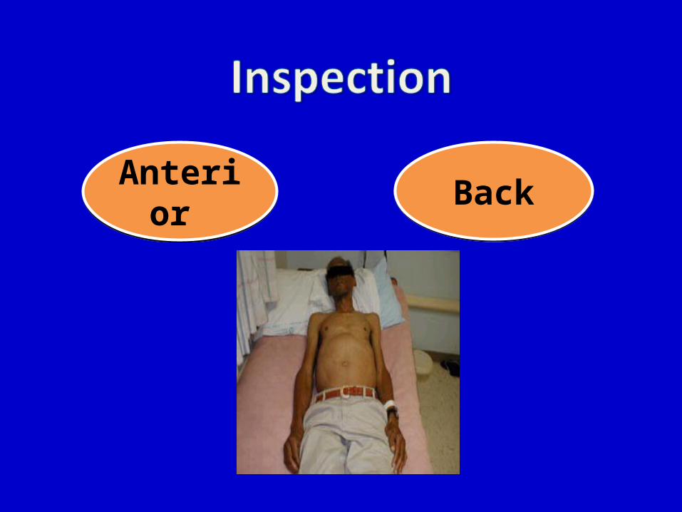
Anterior Back

Swelling Deformity Loin masses Pigmentation tuft of hair
Inspection of the Back
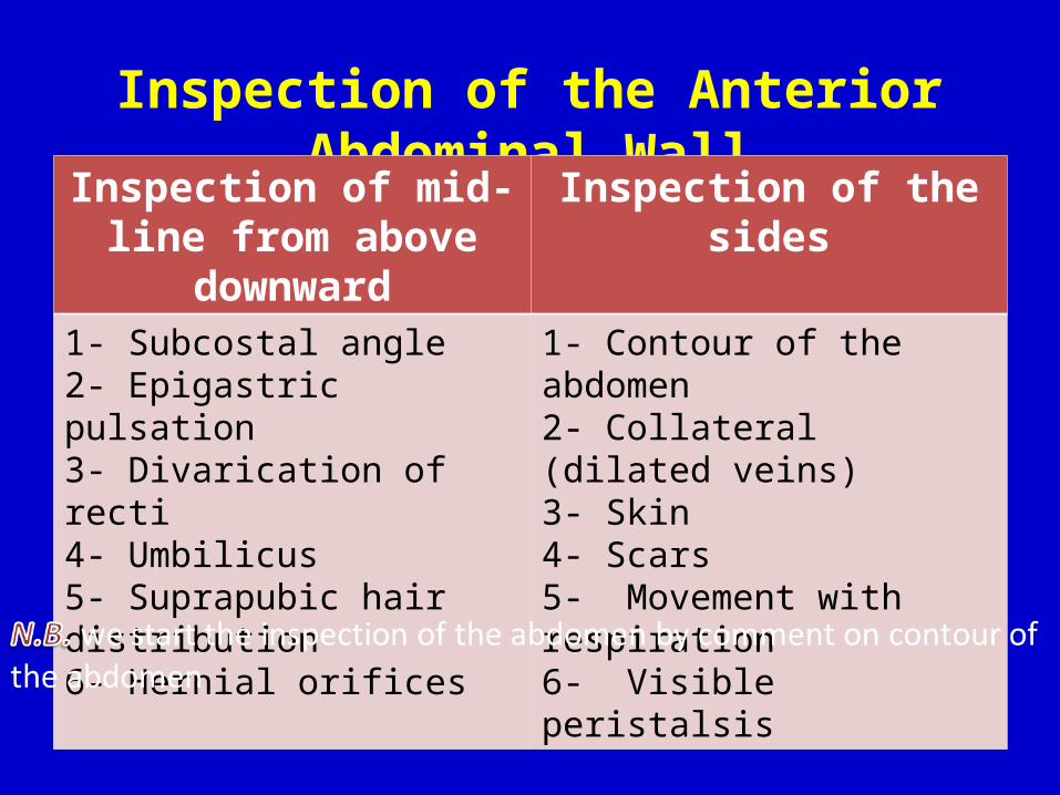
Inspection of the Anterior Abdominal Wall
Inspection of mid-line from above downward
Inspection of the sides
1- Subcostal angle2- Epigastric pulsation3- Divarication of recti4- Umbilicus5- Suprapubic hair distribution6- Hernial orifices
1- Contour of the abdomen2- Collateral (dilated veins)3- Skin4- Scars5- Movement with respiration6- Visible peristalsis


III. Hernia Expansile impulse in cough
IV. Dilated veins Caput medusa in portal hypertension
V. Skin Pigmentation around umbilicus (T.B. peritonitis, Addison dis.) Nodules “sister Mary-Joseph nodules” (abd. malignancy) Ecchymosis “Cullen's sign” (hemorrhagic pancreatitis and
internal hemorrhage)
VI. Discharge: Pus inflammation Stool intestinal fistula Urine patent urachus


Scaphoid abdomenslightly full abdomen but not distended

• examination of abdominal contours– Standing at the foot of the table– Lower yourself until the anterior
abdominal wall– ask the patient to breathe
normally while you are inspect the abdomen.

Generalized abdominal distension
Localized abdominal distension
1- Fluid (ascites)2- Fat (obesity)3- Flatus and Faeces 4- Foetus (pregnancy)5- Full urinary bladder
1- Site2- Shape and size3- Pulsate on cough (hernia or not)4- Movement with respiration5- Extra-abdominal or Intra-abdominal (by asking the pt. to sit up in bed unsupported)

Localized bulge

Generalized abdominal distension

IVC obstruction Portal vein obstruction1- Site of collaterals
Laterally (Sides) Around umbilicus (caput medusa)
2- Blood flow
From below upwards “towards the head”(to bypass the obstruction the blood bypass the IVC via abdominal wall veins to the thorax)
Away from the umbilicus”towards the legs” (the blood pass from the left branch of portal vein to para umbilical vein to anterior abdominal wall veins through the umbilicus)
3- cause in hepatic Pt
Functional compression on IVC by tense ascites
Intra-hepatic causes of portal hypertension

Methods of Detection- The 2 index fingers of both hands are used to milk the blood away from one segment of a dilated vein then, applying firm pressure on both ends of the segment the fingers then can be lifted one by one, while observing the rate of filling at which the vein fills from each direction the blood will be seen coming more rapidly from the direction of blood flow.

Caput medusa
Head of medusa

Caput medusae accentuated by marked ascites. An extensive plexus of veins is seen radiating from the umbilical region and radiating across the anterior abdominal wall. Note the large vein coursing inferiorly along the right flank (arrows). This is the superficial epigastric vein.


It is often difficult to understand whether tiny red spots arising on skin surface are Petechiae or Purpura. However, Petechiae spots have a very small diameter that is maximum 3 mm in size. Purpura rashes are larger in size. These have a diameter that is about 5 mm. A spot that is bigger than Purpura is known as common bruise or echymosis
Echymosis
Abdominal petichae


General rules for palpation

General rules for palpation

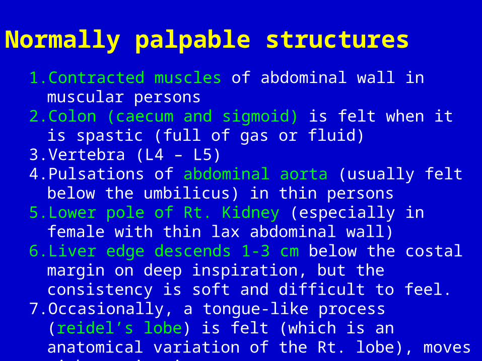
Normally palpable structures1. Contracted muscles of abdominal wall in muscular persons2. Colon (caecum and sigmoid) is felt when it is spastic (full of gas or
fluid)3. Vertebra (L4 – L5)4. Pulsations of abdominal aorta (usually felt below the umbilicus)
in thin persons5. Lower pole of Rt. Kidney (especially in female with thin lax
abdominal wall)6. Liver edge descends 1-3 cm below the costal margin on deep
inspiration, but the consistency is soft and difficult to feel.7. Occasionally, a tongue-like process (reidel’s lobe) is felt (which is
an anatomical variation of the Rt. lobe), moves with respiration

Types of Palpation
Superficial Deep

For: -Confidence of the patient-Superficial masses-Tenderness -Rigidity-Temperature
“from the Lt. iliac fossa in anticlockwise directiontill the suprapubic area”
Superficial Palpation

• Technique – Use pads of three fingers (palmar surface of fingers) of one hand and a
light, gentle, dipping maneuver to examine abdomen– Abdominal wall depressed approximately 1 cm

Palpating the abdomen – Light palpation

Palpating the abdomen – Light palpation

Deep PalpationFor :-Organs “liver, spleen, gall bladder, kidney, colon, urinary bladder”- Masses (ask the patient to flexes his neck as this contracts rectus muscles)-Areas of deep tenderness and rebound (pain induced or increased by letting go)
Deep palpation include the following methods-Ordinary technique “classic”-2 handed method-Bimanual -Dipping-Hooking-Rolling

• Technique – Entire palm (use palmar surface of fingers of one hand; greatest number of fingers) and a deep, firm, gentle maneuver to examine abdomen– Either one- or two handed technique is acceptable (When deep palpation is difficult, examiner may want to use left hand placed over right
hand to help exert pressure)– Palpate tender areas last– Palpate deeply with finger pads (do not “dig in” with finger tips)– Abdominal wall depressed around 4 cm or Push as deeply as patient will allow without significant discomfort.

Palpating the abdomen – Deep palpation
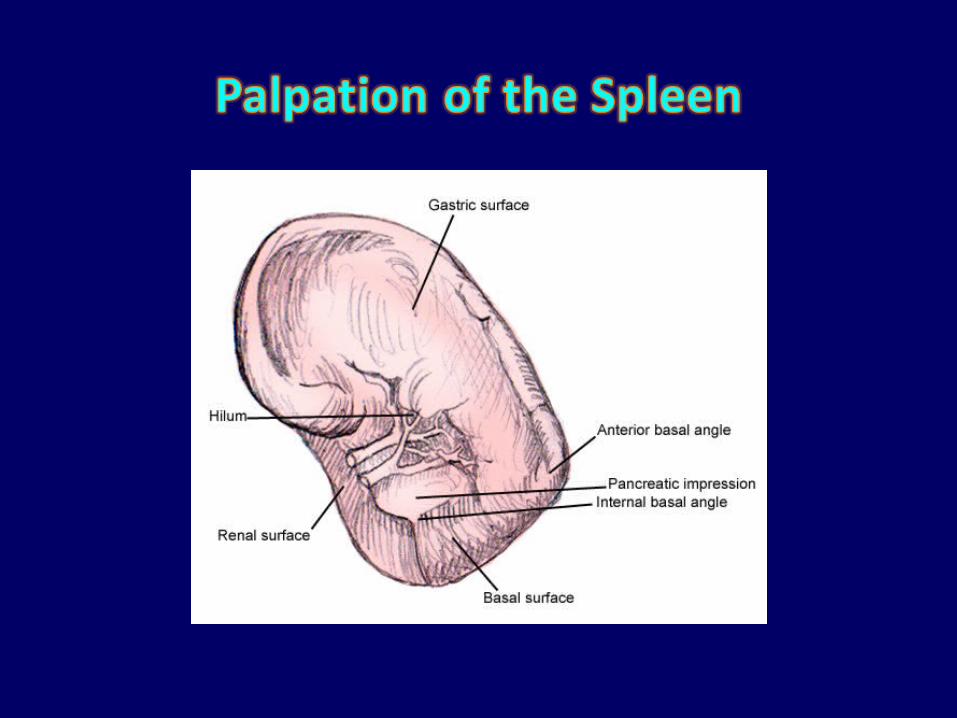


Surface anatomy of the Spleen
11th rb
Medial end
Lateral end
10th rb
9th rb
10th rb

Diaphragmatic surface
Visceral surface
upper border
Lower border

The spleen is not normally palpable It has to be enlarged 2-3 times its usual size to be palpable
under the subcostal margin Enlargement occurs superiorly and posteriorly before it
becomes palpable subcostaly Once the spleen has appeared in this situation, the
direction of further enlargement is downward and towards the Rt. Iliac fossa
The spleen which is not felt doesn’t exclude splenomegaly but it can be said that the spleen is not felt
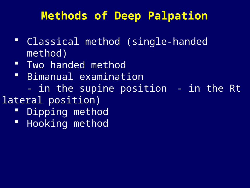
Methods of Deep Palpation
Classical method (single-handed method) Two handed method Bimanual examination
- in the supine position - in the Rt lateral position) Dipping method Hooking method

Classical method (single-handed method)

Two handed method

Bimanual examination in supine position

Palpating the spleen – Bimanual palpation in supine position

Palpating the spleen – Bimanual palpation in supine position

With the patient in the right lateral position, minimal splenic enlargement can be detected
Palpating the spleen – Bimanual palpation in Rt. Lateral position

Palpating the spleen – Bimanual palpation in Rt. Lateral position

Palpating the spleen – Bimanual palpation in Rt. Lateral position

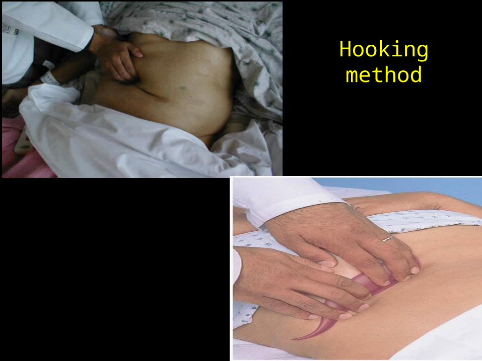
Examining for the spleen from behind the patient, in the right lateral position. In this case, the fingers are "hooked" over the costal margin.
Hooking method

Nature of this palpable spleen (put a comment on):1. Size
Mild (just palpable to 5cm) Moderate (5 – 10 cm) Huge (more than 10 cm, below the umbilicus)
2. Border3. Surface4. Consistency5. Tenderness (e.g. due to splenic infarction, septicemia,
SBE)

Applied anatomy and physiology of the spleenThe spleen is composed predominantly of lymphoid and R.E. tissues, so, any condition “infectious; immunologic; metabolic; malignant or idiopathic” that causes hyperplasia of the lymphoid/RES may cause splenomegalyThe spleen is expansile organ containing many sinusoids, so, interference with its venous drainage as in portal hypertension will cause splenomegaly “congestive splenomegaly”The spleen is a blood forming organ in fetal life and a potential blood forming organ throughout life, so, in myelosclerosis and myelofibrosis, extramedullary hematopoiesis may occur in the spleen with splenomegalyThe spleen destroys senile and defective RBCs, so, in hemolytic anemias, this function is increase with splenomegaly “except in sickle cell anemia”

Causes of Huge Spleen (below the umbilicus) Bilharzial splenomegaly Kala azar “visceral leishmaniasis” Chronic malaria causing TSS “Tropical splenomegaly syndrome” CML Myelofibrosis and Myelosclerosis Polycythemia rubra vera Beta-thalassemia major Amyloidosis Gaucher’s disease
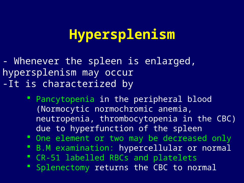
Hypersplenism
- Whenever the spleen is enlarged, hypersplenism may occur-It is characterized by
Pancytopenia in the peripheral blood (Normocytic normochromic anemia, neutropenia, thrombocytopenia in the CBC) due to hyperfunction of the spleen
One element or two may be decreased only B.M examination: hypercellular or normal CR-51 labelled RBCs and platelets Splenectomy returns the CBC to normal

Characters of splenic swelling to be differentiated from the Lt. kidney
-By inspection Moves with respiration down and medially-By palpation it has a notch on the lower part of the anterior
(upper) border “PATHOGNOMONIC” hand can't be insinuated between the mass and the costal margin to get above its upper pole negative ballottement (can’t be pushed in the renal angle)
-By percussion dull on percussion and continuous with the splenic dullness


Upper border is marked by joining the following points:1st point Lt. 5th intercostal space in the MCL “apex of the heart”2nd point Xiphisternal joint.3rd point Upper border of 5th rib in Rt. MCL4th point 7th rib at RT MAL.5th point 9th rib at RT scapular line.
Lower border is marked by curved line joining the following points:1st point Lt. 5th intercostal space in the MCL “apex of the heart”2nd point 8th costal cartilage in the Lt. parasternal line.3rd point midway between xiphisternal junction and the umbilicus4th point 9th costal cartilage in the Rt. MCL.5th point 10th rib in the Rt. MAL.6th point 12th rib in Rt. Scapular line

Xiphisternal junction
Rt. 5th rib
Rt. 7th rib
Rt. 9th rib
LT. 5th space
umbilicus

Rt. 9th costal cartilage
LT. 5th space
LT. 8th costal cartilage
Midway between umbilicus &xiphisternum
umbilicusRt. 10th rib

Technique of detecting the liver Upper border is detected by heavy percussion “hepatic
dullness” Lower border is detected by deep palpation and light
percussionAfter palpation of the lower border of the liver, you must comment on
I. Liver span : Distance between the upper and lower borders of the liver; which is
4 – 8 cm in the middle line “represents the Lt. lobe”
9 – 14 cm in the Rt. MCL “represents the RT. lobe”

II.Nature of this palpable liver (put a comment on):
1. Size “in finger breadth or cm” Normally: not felt below the costal margin Abnormally: enlarged “causes of hepatomegaly” or shrunken
“liver cirrhosis and fibrosis”2. Surface
Normally: smooth Abnormally:
- smooth “congestion, inflammation, infiltration”
- fine irregular “cirrhosis”- nodular “malignancy”
3. Edge Normally: sharp Abnormally:
- sharp “cirrhosis, fibrosis”- rounded “congestion, inflammation,
infiltration”

4. Consistency Normally: soft Abnormally:
- soft “congestion, inflammation, infiltration”- firm “cirrhosis, fibrosis”- hard “malignancy”
5. Tenderness: congestion, inflammation, infiltration, malignancy
6. Pulsation: TI, TS, hemangioma

Methods of Palpation
Classical method (single-handed palpation) Two-handed method Bimanual examination Dipping method Hooking method
- Single-handed palpation is used for lean individuals, while the bimanual technique is best for obese or muscular individuals. Using either technique, the liver is felt best at deep inspiration.

Single-handed method
- For single-handed palpation, the examiner's right hand is initially placed on the patient's abdomen in the right lower quadrant and parallel to the rectus muscle in the MCL. This is done so that palpation of the rectus is not confused with palpation of the underlying and adjacent liver
- Gently pressing in and up, ask the patient to take a deep breath. Palpating hand is held steady while patient inhales Palpating hand is lifted and moved while the patient breathes out If the liver is enlarged, it will come downward to meet your fingertips and will
be recognizable.

Another method of palpating the liver uses the radial border of the index finger. In this method the anterior hand is placed flat on the anterior abdominal wall with fingers parallel to the costal margin

the left hand is held posteriorly, between the 12th rib and the iliac crest. It is lifted gently upward to elevate the bulk of the liver into a more easily accessible position, while the right hand is held anterior and lateral to the rectus musculature. The right hand moves upward using gentle, steady pressure until the liver edge is felt.
Bimanual palpation of Liver

Bimanual palpation of Liver

– Is useful when the patient is obese or when the examiner is small compared to the patient.
– Stand by the patient's chest.
– "Hook" your fingers just below the costal margin and press firmly.
Hooking method
Hooking method

Causes of ptosed liver Emphysema Pneumothorax Pleural effusion Subphrenic abscess
Causes of upward displacement of the liver Lung fibrosis/collapse Diaphragmatic paralysis Ascites / abdominal tumours

Percussion is a method of tapping on a surface to determine the underlying structure

Technique-It is done with the middle finger of Rt. hand (plexor) tapping on DIP of the middle finger of the Lt. hand (pleximeter) using a wrist action. -The non striking finger (pleximeter) is placed firmly on the abdomen, remainder of hand not touching the abdomen.
-Remember that it is easier to hear the change from resonance to dullness – so proceed with percussion from areas of resonance to areas of dullness.
pleximeter
plexor

There are two basic sounds– Resonant sounds indicates hollow, air-filled structures. The
abdomen gives resonant note which varies according to the amount of gas present in the intestine.
– Dull sounds indicates the presence of a solid structure (e.g. liver) or fluid (e.g. ascites) lies beneath the region being examined

Percussion of the abdomen-The abdomen gives a resonant note which varies according to the amount of gas present in the intestine-Type of percussion: Light percussion-Values:
Deleneation of borders of abdominal organs (& assessing for organomegaly).
Decetction of ascites Detection of gaseous distension “tympanic resonant note” Detection of acute abdomen (obliteration of normal liver
dullness) in; - Perforated peptic ulcer and colon- Subphrenic abscess with gas forming organisms

• The two solid organs which are percussable in the normal patient– Liver: will be entirely covered by
the ribs. – Spleen: The spleen is smaller and
is entirely protected by the ribs.

Percussion “liver” Upper border by deep percussionLower border by light percussion
Upper border Define the sternal angle “angle of Louis” (2nd rib), then start
percussing the 2nd intercostal space in the Rt. MCL (Start just below the Rt. breast in RT. MCL). Percussion in this area should produce a relatively resonant note
Percussing in the chest moving down towards the abdomen about ½ to 1 cm at a time (in the intercostal spaces).
Note where the percussion notes change from resonant to dull. The normal hepatic dullness will be reached at the 5th intercostal
space in the RT. MCLLower border
Begin percussion below the umbilicus, in the Rt. MCL and proceed upward until dullness is encounter.

The liver span is estimated by percussionThe distance between the two areas where dullness is first encountered is the liver span.

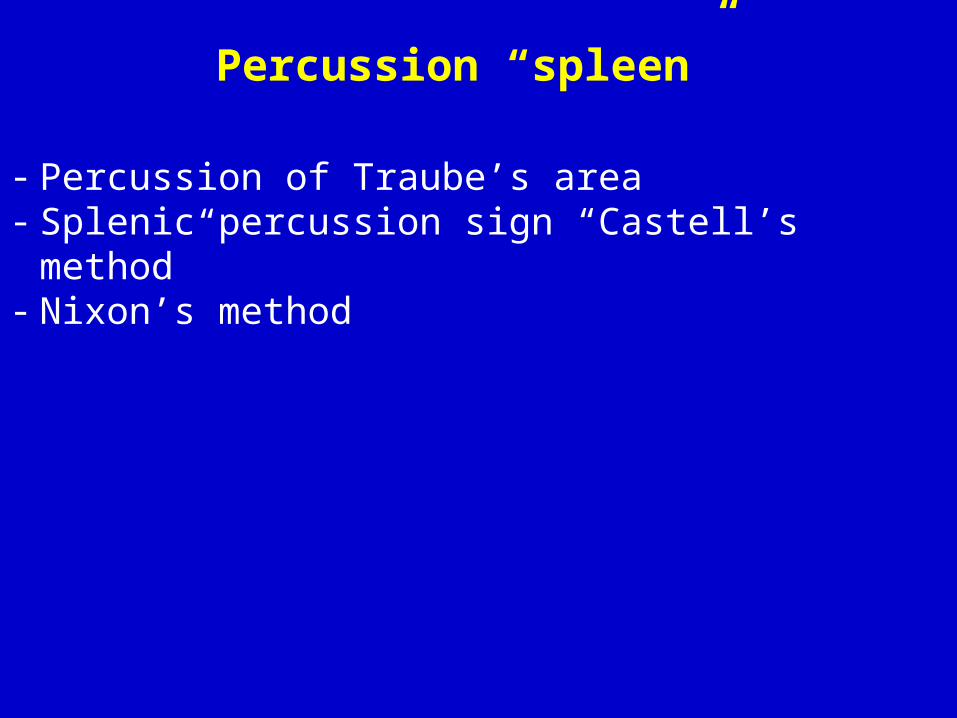
Percussion “spleen”
- Percussion of Traube’s area- Splenic percussion sign “Castell’s method”- Nixon’s method
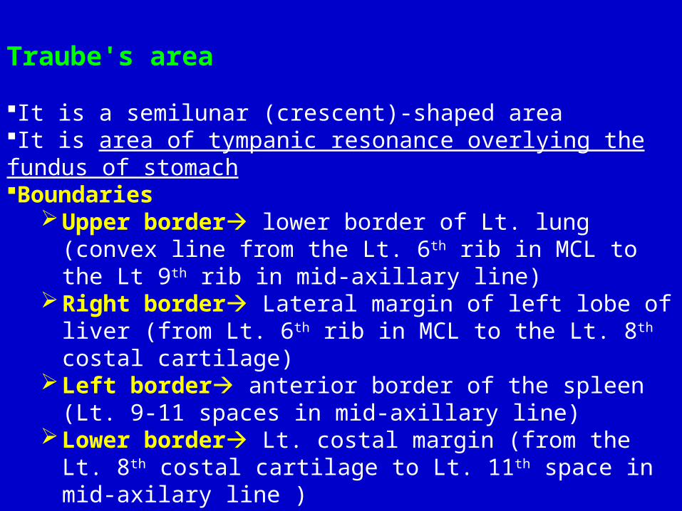
Traube's area
It is a semilunar (crescent)-shaped areaIt is area of tympanic resonance overlying the fundus of stomachBoundaries
Upper border lower border of Lt. lung (convex line from the Lt. 6th rib in MCL to the Lt 9th rib in mid-axillary line)
Right border Lateral margin of left lobe of liver (from Lt. 6th rib in MCL to the Lt. 8th costal cartilage)
Left border anterior border of the spleen (Lt. 9-11 spaces in mid-axillary line)
Lower border Lt. costal margin (from the Lt. 8th costal cartilage to Lt. 11th space in mid-axilary line )


Causes of dullness of Traube’s area:1. Full stomach/ gastric tumours.2. Left sided Pleural effusion / pericardial effusion “from above”.3. Ascites/abdominal tumour “from below”4. Splenomegaly “from left side”.5. Enlargement of left lobe of liver “from the right side”.

Castell’s method “Splenic percussion sign”Put the patient in the supine positionLeft anterior axillary line identifiedLeft lower costal margin identified Percuss in the lowest Left intercostal space in the anterior axillary
line (usually the 8th or 9th IC space) while patient inhales and exhales deeply
This space should remain resonant during full inspirationDullness on full inspiration indicates possible splenic enlargement (a positive Castell’s sign)

Castell’s point


Nixon’s methodPlace the patient in Right lateral decubitusBegin percussion midway along the Left costal marginProceed in a line perpendicular to the Left costal marginIf the upper limit of dullness extends >8 cm above the Left costal margin, this indicates possible splenomegaly

Ascites is free collection of fluid within the peritoneal cavity.The classical signs of ascites include; abdominal distension, shifting dullness, fluid thrill.
Minimal ascites detected in the knee elbow positionModerate ascites detected by the bilateral shifting dullnessTense ascites detected by transmitted fluid thrill “fluid wave”

Bilateral shifting dullness 1.The patient is examined in the supine position. 2.Percussion is done over the abdomen, from the umbilicus to one flank. 3.The spot of the transition from tympany to dullness is detected. 4.The patient is then turned to the opposite side, while the examiner keeps his hand unmoved.5. Percussion of the same spot (which is top now) gives a tympanic note.Note: The tympany over the umbilicus occurs in ascites because bowel floats to the top of the abdominal fluid.
air
air
fluid
fluid

Transmitted fluid thrillPathognomonic foe ascites when the amount of fluid is large
1.The patient is examined in the supine position. 2.The patient or an assistant places one hand in the midline and presses firmly with the ulnar border of the hand , so cut off any vibrations transmitted by the abdominal wall.
3.The examiner places one palm on one flank, while giving a sharp tap with the finger tips on the opposite flank.
4.Positive test: a definite wave “impulse” will be distinctly felt by the receiving hand.


• Diaphragm of stethoscope used• Skin depressed to approximately 1 cm • Listening in one spot is usually sufficient• Listening for 15-20 or 30-60 seconds

Values of auscultation
1.To hear intestinal sounds characteristic gurgling bubbling (gas and fluid in intestine) sounds.
Increase in: acute diarrhea (↑motility) and in early intestinal obstruction
Absent in: paralytic ileus
N.B. Bowel sounds cannot be said to be absent unless they are not heard after listening for 3-5 minutes.

2. To hear vascular sounds
Arterial bruit Venous hum (Wind at sea shore)
Systolic murmur Systolic and diastolic sound in the epigastrium, and Lt. hypochondrial region “Kenawy sign”
Occurs in cases of-Abdominal aortic aneurysm-Renal artery stenosis-Over very vascular tumour “e.g. hemangioma”
Occurs in cases of - portal hypertension due to porto-systemic anastomosis (collateral)

3. Friction rub a dry, grating sound heard with a stethoscope during auscultation; may
be heared over enlarged liver or spleen
Splenic rub: in Lt. hypochondrium; due to splenic infarction and perisplenitis
Hepatic rub: in Rt. Hypochondrium; due to hepatic malignancy with perihepatitis (inflammatory changes or infection in or adjacent to the liver). If detected in a young woman, the examiner should consider gonococcal peritonitis of the upper abdomen (Fitz–Hugh–Curtis syndrome).
N.B. A hepatic rub and bruit in the same patient usually indicates cancer in the liver. A hepatic rub, bruit, and abdominal venous hum would suggest that a patient with cirrhosis had developed a hepatoma.

4. To detect lower border of the liver (scratch method) Place the diaphragm over the area of the liver scratch parallel to
the costal margin in MCLWhen the liver is encountered, the scratching sound heard in the stethoscope will increase significantly
5. To detect minimal ascites (Puddle’s sign)It is useful for detecting small amounts of ascites (as small as 120 mL;
shifting dullness and bulging flanks typically require 500 mL).
The steps are outlined as follows: Patient lies prone for 5 minutes Patient then rises onto elbows and knees Apply stethoscope diaphragm to most dependent part of the abdomen Examiner repeatedly flicks near flank with finger. Continue to flick at same spot on abdomen Move stethoscope across abdomen away from examiner Sound loudness increases at farther edge of puddle

Scratch Test Start in the same areas above and below the liver as you would with percussion. Instead of percussing lightly, scratch moving your finger back and forth while listening over the liver. Since sound is conducted better in solids than in air, when the louder sounds are heard you are over the liver. Mark the superior and inferior boarders of the liver span in the midclavicular line

6. Succusion splash in case of pyloric obstruction (distended stomach with gas and fluid) placing the stethoscope over the upper abdomen rocking the
patient back and forth at the hips Retained gastric material >3 hours after a meal will generate a splash sound.
7. To detect pregnancy fetal heart sounds.
