liver trauma - medical imaging worldmedicalimagingworld.weebly.com/.../-liver-trauma.pdf · liver...
TRANSCRIPT


Liver Trauma
Dr Shafiq Chughtai
Resident Surgeon , SU II
Holy Family Hospital Rawalpindi

Over View
Anatomy Frequency / Mortality / Morbidity Age / Sex Pathophysiology Presentation Work Up Management

Anatomy
Largest Solid Organ
1.2-1.6 Kg in adult
2 Lobes , 2 Accesory lobes , 4 Sectors , 8 Segments
4 Ligaments


Hepatic viens divide the liver into 4 sectors and form 3 portal scissura
Transverse scissura is the plane of division of leftand right portal vien

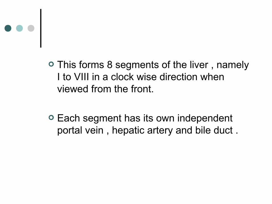
This forms 8 segments of the liver , namely I to VIII in a clock wise direction when viewed from the front.
Each segment has its own independent portal vein , hepatic artery and bile duct .


Each segment is getting its own duct , artery and vein.

Caudate lobe is segment I , also called separate liver.
It drains directly to IVC.
35% RHA , 12% LHA in remaining both arteries
Rt portal vien branch supply this lobe

Vital structures enter the liver through its hilum , hepatic artery , bile duct and portal vien.
Liver is its self related to right kidney , colon , stomach , IVC and stomach.


Portal Vien
Formed behind neck of pancrease by union of SMA and splenic vien.
Delivers 50% O2 and 75% blood flow to the liver .


Hepatic Vien
Drains blood to IVC
Portal Scissura
LHV 2,3,4 , MHV 5 , RHV 6,7,8


Hepatic Artery
Branch of celiac trunk
25% blood , 50% O2


Cuadate lobe
Separate liver
Multiple small veins directly drains into IVC
Comprises segment I of liver
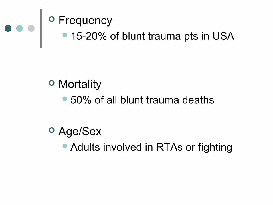
Frequency 15-20% of blunt trauma pts in USA
Mortality 50% of all blunt trauma deaths
Age/SexAdults involved in RTAs or fighting

Pathogenesis

Predisposition to injury due to
Large sizeFixed position relative to ribcage and
spineThin capsuleFriable parenchyma
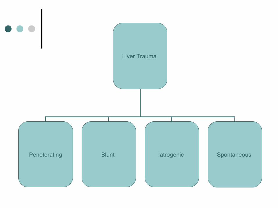
Liver Trauma
Peneterating Blunt Iatrogenic Spontaneous

Peneterating
Gun shot , shot guns , knives , daggers
Fire arm injury corresponds with the KE of the missle at entry and exit wound
20% of such injuries cause heamobilia , biloma , AV fistula or even abcess.

Blunt Injuries
Compressive injuries
Acceleration / deceleration injuries

Compressive Injuries High elastin content in artys > viens,
biliary channels > hepatic parenchyma
Hepatic parenchyma thus is more prone to blunt trauma
Parenchymal injuries cause deep lacerations also called fractures mostly running along vascular channels

Horizontal # running parallel to each other Bear Claw Injury
Transverse # can cause trasection of the liver
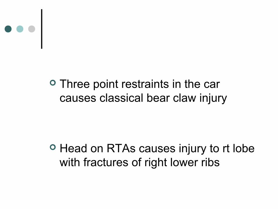
Three point restraints in the car causes classical bear claw injury
Head on RTAs causes injury to rt lobe with fractures of right lower ribs

Acceleration / Deceleration Injuries Falls from height
Hepatic viens can get avulsed from its attachment causing severe He
Laceration can extend deep into parenchyma due to accompanying compression secondary to fall.

>85% injuries involve right lobe , segment 6,7,8 due to less protected location
Incidence is high in children due to elastic rib cage and more friable parenchyma

Blunt abdominal injury
Isolated liver injury 50%
Liver with splenic injury 45%
Liver with rib # 33%

Iatrogenic
Percuteneous Biopsy
ERCP
TIPS

Spontaneous / High Risk
Liver tumors , HCC Scikle Cell Disease Collagen Vascular Disease Coagulopathy CRF Third trimester eclampsia and pre
eclampsia

Nature Of Hepatic Injuries Hematomas (subcapsular , parenchymal)
Lacerations
Contusions
Vascular Disruption
Biliary Tract Injuries

Grades
Mild Less then 25% , one lobe , 3 Months
Moderate 25-50 % , one lobe , 6 months
Severe >50% , one lobe , 9-15 months

Presentations With or without shock
Pain right upper quadrant , Lower chest (broken ribs)
Peritonism due to heamoperitoneum takes time to develop.In 50% cases , abdominal examination is normal in the presence of heamoperitoneum.*
*Mastery Of Surgery

However , bilary tract injuries cause rapid and severe peritonitis due to bile in the peritoneal cavity.

Management
ABCDE
Base line / Grouping and cross match.
CXR,AXR Associated skeletal injuries , R/P air ,
gas under diaphragm

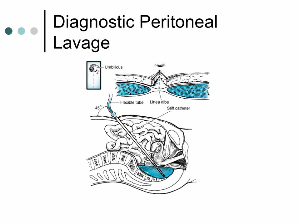
Diagnostic Peritoneal Lavage
95% sensitivity , 1-2 % complications , no role in retroperitoneal injuries.
>10 ml frank blood is considered positive
If nothing comes on insertion of the catheter , N/S can be instilled and the fluid thus aspirated can be send for Lab analysis
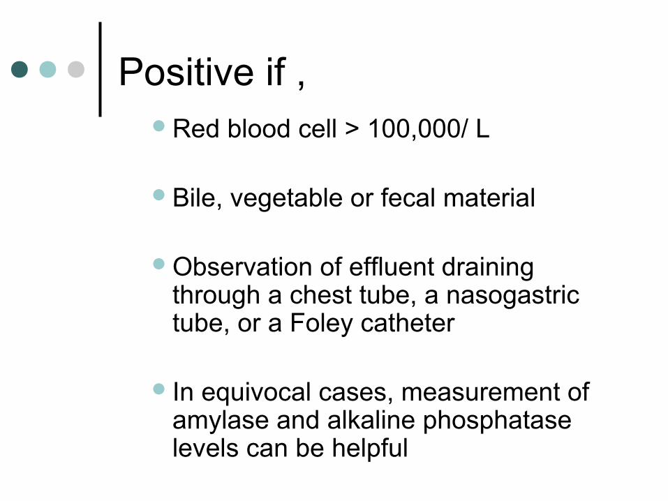
Positive if , Red blood cell > 100,000/ L
Bile, vegetable or fecal material
Observation of effluent draining through a chest tube, a nasogastric tube, or a Foley catheter
In equivocal cases, measurement of amylase and alkaline phosphatase levels can be helpful
Diagnostic Peritoneal Lavage

USG (FAST)
Easily avaliable and quick results
46% sensitive , 94% specific in peneterating abdominal injuries
72% sensitive in blunt abdominal injuries


CT scan Can only be done in stable patientsInvestigation of choice in abdominal
trauma
Angiography Diagnostic and therupaticUse only in stable patientsTranscatheter embolization / stenting

For Bile Leak Radiolabelled Tc 99m scan
RBCs labelled for active bleed , non labelled for bile duct injuries
Can be used for follow up as non invasive investigations or where CT scan is contraindicated

MRCPFor diagnosis and follow up of bile duct
injuries
MRINo advantage over CT scanOnly useful in young pregnant females

Grades As Per CT Scan

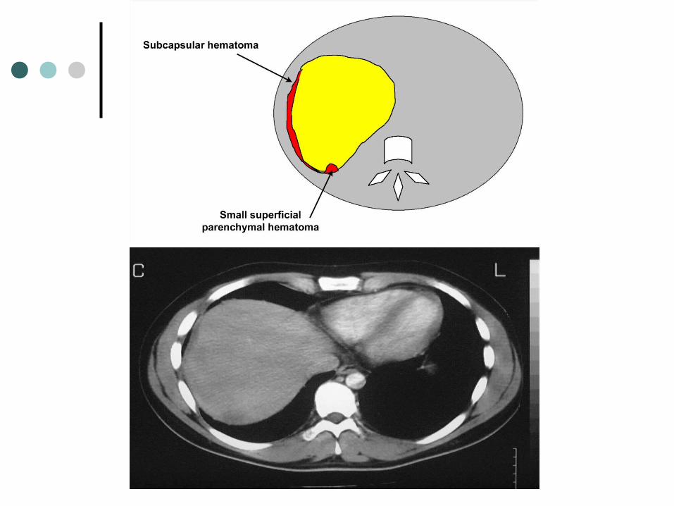


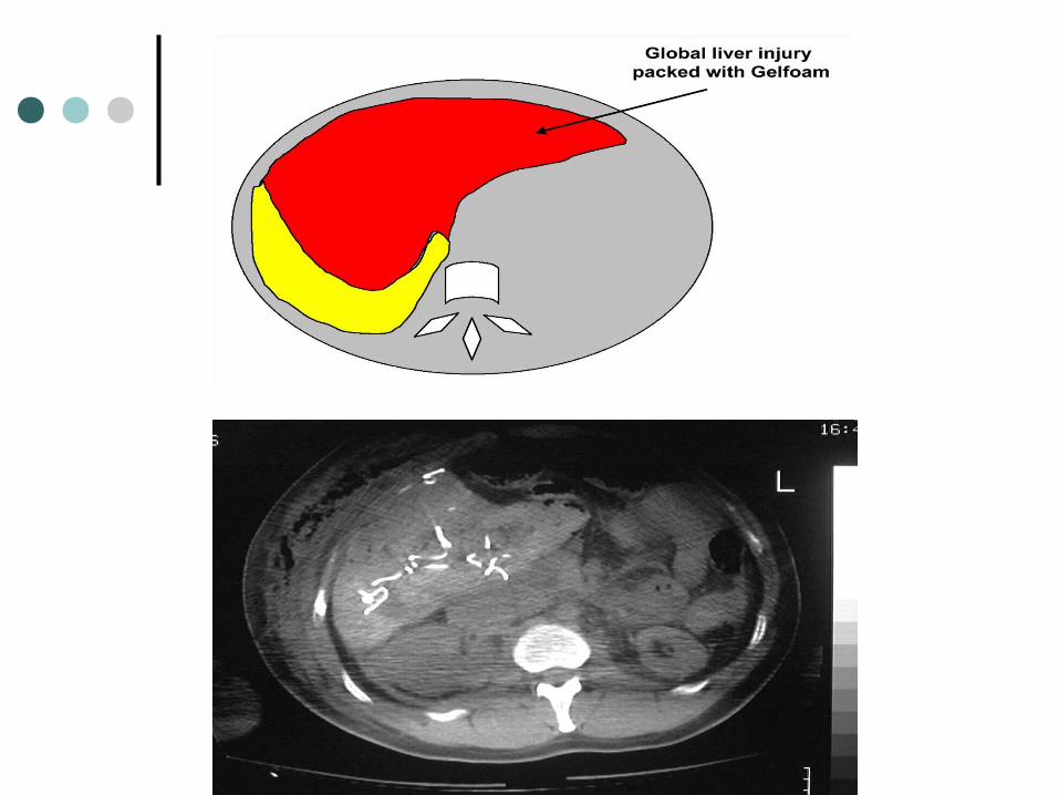

In trauma , always advice contrast enhance CT scan
In active Hge , extravasation of the contrast is seen
Active Hge , attenuation value 85-350HU , in clotted heamatoma 40-70HU.

Remmember , anatomical grading does not correspond with heamodynamic stability of the patient
Generally Grade III , IV & V have mortality of
25,46 & 80%

Approach
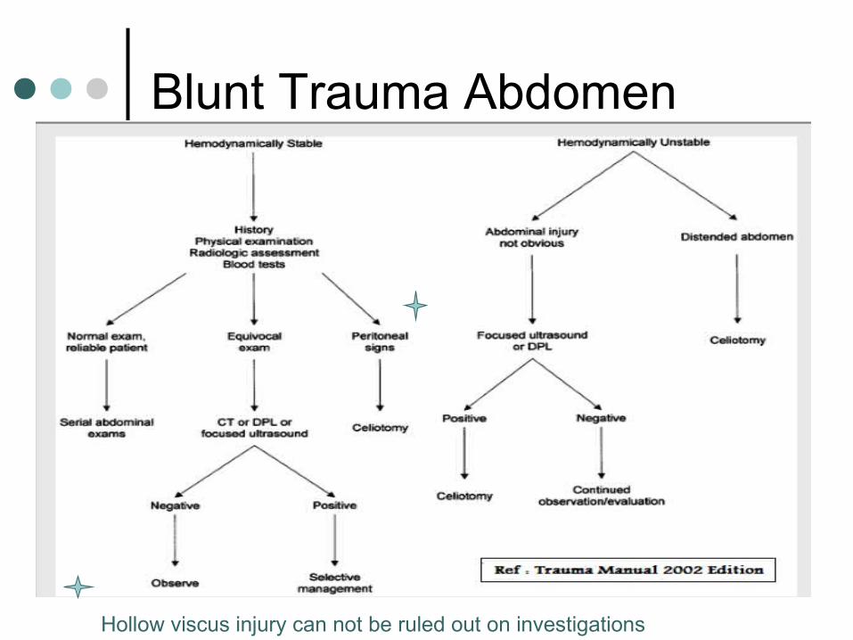
Blunt Trauma Abdomen
Hollow viscus injury can not be ruled out on investigations


Peneterating Rt Upper Abdominal
Trauma
Hollow ViscusInjury Un likely
Hollow Viscus Injury Likely
Peritoneal Breach
Pt Stable Pt Unstable
OperateSerial Exam / CXR
/ CT Scan
Deterioates / +ve findings

Stable patient Management is with serial
examination and monitoring
Unstable patientIntervention is done in a methodical
way

Major cause of death in child and adult is rapid intraoperative bleed related to torn intrahepatic / juxtahepatic vasculature and FAILURE to control blood loss in antacipation to early surgical repair


Preperation
Midneck to mid thigh , table top to table top laterally
Draping should be done before induction of anasthesia because it can cause profound drop in blood pressure

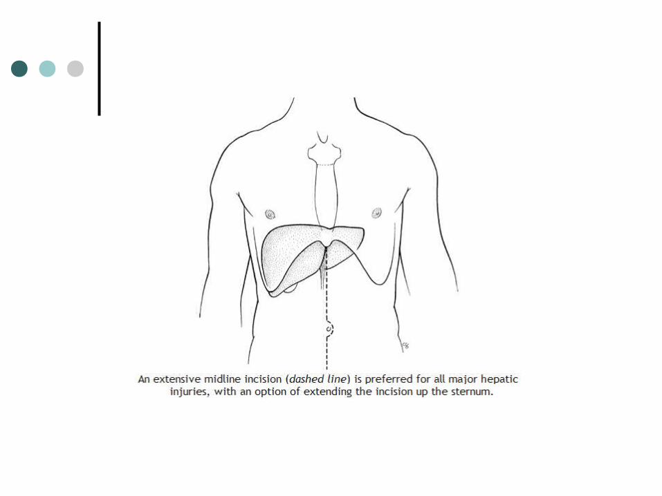
Incision
Xiphoid to below umblicus
Blood in peritoneal cavity , extend incision to suprapubic area

Evacuate & Pack
Evacuate immediately as much blood/clots as possible to temporarily control He
Pack all four quadrants
Allow anasthetist to replace lost volumes

Resuscitation
Wait for heamodynamic stability
Operation Theter thermostat 85 F
Vent humidifier 105F
Transfuse fluids

Reassess abdomen
Remove packs from lower abdomen , look for associated bleed , fecal contamination and Gut injury
If no fecal contamination , autotransfusion can be considered

Contd
Autotransfusion involves collection of blood from body cavities and drains and adding anticoagulant
Its reinfused after washing/filtering
Dilutional coagulopathy / DIC can occour

Contd
Remove pack in left upper quadrant , if spleenic injury present deal with it.
If required proceed with splenectomy

Direct Pressure

Approach Liver
Remove packs in RHC
Assess liver
Gentelly retract dome of liver rostrally , gush of blood from central area means hepatic vien injury
Vascular Isolation
Apply vascular clamp across porta hepatis (pringle) , if this control Hge , bleed is from hepatic arty / portal vien
If pringle maneuver is positive , dissect porta hepatis , take control of left and right portal vien / hepatic artery for selective control

Porta Hepatis


Contd
If pringle maneuver fails to control Hge , the blood is coming from retrohepatic IVC or hepatic viens
Heaney’s maneuver can be done to control Hge.
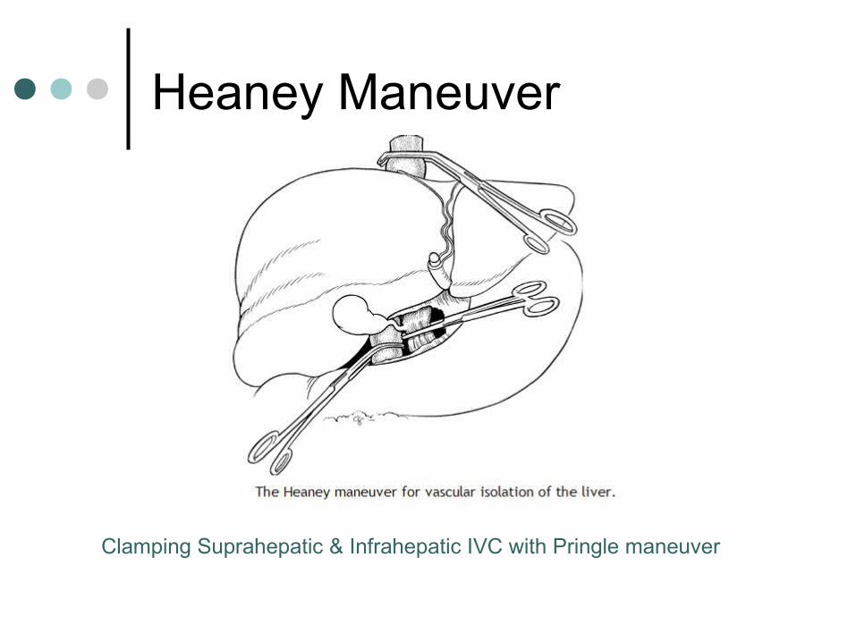
Heaney Maneuver
Clamping Suprahepatic & Infrahepatic IVC with Pringle maneuver

By passing the venous blood from kidneys nad lower extrimities can be done now to reduce morbidity .
Two methods Atrio caval bypassVenoveneous bypass
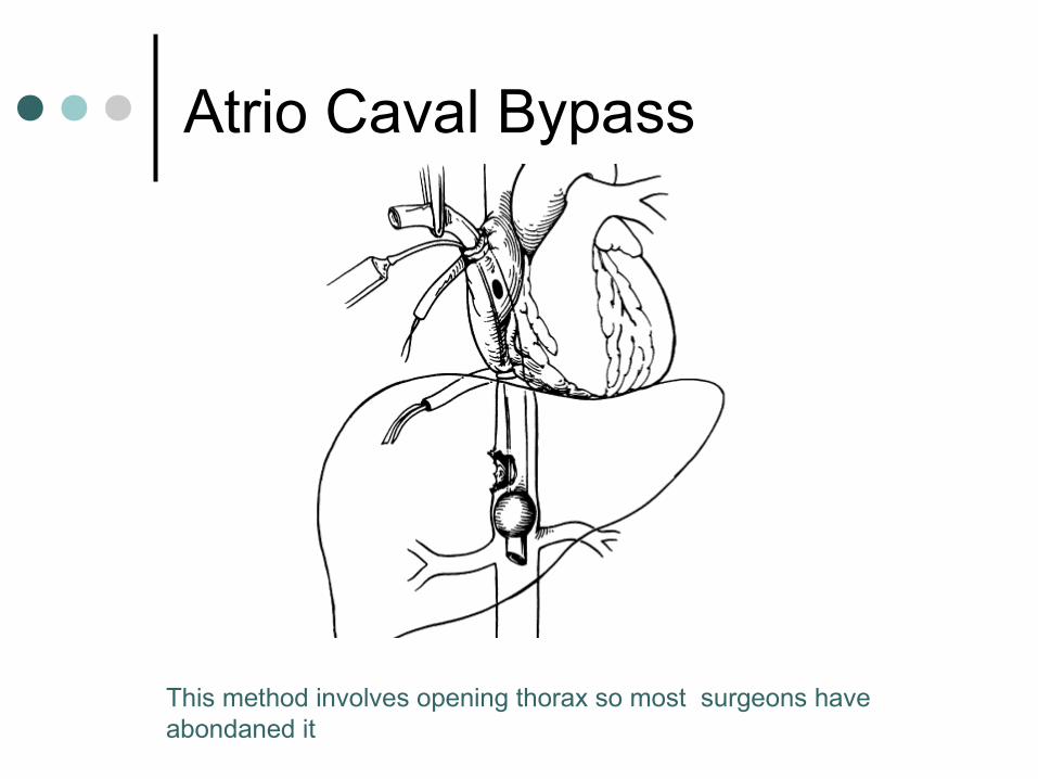
Atrio Caval Bypass
This method involves opening thorax so most surgeons have abondaned it

Veno venous bypass
Axillary and femoral vien catherization can decrease morbidity of vascular isolation and renal injury

Mobilize Liver
Sharply divide falciform , both leaves of coronary ligament and left triangular ligament.
Lesser sac should be entered by dividing gastrohepatic ligament

Definative Control >86% of liver injuries involve rt lobe ,
segment 6,7,8.
Assistant stands on left , extend left hand underneath the right lobe , compress the dome or the uninjured part with the right hand
Surgeon deepens laceration with finger fracture technique

Finger Fracture / Scalpel Handle
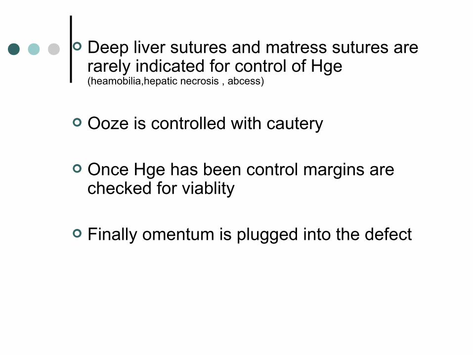
Deep liver sutures and matress sutures are rarely indicated for control of Hge (heamobilia,hepatic necrosis , abcess)
Ooze is controlled with cautery
Once Hge has been control margins are checked for viablity
Finally omentum is plugged into the defect

Pringle maneuver can be done in elective surgery for 1 hr
In compromised pt in trauma , no studies have been done.However , it should be restricted to 15 mins and at the most to ½ hr.

Next
Resectional Debridement
All non viable liver tissue shuld be resected using scalpel or finger fracture technique
When resistence is encountered , its vessel or biliary tract which should be ligated

Oozing should be controlled with cautery
In case of viablity of the tissue is doubtful , 2nd look lap should be done after 24-36 Hrs

Still Bleeding
Less then 1%
Ligate one vessel , right or left hepatic artery or portal vien by dissectin through porta hepatis
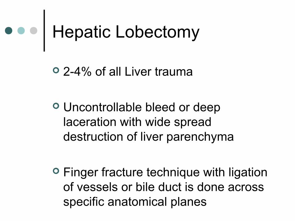
Hepatic Lobectomy
2-4% of all Liver trauma
Uncontrollable bleed or deep laceration with wide spread destruction of liver parenchyma
Finger fracture technique with ligation of vessels or bile duct is done across specific anatomical planes

In trauma , there are 3 options
Left lateral segmentectomy
Left lobectomy
Right lobectomy

Left lateral segmentectomy Segment II & III are taken out.
Resection is carried out to the left of falciform ligament
Care is taken not to divide vessels supplying segment IV
On reaching proximal part of left hepatic vien , be careful not to ligate middle hepatic vien

Left lobectomy
Carried out to the left of GB fossa
Right Lobectomy
Carried out to the right of GB fossa
After left or right lobectomies , large area should be cauterized and omentum should be placed on it.


Packing
It was famous in WWII but results are poor
Buys time in very compromised patient
Only indicated in patient who is coagulopathic or hypothermic , bleeds from large surface and bleed is veneous.

Pack & Re Look after 48 hrs , I/A pressure should be less then 40mmHg

Contd
Placement of pack should not puch diaphram upwards to compromise breathing or compress IVC to compromise veneous return
BP & CVP monitoring is done to aviod these complications

If either of these values fall , compression of IVC is likely.
Once done successfully , pt is resussicitated in icu. Mean while venoveneous bypass or transport to hepatiobiliary surgical unit is planned

Intrahepatic balloon Done in unstable pt , to gain time in
which peneterating injuries form a tract through liver parenchyma
A catheter is placed with a balloon inside , to control bleed via temponade effect
Temporary measure , re Lap is indicated after stabilizing patient.
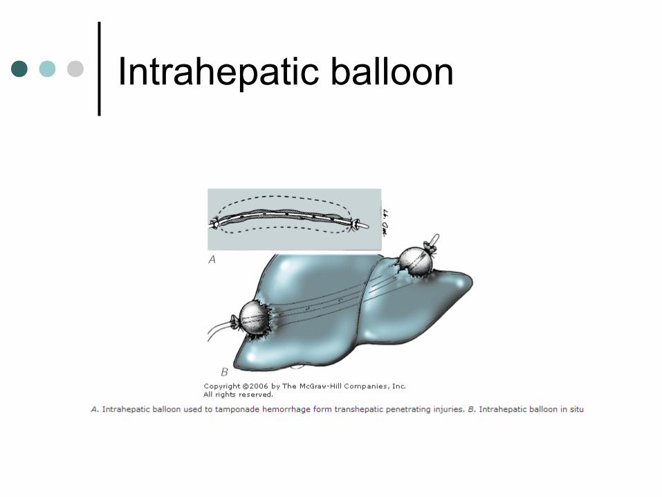
Intrahepatic balloon
Hepatic transplant
If there is severe hepatic injury , causing hepatic avulsion , severe nonreconstructable injury to porta hepatis or both lobes , hepatic transplantation can be considered

Porta Hepatis Injuries
Vascular injuries
Bliary tract injuries

Vascular Injuries Extended Kocker’s maneuver to
mobilize hepatic flexture and duodenum
Expose IVC , Aorta , Duodenum & PV
Proper hepatic arty repair with reverse sapheneous graft
Portal vien Repair with IJV / Splenic vien*
*Only if splenectomy has been done

Biliary Injuries Unstable patient
Exteriorize biliary drainage using catheter
Stable patient
No segmental loss end to side anastamosis
Segmental loss Modified Carrel Patch

Modified Carrel Patch (choledocojejunostostomy)
Post Operative Complications
Coagulopathy Pulmunary Failure Juaindice Bile Leaks Heamatobilia Sepsis (7-12%pneumonia , acalcolous cholecystitis)

Thank you


BloodLoss
Heart rate BloodPressure
CapillReturn
Resp Rate MentalState
<750 <100 Normal Normal Normal Normal
750-1500 >100 SystolicNormal
Prolonged 20-30 MildlyAnxious
>1500-2000
>120 Decreased Prolonged 30-40 AnxiousConfused