liver trauma final
TRANSCRIPT
Liver TraumaProf. Ygber González de la Cruz, MD. MsC
Department of Medicine and Therapeutic. SMAHS-UTG

Lecture overview
• Review the main anatomical and
physiological characteristic of the liver.
• Classify the traumatic liver injury.
• Describe the main clinical and
radiological characteristics of the liver
trauma.
• Define the approach to the patient with
a suspected liver trauma at the A & E
Background
• Largest solid abdominal organ,fixed
position
• Second most common injured, but most
common cause of death after
abdominal trauma
• Blunt MVA most common
• 80% adults, 97% children-conservative
rx
Pathophysiology
• Friable parenchyma, thin capsule, fixed position in relation to spine.
• Right lobe gets hit more since its larger, and closer to ribs.
• 85% injuries involve segments 6,7,8 from compressioin against ribs, spine, abd wall.
• Shear forces at attachments to diaphragm
• Transmission thru right hemithorax.
Pathophysiology
• Liver injured easily in children since ribs
are compliant, force transmitted.
• Liver not as developed in children, with
weaker connective tissue framework.
• Iatrogenic injuries by biopsies, biliary
drainage, TIPS, can cause capsular
tears and bile leaks, fistulas,
hemoperitoneum.
Injuries
• Subcapsular hematoma or intrahepatic hematoma.
• Laceration
• Contusion
• Hepatic vascular disruption
• Bile duct injury
• 86% of injuries have stopped bleeding at time of exploration.
• Decreased transfusion req.With conservative.
Injuries
• Mild hepatic injuries involving < 25% of
one lobe heal in 3 mos.
• Moderate injuries involving 25-50% of
one lobe heal in 6 mos.
• Sever injuries require 9-15 mos to heal.
• Gallbladder injuries rare, with
contusons being most common,
avulsions next most.
Anatomy
• Cantile described main divisions along
a main plane from GB fossa to IVC.
Divides liver into equal halves.
• Couinaud developed 4 sectors and 8
segments, divided into vertical and
oblique planes, defined by the 3 main
hepatic veins and transverse plane thru
right and left portal branches.
Anatomy
• Hepatic veins lie between segments.
• Left hepatc vein divides left lobe into
medial and lateral segments.
• Middle hepatic vein divides liver into left
and right lobes.
Anatomy
• Right hepatic vein divides right lobe into
anterior and posterior segments.
• A horizontal line thru left and right main
portal veins is used to divide lobes into
inferior and superior segments.
• The 8 liver segments are numbers
clockwise on the frontal view.
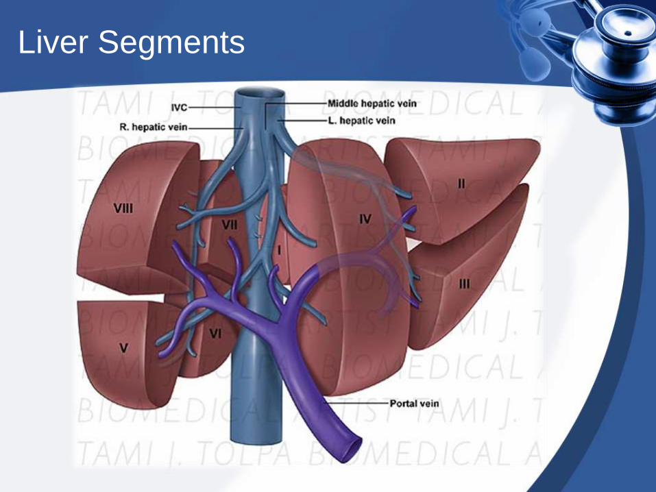
Liver Segments
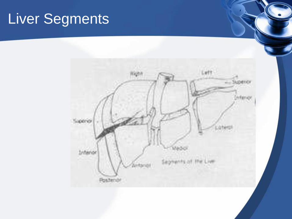
Liver Segments
Clinical Details
• Symptoms of injury are related to blood
loss, peritoneal irritation, RUQ
tenderness, and guarding.
• Unrecognized delayed abcess
• Bilomas
• Signs of blood loss may dominate the
picture.
Clinical Details
• Elevated liver tests
• Biliary peritonitis (nausea, vomiting, abd pain).
• DPL has high sensitivity, 1-2% complication rate.
• Plain x-rays non-specific.
• CT scan diagnostic procedure of choice.
• Hida for leaks, angio for hemorrhage.
Limitations
• FAST sensitivity highest (98%) for grade 3
injuries or greater. Negative findings do not
exclude hepatic injury.
• Emergency sono findings demonstrating free
fluid, parenchymal injury, or both
demonstrate overall sensitivity for detection
of blunt abdominal trauma of 72%.
• Angiogram may fail to detect active bleeding.
CT Scans
• Accurate in localizing the site of liver injury, associated injuries.
• Used to monitor healing.
• CT criteria for staging liver trauma uses AAST liver injury scale
• Grades 1-6
• Hematoma,laceration,vascular,acute bleeding,gallbladder injury,biloma.
Classification
• I-Subcapsular hematoma<1cm,
superficial laceration<1cm deep.
• II-Parenchymal laceration 1-3cm deep,
subcapsular hematoma1-3 cm thick.
• III-Parenchymal laceration> 3cm deep
and subcapsular hematoma> 3cm
diameter.
Classification
• IV-Parenchymal/supcapsular
hematoma> 10cm in diameter, lobar
destruction, or devasularization.
• V- Global destruction or
devascularization of the liver.
• VI-Hepatic avulsion
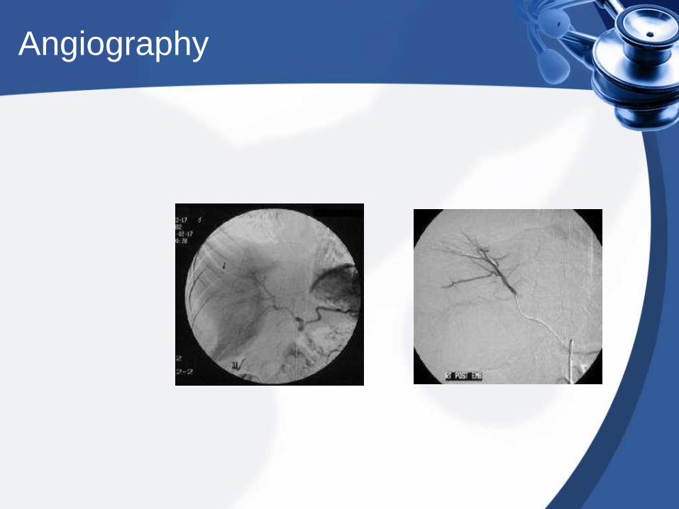
Angiography
• Demonstrates active bleeding
• Transcatheter embolization may be the
only treatment required.
• Findings include contusion, laceration,
hematoma, pseudoaneurysms, fistulas.
• Embolization can reduce transfusion
requirements, stenting for fistulas.
Angiography
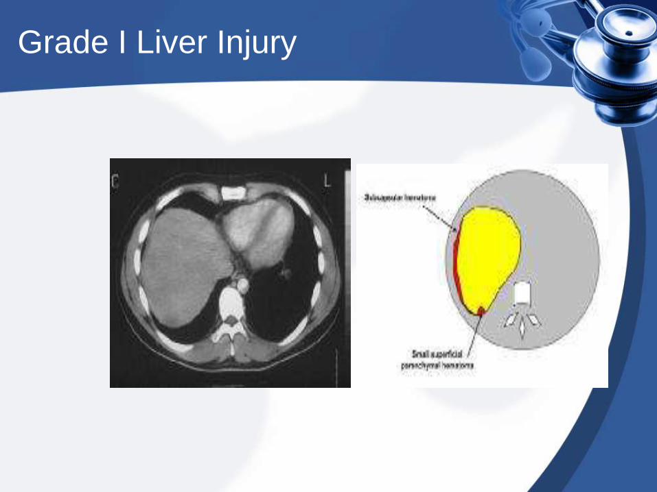
Grade I Liver Injury
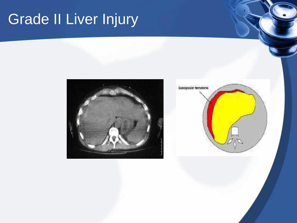
Grade II Liver Injury
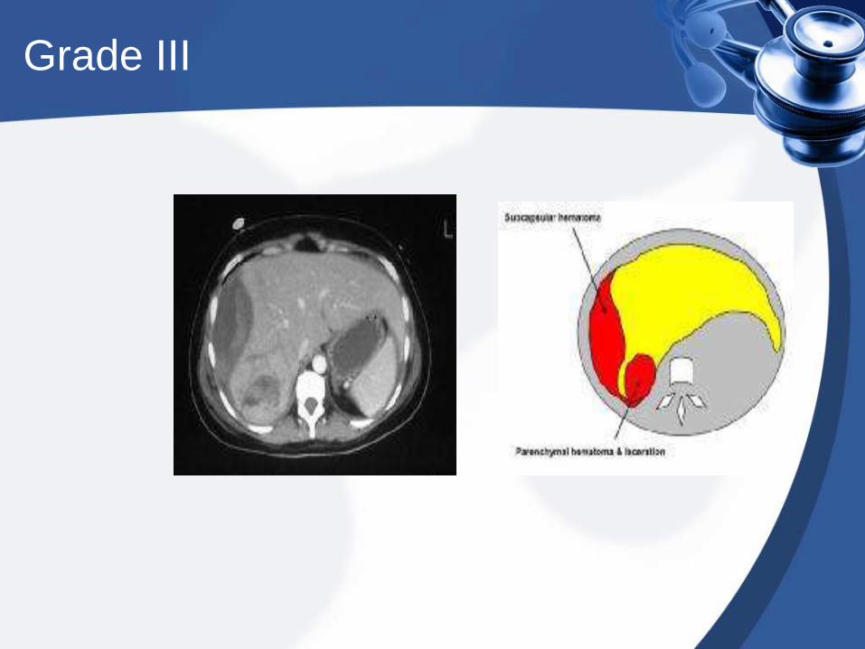
Grade III
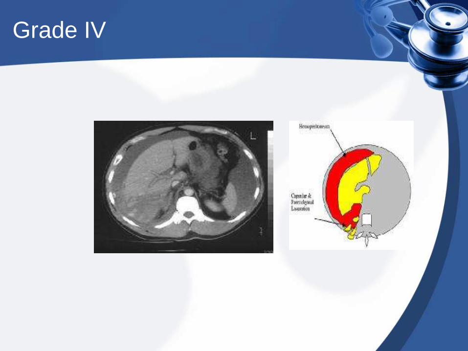
Grade IV
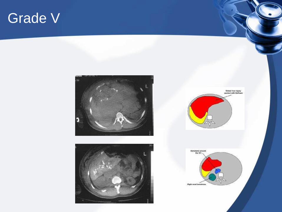
Grade V