litomosoides (nemata: filarioidea) of bats from bolivia
TRANSCRIPT

Southern Illinois University CarbondaleOpenSIUC
Publications Department of Zoology
8-1-2010
Litomosoides (Nemata: Filarioidea) of Bats fromBolivia with Records for Three Known Species andthe Description of a New SpeciesJuliana Notarnicola
F Agustín Jiménez-RuízSouthern Illinois University Carbondale, [email protected]
Scott L Gardner
Follow this and additional works at: http://opensiuc.lib.siu.edu/zool_pubsPublished in the Journal of Parasitology, Vol. 96, No. 4 (2010): 775-782.Copyright 2006, American Society of Parasitologists.Used by permission.
This Article is brought to you for free and open access by the Department of Zoology at OpenSIUC. It has been accepted for inclusion in Publicationsby an authorized administrator of OpenSIUC. For more information, please contact [email protected].
Recommended CitationNotarnicola, Juliana, Jiménez-Ruíz, F Agustín and Gardner, Scott L. "Litomosoides (Nemata: Filarioidea) of Bats from Bolivia withRecords for Three Known Species and the Description of a New Species." 96, No. 4 (Aug 2010).

LITOMOSOIDES (NEMATA: FILARIOIDEA) OF BATS FROM BOLIVIA WITH RECORDS FOR
THREE KNOWN SPECIES AND THE DESCRIPTION OF A NEW SPECIES
Juliana Notarnicola, F. Agustın Jimenez Ruız*, and Scott L. Gardner�Centro de Estudios Parasitologicos y de Vectores–CEPAVE–CCT La Plata-CONICET, Calle 2 #584 (1900) La Plata, Argentina. e-mail:[email protected]
ABSTRACT: Herein, we describe Litomosoides salazari n. sp. collected from the body cavity of the yellow bat, Lasiurus ega, fromBolivia. This new species of filarioid nematode is most closely related to the carinii group and is characterized by its relatively small sizewith the vulva located slightly posterior to the esophago-intestinal junction; an arrow-shaped buccal capsule; and a short, rounded tail.New host and locality records for both Litomosoides hamletti and L. chandleri in phyllostomid bats, and L. brasiliensis occurring inboth phyllostomid and vespertilionid bats, are provided. The morphological variability of the specimens is documented by providingcomparative measurements of 3 known species. Litomosoides brasiliensis occurs in 6 species of bats from Bolivia and was the mostcommon species of filarioid nematode encountered. All 4 species of nematodes, including the new species, were found in sympatry atChijchijpa, a locality in the Yungas of Bolivia.
Bolivia is a country of great biological diversity (Anderson,
1997), with more than 300 species of mammals occurring there. Of
these, approximately 5% are endemic, occurring nowhere else
(Anderson, 1997). More than 100 species of bats have been
identified from within the country (Anderson, 1997; Simmons,
2005). This great diversity is a result of a confluence of several
biomes conjoining in the area, including high-altitude Andes and
Puna-Altiplano, the Yungas in the eastern foothills of the Andes,
Amazon tropical forest in the northeast, and a mixture of
grassland and chaco thorn forest coming in from the southern and
eastern lowlands (Unzueta, 1975).
Filarioid nematodes of Litomosoides Chandler, 1931 occur in
the body cavities of bats, marsupials, and sigmodontine and
hystricognath rodents throughout the Neotropical and southern
Nearctic regions. Showing relatively high taxonomic diversity
with approximately 30 described species, these nemas exhibit an
indirect pattern of transmission between mammalian hosts, with
all known life cycles requiring an arthropod vector or interme-
diate host to transfer microfilariae between and among definitive
hosts (Bain et al., 1980).
As a result of the continuing work on the biodiversity of
mammals of Bolivia as part of the Bolivian Parasite Biodiversity
Project, we report herein the results of our work on the filarioid
nematodes of bats from Bolivia. This work, funded by the
National Science Foundation, was undertaken as a collaborative
project by the Department of Mammalogy, the American
Museum of Natural History (New York, New York); Division
of Mammals in the Museum of Southwestern Biology (Depart-
ment of Biology, University of New Mexico, Albuquerque, New
Mexico); the Harold W. Manter Laboratory of Parasitology
(HWML, University of Nebraska–Lincoln, Lincoln, Nebraska);
and the Coleccion Boliviana de Fauna (CBF, La Paz, Bolivia).
The results of investigations on filarioid nematodes include the
description of several new species associated with bats, rodents,
and primates (Brant and Gardner, 1997; Notarnicola et al., 2007,
2008), and a phylogeny of Litomosoides spp. based on morpho-
logical characters (Brant and Gardner, 2000). Brant and Gardner
(2000) also provide a preliminary list of species of Litomosoides
and their hosts and a discussion of their phylogenetic relation-
ships.
Herein, we describe a new species of Litomosoides parasitizing
the yellow bat, Lasiurus ega (Gervais, 1856), and we provide data
on the morphology, localities, and hosts used by 3 other species in
the genus occurring in bats from Bolivia.
MATERIALS AND METHODS
Specimens were collected following the methods outlined in Gardner(1996) and Gardner and Jimenez (2009). Filarioids were collected andplaced directly in either 70% ethanol, 10% formalin solution, or 95%ethanol. They were transported in these solutions back to the Harold W.Manter Laboratory (Lincoln, Nebraska) and stored. For study ofmorphological characters, specimens were cleared in lactophenol andstudied with an Ultraphot or Axiophot digital microscope (Carl Zeiss,Jena, Germany) using digital measuring software. To examine the oralpapillae, an apical view of the head was prepared. The lateral cuticularinternal ridge was used to identify lateral fields and the Y-shaped sectionof the lumen of the esophagus was used to identify the dorsal side. Todetermine the shape of the lateral chords, a cross section of a female wasmade posterior to the vulva. Microfilariae were dissected from the uterusof fixed females or studied from blood smears. Illustrations were madewith the aid of a drawing tube. Measurements are presented as follows:holotype, male paratypes, allotype, and female paratypes. If more than 3paratypes were examined, mean values and SDs are presented, with rangesin parentheses. Measurements are given in micrometers unless otherwisestated. Prevalence and mean intensity of infection also are provided foreach species.
RESULTS
During our work throughout Bolivia, more than 600 individual
bats were collected, primarily by netting, and then they were
examined for both ecto- and endoparasites, including represen-
tative species from the Phyllostomidae, Vespertilionidae, Thy-
ropteridae, and Mollosidae. Those bats positive for filarioids are
given in Table I. We recovered 1 undescribed species and 3
previously known species, including Litomosoides chandleri
Esslinger, 1973; L. hamletti Sandground, 1934; and L. brasiliensis
Lins de Almeida, 1936.
We encountered filariod nematodes in bats from the following
list of localities in Bolivia: Departamento Santa Cruz: San Rafael
de Amboro (17u369S, 63u329W; 21 July–2 August 1985), San
Miguel Rincon (17u239S, 63u329W; 14 August 1984), Estancia
Cachuela Esperanza (16u479S, 63u149W; 22 August 1984), 10 km
Received 29 October 2009; revised 9 February 2010; accepted 12February 2010.
*Department of Zoology, Southern Illinois University, Carbondale,Illinois 62901-6501.
{Harold W. Manter Laboratory of Parasitology, W-529 Nebraska Hall,University of Nebraska–Lincoln, Lincoln, Nebraska 68588-0514.DOI: 10.1645/GE-2371.1
J. Parasitol., 96(4), 2010, pp. 775–782
F American Society of Parasitologists 2010
775
N San Ramon (16u369S, 63u149W; 7 August 1987), 6 km by road
W of Ascencion (15u439S, 63u99W; 13 August 1985), 4 km SW
Buena Vista (17u289S, 63u429W; 7 August 1987), and 2 km S
Caranda (17u339S, 63u329W; 12 August 1987); Departamento El
Beni: Rıo Beni (13u279S, 67u 219W; 8 September 1985), km 35 NW
of Yucumo (14u529S, 67u079W; 11 July 1992), and Serranıa de
Pilon (15u179S, 67u049W; 18–22 July 1992); Departamento La
Paz: La Reserva (15u449S; 67u319W; 22–26 July 1992) and
Chijchijpa (16u099S, 67u449W; 5–7 July 1992); Departamento
Cochabamba: Sajta (17u069S, 64u469W; 20–23 June 1993) and El
Palmar-Rıo Cochi Mayu (17u069S, 65u329W; 5 July 1993); and
Departamento Tarija: Tapecua (21u269S, 63u559W; 3 June 1995).
See Anderson (1997) for a complete list of localities from which
mammals were collected up to approximately 1995.
DESCRIPTION
Litomosoides salazari n. sp.(Figs. 1–18)
General description (based on 5 males, 7 females): Males 1.5 timessmaller than females. Anterior extremity rounded, dome-like. Fourconspicuous labial papillae forming rectangle, stretched dorso-ventrally,
2 cephalic ventral papillae and small amphids present (Fig. 5). Arrow-shaped buccal capsule well cuticularized. Esophagus undivided or slightlyglandular at posterior end.
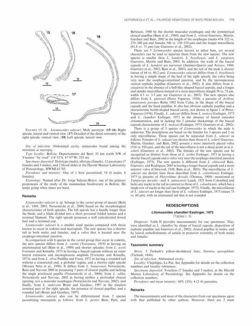
Male: Posterior region coiled with 3–7 loops (Fig. 13). Left spiculeswith handle longer than blade; blade consisting of filament andmembranous alae. Right spicules cuticularized, with prominent dorsalheel and terminal cap (Figs. 15–17). Tail short and rounded. Posterior endwith 3 pairs of subventral cloacal papillae. Area rugosa begins anterior tocloaca, consisting of transverse ridges made of small longitudinal crests,generally extending posteriad through coiled region.
Holotype: Length 12.231 mm; maximum width 100; at level ofesophagus 50; buccal capsule 17 long, external diameter 10, buccal cavity3; esophagus 330 long. Nerve ring 130 from anterior extremity. Tail 100long. Left spicule 205 long, with handle 110 long; right spicule 75; spicularratio 1:2.7. Area rugosa beginning 300 to 1,700 anterior from tail end;1,400 long, width of rows 100–120; crests 5–7 in height, spaced 25–30.
Paratypes (n 5 5): Length 9.76 ± 0.9 (8.92–10.8) mm; width 95 ± 21.7(75–120); buccal capsule 18.2 ± 1.6 (17–21) long and external diameter 8.2± 1.4 (6–10); esophagus 310 ± 36.7 (270–360) long; nerve ring 160 and 220from anterior end (n 5 2); tail 80.2 ± 10.6 (70–95); left spicule 207.5 ± 9.5(200–220) long, handle 110 ± 8.1 (100–120); right spicule 77.5 ± 6.4 (70–85); spicular ratio 2.69 ± 0.34 (2.35–3.14). Area rugosa from 2 specimens1,100 and 1,570 long; beginning 190 and 230 to 1,100 and 1,570,respectively, from tail end.
Female: Vulva slightly posterior to esophago-intestinal junction; vaginaglobular; ovejector directed posteriad. Tail short, rounded. No cuticular
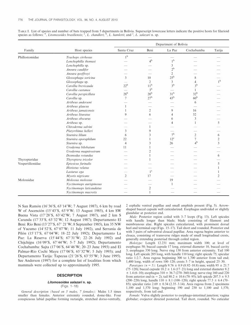
TABLE I. List of species and number of bats trapped from 5 departments in Bolivia. Superscript lowercase letters indicate the positive hosts for filarioidspecies as follows: b, Litomosoides brasiliensis; c, L. chandleri; h, L. hamletti; and s, L. salazari n. sp.
Family Host species
Department of Bolivia
Santa Cruz Beni La Paz Cochabamba Tarija
Phillostomidae .Trachops cirrhosus 1b — — — —
.Lonchophilla thomasi — 4b 1b — —
.Lonchophilla sp. — — 3 — —
.Anoura caudifer — — 2b — —
.Anoura geoffroyi — — 2 — —
.Glossophaga soricina 3 10 25h 8 —
.Glossophaga sp. — 2 3 4 1h
.Carollia brevicauda 22b 11b 3b 3b —
.Carollia castanea — 3b — 1 —
.Carollia perspicillata 28b 20b 31b 32b —
.Carollia sp. 7 27b 43b 60b —
.Artibeus anderseni — — — 6 —
.Artibeus glaucus 1 — — — —
.Artibeus jamaicensis 1 2 9 16 —
.Artibeus lituratus 9 6 4 32 —
.Artibeus obscurus — — 6 3 —
.Artibeus sp. — — 4 1 5
.Chiroderma salvini — 1 — — —
.Platyrrhinus helleri 3 9 — — —
.Sturnira lilium 6 3 7b 2 6
.Sturnira oporaphilum 22 1 12c 7 2
.Sturnira sp. 4 3 26 4b 3
.Uroderma bilobatum 11 1 — — —
.Uroderma magnirostrum — 2 — — —
.Desmodus rotundus — 1 — — 6
Thyropteridae .Thyroptera tricolor — 1 — — —
Vespertilionidae .Eptesicus furinalis — — — — 5
.Histiotus velatus — — — — 1
.Lasiurus ega — 1s — — 1
.Myotis nigricans 2 17 — — 2
Molossidae .Molossus molossus — — 19 — —
.Nyctinomops aurispinosus — — 1 — —
.Nyctinomops laticaudatus — 2 .— — —
.Nyctinomops macrotis — 1 .— — —
776 THE JOURNAL OF PARASITOLOGY, VOL. 96, NO. 4, AUGUST 2010

ornamentations observed. In transversal section, lateral hypodermicchords well developed, dome-shaped (Fig. 6).
Allotype: Length 20.53 mm; maximum width 170; with at level of vulva100; buccal capsule 16 long, and external diameter 8; buccal cavity 3; nervering 260 from anterior end; esophagus 370 long; vulva from apex 480; vulvalocated 100 from esophago-intestinal junction; ovejector 80 long; tail 115 long.
Paratypes (n 5 8): Length 14.7 ± 5.1 (10.12–24.6); maximum width137.6 ± 28.1 (106–170); buccal capsule 21.1 ± 1.4 (19–23) long, externaldiameter 8.3 ± 0.7 (7–9); nerve ring 200 from apex (n 5 2); esophagus412.5 ± 54.4 (330–510) long; vulva 510.6 ± 35.5 (480–580) from apex;distance of vulva to esophago-intestinal junction 118 ± 70.8 (30–200); tail127.1 ± 16.5 (110–160) long.
FIGURES 1–8. Litomosoides salazari. Female. (1) Anterior region, ventral view. (2) Female allotype anterior end median view. (3–5) Female paratype,median, lateral, and apical views. (6) Cross section posterior to the vulva. (7) Tail, ventral view. (8) Uterine microfilaria.
NOTARNICOLA ET AL.—FILARIOID NEMATODES OF BATS FROM BOLIVIA 777
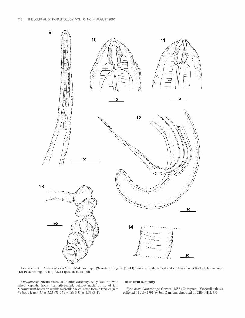
Microfilariae: Sheath visible at anterior extremity. Body fusiform, withsalient cephalic hook. Tail attenuated, without nuclei at tip of tail.Measurement based on uterine microfilariae collected from 2 females (n 5
6): body length 75 ± 5.25 (70–85); width 3.33 ± 0.51 (3–4).
Taxonomic summary
Type host: Lasiurus ega Gervais, 1856 (Chiroptera, Vespertilionidae),collected 11 July 1992 by Jon Dunnum, deposited at CBF NK25336.
FIGURES 9–14. Litomosoides salazari. Male holotype. (9) Anterior region. (10–11) Buccal capsule, lateral and median views. (12) Tail, lateral view.(13) Posterior region. (14) Area rugosa at midlength.
778 THE JOURNAL OF PARASITOLOGY, VOL. 96, NO. 4, AUGUST 2010
Site of infection: Abdominal cavity, nematodes found among theintestines at necropsy.
Type locality: Bolivia: Departamento del Beni: 35 km north NW ofYucumo ‘‘by road’’ (14u529S, 67u079W; 253 m).
Specimens deposited: Holotype (male), allotype (female), 12 paratypes (7females and 5 males), and 2 blood slides at the Harold Manter Laboratoryof Parasitology, HWML61785.
Prevalence and intensity: One of 1 host parasitized; 14 (6 males, 8females).
Etymology: Named after Dr. Jorge Salazar-Bravo, one of the primaryproponents of the study of the mammalian biodiversity in Bolivia. Hekeeps going when times are hard.
Remarks
Litomosoides salazari n. sp. belongs to the carinii group of species (Bainet al., 1989, 2003; Notarnicola et al., 2000) based on the morphologicalcharacteristics of both spicules. The left spicule has a handle longer thanthe blade, and a blade divided into a short proximal folded lamina and aterminal filament. The right spicule possesses a well cuticularized dorsalheel and a terminal cap.
Litomosoides salazari is a smaller sized species compared with thoseknown to occur in rodents and marsupials. The new species has a shortertail in both males and females, and a vulva that is located near theesophago-intestinal junction.
In comparison with 6 species in the carinii group that occur in rodents,the new species differs from L. carinii (Travassos, 1919) in having anunattenuated tail (Bain et al., 1989) and shorter spicules; from L. scottiForrester and Kinsella, 1973 in having a buccal capsule without an outerlateral extension and inconspicuous amphids (Forrester and Kinsella,1973); and from L. silvai Padilha and Faria, 1977 in having a rounded tailwithout a constricted end, a globular vagina, and a shorter right spicule(Moraes Neto et al., 1996). It differs from L. bonaerensis Notarnicola,Bain and Navone 2000 in possessing 3 pairs of cloacal papilla and lackingthe single precloacal papilla (Notarnicola et al., 2000); from L. odilaeNotarnicola and Navone, 2002 in having neither a protruded cloacalopening, nor a muscular esophagus (Notarnicola and Navone, 2002); andfinally, from L. andersoni Brant and Gardner, 1997 in the simplestterminal part of the right spicule, the presence of cloacal papillae, and arounded tail (Brant and Gardner, 1997).
Litomosoides salazari also can be differentiated from 2 speciesparasitizing marsupials as follows: from L. petteri Bain, Petit, and
Berteaux, 1980 by the shorter muscular esophagus and the symmetricalcloacal papillae (Bain et al., 1980); and from L. wilsoni Guerrero, Martin,Gardner and Bain, 2002 in the length of the esophagus (males 474–522 vs.270–360 mm and females 546 vs. 330–510 mm) and the longer microfilaria(61.8 vs. 75 mm) (see Guerrero et al., 2002).
There are 5 Litomosoides species known to infect bats, yet severalcharacters can be used to separate them from the new species. The newspecies is smaller than L. hamletti, L. brasiliensis, and L. yutajensisGuerrero, Martin and Bain, 2003. In addition, the walls of the buccalcapsule of L. hamletti are narrower (Jimenez-Quiros and Arroyo, 1960;Guerrero et al., 2002; Bain et al., 2003), and the tail of the male is shorter(mean of 64 vs. 80.2 mm). Litomosoides salazari differs from L. brasiliensisin having a simple shape of the heel of the right spicule, the vulva beingvery near the esophago-intestinal junction, and by the inconspicuousventral cephalic papillae (Guerrero et al., 2002). It also differs from L.yutajensis in the absence of a bell-like–shaped buccal capsule, and a longerand slender microfilaria instead of a stout microfilaria (length 58 vs. 75 mm,width 4.3 vs. 3.3 mm; see Guerrero et al., 2003). The new species alsodiffers from L. guiterasi (Perez Vigueras, 1934), a parasite of Artibeusjamaicensis parvipes Rehn 1902 from Cuba, in the shape of the buccalcapsule and the head papillae. It also has obvious cephalic papillae and acharacteristic bottle-shaped buccal cavity, not shown in figure 1 of Perez-Vigueras (1934). Finally, L. salazari differs from L. molossi Esslinger, 1973and L. chandleri Esslinger, 1973 in the absence of lateral cuticularornamentation, and in lacking the 2 annular thickenings of the buccalcapsule characteristic of L. molossi (Esslinger, 1973; Guerrero et al., 2002).
There is a group of 5 species of Litomosoides in which the male isunknown. The descriptions are based on the females for 3 species and 2 ononly microfilariae. Those species can be separated from L. salazari asfollows. Females of L. salazari are shorter than those of L. solarii Guerrero,Martin, Gardner, and Bain, 2002, possess a more anteriorly placed vulva(510 vs. 910 mm), and the tail of the microfilaria is not a sharp point as in L.solarii (Guerrero et al., 2002). The females of the new species can berecognized as distinct from those of L. artibei Esslinger, 1973 in having ashorter buccal capsule and a vulva very near the esophago-intestinal junction(Esslinger, 1973). The new species is different from L. chitwoodi Bain,Guerrero, and Rodriguez, 2003 in having a longer buccal capsule and a moreanterior vulva (Chitwood, 1938; Bain et al., 2003). The microfilariae of L.salazari are shorter than those described from L. colombiensis Esslinger,1973 [a parasite of Platyrrhinus dorsalis (Thomas, 1900)—mentioned asVampyrops dorsalis—and A. jamaicensis Leach, 1821 from Colombia] andlacks any nuclei in the tail in contrast to those of L. colombiensis, which has asingle row of nuclei in the tail (see Esslinger, 1973). Finally, the microfilariaeof L. salazari are longer than those of L. caliensis Esslinger, 1973 (mean 75vs. 60 mm), with an attenuated tail that is not rounded.
REDESCRIPTIONS
Litomosoides chandleri Esslinger, 1973(Tables I, II)
Diagnosis: Table II provides measurements for our specimens. Theywere identified as L. chandleri by shape of buccal capsule, disposition ofcephalic papillae (see Guerrero et al., 2002), cloacal papillae in males, andby lateral embellishments of cuticle in posterior extremity of both malesand females.
Taxonomic summary
Hosts: 3 Tschudi’s yellow-shouldered bats, Sturnira oporaphilum(Tschudi, 1844).
Site of infection: Abdominal cavity.Locality: Chijchijpa, La Paz. See Appendix for details on the collection
numbers and locality references.Specimens deposited: Vouchers (7 females and 5 males), at the Harold
Manter Laboratory of Parasitology. See Appendix for details on thecollection numbers.
Prevalence and mean intensity: 60% (3/5); 4 (2–6) parasites.
Remarks
The measurements and most of the characters from our specimens agreewith that published by other authors. However, there are 2 main
FIGURES 15–18. Litomosoides salazari. Male paratype. (15–16) Rightspicule, lateral and ventral view. (17) Detailed of the distal extremity of theright spicule, ventral view. (18) Left spicule, lateral view.
NOTARNICOLA ET AL.—FILARIOID NEMATODES OF BATS FROM BOLIVIA 779

differences between our specimens and those described by Esslinger (1973)and Guerrero et al. (2002): the buccal capsule from our specimens is longer(males 14.4 and 13.7 mm, respectively, vs. 18.8 mm; females 15.4 and15.8 mm, respectively, vs. 19.1 mm). However, the shape of the buccalcapsule and cavity resembles figure 11 in Esslinger (1973) and also figure59 from Guerrero et al. (2002). The vulva of the females of our specimensis located posterior to the esophago-intestinal junction, a structure thatwas usually reported near, or slightly anterior, to that structure.
Litomosoides chandleri has been reported from Artibeus jamaicensisLeach, 1821 from Colombia, Venezuela, French Guiana, and Peru (in thislast country, mentioned as Artibeus planirostris) (Esslinger, 1973; Guerreroet al., 2002) and Phyllonycteris poeyi Gundlach, 1860 and Nyctinomopslaticaudatus (E. Geoffroy, 1805) from Cuba (Rutkowska, 1980). Tenspecies of bats were trapped in Chijchipa, La Paz, including Artibeusjamaicensis (n 5 8), A. lituratus (n 5 4), and A. obscurus (n 5 2), but nofilarioid worms were found. The presence of this species in S. oporaphilumreported herein is a new host record.
Litomosoides hamletti Sandground, 1934(Tables I, II)
Diagnosis: Measurements for these specimens are included in Table II.The specimens were identified as L. hamletti because of the thin andnarrow buccal capsule, presence of a single cephalic papilla, vulva at thelevel of the esophagus, short tail in both sexes, and no more than 2 pair ofcloacal papillae.
Taxonomic summary
Hosts and localities: 4 Pallas’s Long-tongued bats, Glossophaga soricina,from Chijchijpa; and 1 Glossophaga sp. from Tapecua, Tarija. SeeAppendix for details on the collection numbers and locality references.
Site of infection: Abdominal cavity.Specimens deposited: Vouchers (20 females and 16 males) at the Harold
Manter Laboratory of Parasitology. See Appendix for details on thecollection numbers.
Prevalence and mean intensity: From G. soricina 16.6% (4/24); 5 (1–9)parasites; from Glossophaga sp. 100% (1/1); intensity 5 16 parasites.
Remarks
After re-examination of the type material, Bain et al. (2003) suggested 2subspecies for L. hamletti, i.e., Litomosoides hamletti hamletti to includethe specimens of Sandground (1934), Rego (1961), Esslinger (1973), andGuerrero et al. (2002)—mentioned as L. guiterasi—and L. hamletti penaito include the specimens of Jimenez-Quiros and Arroyo (1960)—mentioned as L. penai. The justification is that because females of thelatter subspecies are longer, the vulva is located posterior to the esophago-intestinal junction, and the tail is more attenuated. Our material is inagreement with the morphologic and metric characters of L. hamlettihamletti. Some males and females presented longer buccal capsules (35 and40, respectively), and most of the males examined had an adcloacal pair ofpapillae and 1 pair of asymmetric post-cloacal papillae. This subspecieshas been reported from G. soricina from Brazil, Mexico, Colombia, andVenezuela (Sandground, 1934; Chitwood, 1938; Rego, 1961; Esslinger,1973; Guerrero et al., 2002). It represents the first record for the species inBolivia.
Litomosoides brasiliensis Lins de Almeida, 1936(Tables I, III)
Diagnosis: Specimens identified as L. brasiliensis. Table III containsmeasurements of specimens recovered from C. pespicillata and C.brevicauda, the host species most frequently found infected. Males possess
TABLE II. Measurements of Litomosoides chandleri and L. hamletti from Bolivia.
Host species:
Litomosoides chandleri Litomosoides hamletti
Sturnira oporaphilum Glossophaga soricina
Males (n 5 5) Females (n 5 7) Males (n 5 10) Females (n 5 15)
Length (mm) 11.6 (9.9–14) 23.4 (17.4–27.6) 16.8 (14.7–24) 50.05 (39–56)
Width 97.6 (88–116) 162.1 (116–193) 81.2 (57–99) 164.8 (136–257)
Buccal capsule 18.8 (17–21) 19.1 (17–21) 27.4 (18–35) 29.2 (23–40)
Esophagus 424.4 (390–473) 483.8 (400–550) 531.3 (390–752) 728.5 (509–1,038)
Tail 116 (95–140) 284 (230–350) 66.7 (55–78) 161.1 (105–233)
Left spicule 216.5 (213–220) — 218.1 (190–274) —
Handle 110 (100–120) — 127.2 (110–142) —
Right spicule 61.6 (60–65) — 73.2 (60–93) —
Vulva to apex — 706.1 (565–850) — 584.4 (455–829)
Microfilaria length (n 5 2) 63; 64 (n 5 12) 51.66 (48–55)
Width 3; 3 5
TABLE III. Measurements of Litomosoides brasiliensis from Bolivia.
Host species: Carollia brevicauda Carollia perspicillata
Males (n 5 7) Females (n 5 9) Males (n 5 21) Female (n 5 26)
Length (mm) 56.6 (39.2–83) 134.52 (79.2–174) 52 (36.2–81.1) 124.1 (72.3–156.7)
Width 143.8 (130–158) 232.3 (135–297) 138 (94–188) 230.8 (149–307)
Buccal capsule 22.12 (17–25) 22.44 (17–30) 20.7 (15–25) 22.6 (16–28)
Esophagus 663 (534–860) 775.4 (645–1,048) 750 (572–1,127) 863.6 (620–1,211)
Tail 239 (189–318) 340.6 (193–505) 225.5 (149–346) 351.4 (219–465)
Left spicule 511.5 (383–616) — 454.6 (299–663) —
Handle 197.7 (141–257) — 207.6 (148–314) —
Right spicule 175.5 (142–213) — 157.1 (101–220) —
Vulva to apex — 2,843.4 (2,330–3,657) — 2,603.8 (1,706–3,185)
Microfilaria length — (n 5 5) 56.4 (48–61)
780 THE JOURNAL OF PARASITOLOGY, VOL. 96, NO. 4, AUGUST 2010

a distinctive right spicule, with a folded heel, buccal capsule with 2 rings(as shown in figures 20 and 21 in Guerrero et al., 2002), vaginasubglobular, posteriorly elongated, cloacal papillae disposed in tandemon ventral longitudinal line.
Taxonomic summary
Hosts and localities: Trachops cirrhosus (Spix, 1823), Lonchophilathomasi Allen, 1904, Anoura caudifer (E. Geoffroy, 1818), Carolliabrevicauda (Schinz, 1821), C. castanea H. Allen, 1890, C. pespicillata(Linnaeus, 1758), Carollia sp. Gray, 1838, and Sturnira lilium (E.Geoffroy, 1810) in 4 of the 5 Departments surveyed. See Table I andAppendix for detailed localities.
Site of infection: Abdominal cavity.Specimens deposited: Vouchers (111 females and 108 males) and blood
slides at the Harold Manter Laboratory of Parasitology. See Appendix fordetailed numbers of collection.
Prevalence and mean intensity: From T. cirrhosus 100% (1/1), Intensity5 3 parasites; from L. thomasi 100% (5/5), 6.5 (4–10) parasites per host;from A. caudifer 50% (1/2), Intensity 5 1; from C. brevicauda 28.5% (11/39), 4 (1–10) parasites per hosts; from C. castanea 33% (1/3), Intensity 5 1;from C. pespicillata 33% (37/111), 4.4 (1–21) parasites per hosts; fromCarollia sp. 8% (11/137), 5.9 (1–11) parasites per hosts; from S. lilium 4%(1/24), intensity 5 1.
Remarks
Worms of L. brasiliensis are longer compared with the 3 filarioid speciescollected in this survey. Moreover, the rows of the area rugosa are closer,one from another, compared with L. chandleri and L. hamletti, which aremore spaced. The morphological character as well as the measurementsfrom our specimens fit with those reported by Caballero (1947), Esslinger(1973), and Guerrero et al. (2002). However, microfilariae reported bythese authors are longer than ours (92 from Esslinger, 1973 and 71.8–94.5from Guerrero et al., 2002 vs. 56.4 from present study). This differencecould be explained because our measurements provide from blood smearsinstead of mature females or blood concentrated 2% aqueous formalinand was reported that microfilariae frequently shrink with this technique(Esslinger, 1973).
This filarioid was the most common species found in the present survey.Its ubiquitous presence is due to its ability to infect both phyllostomid andvespertillionid bats (Lins de Almeida, 1936; Caballero, 1947; Diaz-Ungria,1963; Guerrero et al., 2002).
Litomosoides brasiliensis has been frequently reported from severalspecies of Carollia from Central and South America. Recently, Guerreroet al. (2002) added Lyonycteris spurrelli Thomas, 1913, A. caudifer, and S.lilium, to the list of hosts. The latter 2 host species also were recorded inBolivia infected by this species. In addition, the presence of L. brasiliensisin T. cirrhosus and L. thomasi represent new host records.
DISCUSSION
Several authors have found small variability in both quantita-
tive and qualitative morphological characters of some species of
Litomosoides (Esslinger, 1973; Guerrero et al., 2002; Notarnicola,
2005); however, the landmarks for each species remain stable. The
4 species recovered in the present study were identifiable by the
combination of characters such as the buccal capsule, the shape of
the spicules, the caudal papillae in males, and the vulva.
An examination of the gazetteer and list of species of mammals
known from Bolivia in Anderson (1997) shows that the species of
mammals that we have reported on up to the present time
represents a very small proportion of the total. Therefore, the
results presented in the present paper, as well as those presented
by Dick et al. (2007), which focus only on ectoparasites, fall short
in summarizing the diversity of parasites infecting the bats of
Bolivia. Additional students and researchers are encouraged to
continue to work with these specimens, especially as many of the
areas in which we made our collections from 1984 to 2000 have
been reduced from diverse forest ecosystems to monotonous
cleared landscapes covered with introduced grasses and populated
mostly by bovids.
ACKNOWLEDGMENTS
We thank Marıa Cristina Estivariz (CEPAVE) for preparing thedrawings. This article is a result of the National Science Foundationgrants BSR8612329 (to S.L.G., T. L. Yates, and D. W. Duszynski) andBSR9024816, DEB9496263, DEB9631295, and DBI0097019 (to S.L.G.).J.N.’s work in the HWML was sponsored by Fulbright-CONICET.
LITERATURE CITED
ANDERSON, S. 1997. Mammals of Bolivia, taxonomy and distribution.Bulletin of the American Museum of Natural History 231: 1–652.
BAIN, O., R. GUERRERO, B. RODRIGUEZ, S. BABAYAN, AND N. JOUVENET.2003. Examination of the type material of two species of Litomosoides(Filarioidea: Onchocercidae), parasites from bats: Taxonomic conse-quences. Parasite 10: 211–218.
———, G. PETIT, AND S. BERTEAUX. 1980. Description de deux nouvellesFilaires du genre Litomosoides et de leurs stades infestants. Annalesde Parasitologie Humaine et Comparee 55: 225–267.
———, ———, AND M. DIAGNE. 1989. Etude de quelques Litomosoidesparasites de rongeurs; consequences taxonomiques. Annales deParasitologie Humaine et Comparee 64: 268–289.
BRANT, S. V., AND S. L. GARDNER. 1997. Two new species of Litomosoides(Nemata: Onchocercidae) from Ctenomys opimus (Rodentia: Ctenomyi-dae) on the altiplano of Bolivia. Journal of Parasitology 83: 700–705.
———, AND ———. 2000. Phylogeny of the species of the genusLitomosoides (Nemata: Onchocercidae): Evidence of rampant hostswitching. Journal of Parasitology 86: 545–554.
CABALLERO, C. 1947. Algunas filarias de mamıferos y de reptiles de lasRepublicas de Colombia y Panama. Anuales del Instituto de Biologıade Mexico 18: 169–188.
CHITWOOD, B. G. 1938. Some nematodes from the caves of Yucatan.Carnegie Institution of Washington Publications 491: 51–66.
DIAZ-UNGRIA, C. 1963. Nematodes parasites, nouveaux ou interessants, duVenezuela. Annales de Parasitologie Humaine et Comparee 38: 893–914.
DICK, C. W., D. GETTINGER, AND S. L. GARDNER. 2007. Bolivianectoparasites: A survey of bats (Mammalia Chiroptera). ComparativeParasitology 74: 372–377.
ESSLINGER, J. H. 1973. The genus Litomosoides Chandler, 1931 (Filarioi-dea: Onchocercidae) in Colombian bats and rats. Journal ofParasitology 59: 225–246.
FORRESTER, D. J., AND J. M. KINSELLA. 1973. Comparative morphologyand ecology of two species of Litomosoides (Nematoda: Filarioidea)of rodents in Florida, with a key to the species of LitomosoidesChandler, 1931. International Journal for Parasitology 3: 255–263.
GARDNER, S. L. 1996. Essential techniques for collection of parasitesduring surveys of mammals. In Measuring and monitoring biologicaldiversity: Standard methods for mammals, D. E. Wilson, R. Cole, J.D. Nichols, R. Rudran, and M. Foster (eds.). Smithsonian InstitutionPress, Washington, D.C., p. 291–298.
———, AND F. A. JIMENEZ. 2009. Methods for the study of batsendoparasites. In Ecological and behavioral methods for the study ofbats, T. H. Kunz and S. Parsons (eds.). The Johns HopkinsUniversity Press, Baltimore, Maryland, p. 795–805.
GUERRERO, R., C. MARTIN, S. L. GARDNER, AND O. BAIN. 2002. New andknown species of Litomosoides (Nematoda: Filarioidea): Importantadult and larval characters and taxonomic changes. ComparativeParasitology 69: 177–195.
———, ———, AND O. BAIN. 2003 Litomosoides yutajensis n. sp., firstrecord of this filarial genus in a mormoopid bat. Parasite 10: 219–225.
JIMENEZ-QUIROS, O., AND G. ARROYO. 1960. Helmintos de la Republica deCosta Rica. XV. Nematoda 3. Presencia de Litomosoides penai n. sp.en Hemiderma perspicillatum aztecum (Saussure, 1860). Revista deBiologıa Tropical 8: 63–67.
LINS DE ALMEIDA, J. 1936. Sobre parasito de Cheiroptera: Litomosoidesbrasiliensis Lins de Almeida, 1936 (Nematoda: Filariidae). Revista delDepartamento Nacional de Produccion Animal 3: 133–138.
MORAES NETO, A. H. A. DE, R. M. LANFREDI, AND W. DE SOUZA. 1996.Emended description of Litomosoides silvai (Nematoda: Filarioidea)
NOTARNICOLA ET AL.—FILARIOID NEMATODES OF BATS FROM BOLIVIA 781

of Akodon cursor (Rodentia: Muridae). Journal of Parasitology 82:988–991.
NOTARNICOLA, J. 2005. Description of adults and fourth-stage larva ofLitomosoides navonae n. sp. (Nematoda: Onchocercidae), a parasiteof five species of sigmodontine rodents from northeastern Argentina.Systematic Parasitology 62: 171–183.
———, AND G. T. NAVONE. 2002. A new species Litomosoides odilae n. sp.(Nemadoda: Onchocercidae) from Oligoryzomys nigripes (Rodentia:Muridae) in the rain forest of Misiones, Argentina. Journal ofParasitology 88: 967–971.
———, O. BAIN, AND G. T. NAVONE. 2000. Two new species ofLitomosoides (Nematoda: Filarioidea) in sigmodontines (Rodentia:Muridae) from Rio de La Plata marshland, Argentina. Journal ofParasitology 86: 1318–1325.
———, F. A. JIMENEZ, AND S. L. GARDNER. 2007. A new species ofDipetalonema (Filarioidea: Onchocercidae) from Ateles chamek fromthe Beni of Bolivia. Journal of Parasitology 93: 661–667.
———, M. PINTO, AND G. T. NAVONE. 2008. Host occurrence andgeographical distribution of Dipetalonema spp. (Nematoda: Oncho-cercidae) in Neotropical monkeys and the first record of Dipetalo-nema gracile in Ecuador. Comparative Parasitology 75: 61–68.
PEREZ-VIGUERAS, I. 1934. Notas sobre las especies de Filarioidea(Nematoda) encontradas en Cuba. Memorias de la Sociedad Cubanade Historia Natural ‘Felipe Poey’ 8: 55–60.
REGO, A. A. 1961. Nota sobre a especies Litomosoides guiterasi Vigueras,1934 (Nematoda, Filarioidea). Atas de la Sociedad de Biologia de Riode Janeiro 5: 13–14.
RUTKOWSKA, M. A. 1980. The helminthofauna of bats (Chiroptera) fromCuba. I. A review of nematodes and acanthocephalans. ActaParasitologica Polonica 26: 153–186.
SANDGROUND, J. H. 1934. Description of a species of the filariid genusLitomosoides from Glossophaga soricina (Cheiroptera). Annales andMagazine of the Natural History Series10 14: 595–599.
SIMMONS, N. B. 2005. Order Chiroptera. In Mammal species of the world,3rd ed., D. E. Wilson and D. M. Reeder (eds.). Johns HopkinsUniversity Press, Baltimore, Maryland, p. 312–529.
UNZUETA, O. Q. 1975. Mapa ecologico de Bolivia. Ministerio de Asuntoscampesinos y Agropecuarios, La Paz, Bolivia, 432 p.
APPENDIX
Locality data and host species of the filarioid nematodes.
HWML: Collection number of the filarioid specimens at Harold
Manter Laboratory. NK: Field number of the host species.
Litomosoides chandleri
Departamento La Paz: Chijchijpa (16u099S, 67u449W) 6 July
1992: Sturnira oporaphilum NK25260; HWML61758 S. oporaphi-
lum NK25262; HWML61771 S. oporaphilum NK25285.
Litomosoides hamletti
Departamento La Paz: Chijchijpa (16u099S, 67u449W) 5 July
1992: HWML61750 Glossophaga soricina NK25221;
HWML61752 G. soricina NK25227; HWML61753 G. soricina
NK25228; HWML61776 G. soricina NK25295.
Departamento Tarija: Tapecua (21u269S, 63u559W) 5 June
1995: Glossophaga sp. NK30829.
Litomosoides brasiliensis
Departamento Santa Cruz: San Miguel Rincon (17u239S,
63u329W) 14 August 1984: HWML63396 Carollia perspicillata
NK11652; HWML63397 C. perspicillata NK11654; Estancia
Cachuela Esperanza, Santa Cruz (16u479S, 63u149W) 22 August
1984: HWML63398 C. perspicillata NK11697; HWML63399 C.
perspicillata NK11700; HWML63400 C. perspicillata NK11710;
HWML63401 C. perspicillata NK11716; HWML63402 C. perspi-
cillata NK11717; HWML63403 C. perspicillata NK11767;
HWML63404 C. brevicauda NK12739; HWML63405 C. brevi-
cauda NK12783; HWML63406 C. brevicauda NK12794;
HWML63407 C. perspicillata NK12834; HWML63408 C. brevi-
cauda NK12937; HWML63409 Carollia sp. NK12954. Ten km N
San Ramon (16u369S, 63u149W) 7 August 1987: HWML63410 C.
brevicauda NK12981. Six kilometers by road W of Ascencion
(15u439S, 63u99W) 13 August 1985: HWML63411 Trachops
cirrhosus NK13096. Four kilometers SW Buena Vista (17u289S,
63u429W) 7 August 1987: HWML63413 Carollia sp. NK15230.
Two kilometers S Caranda (17u339S, 63u329W) 12 August 1987:
HWML63414 C. perspicillata NK15263; HWML63415 Carollia
sp. NK15265; HWML63416 Carollia sp. NK15271; HWML63417
C. perspicillata NK15273. Eight kilometers SE Santa Cruz
(17u449S, 63u179W) 9 June 1988: HWML63418 C. brevicauda
NK15642.
Departamento La Paz: Chijchijpa (16u099S, 67u449W) 5 July
1992: HWML63419 C. brevicauda NK25238; HWML63420
Carollia sp. NK25244; HWML61773 C. perspicillata NK25290;
Sturnira lilium NK25319. La Reserva (15u449S, 67u319W) 24 July
1992: HWML63435 C. perspicillata NK25592; HWML63436 C.
perspicillata NK25597; HWML63437 Carollia sp. NK25627;
HWML63438 Anoura caudifer NK25628; HWML63439 C.
perspicillata NK25636; HWML63440 Lonchophylla thomasi
NK25665.
Departamento del Beni: Rio Beni (13u279S, 67u219W) 8
September 1985: HWML63412 Carollia sp. NK13533. Serranıa
de Pilon (15u179S, 67u049W) 19 July 1992: HWML63421
Lonchophylla thomasi NK25399; C. perspicillata NK25404; C.
perspicillata NK25405; HWML63422 Carollia sp. NK25413;
HWML63423 C. brevicauda NK25414; HWML63424 C. perspi-
cillata NK25417; HWML63425 C. perspicillata NK25420; Car-
ollia sp. NK25421; L. thomasi NK25423; HWML63426 C.
perspicillata NK25424; HWML63427 L. thomasi NK25429;
HWML63428 L. thomasi NK25430; C. perspicillata NK25431;
C. perspicillata NK25432; HWML63429 C. perspicillata
NK25435; C. perspicillata NK25436; HWML63430 C. perspicil-
lata NK25437; HWML63431 C. perspicillata NK25438;
HWML63432 C. castanea NK25439; HWML63433 Carollia sp.
NK25447; HWML63434 C. brevicauda NK25450.
Departamento Cochabamba: Sajta (17u069S, 64u469W) 22 June
1993: HWML63441 C. perspicillata NK29945; HWML63442 C.
perspicillata NK30014; HWML63443 Carollia sp. NK30065;
HWML63444 C. perspicillata NK30067; HWML63445 C. perspi-
cillata NK30068; HWML63446 Carollia sp. NK30080. El Palmar-
Rio Cochi Mayu (17u069S, 65u329W) 5 July 1993: HWML63447
C. perspicillata NK30174; HWML63448 C. perspicillata
NK30195; HWML63449 C. perspicillata NK30199; HWML63450
C. brevicauda NK30200.
782 THE JOURNAL OF PARASITOLOGY, VOL. 96, NO. 4, AUGUST 2010