cuticle fine structure of nine species in the genus...
TRANSCRIPT
Fundam. app/. NemaLO/., 1993,16 (2),137-149
Cuticle fine structure of nine species in the genusTylenchorhynchus Cobb, 1913 (Nemata: Belonolaimidae)
Danamou MOUNPORT *, Pierre BAuJARD ** and Bernard MARTINY **
" Département de Biologie Animale, Faculté des Sciences, Université Cheikh Anta Diop, Dakar, Sénégal,and "" Laboratoire de Nématologie, Centre ORSTOM, B.P. 1386, Dakar, Sénégal.
Accepted for publication 29 May 1992.
Summary - The cuticle fine structure of females of nine species in me genus Tyleru:horhynchus, belonging ta three morphologicaltypes according ta external cuticular ornamentations outside lateral fields, is described : first type of species wimout longirudinalridges and longirudinal incisures (T. indicus, T. vemra/is, T. annu/alUs and T. vu/garis); second type wim longirudinal ridges andincisures (T. gladiolalUs); third type wimlongirudinal ridges but wimout longirudinal incisures (T. germaniz~ T. microphasmis, T.sulœtus and an unidentified species). Six groups are identified in me nine Tyleru:horhynchus species srudied according to cuticle finestrucrure criteria.
Résumé - Ultrastructure de la cuticle chez neufespèces du genre Tylenchorhynchus Cobb, 1913 (Nernata : Belonolaimidae) - L'ultrastrucrure de la cuticule de neuf espèces du genre Ty/enchorhyru:hus appanenant à trois types morphologiques- suivant les ornementations externes de la cuticule en dehors des champs latéraux - est décrite. Le premier type comprend desespèces sans crêtes ni incisures longirudinales (T. indicus, T. annulatus, T. ventra/is et T. vu/garis). Le deuxième type, des espèces aveccrêtes et incisures longirudinales (T. gladiolalUS). Le troisième type, des espèces avec crêtes et sans incisures longirudinales (T.gennanii, T. microphasmis, T. su/calUs et une espèce non déterminée). Six groupes sont identifiés dans l'ensemble des neuf espècesérudiées sur la base de trois critères se rapportant à l'ultrastrucrure de la cuticule: nombre de couches dans la zone mèdiane endehors des champs latéraux, nombre de couches dans la zone basale au niveau des champs latèraux et aspect de la couche basalestriée. La strucrure de la cuticule au niveau des champs latéraux chez Ty/eru:horhyru:hus sp. rappelle celle observée chez lesHoplolaiminae.
The family Belonolaimidae Whitehead, 1960 was redifmed by Fortuner and Luc (1987) and the followingnine genera were proposed as synonyms of Tylenchorhynchus Cobb, 1913 : Bitylenchus Filip'ev, 1934; TelOlylenchus Siddiqi, 1960; Quinisulcius Siddiqi, 1971; Dolichorhynchus Mulk & Jairajpuri, 1974; Trilineellus Lewis& Golden, 1981; Divittus Jairajpuri, 1984; MorasinemaJaved, 1984; Tessellus Jairajpuri & Hunt, 1984; Neodolichorhynchus Jairajpuri & Hunt, 1984. Morphobiometriccriteria used in the generic diagnosis were tao variableand so could not be used in the characterization of therejected genera. The taxonomie importance of cuticlelayering was demonstrated by Shepherd, et al. (1972)and Maggenti (1979). Mounport et al. (1990, 1991a, b)confirmed the constancy of cuticle layering in the generaCriconemella, Pratylenchus and Scutellonema. The aim ofthis paper is to appraise the variability of cuticle ultrastructure in the genus Tylenchorhynchus as redefined byFortuner and Luc (1987).
Material and methods
MATERIAL
The nine available species studied here belong to threemajor types according to morphological features of thecuticle :
Type 1 : longitudinal ridges and incisures absent outsidelateral fields.
Tylenchorhynchus annulatus (Cassidy, 1930) Golden,1971=Tylenchorhynchus martini Fielding, 1956.
Tylenchorhynchus indicus (Siddiqi, 1960) Fortuner &Luc, 1987=Telotylenchus indicus Siddiqi, 1960.
Tylenchorhynchus ventralis (Loof, 1963) Fortuner &Luc, 1987=Telotylenchus ventralis Loof, 1963.
Tylenchorhynchus vulgaris Upadhyay, Swarup &Sethi, 1972=Bitylenchus vulgaris (Upadhyay, Swarup & Sethi,1972) Siddiqi, 1986.
Type 2 : longitudinal ridges and incisures present outside lateral fields.
Tylenchorhynchus gladiolalUS Fortuner & Amougou,1974=Dolichorhynchus (Neodolichorhynchus) gladiolatus(Fortuner & Amougou, 1973) Mulk & Siddiqi,1982;= Neodolichorhynchus gladiolatus (Fortuner &Amougou, 1973) Jairajpuri & Hunt, 1984.
ISSN 1164-5571/93/02/137 13 S 3.30/ © Gauthier-Villars - ORSTOM 137

D. Mounporl et al.
Type 3 : longitudinal ridges present and longitudinalmCIsure absent outside lateral fields.
Tylenchorhynchus germanii (Germani & Luc, 1984)Fortuner & Luc, 1987=Dolichorhynchus (Dolzchorhynchus) elegans Germani & Luc, 1984;= Tylenchorhynchus elegans (Germani & Luc, 1984)Fortuner & Luc, 1987.
Tylenchorhynchus microphasmis Loof, 1960= Dolichorhynchus (Neodolichorhynchus) microphasmis (Loof, 1960) Mulk & Siddiqi, 1982= Neodolichorhynchus microphasmis (Loof, 1960)]airajpuri & Hunt, 1984.
Tylenchorhynchus sulcalUs de Guiran, 1967= Dolzchorhynchus (Neodolichorhynchus) sulcaluS (deGuiran, 1967) Mulk & Siddiqi, 1982;= Neodolichorhynchus sulcalUs (de Guiran, 1967)]airajpuri & Hunt, 1984.
Tylenchorhynchus sp.
LOCALITY AND DATE OF COLLECTION OF SPECIES
T. annulalus : Richard-ToU (Senegal) in 1982.T. germanii : toporypes from Patar (Senegal) in 1984.T. gladiO/alUS : Nebe (Senegal) in 1986.T. indicus : Thienaba (Senegal) in 1988.T. sulcalUs : N'Dindy (Senegal) in 1982.T. venlralis : Louga (Senegal) in 1982.T. vulgaris : Agadez region (Niger) in 1987.Tylenchorhynchus sp.: Aogadut region (Niger) in
1987.These species are cultured on Sorghum vulgare in the
laboratory from the sampling date. Specimens of T. microphasmis were obtained from Dr. F. C. Zoon (TheNetherlands) in FP 4: 1 fixative.
PROCESSING TECHNIQUES FOR ELECTRON MICROSCO
PY
Females of ail nine Tylenchorhynchus species werefixed overnight at 4 oC in glutaraldehvde 2.5 % in a0.1 M sodium cacodylate buffer at pH 7.2; they werethen cut into rwo or three pieces and prepared for embedding in low viscosity epoxy resin (Spurr, 1969) aspreviously described by Mounport el al. (1990). Ultrathin sections were cut with a diamond knife on aSorvaU Porter Blum MTl ultramicrotome; grids werecontrasted (Reynolds, 1963) and observed in a ]eol 100CXII electron microscope operating at 80 kV. Crossand longitudinal sections of at least ten females of eachspecies were examined.
DEFINITIONS
The following definitions will be used for incisuresand ridges observed in the different species :
incisures = longitudinal invaginations of the externalcortical layer;
ridges = longitudinal band on the cuticle appearing
138
more or less hemispherical in cross-section, demarcatedor not by incisures.
Results
SPECIES WITHOUT LONGITUDINAL RIDGES Al"lD INCI
SURES OUTSIDE LATERAL FIELDS
Cross-sections at mid-body show that the cuticle isthinner in T. vulgaris (0.50 fLm) than in the other species(0.80 fLm). Cuticle ultrasrructure of T. indicus and T.venlralis is sirnilar and observations on the latter speciesare presented.
The cuticle outside lateral fields (Fig. 1 B, Ci Fig. 2 B,Ci Fig. 3 B, C) is composed of three major zones:
The cortical zone, consists of i) an external rrilaminatelayer whose thickness is constant (0.03 fLm); il) an internai granular layer averaging 0.20 fLm in T. vulgarisand 0.17 fLm to 0.25 fLm in thickness in the other species.
The median zone, consists of an electron-lucent orelectron-dense layer, which is very thin in T. vulgaris(0.05 fLm vs 0.20 fLm in the other species). Infiltrationsof granular material from the inner cortical layer areobserved in this layer in T. venlralis (Fig. 2 B, D) and T.annu/alUs (Fig. 1 C). In T. venlralis tangential section ofthe oesophagus (Fig. 2 E) shows that theses infùtrationsof circular section are arranged in four rows under eachannulation; at the mid-body these rows are less obvious(Fig. 2 F). Near the vulva the median layer is thicker(l.20 fLm), resulting in a cuticular ridge (Fig. 2 D).
The basal zone, consists of a striated layer, rangingfrom 0.20 fLm in thickness in T. vulgaris to 0.40 fLm inthe other species (Fig. 1 C; Fig. 2 C; Fig. 3 C). Striations are perpendicular to the somatic muscles and theirperiodicity in longitudinal sections is higher than incross-sections. We observed a particular pattern in T.indicus, T. venlralis and T. vulgaris where the striatedlayer is regularly interrupted by parallel bands withoutstriation; these bands correspond to the body annulations (Fig. 2 Gi Fig. 3 D). The striated layer in aU species is attached to somatic muscles by hemidesmosomes(Fig. 1 C; Fig. 2 B).
The cuticle at the level of the lateral fields is shown inFig. 1 Di Fig. 2 Hi Fig. 3 E, F and Fig. 4. Lateral fieldsare not prominent (Fig. 2 A, H; Fig. 3 A) except in T.annulalus (Fig. 1 A). The cuticle is composed of:
The cortical zone, whose ultrasrructure and thickness isidentical to that outside the lateral fields, except in T.annu/alUs where the inner cortical layer is thicker andseems to be stratified (Fig. 1 D).
The median zone, which in cross-sections of T. annu/alUS and T. venlralis reveals a discontinuity of the medianlayer, located only berween the incisures (Fig. 1 D;Fig. 2 H); the layer is continuous in T. vulgaris(Fig. 3 F).
Fundam. appl. Nemalol.

Cutic/e fine stTUcture ofTylenchorhynchus spp.
•..'"--.....,;"
®
@
Fig. 1. Cuticle fine strucrure of Ty/enchorhynchus gladiolatus females in longirudinal (LS) and cross (CS) sections. A : CS nearmid-body; B : LS between two incisures; C : LS at level of an incisure; D : CS between two incisures, E : CS of an external incisureof a lateral field; F: CS of a lateral field; G : Detail of F showing layers of the basal zone. (Bars: A =5 fJ.rn; B-G =0.5 fJ.rn).
List ofabbreviations used in the Figures. - btl =basal fibrillar layer; bs =basal striated layer; bv, yb, vbl =basal vacuolar layer;ec =external cortical layer; tl =fibrillar layer; fm =fibrillar median layer; gl =gramùar layer; he =hemidesmosome; hy =hypodermis; i =longirudinal incisure; ic =internal cortical layer; 1=line; li = intestine lumen; lm =limiting membrane; md =median denselayer; ml = median layer; sm =somatic muscles; u = uterine wail.
Vol. 16, n° 2 - 1993 139

D. Mounporl et al.
®
Fig. 2. Cuticle fine structure of Tylenchorhynchus venLralis females in longitudinal (LS), cross (CS) and tangential (TS) sections. A :CS near mid-body; B : LS at mid-body; C : CS at mid-body; D : LS of the prevulvar zone; E, F : TS showing median layer; G : TSof striated layer, showing transverse bands (arrowheads) without striae; H : CS of a lateral field. (Bars: A = 5 fLm; B-H = 0.5 fLm).
Abbreviations : see Fig. \.
140 Fundam. appl. Nematol.

The basal zone, whose basal striated layer becomesforked in cross-section in ail species at the level of thetwo outer incisures. In T. annulatus this layer is replacedby two fibrillar layers and averages 1.20 f.Lm in thickness.In T. indicus and T. ventralis it is replaced by twa outergranular and two inner fibrillar layers and averages1.00 f.Lm (Fig. 2 H). In T. vulgan's five layers are present; the granular and fibrillar layers are separated by anelectron-Iucent layer; the total thickness of these fivelayers is about 0.60 f.Lm (Fig. 3 E, F). The fibres in eachfibrillar layer are oriented obliquely, forming an angle ofabout 30° with the longitudinal axis in T. annulatus(Fig. 1 F).
SPECIES WITH LONGITUDINAL RIDGES AND INCISURES
OUTSlDE LATERAL FIELDS
In T. g!adio!atus, the only species examined in thisgroup, cross-sections at mid-body reveal 28 longitudinalincisures (Fig. 5 A); areas between these incisures appear in cross-section as ridges and T. g!adiolalUs shauldbe considered as a species with adjacent longitudinalridges : the cuticle thickness ranges from 0.16 f.Lm at thelevel of incisures ta 0.50 f.Lm between them.
CUliâe fine slnlClure ofTylenchorhynchus spp.
The cuticle outside lateral fields is composed of thefollowing elements :
The cortical zone, consists of an external (0.03 f.Lm)and an internai granular layer, ranging from 0.03 f.Lmunder the incisures ta 0.13 f.Lm between them (Fig. 5 BD).
The median zone, is represented by an electron densevacuolar layer of 0.20 f.Lm in thickness (Fif. 5 B, D); thelayer is absent under the incisures (Fig. 5 C, D).
The basal zone, consists of a striated layer averaging0.13 f.Lm in thickness; striations are radial and the layer isattached ta somatic muscles by hemidesmosomes(Fig. 5 B, D).The cuticle at the level of the lateral fields (Fig. 5 E-G;Fig. 4) shows thickness and incisures deepness identicalto those outside lateral fields.
The cortical zone, is similar to previous observationsoutside lateral fields.
The median zone, remains unchanged.
The basal zone, shows a striated layer in cross-sectionsthat becomes forked under the two outer incisures of the
®
®
Fig. 3. Cuticle fine srructure of Tylenchorhynchus vulgaris females in longitudinal (LS), cross (CS) and tangential (TS) sections. A :CS near mid-body; B : LS at mid-body level; C : CS at mid-body level; D : TS of basal striated layer; E : CS of a lateral field; F : CSshowing an enlargement of the first incisure. (Bars: A =5 !-lm; B-F = 0.5 jJ..m).
Abbreviations : see Fig. 1.
Vol. 16,no2-1993 141

D. Mounporl et al.
T. annulafusT. germa');; _ T. sulcafus
T. microphasmis
"'::::::i:::::::::;':::::'
~>~miW(ll(î~ffi1~~*~i.::; ..:l~~;:(jl\'~~!W_
T. vulgaris
T. gkldiolafus\\11111111 ~ FLas
[ill]: .. ;;::::::
!IV ~~~~~~:!!!! GL..... :::::::::::
Fig. 4. Diagrams oflateral fields cross sections in females of the nine examined Tylendwrhym:hus species showing the ultrastrucrureof the basal zone.
Abbreviations : see Fig. 1.
lateral fields; there it is replaced by five layers : two outergranular layers, an intermediate vacuolar layer appearing only between incisures and two fibril1ar layers thatjoin the hypodermis (Fig. 5 F, G).
SPECIES WITH LONGITUDINAL RIDGES OUTSIDE LAT
ERAL FIELDS
The ultrastrucrure of the cuticle will be described for:i) areas between ridges outside lateral fields, ii) the ridgesoutside lateral fields, iii) the ridges of the lateral fields.Cuticle ultrastructure and thickness of T. germanii andT. sulcalus are similar and observations on the formerspecies will be presented. T. germanii, T. microphasmis
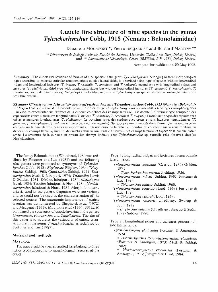
and T. sulcalus have sixteen longitudinal ridges extending over the entire body. Three ridges form each lateralfield, the median one being larger than the two outerones. The dorsal and ventral ridges are bordered onboth sides by two sublateral ridges (Fig. 6 A; Fig. 7 A).Cross-sections of Tylenchorhynchus sp. at mid-body reveal twelve longitudinal ridges (Fig. 8 A). Only oneridge occurs in each lateral field.
T. germanii and T. SUlcalUS
Cross-sections at the level of the anterior part of theintestine show that the two ridges bordering immediateIy the dorsal and ventral ridges on both sides are less
142 Fundam. appl. NemalOl.

®
@
CUlicle fine slrUClUre ofTylenchorhynchus spp.
®
Fig. S. Cuùcle fine structure of Tylenchorhynchus annu/alUs females in longitudinal (LS), cross (CS) and tangenùal (TS) secùons.A : CS near mid-body; B : LS at mid-body; C : CS at mid-body; D : CS of a lateral field showing rwo basal fibrillar layers; E : CSshowing an eruargement of the fust incisure; F : TS in fibrillar layers. (Bars: A =5 f.lm; B-F =0.5 f.lm).
Abbreviaùons : see Fig. 1.
prominent than the other ridges (Fig. 6 A). Near thevulva (Fig. 6 B), the ventral ridge disappears and a1lridges are of more or less equal size. The cuticle thickness ranges from 0.40 /-lm to 2.20 /-lm at the level ofridges.
The cuticle between ridges outside lateralfields (Fig. 6 C,D) consists of: 1) a trilaminate extemal and a granularinternai cortical layer of respectively 0.03 /-lm and0.13 /-lm in thickness; il) a vacuolar median layer, av-
Vol. 16, n° 2 - 1993
eraging 0.12 /-lm in thickness and appearing in longitudinal sections as (Wo electron-lucent ovoid structuresunder each annule (Fig. 6 D); iii) a basal striated layer,ranging from 0.22 /-lm to 0.33 /-lm in the middle of theannules. The periodicity of the striae in longitudinal andcross-sections is 0.02 /-lm and 0.03 /-lm respectively andthe layer is attached ta underlying somatic muscles byhemidesmosomes (Fig. 6 C, E).
The cuticle of the n'dges outside lateralfields (Fig. 6 E-G)
143
®
D. Mounporl et al.
®
•
®
o
Fig. 6. Cuticle fine structure of Tylenchorhynchus germami females in longitudinal (LS), cross (CS) and tangential (TS) sections. A :CS through anterior pan of intestine; B : CS near vulva; C : CS between two ridges; D : LS between two ridges; E : CS of ridge; F :LS of a ridge; G : TS of a ridge; H : TS of basal striated layer; 1 : CS showing an enlargemem of forked striated layer; J : CS of themedian (at right) and one outer ridge of a lateral field. (Bars: A, B =5 !-Lm; C-J =0.5 !-Lm).
Abbreviations : see Fig. 1.

consists of: 1) an external and internai cortical layer; il) amedian zone with an outer fibrillar layer of 0.50 /Lm inthickness and an inner vacuolar layer, averaging0.95/Lm. Cross and tangential sections (Fig. 6 E-G)show electron-Iucent structures in a fibrillar matri.x; iii) abasal striated layer as previously described betweenridges.
The cUlide of lhe n'dges of laleralfields (Fig. 4; Fig. 6 l,D consists of: i) a typical cortical zone; il) a vacuolarmedian layer, averaging 0.27 /Lm in the outer ridges and0.50 /Lm in the middle one; iii) a basal zone, which under both the outer incisures, becomes forked in crosssections and is replaced by five layers : two outer granular layers averaging 0.23 /Lm in thickness in the outerridges and 0.70 /Lm in the median one; an intermediatevacuolar layer of 0.20 /Lm, appearing only in the outerridges and two inner fibrillar layers averaging 0.42 /Lmin thlckness and in contact with the hypodermis.
T. mùrophasmis
The cuticle thickness ranges from 0.90 /Lm betweenthe ridges to 1.80 /Lm at their level; the longitudinalridges are less prominent than in T. germanii and T.sulcatus (Fig. 7 A). The median ridge of each lateral fieldaverages 2.30 /Lm in thickness. The cuticle ultrastructure outside lateral fields is similar to that of T. germaniiand T. sulcaluS females. A slight difference in the basalzone of the lateral fields is revealed by the continuity ofthe intermediate vacuolar layer along the three ridges(Fig. 4, Fig. 7 G); this layer appears embedded in one ofthe granular layers (Fig. 7 F, G).
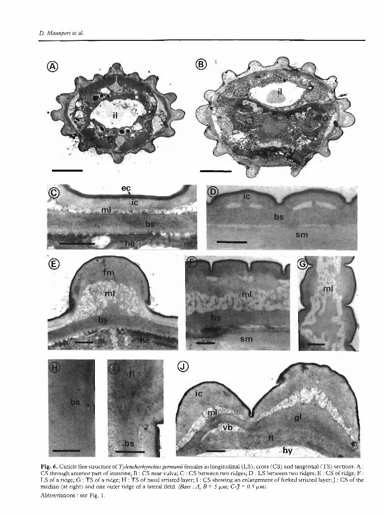
Tylenchorhynchus sp.
The cuticle thickness ranges from 0.65 /Lm betweenthe ridges to 2.50 f.lm at the level of the prominent longitudinal ridges (Fig. 8 A).
The cutide belween ridges oUlside laleralfields (Fig. 8 B,D) consists, as in T. germanil~ of: i) an external andinner cortical layer, averaging 0.30 f.lm in thickness inthe middle of the annules; ii) a discontinuous electronlucent median layer and iii) a striated basal layer, rangingfrom 0.18 f.lm to 0.35 /Lm in the middle of annules.
The cUlide of lhe ridges oUlside laleral fields (Fig. 8 C,E-G) can be distinguished into two types:
The cuticle of the dorsal and ventral ridges and theirbordering ridges on both sides (total =6) consists of: 1) atypical cortical zone; il) a median zone with a thlck outerelectron lucent fibrillar layer (1. 00 f.lm) and an innerosmophilic layer whose thickness ranges from 0.12 f.lmin the middle of annules ta 1.00 f.lm between them(Fig. 8 C); iil) a basal striated layer as previously described between ridges.
The ultrastructures of the cortical and median zone ofthe four ridges adjacent to the two single ridges of thelateral fields (Fig. 8 F) are simiJar ta those of the otherridges; the basal striated layer, however, is forked in
Vol. 16,no2-1993
Cucicle fine structure ofTylenchorhynchus spp.
cross-section and replaced by two granular and two fibrillar Jayers (Fig. 4; Fig. 8 H).
The CUlide of laleral fields ridges (Fig. 4; Fig. 8 H). Themain difference concerns the basal zone which consistsof i) two outer granular layers separated by a thin electron-dense layer; ù) an intermediate electron-dense fibrillar layer; iiz) an inner electron-Iucent fibrillar layer incontact with hypodermis.
Discussion
The cuticle ultrastructure outside lateral fields in thefour Tylenchorhynchus species without longitudinalridges and incisures corresponds to previous observations on T. annulaLUs (Ibrahim, 1967) and T. dubius(Byers & Anderson, 1972) but parallel bands withoutstriae in the basal striated layer occuring in T. indicus, T.ventralis and T. vulgaris were not observed by the previous authors.
Granular and fibrillar layers replacing the basal striated layer beneath the lateral fields were not observed inother phytoparasitic nematodes belonging to the Tylenchida (Kiesel et al., 1972; Durnez el al., 1973; B'Chir,1979; Mounport el al., 1991 b) except in Hoplolaiminae(Mounport el al., 1991 a). However, few studies are reported on the ultrastructure of the lateraJ fields of Tylenchina. In belonolaimids, Ibrahim (1967) did not examine their fine structure in T. annulatus and Byers andAnderson (1972) observed an electron-Iucent fibrillarlayer in the basal part of the lateral fields of T. dubius.
T. gladiolalus, a species with longitudinal adjacentridges, is distinct from the four other species with longitudinal ridges by i) the uniformity of the cutic1e thicknessail around the body, ii) the absence of the median layerbeneath incisures and iii) the absence of the medianfibrillar layer beneath ridges. Cross-sections of specieswith longitudinal ridges show in T. germanii two incisures in the lateral fields which are composed of threeridges instead of one (Germani & Luc, 1984). In T.sulcaLUs the longitudinal ridges are not adjacent but isolated from each other as in T. germami and T. microphasmis. These ridges result from a thickening of the medianzone of the cutic1e. Tylenchorhynchus sp. is distinct fromthe three other species with longitudinal ridges by i) thefine structure of the median layer of the cuticie, ii) thebasal layers of the lateral fields and iil) the absence ofincisure in the lateral fields. In most phytoparasitic nematodes belonging to Tylenchida, lateral fields aremarked in cross-sections by an interruption of the basalstriated layer under both outer incisures Qohnson el al.,1970; Byers & Anderson, 1972; Johnson, 1981; Mounport el al., 1990). Only one ridge occurs in each lateralfield of Tylenchorhynchus sp. and the basal striated layerin cross-sections stops under both ridges next to thelateral ridge. The fibrillar layers observed in the basalpart of the lateral fields in Tylenchorhynchus sp. are different from those in the other species; these layers re-
145

D. Mounporl er al.
®
@
®
fm
Fig. 7. Cuucle fine srrucrure of Tylenchorhynchus rmcrophasrnis females in longirudinal (LS), and cross (CS) sections. A : CS armid-body; B : LS bet:\'1een rwo ridges; C : LS of a ridge; D : CS of a ridge; E : CS berween (Wo ridges; F : CS of a larerai field; G : CSar the Ievel of the firsr incisure showing the forked srriared layer. (Bars =A =5 fLrn; B-G = 0.5 fLm).Abbreviarions : see Fig. 1.
146 Fundam. appl. Nernalol.
Culicle fine slruClure ofTylenchorhynchus spp.
®
®
Fig. 8. Cuticle fine structure of Tyleruhorhyruhus sp. females in longitudinal (LS), cross (CS) and rangential (TS) sections. A : CSar mid-body; B : LS berween rwo ridges; C : LS of a ridge; D : CS berween rwo ridges; E : CS of a ridge be[\'leen rwo annules; F : CSof a ridge in the middJe of an annule; G : TS of a ridge; H : CS of a lareral field. (Bars: A = 5 fl.m; B-H = 0.5 fl.1n).
Abbreviarions : see Fig. 1. 147

D. Mounporl et al.
semble those observed in the basal part of Hoplolaiminae, such as in Helicotylenchus and Pararotylenchus(unpubl.).
ConclusionFive groups (Table 1) may be defined from the nine
species studied on the basis of: i) number oflayers in themedian zone outside lateral fields; ii) pattern of the basalstriated layer and iii) number and ultrastructure oflayersreplacing the striated basal layer under the lateral fields.Species of the first three groups were originally described in three different genera; species of two othergroups include species whose generic diagnosis wasmodified several times during the last years (Tylenchorhynchus ...... Dolichorhynchus ...... Neodolichorhynchus ......Tylenchorhynchus).
Previous studies on phytoparasitic nematodes belonging to the Tylenchida reported a constancy of cuticlelayering at the generic level (Kisiel et a!., 1972; Shepherdet al., 1972; De Grisse & Roose, 1975; Mounport et al.,1990, 1991a, b). Several genera were synonymized withthe genus Tylenchorhynchus by Fortuner and Luc(1987) based on the use of invalid criteria in the originaldescriptions. The present observations on nine speciesof Tylenchorhynchus let us suppose that the genus Tylenchorhynchus (Cobb, 1913) Fortuner & Luc (1987)might be composed of several genera which must beredefined. Morpho-biometrical criteria from light microscopie studies and morphological criteria from scanning electron microscopy are currently used in taxonomy, but little SEM information is available on the genusTylenchorhynchus (Sher & Bell, 1975). Additional crite-
Table 1. Groups of species identified on the basis of thecuticule fine structure. C = one layer between the ridges andtwo layers under the ridges; ** = hoplolamid pattern of thebasal fibrous layers).
Group Species Number of Number of Panern oflayers ln the layers in the the snialedmedian zone basal zone basal layeroULSide the the level oflaIera! field laIerai fields
T. annuialUs 2 continuous
T. indicus 4 discontinuous
T. venlraiis
T. vuigaris discontinuous
T gladiolalUS continuous
T gennanii 1X, 2x continuousT sU/i:QlUST mitrophasmis
Tsp. 1,,2" 5.,., continuous
148
ria obtained from transmission electron microscope(e.g. ultrastructure of the intestine, reproductive systemcuticle) may be usefully considered for a better diagnos~is of the genera in the Belonolaimidae.
References
B'CHIR, M. M. (1979). COnlribulion à l'élUde morphologique,anaLOmique el &ioécologique de quelques espéces du genre Aphelenchoides (NemaLOda .' Aphelenchoidea). Thèse, Univ. Montpellier, 206 p.
BYERS, J. R. & ANDERSON, R. V. (1972). Uluastrueturalmorphology of the body wall, stoma and stomatostyleof the nematode Tylenchorhynchus dubius (Buetschli, 1873)Filip'ev, 1936. Canad. J. Zool., 50: 457-465.
DE GRISSE, A. & ROOSE, D. (1975). The uluastructure of thevulva region in Sculellonema cavenessi Sher, 1964 (Nematoda: Hoplolaimidae). Meded. Fac. Landbouww. Rijksuniv.Genl, 40: 501-510.
DURNEZ, c., DE GRISSE, A & GILLARD, A. (1973). Elektronen mikroskopische studie van de cuticulastruktur van delichaamswand bij ROlylenchus robuslUs (Nematoda : Hoplolaimidae). Meded. Fac. Landbouww. Rijksuniv. Genl, 38:1329-1350.
FORTUNER, R. & Luc, M. (1987). A reappraisal of Tylenchina (Nemata). 6. The famiJy Belonolaimidae Whitehead,1960. Revue NémalOl., 10: 183-202.
GERMANl, G. & Luc, M. (1984). Description de Dolichorhynchus elegans n. sp. et Aphasmalylenchus variabilis n. sp.(Nematoda: Tylenchida). Revue NémaLOl., 7 : 81-86.
IBRAHIM, 1. K. A. (1967). Morphological differences betweenthe cuticle of swarming and non-swarming TylenchorhynchusmaTlini. Proc. helminlh. Soc. Wash., 34: 18-20.
JAJRi\JPURI, M. S. & HUNT, D. J. (1984). The taxonomy ofTylenchorhynchinae (Nematoda : Tylenchida) with longitudinallines and ridges. SYSI. Parasilol., 6 : 261-268.
JOHNSON, P. W. (1981). Observations on the cuticle ultrastructure of Meloidogyne hapla males. J. NemaLOl., 13: 231233.
JOHNSON, P. W., VAN GUNDY, S. D. & THOMSON, W. W.(1970). Cuticle ultrastructure of Hemicycliophora arenaria,Aphelenchus avenae, Hirschmanniella graâlis and Hirschmanniella belli. J. NemaLOl., 2 : 42-58.
KISIEL, M., HIMMELHOCH, S. & ZUCKERMAN, B. M. (1972).Fine structure of the body wall and vulva area of Pralylenchus penelrans. NemaLOlogica, 18: 234-238.
MAGGENTI, A R. (1979). The role of cuticle strata nomenclature in the systematics of Nemata. J. NemaLOl., 11 : 94-98.
MOUNPORT, D., BAU)ARD, P. & MARTINY, B. (1990). Étudeultrastructurale de la cuticule de Pralylenchus /rrcu:hyunls, P.loosi et P. sefaensis (Nemata : Pratylenchidae). Revue NémaLOl., 13 : 249-254.
MOUNPORT, D., BAU)ARD, P. & MARTINY, B. (1991a). Ulrrastrueture de la cuticule et de la région vaginale de Sculellonema bradys, S. cavenessi et S. clalhricaudalUm (Nemata, Hoplo1aimidae). Revue NémaLOl., 14: 291-297.
Fundam. appl. NemaLOI.

MOUNPORT, D" BAUJARD, P, & MARTTNY, B. (199Ib), Cu ticle ultrastructure of CriconemeUa CUI'Vala and Criconemel/asphaerocepha/a (Nemata : Criconematidae). J. NemaLO/., 23 :99-103.
MULK, M, M, &JAlRAJPURl, M, S, (1974), Proposa! ofa newgenus Dolichorhynchus and a new species Dolichorhynchusnigericus (Nematoda: Dolichodoridae), Indian J. Zoo/., 2 :15-18.
REYNOLDS, E. S, (1963). The use of lead citrate at high pH asan electron-opaque stain in electron microscopy. J. CeUBiol" 17 : 208-212,
VoI.16,no2-1993
CUlicLe fine SlruClure ofTylenchorhynchus spp,
SHEPHERD, A. M" CLARK, S. A. & DART, P, l (1972). Cuticie structure in the genus Helerodera. NemaLOlogica, 18: 117,
SHER, S, A. & BELL, A. H. (1975), Scanning electron micrographs of the anterior region of sorne species of Tylenchoidea (Tylenchida : Nematoda), J, NemaLOI" 7 : 69-83,
SPURR, A, R. (1969), A low-viscosity epoxy resin embeddingmedium for electron microscopy, J. Ultraslr, Res., 26: 3143,
149