lisfranc injuries lecture - ota.org · • 30 degree oblique
TRANSCRIPT

4/19/2016
1
Orthopaedic Trauma Association
Protection and/or confidentiality of contents statement, this statement may also include a corporate copyright notice.
Lisfranc InjuriesMidfoot Injuries
Resident Comprehensive Fracture Course
Introduction• Uncommon Injuries
• Frequently missed• Up to 20%
• Highly Variable Injuries• Sports• Trauma
Osseous Anatomy• Trapezoidal
configuration
• *Recessed 2nd TMT serves as the “keystone” *
• Individual joints are “flat on flat”• Little inherent
stability Siddiqui et al. Evaluation of the tarsometatarsal joint using conventional radiography, CT, and MR imaging. Radiographics. 2014
Ligamentous Anatomy• Transverse
Intermetatarsal M2-M5
• *Interosseous C1-M2 ligament = Lisfranc ligament*
• Plantar ligaments stronger than dorsal ligaments Panchbhavi et al. Three-dimensional, digital, and gross anatomy
of the Lisfranc ligament. Foot Ankle Int. 2013
Functional Anatomy• Column Theory
• Medial column• First TMT and NC joints • Limited mobility
• Intermediate column• 2nd , 3rd TMT joints and
NC joints• Rigid
• Lateral Column• 4th and 5th TMT joints• Mobile
• Medial and Intermediate Columns function as a rigid lever for propulsion
• Lateral column mobile for shock absorption
Reid JJ, Early JS. Osseous anatomy of the midfoot. In: Bucholz et al., editors. Rockwood and Green’s fractures in adults. 7th ed
Initial Evaluation• Careful History
• Physical Exam• *Plantar arch
ecchymosis*
• Gap sign
• Provocative maneuvers
• Pronation abduction stress

4/19/2016
2
Significance of Lisfranc Injuries
• Soft tissue injury
• Minimal bony displacement may understate ligamentous injury
• Diagnosis is missed or delayed in up to 20% of cases
• LISFRANC UNTIL PROVEN OTHERWISE
Normal X-rays• AP
• Up to 3 mm normal between 1st and 2nd
metatarsal bases
• Lateral base 1st MT in-line with lateral aspect of medial cuneiform
• Medial base 2nd MT in-line with medial aspect of middle cuneiform
AP
Normal x-rays• 30 degree oblique
• Medial base 3rd
MT in-line with medial aspect of lateral cuneiform
• Medial base 4th
MT in-line with medial aspect of cuboid
Normal x-rays• Lateral: A metatarsal should never be more
dorsal than its respective tarsal bone
Imaging of Lisfranc Injuries
“Fleck” sign
• Dynamic Joints• Weight bearing radiographs crucial
Imaging of Lisfranc Injuries

4/19/2016
3
• Contralateral views
Imaging of Lisfranc Injuries
Stress Imaging
Advanced Imaging• CT scan
• Articular comminution
• Non displaced fracture lines
• Helpful for preop planning
• NOT DYNAMIC
Advanced Imaging• MRI
• Ligamentous injury
• *Plantar Oblique Ligament*
• C1-M2-M3
• Disruption predictive of intraoperative instability
• Raikin et al, JBJS 2009
• Not dynamic
Initial Management• Urgent if tenting the
skin
• Can be blocked by tendons, ligaments, bone
• External Fixation?• High energy patterns
• Temporize until soft tissue appropriate
Initial Management

4/19/2016
4
Management
• Nonoperative• Rule out occult instability
• Negative weight bearing or stress imaging
• EUA if necessary
• Operative• Multiple base fractures
• Articular involvement
• Instability (>2 mm)
Definitive Management• ORIF
• Do not eliminate a motion segment of the foot
• Hard to make the multiple fractures and a fusion heal
• Traditional treatment with reasonable outcomes
• Primary Arthrodesis• Medial column of the
midfoot functions rigidly during gait for stability
• “non-essential joints”• Lateral column (4th, 5th
TMT) is the mobile midfoot segment
• One operation, one recovery period
• Fusion after ORIF is technically more difficult
• With worse outcomes
Fixation• What to use?
• Screws• 3.5mm or 4.0mm
cortical
• Cannulated screws
• Spanning Plates• comminution
Operative Technique
• Incisions
Operative Technique• Reduction/Temporary Fixation
Operative Technique• Where to start?
• Medial to lateral?
• Lateral to medial?
• Proximal to distal?

4/19/2016
5
Inter-cuneiform instability Operative Technique
Pin 4th and 5th if unstable Post-Op
• Remove K wires at 4-6 weeks
• NWB Short leg cast X 8-12 weeks
• Gradual WB over next 2-4 weeks +/-arch support
• PT
Implant Removal?
• Indications?• When?
Results of ORIF• Arntz et al, JBJS 1988• 41 injuries - 3.4 yr followup
• 33 of 35 who had “good to excellent result” -> anatomic reduction
• 6 had fair/poor result• 5 open injuries/1 malreduction
• Kuo et al, JBJS 2000• 48 patients – 55 mo followup
• Avg AOFAS score 77• 12 post-traumatic OA (6 fusion)
• 6 of 15 with ligamentous injury• Better results with anatomic reduction

4/19/2016
6
Alternatives to ORIF? WHY Primary Arthrodesis?
Kuo et al JBJS am 2000
Worse outcomes in primarily ligamentous injuries
Komenda et al JBJS am 1996
Significant improvement after midfoot arthrodesis for PTOA
Sangeorzan et al FAI 1990
Improved results with early fusion for PTOA midfoot
Primary Arthrodesis(PA) vs ORIF PA vs ORIF
• Level I, 41 patients (21 ORIF, 20 PA)
• 2 year followup
• All results in favor of PA• AOFAS Midfoot score in favore of PA
• 15 of 21 ORIF with radiographic arthritis
• 5 of 21 converted to arthrodesis
• 16 of 21 with second surgery for screw removal
Summary
• Complex injuries
• Wide range of injury from subtle athletic to high energy crush
• DO NOT MISS THE SUBTLE LISFRANC
• Goal of fixation Stable painless plantigrade foot
• Fuse vs. ORIF – still controversial
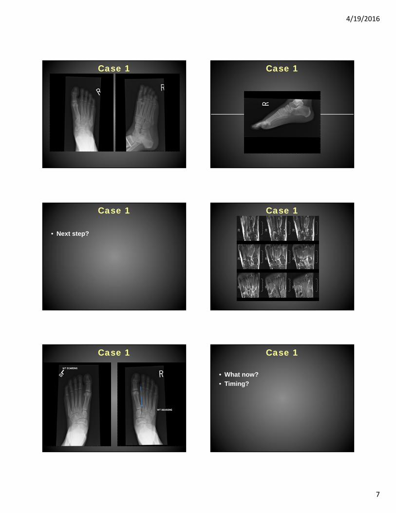
Case 1
• 17 yo female rugby player with r foot sprain 4 weeks ago
4/19/2016
7
Case 1 Case 1
Case 1
• Next step?
Case 1
Case 1 Case 1
• What now?
• Timing?

4/19/2016
8
Case 1 Case 1
Case 2
• 50 yo male who jumped from motel roof presents to ED with R foot pain as only complaint
• PMHx: Substance abuse, Hepatitis C, Bipolar disorder, smoker
• PE: Foot swollen, ecchymosis. Pulses intact sensation intact
Case 2
Case 2 Case 2

4/19/2016
9
Case 2 Case 2
Case 2 – 11 months Case 3
• 27 year old male
• MVC
• Isolated left foot injury
• NV intact
Case 3 Case 3
• What now?
• Initial Management?
• Definitive Management?• ORIF? Arthrodesis?

4/19/2016
10
Case 3 Case 3: 2 year follow up
Thank You