lippincott's illustrated reviews:...
TRANSCRIPT

Antiarrhythmics 17I. OVERVIEW
In contrast to skeletal muscle, which contracts only when it receives a stimulus, the heart contains specialized cells that exhibit automaticity. This means that they can intrinsically generate rhythmic action poten-tials in the absence of external stimuli. These “pacemaker” cells differ from other myocardial cells in showing a slow, spontaneous depolarization during diastole (Phase 4), caused by an inward positive current carried by sodium- and calcium-ion flows. This depolarization is fastest in the sino-atrial (SA) node (the normal initiation site of the action potential), and it decreases throughout the normal conduction pathway through the atrioventricular (AV) node to the bundle of His and the Purkinje system. Dysfunction of impulse generation or conduction at any of a number of sites in the heart can cause an abnormality in cardiac rhythm. Figure 17.1 summarizes the drugs used to treat cardiac arrhythmias.
II. INTRODUCTION TO THE ARRHYTHMIAS
The arrhythmias are conceptually simple. Dysfunctions cause abnormali-ties in impulse formation and conduction in the myocardium. However, in the clinic, arrhythmias present as a complex family of disorders that show a variety of symptoms. For example, cardiac arrhythmias may cause the heart to beat too slowly (bradycardia) or too rapidly (tachycardia) and to beat regularly (sinus tachycardia or sinus bradycardia) or irregularly (atrial fibrillation). The heart cavity from which the arrhythmia originates gives the name to the arrhythmia. Thus, atrial tachycardia names a rapid arrhyth-mia originating in the atria. Impulses originating from sites other than the SA node and impulses traveling along accessory (extra) pathways that lead to deviant depolarizations (AV reentry, Wolff-Parkinson-White syn-drome) may also trigger arrhythmias. To make sense of this large group of disorders, it is useful to organize the arrhythmias into groups accord-ing to the anatomic site of the abnormality: the atria, the AV node, or the ventricles. Figure 17.2 summarizes several commonly occurring atrial, AV junction, and ventricular arrhythmias. Although not shown here, each of these abnormalities can be further divided into subgroups depending on the electrocardiogram (ECG) findings
A. Causes of arrhythmias
Most arrhythmias arise either from aberrations in impulse generation (abnormal automaticity) or from a defect in impulse conduction.
CLASS I (Na+-channel blockers)Disopyramide (IA) NORPACEFlecainide (IC) TAMBOCORLidocaine (IB) XYLOCAINE Mexiletine (IB) MEXITILProcainamide (IA) PRONESTYL, PROCANPropafenone (IC) RYTHMOLQuinidine (IA) QUINIDEX
CLASS II (ß-adrenoreceptor blockers)Esmolol BREVIBLOCMetoprolol LOPRESSOR, TOPROL-XLPropranolol INDERAL
CLASS III (K+ channel blockers)Amiodarone CORDARONE, PACERONEDofetilide TIKOSYNDronedarone MULTAQ Sotalol BETAPACE, SORINE
CLASS IV (Ca2+ channel blockers)Diltiazem CARDIZEM, CARTIA XT, DILACOR XR, DILTIA XT, Verapamil CALAN, COVERA-HS, ISOPTIN SR, VERELAN
OTHER ANTI-ARRHYTHMIC DRUGSAdenosine ADENOCARD, ADENOSCANDigoxin LANOXIN
Figure 17.1Summary of antiarrhythmic drugs.
Pharm 5th 3-21-11.indb 207 3/21/11 2:23:43 PM

208 17. Antiarrhythmics
Figure 17.2Therapeutic indications for some commonly encountered arrhythmias. AV = atrioventricular.
VerapamilATRIAL
FLUTTER
Class I Class II Class III Class IV Other
Verapamil AV NODALREENTRY
LidocaineACUTE VENTRICULAR
TACHYCARDIA
Lidocaine
DofetilideQuinidine Diltiazem
TYPE OFARRHYTHMIA
ANTIARRHYTHMIC DRUGS
Propranolol
Propranolol
Propranolol
Anticoagulanttherapy
Verapamil Adenosine
VENTRICULARFIBRILLATION
(not responding to electrical de�brillation)
ACUTE SUPRA-VENTRICULARTACHYCARDIA
ATRIALFIBRILLATION
ATRIALFLUTTER
Class I
Quinidine
TYPE OFARRHYTHMIA
ATRIALFIBRILLATION
Digoxin
Digoxin
Epinephrine
LidocaineTACHYCARDIA
LidocaineVENTRICULARFIBRILLATION
(not responding to electrical de�brillation)
This arrhythmia is a common cause of death in patients who have had a myocardial infarction. Cardiac output is impaired, and tachycardia may deteriorate into ventricular �brillation. Therefore, ventricular tachycardia requires prompt management.
Drug name
Drug name Commonly used drugs
Alternative drugs
Key:
This common arrhythmia involves multiple ectopic foci of atrial cells, creating a chaotic movement of impulses through the atria. The ventricular response is rapid (100–150 beats per minute) and irregular. Cardiac output is decreased and exercise intolerance is common.
Verapamil Digoxin
Conduction isslowed through the AV node with propranolol, verapamil, or digoxin.
ATRIALARRHYTHMIAS
SUPRAVENTRICULARTACHYCARDIAS
VENTRICULARTACHYCARDIAS
Verapamil
Class III Class IV Other
Diltiazem
ANTIARRHYTHMIC DRUGS
Propranolol
Propranolol
Anticoagulanttherapy
Digoxin
β-Blockers are used in atrial �brillation, because they decrease heart rate and promote conversion to sinus rhythm. Long-term, low-dose anticoagulant therapy reduces the risk of stroke that is associated with atrial �brillation.
Amiodarone
Amiodarone
Amiodarone
Pharm 5th 3-21-11.indb 208 3/21/11 2:23:46 PM

II. Introduction To The Arrhythmias 209
Figure 17.3Schematic representation of reentry.
Ventriclewall
Nerveimpulse
A. Normal
B. Unidirectional Block
Impulse isblockedin one
direction
Impulse travels in theretrograde directionand reenters theconduction pathway,causing an extra orirregular heart beat.
12
1. Abnormal automaticity: The SA node shows the fastest rate of Phase 4 depolarization and, therefore, exhibits a higher rate of discharge than that occurring in other pacemaker cells exhibiting automaticity. Thus, the SA node normally sets the pace of contrac-tion for the myocardium, and latent pacemakers are depolarized by impulses coming from the SA node. However, if cardiac sites other than the SA node show enhanced automaticity, they may generate competing stimuli, and arrhythmias may arise. Abnormal automatic-ity may also occur if the myocardial cells are damaged (for example, by hypoxia or potassium imbalance). These cells may remain par-tially depolarized during diastole and, therefore, can reach the firing threshold earlier than the normal SA cells. Abnormal automatic dis-charges may thus be induced.
2. Effect of drugs on automaticity: Most of the antiarrhythmic agents suppress automaticity by blocking either Na+ or Ca2+ channels to reduce the ratio of these ions to K+. This decreases the slope of Phase 4 (diastolic) depolarization and/or raises the threshold of discharge to a less negative voltage. Antiarrhythmic drugs cause the frequency of discharge to decrease. This effect is more pronounced in cells with ectopic pacemaker activity than in normal cells.
3. Abnormalities in impulse conduction: Impulses from higher pace-maker centers are normally conducted down pathways that bifur-cate to activate the entire ventricular surface (Figure 17.3). A phe-nomenon called reentry can occur if a unidirectional block caused by myocardial injury or a prolonged refractory period results in an abnormal conduction pathway. Reentry is the most common cause of arrhythmias, and it can occur at any level of the cardiac conduc-tion system. For example, consider a single Purkinje fiber with two conduction pathways to ventricular muscle. An impulse normally travels down both limbs of the conduction path. However, if myo-cardial injury results in a unidirectional block, the impulse may only be conducted down Pathway 1 (see Figure 17.3). If the block in Pathway 2 is in the forward direction only, the impulse may travel in a retrograde fashion through Pathway 2 and reenter the point of bifurcation. This short-circuit pathway results in re-excitation of the ventricular muscle, causing premature contraction or sustained ven-tricular arrhythmia.
4. Effects of drugs on conduction abnormalities: Antiarrhythmic agents prevent reentry by slowing conduction (Class I drugs) and/or increasing the refractory period (Class III drugs), thereby converting a unidirectional block into a bidirectional block.
B. Antiarrhythmic drugs
As noted above, antiarrhythmic drugs can modify impulse generation and conduction. More than a dozen such drugs that are potentially use-ful in treating arrhythmias are currently available. However, only a lim-ited number of these agents are clinically beneficial in the treatment of selected arrhythmias. For example, the acute termination of ventricular tachycardia by lidocaine or of supraventricular tachycardia by adenos-ine or verapamil are examples in which antiarrhythmic therapy results in decreased morbidity. In contrast, many of the antiarrhythmic agents are now known to have dangerous proarrhythmic actions—that is, to cause arrhythmias. The efficacy of many antiarrhythmic agents remains
Pharm 5th 3-21-11.indb 209 3/21/11 2:23:46 PM

210 17. Antiarrhythmics
unproven in placebo-controlled, random trials. [Note: Implantable car-dioverter defibrillators are becoming more widely used to manage this condition.]
III. CLASS I ANTIARRHYTHMIC DRUGS
The antiarrhythmic drugs can be classified according to their predomi-nant effects on the action potential (Figure 17.4). Although this classifica-tion is convenient, it is not entirely clear-cut, because many of the drugs have actions relating to more than one class or may have active metabolites with a different class of action. Class I antiarrhythmic drugs act by block-ing voltage-sensitive sodium (Na+) channels via the same mechanism as local anesthetics. The decreased rate of entry of sodium slows the rate of rise of Phase 0 of the action potential. [Note: At therapeutic doses, these drugs have little effect on the resting, fully polarized membrane because of their higher affinity for the active and inactive channels rather than for the resting channel.] Class I antiarrhythmic drugs, therefore, generally cause a decrease in excitability and conduction velocity. The use of sodium channel blockers has been declining continuously due to their possible proarrhyth-mic effects, particularly in patients with reduced left ventricular function and ischemic heart disease.
A. Use-dependence
Class I drugs bind more rapidly to open or inactivated sodium chan-nels than to channels that are fully repolarized following recovery from the previous depolarization cycle. Therefore, these drugs show a great-er degree of blockade in tissues that are frequently depolarizing (for example, during tachycardia, when the sodium channels open often). This property is called use-dependence (or state-dependence) and it enables these drugs to block cells that are discharging at an abnormal-ly high frequency, without interfering with the normal, low-frequency beating of the heart. The Class I drugs have been subdivided into three groups according to their effect on the duration of the action potential. Class IA agents slow the rate of rise of the action potential (thus slowing conduction), prolong the action potential, and increase the ventricular
IA
IB
IC
II
III
IV
Na+ channel blocker
Na+ channel blocker
Na+ channel blocker
-Adrenoreceptor blocker
K+ channel blocker
Ca2+ channel blocker
β
Slows Phase 0 depolarization in ventricular muscle �bers
Shortens Phase 3 repolarization in ventricular muscle �bers
Markedly slows Phase 0 depolarization in ventricular muscle �bers
Inhibits Phase 4 depolarization in SA and AV nodes
Prolongs Phase 3 repolarization in ventricular muscle �bers
Inhibits action potential in SA and AV nodes
Figure 17.4Actions of antiarrhythmic drugs. SA = sinoatrial; AV = atrioventricular.
CLASSIFICATIONOF DRUG
MECHANISM OF ACTION COMMENT
Pharm 5th 3-21-11.indb 210 3/21/11 2:23:47 PM

III. Class I Antiarrhythmic Drugs 211
eff ective refractory period. They have an intermediate speed of asso-ciation with activated/inactivated sodium-channels and an interme-diate rate of dissociation from resting channels. Prolongation of dura-tion of the action potential and increased ventricular eff ective period are due to concomitant Class III activity. Class IB drugs have little eff ect on the rate of depolarization; rather, they decrease the duration of the action potential by shortening repolarization. They rapidly interact with sodium channels. Class IC agents markedly depress the rate of rise of the membrane action potential. Therefore, they cause marked slowing of conduction but have little eff ect on the duration of the membrane action potential or the ventricular eff ective refractory period. They bind slowly to sodium channels.
B. Arrhythmias
Inhibition of potassium channels (Class III activity) widens the action potential, leading to a prolonged QT interval on the electrocardio-gram. Such an eff ect is associated with increased risk of developing life-threatening ventricular tachyarrhythmias (torsades de pointes). The most common cause of QT prolongation is drug-induced, although it may also be genetic. QT prolongation is not only seen with Class III antiarrhythmics. Drugs such as cisapride, grepafl oxacin, terfenadine, and astemizole were withdrawn from the market because of severe and fatal arrhythmias. Erythromycin, clarithromycin, pentamidine, moxifl oxa-cin, levofl oxacin, imipramine, desipramine, amitriptyline, doxepin, thior-idazine, mesoridazine, haloperidol, risperidone, ziprasidone, and quetia-pine are some of the drugs known to prolong the QT interval. Caution should be exerted when combining several drugs with eff ects on the QT interval (for example, quinidine with levofl oxacin) or when giving these drugs combined with azole antifungals (fl uconazole and itracon-azole). The latter are known to inhibit drug metabolism, leading to large increases in plasma drug concentrations.
C. Quinidine.
Quinidine [KWIN-i-deen] is the prototype Class IA drug. Because of its concomitant Class III activity, it can actually precipitate arrhythmias such as polymorphic ventricular tachycardia (torsades de pointes), which can degenerate into ventricular fi brillation. Because of the toxic potential of quinidine, calcium antagonists, such as amiodarone and verapamil, are increasingly replacing this drug in clinical use.
1. Mechanism of action: Quinidine binds to open and inactivated sodium channels and prevents sodium infl ux, thus slowing the rapid upstroke during Phase 0 (Figure 17.5). It also decreases the slope of Phase 4 spontaneous depolarization and inhibits potassium chan-nels. Because of these actions, it slows conduction velocity and increases refractoriness.
2. Therapeutic uses: Quinidine is used in the treatment of a wide vari-ety of arrhythmias, including atrial, AV-junctional, and ventricular tachyarrhythmias. Quinidine is used to maintain sinus rhythm after direct-current cardioversion of atrial fl utter or fi brillation and to pre-vent frequent ventricular tachycardia.
3. Pharmacokinetics: Quinidine sulfate is rapidly and almost com-pletely absorbed after oral administration. It undergoes extensive metabolism by the hepatic cytochrome P450 enzymes, forming active metabolites.
Ca2+
Ca2+
Ca2+
Ca2+
Ca2+
K+
K+ K+
K+
K+
Actionpotentialcurrents
Na+
Diastolic currents
Na+
Figure 17.5Schematic diagram of the e�ectsof Class IA agents. INa and IK aretransmembrane currents due to themovement of Na+ and K+, respectively.
-85 mV
0 mV
Phase 0(INa)
Nodrug
Phase 3 (IK)
E�ectiverefractory period
0 mV
Nodrug
Class IA drugs slow Phase 0 depolarization. In addition, because of their Class III activity, these drugs prolong the action potential.
Membrane
Inside
Outside
Ca2+
Ca2+
Ca2+
Ca2+
Ca2+
Membrane
side
Quinidine, procainamide,and disopyramide blockopen or inactivated sodium channels. These drugs have an intermediate rate of association with sodium channels.
Pharm 5th 3-21-11.indb 211 3/21/11 2:23:55 PM

212 17. Antiarrhythmics
4. Adverse effects: A potential adverse effect of quinidine (or of any antiarrhythmic drug) is development of arrhythmia (torsades de pointes). Quinidine may cause SA and AV block or asystole. At toxic levels, the drug may induce ventricular tachycardia. Nausea, vom-iting, and diarrhea are commonly observed. Large doses of quini-dine may induce the symptoms of cinchonism (for example, blurred vision, tinnitus, headache, disorientation, and psychosis). The drug has a mild α-adrenergic blocking action as well as an atropine-like effect. Quinidine can increase the steady-state concentration of digoxin by displacement of digoxin from tissue-binding sites (minor effect) and by decreasing digoxin renal clearance (major effect).
D. Procainamide
1. Actions: This Class IA drug, a derivative of the local anesthetic pro-caine, shows actions similar to those of quinidine.
2. Pharmacokinetics: Procainamide [proe-KANE-a-mide] is well-absorbed following oral administration. [Note: The intravenous route is rarely used, because hypotension occurs if the drug is infused too rapidly.] Procainamide has a relatively short half life of 2 to 3 hours. A portion of the drug is acetylated in the liver to N-acetylprocainamide (NAPA), which has little effect on the maximum polarization of Purkinje fibers but prolongs the duration of the action potential. Thus, NAPA has properties and side effects of a Class III drug. NAPA is eliminated via the kidney, and dosages of procainamide may need to be adjusted in patients with renal failure.
3. Adverse effects: With chronic use, procainamide causes a high inci-dence of side effects, including a reversible lupus erythematosus–like syndrome that develops in 25 to 30 percent of patients. Toxic concentrations of procainamide may cause asystole or induction of ventricular arrhythmias. Central nervous system (CNS) side effects include depression, hallucination, and psychosis. With this drug, gastrointestinal intolerance is less frequent than with quinidine.
E. Disopyramide
1. Actions: This Class IA drug shows actions similar to those of quini-dine. Disopyramide [dye-soe-PEER-a-mide] produces a negative ino-tropic effect that is greater than the weak effect exerted by quini-dine and procainamide, and unlike the latter drugs, disopyramide causes peripheral vasoconstriction. The drug may produce a clini-cally important decrease in myocardial contractility in patients with preexisting impairment of left ventricular function. Disopyramide is used in the treatment of ventricular arrhythmias as an alternative to procainamide or quinidine. Like procainamide and quinidine, it also has Class III activity.
2. Pharmacokinetics: Approximately half of the orally ingested drug is excreted unchanged by the kidneys. Approximately 30 percent of the drug is converted by the liver to the less active mono-N-dealky-lated metabolite.
3. Adverse effects: Disopyramide shows effects of anticholinergic activity (for example, dry mouth, urinary retention, blurred vision, and constipation).
Pharm 5th 3-21-11.indb 212 3/21/11 2:23:55 PM

III. Class I Antiarrhythmic Drugs 213
F. Lidocaine
Lidocaine [LYE-doe-kane] is a Class IB drug. The Class IB agents rapidly associate and dissociate from sodium channels. Thus, the actions of Class IB agents are manifested when the cardiac cell is depolarized or fi ring rapidly. Class IB drugs are particularly useful in treating ventricular arrhythmias. Lidocaine was the drug of choice for emergency treatment of cardiac arrhythmias.
1. Actions: Lidocaine, a local anesthetic, shortens Phase 3 repolarization and decreases the duration of the action potential (Figure 17.6).
2. Therapeutic uses: Lidocaine is useful in treating ventricular arrhyth-mias arising during myocardial ischemia, such as that experi-enced during a myocardial infarction. The drug does not markedly slow conduction and, thus, has little eff ect on atrial or AV junction arrhythmias.
3. Pharmacokinetics: Lidocaine is given intravenously because of extensive fi rst-pass transformation by the liver, which precludes oral administration. The drug is dealkylated and eliminated almost entirely by the liver; consequently, dosage adjustment may be nec-essary in patients with liver dysfunction or those taking drugs that lower hepatic blood fl ow, such as propranolol.
4. Adverse eff ects: Lidocaine has a fairly wide toxic-to-therapeutic ratio. It shows little impairment of left ventricular function and has no negative inotropic eff ect. CNS eff ects include drowsiness, slurred speech, paresthesia, agitation, confusion, and convulsions. Cardiac arrhythmias may also occur.
G. Mexiletine and tocainide
These Class IB drugs have actions similar to those of lidocaine, and they can be administered orally. Mexiletine [MEX-i-le-teen] is used for chronic treatment of ventricular arrhythmias associated with previous myocar-dial infarction. Tocainide [toe-KAY-nide] is used for treatment of ven-tricular tachyarrhythmias. Tocainide has pulmonary toxicity, which may lead to pulmonary fi brosis.
H. Flecainide
Flecainide [FLEK-a-nide] is a Class IC drug. These drugs slowly dissoci-ate from resting sodium channels, and they show prominent eff ects even at normal heart rates. They are approved for refractory ventricular arrhythmias and for the prevention of paroxysmal atrial fi brillation/fl ut-ter associated with disabling symptoms and paroxysmal supraventricu-lar tachycardia. However, recent data have cast serious doubts on the safety of the Class IC drugs.
1. Actions: Flecainide suppresses Phase 0 upstroke in Purkinje and myo-cardial fi bers (Figure 17.7). This causes marked slowing of conduction in all cardiac tissue, with a minor eff ect on the duration of the action potential and refractoriness. Automaticity is reduced by an increase in the threshold potential rather than a decrease in the slope of Phase 4 depolarization.
2. Therapeutic uses: Flecainide is useful in treating refractory ventric-ular arrhythmias. It is particularly useful in suppressing premature
Ca2+
Ca2+
Ca2+
Ca2+
Ca2+
K+
K+
K+
K+
K+
K+
Actionpotentialcurrents
Na+Diastolic currents
Na+
Membrane Inside
Outside
Figure 17.6Schematic diagram of the e�ectsof Class IB agents. INa and IK aretransmembrane currents due to themovement of Na+ and K+, respectively.
Ca2+
Ca2+2+
Ca2+
Ca2+
Ca2+
Membranenside
Lidocaine, mexiletine, and tocainide block open or inactivated sodium channels. These drugs have a rapid rate of association with sodium channels.
-85 mV
0 mV
Phase 0(INa)
Nodrug
Phase 3 (IK)
E�ectiverefractory period
0 mV
Phase 0
drug
Class IB drugs shortenPhase 3 repolarizationand decrease the duration of the action potential.
Pharm 5th 3-21-11.indb 213 3/21/11 2:24:03 PM

214 17. Antiarrhythmics
ventricular contraction. Flecainide has a negative inotropic eff ect and can aggravate congestive heart failure.
3. Pharmacokinetics: Flecainide is absorbed orally, undergoes mini-mal biotransformation, and has a half life of 16 to 20 hours.
4. Adverse eff ects: Flecainide can cause dizziness, blurred vision, head-ache, and nausea. Like other Class IC drugs, fl ecainide can aggravate preexisting arrhythmias or induce life-threatening ventricular tachy-cardia that is resistant to treatment.
I. Propafenone
This Class IC drug shows actions similar to those of fl ecainide. Propafenone [proe-pa-FEEN-one], like fl ecainide, slows conduction in all cardiac tis-sues and is considered to be a broad-spectrum antiarrhythmic agent.
IV. CLASS II ANTIARRHYTHMIC DRUGS
Class II agents are β-adrenergic antagonists. These drugs diminish Phase 4 depolarization, thus depressing automaticity, prolonging AV conduction, and decreasing heart rate and contractility. Class II agents are useful in treat-ing tachyarrhythmias caused by increased sympathetic activity. They are also used for atrial fl utter and fi brillation and for AV-nodal reentrant tachy-cardia. [Note: In contrast to the sodium-channel blockers, β-blockers and Class III compounds, such as sotalol and amiodarone, are increasing in use.]
A. Propranolol
Propranolol [pro-PRAN-oh-lol] reduces the incidence of sudden arrhyth-mic death after myocardial infarction (the most common cause of death in this group of patients). The mortality rate in the fi rst year after a heart attack is signifi cantly reduced by propranolol, partly because of its abil-ity to prevent ventricular arrhythmias.
B. Metoprolol
Metoprolol [me-TOE-pro-lol] is the β-adrenergic antagonist most widely used in the treatment of cardiac arrhythmias. Compared to propranolol, it reduces the risk of bronchospasm. Like propranolol, is extensively metabolized and has extensive CNS penetration.
C. Esmolol
Esmolol [ESS-moe-lol] is a very short-acting β-blocker used for intrave-nous administration in acute arrhythmias that occur during surgery or emergency situations.
V. CLASS III ANTIARRHYTHMIC DRUGS
Class III agents block potassium (K+) channels and, thus, diminish the out-ward potassium current during repolarization of cardiac cells. These agents prolong the duration of the action potential without altering Phase 0 of depolarization or the resting membrane potential (Figure 17.8). Instead, they prolong the eff ective refractory period, increasing refractoriness. All Class III drugs have the potential to induce arrhythmias.
Figure 17.7Schematic diagram of the e�ectsof Class IC agents. INa and IK aretransmembrane currents due to themovement of Na+ and K+, respectively.
Ca2+
Ca2+
Ca2+
Ca2+
Ca2+
K+
K+ K+
K+
K+
Actionpotentialcurrents
Na+Diastolic currents
Na+
Membrane
Inside
Outside
Ca2+
Ca2+
Ca2+
Ca2+
Ca2+
Membrane
side
Flecainide and propafenoneblock open or inactivatedsodium channels. Thesedrugs have a slow rate of association with sodium channels.
-85 mV
0 mV
Phase 0(INa)
Nodrug
Phase 3 (IK)
E�ectiverefractory period
0 mV
Nodrug
Class IC drugs markedly slow Phase 0 depolarization.
Pharm 5th 3-21-11.indb 214 3/21/11 2:24:10 PM

V. Class III Antiarrhythmic Drugs 215
A. Amiodarone
1. Actions: Amiodarone [a-MEE-oh-da-rone] contains iodine and is related structurally to thyroxine. It has complex eff ects, showing Class I, II, III, and IV actions. Its dominant eff ect is prolongation of the action potential duration and the refractory period. Amiodarone has antianginal as well as antiarrhythmic activity.
2. Therapeutic uses: Amiodarone is eff ective in the treatment of severe refractory supraventricular and ventricular tachyarrhythmias. Amiodarone has been a mainstay of therapy for the management of atrial fi brillation (AF). Amiodarone has shown to be eff ective in maintaining sinus rhythm. Despite its side-eff ect profi le, amiodaroneis the most commonly employed antiarrhythmic.
3. Pharmacokinetics: Amiodarone is incompletely absorbed after oral administration. The drug is unusual in having a prolonged half life of several weeks, and it distributes extensively in adipose tissue. Full clinical eff ects may not be achieved until 6 weeks after initiation of treatment, unless loading doses are employed.
4. Adverse eff ects: Amiodarone shows a variety of toxic eff ects. After long-term use, more than half of patients receiving the drug show side eff ects that are severe enough to prompt its discontinuation. However, use of low doses reduces toxicity, while retaining clini-cal effi cacy. Some of the side eff ects include interstitial pulmonary fi brosis, gastrointestinal tract intolerance, tremor, ataxia, dizziness, hyper- or hypothyroidism, liver toxicity, photosensitivity, neuropa-thy, muscle weakness, and blue skin discoloration caused by iodine accumulation in the skin.
B. Dronedarone
Dronedarone [droe-NE-da-rone] is a benzofuran amiodarone derivative, which is less lipophilic, has lower tissue accumulation, and a shorter serum half life than amiodarone. It does not have the iodine moieties that are responsible for thyroid dysfunctions associated with amio-darone. Like amiodarone, it has Class I, II, III, and IV actions. Most of its side eff ects are gastrointestinal in nature and include nausea, vomit-ing, and diarrhea. A recent seven month duration study in patients with atrial fi brillation revealed that dronedarone was less eff ective than ami-odarone in decreasing AF recurrence, but had a better safety profi le, specifi cally with regard to thyroid and neurologic events and a lack of interaction with oral anticoagulants. However, further long-term com-parative studies with amiodarone are needed to defi ne dronedarone’splace in the treatment of atrial fi brillation.
C. Sotalol
Sotalol [SOE-ta-lol], although a class III antiarrhythmic agent, also has potent nonselective β-blocker activity. Sotalol has two stereoiso-mers with diff erent pharmacological activity. The levorotatory isomer (l-sotalol) is responsible for the β-blocker activity, and d-sotalol for the Class III antiarrhythmic action. It is well established that β-blockers reduce mortality associated with acute myocardial infarction.
Figure 17.8Schematic diagram of the e�ectsof Class III agents. INa and IK aretransmembrane currents due to themovement of Na+ and K+, respectively.
Ca2+ Ca2+ Ca2+
K+
K+K+K+
Actionpotentialcurrents
Na+
Na+
Diastolic currents
-85 mV
0 mV
Phase 0 (INa)Phase 3 (IK)
E�ectiverefractory period
Nodrug
Phase 0
Phase 3 (INo
Class III drugs prolong Phase 3 repolarization,without altering Phase 0.
Membrane
Inside
Outside
K+
K+K+K+
Actionpotentialcurrents
currents
Amiodarone, dofetilide,and sotalol blockpotassium channels.
Pharm 5th 3-21-11.indb 215 3/21/11 2:24:15 PM

216 17. Antiarrhythmics
1. Actions: Sotalol blocks a rapid outward potassium current, known as the delayed rectifier. This blockade prolongs both repolarization and duration of the action p otential, thus lengthening the effective refractory period.
2. Therapeutic uses: β-Blockers are used for long-term therapy to decrease the rate of sudden death following an acute myocardial infarction. β-Blockers have a modest ability to suppress ectopic beats and to reduce myocardial oxygen demand. They have strong antifibrillatory effects, particularly in the ischemic myocardium. Sotalol was more effective in preventing recurrence of arrhythmia and in decreasing mortality than imipramine, mexiletine, procain-amide, propafenone, and quinidine in patients with sustained ven-tricular tachycardia (Figure 17.9).
3. Adverse effects: This drug also has the lowest rate of acute or long-term adverse effects. As with all drugs that prolong the QT interval, the syndrome of torsade de pointes is a serious potential adverse effect, typically seen in three to four percent of patients.
D. Dofetilide
Dofetilide [doh-FET-il-ide] can be used as a first-line antiarrhythmic agent in patients with persistent atrial fibrillation and heart failure or in those with coronary artery disease with impaired left ventricular func-tion. Because of the risk of proarrhythmia, dofetilide initiation is lim-ited to the inpatient setting and is restricted to prescribers who have completed a specific manufacturer’s training session. Along with amio-darone and β-blockers, dofetilide is the only antiarrhythmic drug that is recommended by experts for the treatment of atrial fibrillation in a wide range of patients. The half life is 10 hours. Excretion is in the urine, with 80 percent as unchanged drug and 20 percent as inactive or mini-mally active metabolites.
VI. CLASS IV ANTIARRHYTHMIC DRUGS
Class IV drugs are calcium-channel blockers (see p. 236). They decrease the inward current carried by calcium (Ca2+), resulting in a decreased rate of Phase 4 spontaneous depolarization. They also slow conduction in tissues that are dependent on calcium currents, such as the AV node (Figure 17.10). Although voltage-sensitive calcium channels occur in many different tis-sues, the major effect of calcium-channel blockers is on vascular smooth muscle and the heart.
A. Verapamil and diltiazem
Verapamil [ver-AP-a-mil] shows greater action on the heart than on vas-cular smooth muscle, whereas nifedipine, a calcium-channel blocker used to treat hypertension (see p. 236), exerts a stronger effect on the vascular smooth muscle than on the heart. Diltiazem [dil-TYE-a-zem] is intermediate in its actions.
1. Actions: Calcium enters cells by voltage-sensitive channels and by receptor-operated channels that are controlled by the binding of agonists, such as catecholamines, to membrane receptors. Calcium-channel blockers, such as verapamil and diltiazem, are more effec-tive against the voltage-sensitive channels, causing a decrease in the slow inward current that triggers cardiac contraction. Verapamil
Figure 17.9Comparison of sotalol to �ve other drugs with respect to mortality from cardiac arrhythmias.
Cum
ulat
ive
deat
hs
from
arr
hyth
mia
s(%
)
Year0 1 2 3 4
0
100
50
Other drugs: Imipramine Mexiletine Procainamide Propafenone Quinidine
Sotalol
Pharm 5th 3-21-11.indb 216 3/21/11 2:24:15 PM
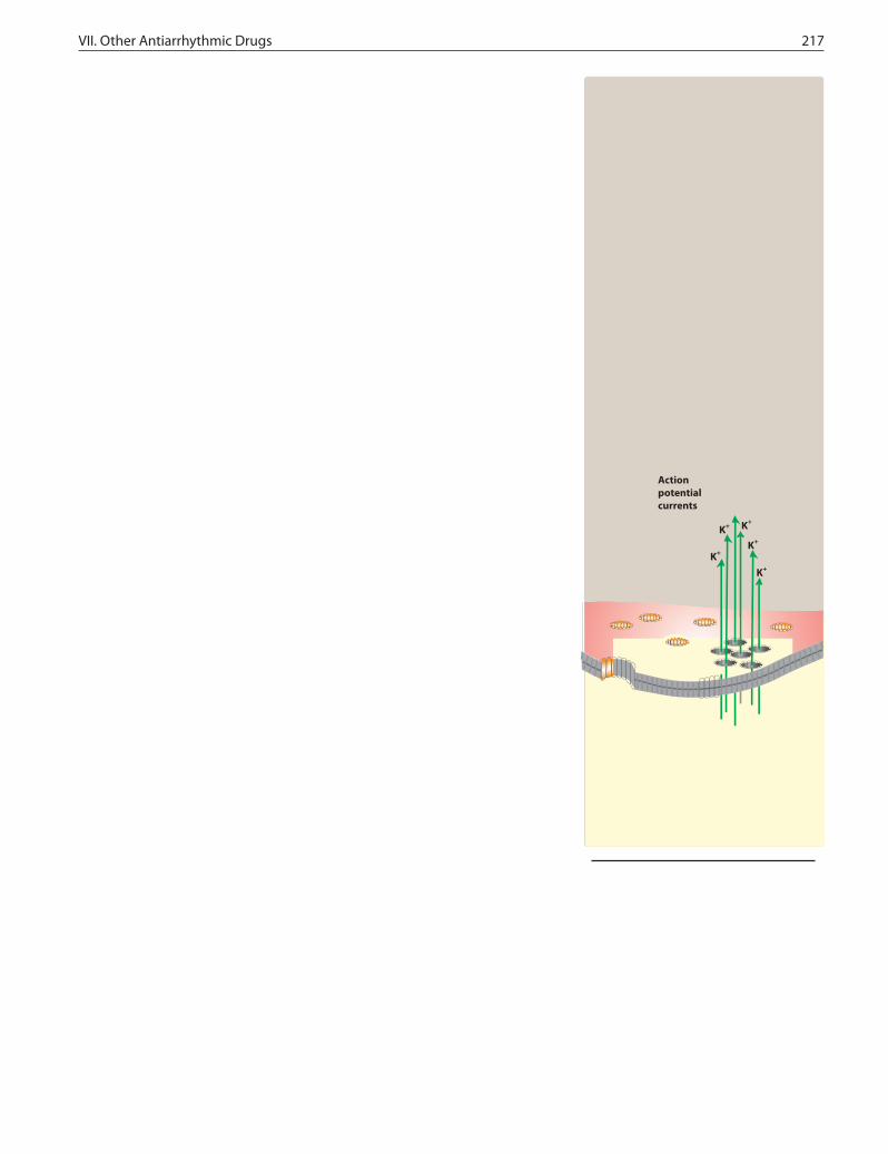
VII. Other Antiarrhythmic Drugs 217
K+
K+ K+
K+
K+
Actionpotentialcurrents
Na+
Diastolic currents
Na+
Membrane
Inside
Outside
Figure 17.10Schematic diagram of the e�ectsof Class IV agents. ICa and IK aretransmembrane currents due to themovement of Ca2+ and K+, respectively.
Class IV drugs slow Phase 4 spontaneous depolarization and slow conduction in tissues dependent on calcium currents, such as the AV node.
-75 mV
0 mV
Phase 0
Phase 2 (ICa and IK)
ICa
Group IVaction
E�ectiverefractory period
Nodrug
Na+Na+
Memb
e
Verapamil and diltiazem block open or inactivated calcium channels.
Note
Ca2+
Ca2+Ca2+
and diltiazem bind only to open, depolarized channels, thus pre-venting repolarization until the drug dissociates from the channel. These drugs are therefore use-dependent; that is, they block most eff ectively when the heart is beating rapidly, because in a normally paced heart, the calcium channels have time to repolarize and the bound drug dissociates from the channel before the next conduc-tion pulse. By decreasing the inward current carried by calcium, verapamil and diltiazem slow conduction and prolong the eff ective refractory period in tissues that are dependent on calcium currents, such as the AV node. These drugs are therefore eff ective in treating arrhythmias that must traverse calcium-dependent cardiac tissues.
2. Therapeutic uses: Verapamil and diltiazem are more eff ective against atrial than against ventricular arrhythmias. They are useful in treating reentrant supraventricular tachycardia and in reducing the ventricular rate in atrial fl utter and fi brillation (ventricular rate reduction). In addition, these drugs are used to treat hypertension and angina.
3. Pharmacokinetics: Verapamil and diltiazem are absorbed after oral administration. Verapamil is extensively metabolized by the liver; thus, care should be taken when administering this drug to patients with hepatic dysfunction.
4. Adverse eff ects: Verapamil and diltiazem have negative inotropic properties and, therefore, may be contraindicated in patients with preexisting depressed cardiac function. Both drugs can also pro-duce a decrease in blood pressure because of peripheral vasodila-tion—an eff ect that is actually benefi cial in treating hypertension.
VII. OTHER ANTIARRHYTHMIC DRUGS
A. Digoxin
Digoxin [di-JOX-in] shortens the refractory period in atrial and ventricu-lar myocardial cells while prolonging the eff ective refractory period and diminishing conduction velocity in the AV node. Digoxin is used to con-trol the ventricular response rate in atrial fi brillation and fl utter. At toxic concentrations, digoxin causes ectopic ventricular beats that may result in ventricular tachycardia and fi brillation. [Note: This arrhythmia is usu-ally treated with lidocaine or phenytoin.]
B. Adenosine
Adenosine [ah-DEN-oh-zeen] is a naturally occurring nucleoside, but at high doses, the drug decreases conduction velocity, prolongs the refrac-tory period, and decreases automaticity in the AV node. Intravenous adenosine is the drug of choice for abolishing acute supraventricular tachycardia. It has low toxicity, but causes fl ushing, chest pain, and hypotension. Adenosine has an extremely short duration of action (approximately 15 seconds).
Pharm 5th 3-21-11.indb 217 3/21/11 2:24:20 PM

218 17. Antiarrhythmics
Study Questions
Choose the ONE best answer.
17.1 A 66-year-old man had a myocardial infarct. Which one of the following would be appropriate prophy-lactic antiarrhythmic therapy?
A. Lidocaine.B. Metoprolol.C. Procainamide.D. Quinidine.E. Verapamil.
17.2 Suppression of arrhythmias resulting from a reentry focus is most likely to occur if the drug:
A. Has vagomimetic effects on the AV node.B. Is a β-blocker.C. Converts a unidirectional block to a bidirectional
block.D. Slows conduction through the atria.E. Has atropine-like effects on the AV node.
17.3 A 57-year-old man is being treated for an atrial arrhythmia. He complains of headache, dizziness, and tinnitus. Which one of the following antiarrhyth-mic drugs is the most likely cause?
A. Amiodarone.B. Procainamide.C. Propranolol.D. Quinidine.E. Verapamil.
17.4 A 58-year-old woman is being treated for chron-ic suppression of a ventricular arrhythmia. After 2 months of therapy, she complains about feeling tired all the time. Examination reveals a resting heart rate of 10 beats per minute lower than her previous rate. Her skin is cool and clammy. Laboratory test results indicate low thyroxin and elevated thyroid-stimulat-ing hormone levels. Which of the following antiar-rhythmic drugs is the likely cause of these signs and symptoms?
A. Amiodarone.B. Procainamide.C. Propranolol.D. Quinidine.E. Verapamil.
Correct answer = B. β-Blockers, such as metoprolol, prevent cardiac arrhythmias that occur subsequent to a myocardial infarction. None of the other drugs has been shown to be particularly effective in pre-venting postinfarct arrhythmias.
Correct answer = C. Current theory holds that a reentrant arrhythmia is caused by damaged heart muscle so that conduction is slowed through the damaged area in only one direction. A drug that prevents conduction in either direction through the damaged area interrupts the reentrant arrhyth-mia. Class I antiarrhythmics, such as lidocaine, are capable of producing bidirectional block. The other choices do not have any direct effects on the direc-tion of blockade of conduction through damaged cardiac muscle.
Correct answer = D. The clustered symptoms of headache, dizziness, and tinnitus are characteristic of cinchonism, which is caused by quinidine. The other drugs have characteristic adverse effects, but not this particular group of effects.
Correct answer = A. The patient is exhibiting symp-toms of hypothyroidism, which is often associated with amiodarone therapy. Propran olol could slow the heart but would not produce the changes in thyroid function. None of the other antiarrhythmics is likely to cause hypothyroidism.
Pharm 5th 3-21-11.indb 218 3/21/11 2:24:21 PM