lipid peroxidation: chemical mechanism, biological ... - …cdn.intechopen.com/pdfs-wm/38477.pdf ·...
TRANSCRIPT

3,350+OPEN ACCESS BOOKS
108,000+INTERNATIONAL
AUTHORS AND EDITORS114+ MILLION
DOWNLOADS
BOOKSDELIVERED TO
151 COUNTRIES
AUTHORS AMONG
TOP 1%MOST CITED SCIENTIST
12.2%AUTHORS AND EDITORS
FROM TOP 500 UNIVERSITIES
Selection of our books indexed in theBook Citation Index in Web of Science™
Core Collection (BKCI)
Chapter from the book Lipid PeroxidationDownloaded from: http://www.intechopen.com/books/lipid-peroxidation
PUBLISHED BY
World's largest Science,Technology & Medicine
Open Access book publisher
Interested in publishing with IntechOpen?Contact us at [email protected]
Chapter 1
© 2012 Repetto et al., licensee InTech. This is an open access chapter distributed under the terms of the Creative Commons Attribution License (http://creativecommons.org/licenses/by/3.0), which permits unrestricted use, distribution, and reproduction in any medium, provided the original work is properly cited.
Lipid Peroxidation: Chemical Mechanism, Biological Implications and Analytical Determination
Marisa Repetto, Jimena Semprine and Alberto Boveris
Additional information is available at the end of the chapter
http://dx.doi.org/10.5772/45943
1. Introduction
Currently, lipid peroxidation is considered as the main molecular mechanisms involved in
the oxidative damage to cell structures and in the toxicity process that lead to cell death.
First, lipid peroxidation was studied for food scientists as a mechanism for the damage to
alimentary oils and fats, nevertheless other researchers considered that lipid peroxidation
was the consequence of toxic metabolites (e.g. CCl4) that produced highly reactive species,
disruption of the intracellular membranes and cellular damage (Dianzani & Barrera, 2008).
Lipid peroxidation is a complex process known to occur in both plants and animals. It
involves the formation and propagation of lipid radicals, the uptake of oxygen, a
rearrangement of the double bonds in unsaturated lipids and the eventual destruction of
membrane lipids, with the production of a variety of breakdown products, including
alcohols, ketones, alkanes, aldehydes and ethers (Dianzani & Barrera, 2008).
In pathological situations the reactive oxygen and nitrogen species are generated at higher
than normal rates, and as a consequence, lipid peroxidation occurs with -tocopherol
deficiency. In addition to containing high concentrations of polyunsaturated fatty acids and
transition metals, biological membranes of cells and organelles are constantly being
subjected to various types of damage (Chance et al., 1979; Halliwell & Gutteridge, 1984). The
mechanism of biological damage and the toxicity of these reactive species on biological
systems are currently explained by the sequential stages of reversible oxidative stress and
irreversible oxidative damage. Oxidative stress is understood as an imbalance situation with
increased oxidants or decreased antioxidants (Sies, 1991a; Boveris et al., 2008). The concept
implies the recognition of the physiological production of oxidants (oxidizing free-radicals
and related species) and the existence of operative antioxidant defenses. The imbalance
Lipid Peroxidation 4
concept recognizes the physiological effectiveness of the antioxidant defenses in
maintaining both oxidative stress and cellular damage at a minimum level in physiological
conditions (Boveris et al., 2008).
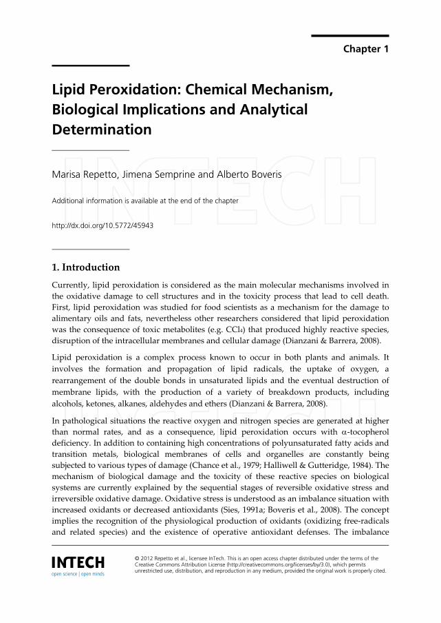
Lipid peroxidation is a chain reaction initiated by the hydrogen abstraction or addition of an
oxygen radical, resulting in the oxidative damage of polyunsaturated fatty acids (PUFA).
Since polyunsaturated fatty acids are more sensitive than saturated ones, it is obvious that
the activated methylene (RH) bridge represents a critical target site. The presence of a
double bond adjacent to a methylene group makes the methylene C-H bond weaker and
therefore the hydrogen in more susceptible to abstraction. This leaves an unpaired electron
on the carbon, forming a carbon-centered radical, which is stabilized by a molecular
rearrangement of the double bonds to form a conjugated diene which then combines with
oxygen to form a peroxyl radical. The peroxyl radical is itself capable of abstracting a
hydrogen atom from another polyunsaturated fatty acid and so of starting a chain reaction
(Halliwell & Gutteridge, 1984) (Fig. 1).
Figure 1. Initiation step of lipid peroxidation process.
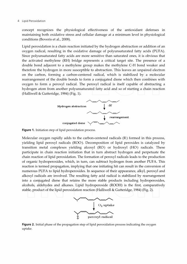
Molecular oxygen rapidly adds to the carbon-centered radicals (R.) formed in this process,
yielding lipid peroxyl radicals (ROO.). Decomposition of lipid peroxides is catalyzed by
transition metal complexes yielding alcoxyl (RO.) or hydroxyl (HO.) radicals. These
participate in chain reaction initiation that in turn abstract hydrogen and perpetuate the
chain reaction of lipid peroxidation. The formation of peroxyl radicals leads to the production
of organic hydroperoxides, which, in turn, can subtract hydrogen from another PUFA. This
reaction is termed propagation, implying that one initiating hit can result in the conversion of
numerous PUFA to lipid hydroperoxides. In sequence of their appearance, alkyl, peroxyl and
alkoxyl radicals are involved. The resulting fatty acid radical is stabilized by rearrangement
into a conjugated diene that retains the more stable products including hydroperoxides,
alcohols, aldehydes and alkanes. Lipid hydroperoxide (ROOH) is the first, comparatively
stable, product of the lipid peroxidation reaction (Halliwell & Gutteridge, 1984) (Fig. 2).
Figure 2. Initial phase of the propagation step of lipid peroxidation process indicating the oxygen
uptake.
Lipid Peroxidation: Chemical Mechanism, Biological Implications and Analytical Determination 5
Reduced iron complexes (Fe2+) react with lipid peroxides (ROOH) to give alkoxy radicals,
whereas oxidized iron complexes (Fe3+) react more slowly to produce peroxyl radicals. Both
radicals can take part in the propagation of the chain reaction. The end products of these
complex metal ion-catalyzed breakdowns of lipid hydroperoxides include the cytotoxic
aldehydes and hydrocarbon gases such as ethane.
The free radical chain reaction propagates until two free radicals conjugate each other to
terminate the chain. The reaction can also terminate in the presence of a chain-breaking anti-
oxidant such as vitamin E (α-tocopherol) (Halliwell & Gutteridge, 1984).
In conditions in which lipid peroxidation is continuously initiated it gives non-radical
products destroying two radicals at a time. In the presence of transition metal ions, ROOH
can give rise to the generation of radicals capable of re-initiating lipid peroxidation by
redox-cycling of these metal ions (Halliwell & Gutteridge, 1984).
Lipid peroxidation causes a decrease in membrane fluidity and in the barrier functions of
the membranes. The many products of lipid peroxidation such as hydroperoxides or their
aldehyde derivatives inhibit protein synthesis, blood macrophage actions and alter
chemotactic signals and enzyme activity (Fridovich & Porter, 1981).
2. Biological implications of lipid peroxidation
The biological production of reactive oxygen species primarily superoxide anion (O2.-) and
hydrogen peroxide (H2O2) is capable of damaging molecules of biochemical classes
including nucleic acids and aminoacids. Exposure of reactive oxygen to proteins produces
denaturation, loss of function, cross-linking, aggregation and fragmentation of connective
tissues as collagen (Chance et al., 1979). However, the most damaging effect is the induction
of lipid peroxidation. The cell membrane which is composed of poly-unsaturated fatty acids
is a primary target for reactive oxygen attack leading to cell membrane damage.
The lipid peroxidation of polyunsaturated fatty acids may be enzymatic and non-enzymatic.
Enzymatic lipid peroxidation is catalyzed by the lipoxygenases family, a family of lipid
peroxidation enzymes that oxygenates free and esterified PUFA generating as a
consequence, peroxy radicals. Non enzymatic lipid peroxidation and formation of lipid-
peroxides are initiated by the presence of molecular oxygen and is facilitated by Fe2+ ions
(Repetto et al., 2010a).
Oxidative breakdown of biological phospholipids occurs in most cellular membranes
including mitochondria, microsomes, peroxisomes and plasma membrane. The toxicity of
lipid peroxidation products in mammals generally involves neurotoxicity, hepatotoxicity
and nephrotoxicity (Boveris et al., 2008). The principal mechanism involves detoxification
process in liver. Toxicity from lipid peroxidation affect the liver lipid metabolism where
cytochrome P-450s is an efficient catalyst in the oxidative transformation of lipid derived
aldehydes to carboxylic acids adding a new facet to the biological activity of lipid oxidation
metabolites. Cytochrome P-450-mediated metabolism operates in parallel with other
metabolic transformations of aldehydes; hence, the P450s could serve as reserve or
Lipid Peroxidation 6
compensatory mechanisms when other high capacity pathways of aldehyde elimination are
compromised due to disease or toxicity. Finally, 4-hydroxynonenal (HNE), unsaturated
aldehydes, such as acrolein, trans-2-hexenal, and crotonaldehyde, are also food constituents
or environmental pollutants, P-450s may be significant in favoring lipid peroxidation that
has significant downstream effects and possibly play a major role in cell signaling pathways.
Oxidized lipids appear to have a signaling function in pathological situations, are pro-
inflammatory agonists and contribute to neuronal death under conditions in which
membrane lipid peroxidation occurs. For example, mitochondrial lipid cardiolipin makes up
to 18% of the total phospholipids and 90% of the fatty acyl chains are unsaturated.
Oxidation of cardiolipin may be one of the critical factors initiating apoptosis by liberating
cytochrome c from the mitochondrial inner membrane and facilitating permeabilization of
the outer membrane. The release of cytochrome c activates a proteolytic cascade that
culminates in apoptotic cell death (Navarro & Boveris, 2009).
Previous results indicate that lipid peroxidation has a role in the pathogenesis of several
pathologies as neurodegenerative (Dominguez et al., 2008; Famulari et al., 1996; Fiszman et
al., 2003), inflammatory (Farooqui & Farooqui, 2011), infectious (Repetto et al., 1996), gastric
(Repetto et al., 2003) and nutritional diseases (Repetto et al., 2010b).
Oxidative damage in liver is associated with hepatic lipid metabolism, and may be affecting
the absorption and transport mechanisms of -tocopherol in this organ. In the liver, the
morphological damage is previous to the lipid peroxidation and the consumption of
endogenous antioxidants. In kidney and heart, indeed, lipid peroxidation and oxidative
damage preceded necrosis (Repetto et al., 2010b).
Lipid peroxidation is a chain reaction process characterized by repetitive hydrogen
abstraction by HO. and RO., and addition of O2 to alkyl radicals (R.) resulting in the
generation of ROO. and in the oxidative destruction of polyunsaturated fatty acids, in which
the methylene group (=RH-) is the main target (Halliwell & Gutteridge, 1984).
The association between increased phospholipid oxidation, free-radical mediated reactions
and pathological states was early recognized (Cadenas, 1989; Verstraeten et al., 1997; Liu et
al., 2003). The contribution by Sies of the concept of oxidative stress followed (Sies,
1991a,1991b) with the implication that increased free-radical mediated reactions, basically
by HO. and RO., would produce phospholipid, protein, lipid, DNA, RNA or carbohydrate
oxidation, whatever is close (Halliwell & Gutteridge, 1984). The increased oxidation of the
cell biochemical constituents is associated with ultra structural changes in mitochondrial
morphology with mitochondrial swelling and increased matrix volume (Boveris et al., 2008).
In human liver, the morphological changes can affect the organ structure and function as it
is the case for the bile canaliculi that are damaged in liver transplanted patients; a fact that is
interpreted as consequence of the oxidative damage that is associated to ischemia-
reperfusion (Cutrin et al., 1996). Interestingly, there are reports in rat liver experimental
models, of increased peroxidation secondary to increased mitochondrial production of O2-
and H2O2 (Fridovich, 1978; Navarro &Boveris, 2007; Navarro et al., 2009).
Lipid Peroxidation: Chemical Mechanism, Biological Implications and Analytical Determination 7
3. Chemical mechanisms for lipid peroxidation process
The spectrum of oxygen reactive species that are considered responsible for biological oxygen
toxicity include the intermediates of the partial reduction of oxygen, superoxide radical (O2.-),
hydrogen peroxide (H2O2), and other reactive species as hydroxyl radicals (HO.), peroxyl
radical (ROO.), nitric oxide (NO), peroxinitrite (ONOO-) and singlet oxygen (1O2).
The biological effects of excess levels of the spectrum of these species are quite similar, and
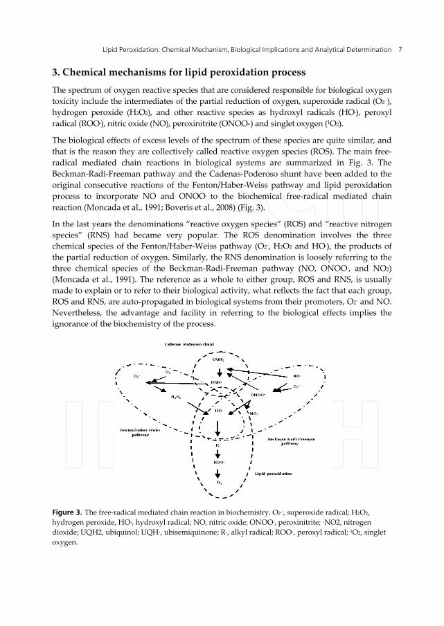
that is the reason they are collectively called reactive oxygen species (ROS). The main free-
radical mediated chain reactions in biological systems are summarized in Fig. 3. The
Beckman-Radi-Freeman pathway and the Cadenas-Poderoso shunt have been added to the
original consecutive reactions of the Fenton/Haber-Weiss pathway and lipid peroxidation
process to incorporate NO and ONOO. to the biochemical free-radical mediated chain
reaction (Moncada et al., 1991; Boveris et al., 2008) (Fig. 3).
In the last years the denominations “reactive oxygen species” (ROS) and “reactive nitrogen
species” (RNS) had became very popular. The ROS denomination involves the three
chemical species of the Fenton/Haber-Weiss pathway (O2-, H2O2 and HO.), the products of
the partial reduction of oxygen. Similarly, the RNS denomination is loosely referring to the
three chemical species of the Beckman-Radi-Freeman pathway (NO, ONOO., and NO2)
(Moncada et al., 1991). The reference as a whole to either group, ROS and RNS, is usually
made to explain or to refer to their biological activity, what reflects the fact that each group,
ROS and RNS, are auto-propagated in biological systems from their promoters, O2- and NO.
Nevertheless, the advantage and facility in referring to the biological effects implies the
ignorance of the biochemistry of the process.
Figure 3. The free-radical mediated chain reaction in biochemistry. O2.-, superoxide radical; H2O2,
hydrogen peroxide, HO·, hydroxyl radical; NO, nitric oxide; ONOO., peroxinitrite; ·NO2, nitrogen
dioxide; UQH2, ubiquinol; UQH·, ubisemiquinone; R·, alkyl radical; ROO·, peroxyl radical; 1O2, singlet
oxygen.
Lipid Peroxidation 8
The individual steps of the free-radical mediated chain reaction of biological systems (Fig. 3)
are in majority non-enzymatic second order reactions with fast reaction rates, about 107 M-1
s-1. The exceptions are the enzymatic dismutation of O2- (1010 M-1 s-1, catalyzed by the
antioxidant enzyme superoxide dismutase, SOD), the first order reaction of decomposition
of ONOO-, and the relatively lower rate (105 M-1 s-1) of the homolysis of H2O2 catalyzed by
Fe2+ (Boveris et al., 2008).
Concerning the molecular mechanisms that produces lipid peroxidation in biological
systems previous, it is accepted that lipid peroxidation may be a consequence of a)
intermediates of the partial reduction of oxygen (homolysis of H2O2 and HO. generation), b)
direct autoxidation of lipids, c) intermediates of the nitric oxide metabolism, and d)
modifications of lipid membrane surface structure (Fridovich & Porter, 1981; Boveris et al.,
2008; Navarro & Boveris, 2009; Repetto et al., 2010a;).
The lipid peroxidation process is induced for the pro-oxidant effect of transition metals. A
vast evidence supports the occurrence of reactions of metal ions with H2O2, and
hydroperoxides in the cytosol and in biological membranes. The latter ones are the main
target of oxidative damage. In other words, by one mechanism, transition metals produce
lipid peroxidation by stimulation of the oxidative capacity of H2O2 by promoting free-
radical mediated processes (Fridovich, 1978; Moncada et al., 1991; Verstraeten et al., 1997;
Repetto et al., 2010a; Repetto & Boveris, 2012), and by another mechanism, they bind to
negatively charged phospholipids which alters the physical properties of the bilayer and
favors the initiation and propagation reactions of lipid peroxidation (Repetto et al., 2010a;
Repetto & Boveris, 2012).
Lipid peroxidation is a chain reaction initiated by hydrogen abstraction or by addition of an
oxygen radical, resulting in the oxidative damage of polyunsaturated fatty acids (PUFA).
Since polyunsaturated fatty acids are more sensitive than saturated ones, it is obvious that
the activated methylene (RH) bridge represents a critical target site. This initiation is usually
performed by a radical of sufficient reactivity (Eq.1):
R1H + R . R1 . + RH (1)
Molecular oxygen rapidly adds to the carbon-centred radical (R.) formed in this process,
yielding the lipid peroxyl radical (ROO) (Eq. 2):
R. + O2 ROO (2)
The formation of peroxyl radicals leads to the production of organic hydroperoxides, which,
in turn, can abstract hydrogen from another PUFA, analogous to reaction 1:
R1H + ROO . R1. + ROOH (3)
This reaction is termed propagation, implying that one initiating hit results in the
conversion of numerous PUFA to lipid hydroperoxides.
In the sequence of their appearance, alkyl, peroxyl, and alkoxyl radicals are generated in the
free radical chain reaction.
Lipid Peroxidation: Chemical Mechanism, Biological Implications and Analytical Determination 9
The alkyl radical is stabilized by rearrangement into a conjugated diene that is a relatively
stable product.
Lipid hydroperoxide (ROOH) is the first stable product of the lipid peroxidation reaction.
Under conditions where lipid peroxidation is continuously initiated, radical anhilation or
termination occurs with the destroying of two radicals at once:
ROO . + ROO . ROH + RO. + 1O2 (4)
In the presence of transition metal ions, ROOH gives raise to the generation of radicals
capable of (re-)initiating the lipid peroxidation by redox-cycling of the metal ions (Repetto et
al., 2010a; Repetto & Boveris, 2012):
ROOH + Me n+ RO . + Me (n-1)+ (5)
ROOH + Me (n-1)+ ROO . + Me n+ (6)
3.1. Autoxidation of lipids: Non-enzymatic lipid peroxidation
Non-enzymatic lipid peroxidation is a free radical driven chain reaction in which one free
radical induces the oxidation of lipids, mainly phospholipids containing polyunsaturated
fatty acids. Autoxidation of lipids in biological systems is a direct process that occurs by
homolysis of endogenous hydroperoxides by scission of ROOH and production of RO. and
ROO..
The polyunsaturated fatty acids such as linoleic and arachidonic acids, which are present as
phosphoglyceride esters in lipid membranes, are particularly susceptible to autoxidation.
Moreover, autoxidation in biological systems has been associated with such important
pathological events as damage to cellular membranes in the process of aging and the action
of certain toxic substance. The autoxidation of most organic substrates in homogeneous
solution is a spontaneous free-radical chain process at oxygen partial pressures above 100
torr (Repetto et al., 2010a).
Lipid hydroperoxides, in presence or absence of catalytic metal ions, produce a large variety
of products including short and long chain aldehydes and phospholipids and cholesterol
ester aldehydes, which provide an equivalent hydrogen abstraction from an unsaturated
fatty acid and formation of free radical. The secondary products can be used to assess the
degree of lipid peroxidation in a system (Sies, 1991a) (Eq. 7 to 9).
Eq. 7 requires some comments. As written is thermodinamically non spontaneous since it
involves the breaking of a C-H bond (435 kJ/mol). However, polyunsaturated fatty acids in
solutions are readily autooxidized, likely catalized by transition metal ions. The R· radicals
reaction with O2 yielding ROO..
RH R. + H. (7)
R. + O2 ROO. (8)
Lipid Peroxidation 10
ROO. + RH ROOH + R. (9)
Transition metal ions Fe2+
and Cu+
stimulate lipid peroxidation by the reductive cleavage of
endogenous lipid hydroperoxides (ROOH) of membrane phospholipids to the
corresponding alkoxyl (RO.) and peroxyl (ROO.) radicals in a process that is known as
ROOH-dependent lipid peroxidation (Eqs. 10 and 11):
Fe2+ + ROOH RO. + OH- + Fe3+ (10)
Fe3+ + ROOH RO2. + H+ + Fe2+ (11)
The mechanisms of these two reactions appear to involve the formation of Fe(II)-Fe(III) or
Fe(II)-O2-Fe(III) complexes with maximal rates of HO· radical formation at a ratio
Fe(II)/Fe(III) of 1 (Repetto et al., 2010a; Repetto & Boveris, 2012).
Cu2+ and Cu+ are known for their capacity to decompose organic hydroperoxides (ROOH) to
form RO. and ROO· (Eqs. 12 and 13) (Sies, 1991a; Repetto et al., 2010a; Repetto & Boveris,
2012).
Cu+ + ROOH RO. + OH- + Cu2+ (12)
Cu2+ + ROOH RO2. + H+ + Cu+ (13)
3.2. Lipid peroxidation generated for intermediates of the partial reduction of
oxygen
The physiological generation of the products of the partial reduction of oxygen, O2- and H2O2,
constitute the biological basis of the process of lipid peroxidation in mammalian aerobic cells.
From a molecular point of view hydroxyl radical (HO·) generation, formed from H2O2 and Fe2+
by the Fenton reaction, has been considered for a long time as the likely rate-limiting step for
physiological lipid peroxidation (Verstraeten et al., 1997; Repetto Boveris, 2012). The Fenton
reaction and Fenton-like reactions (Eq. 14) are frequently used to explain the toxic effects of
redox-active metals (Eq. 5), where M(n)+ is usually a transition metal ion:
Fe2+ + H2O2 [Fe(II)H2O2] Fe3+ + HO- + HO (14)
Trace (nM) levels of cellular and circulating active transition metal ions seem enough for the
catalysis of a slow Fenton reaction in vivo, at the physiological levels of H2O2 (0.1-1.0 M)
(Chance et al., 1979).
Reactive oxygen species mainly include O2.- and H2O2, which are physiologically generated
as by-products of mitochondrial electron transfer. The formation of O2.- is originated from
the auto-oxidation of the ubisemiquinone of complexes I and III and the production of H2O2
occurs by intramitochondrial Mn-SOD catalysis (Navarro Boveris, 2004; Navarro et al.,
2007, 2010). When the electron transfer process is blocked at complexes I and III, electrons
pass directly to O2 producing O2.-. The reactive oxygen and nitrogen species, although kept
Lipid Peroxidation: Chemical Mechanism, Biological Implications and Analytical Determination 11
in low steady-state concentrations by antioxidant systems, are able to react and damage
biomolecules (Fig. 3). Mitochondria are considered the main intracellular source of oxidizing
reactive oxygen species (Navarro Boveris, 2004; Navarro et al., 2005, 2009, 2010).
At low level of H2O2, Fe2+ induces lipid peroxide decomposition, generating peroxyl and
alkoxyl radicals and favoring lipid peroxidation. These results indicate that the onset of the
Fe3+ stimulatory effect on Fe2+-dependent lipid peroxidation is due to reactive oxygen species
production via Fe2+ oxidation with endogenous ROOH (Repetto Boveris, 2012).
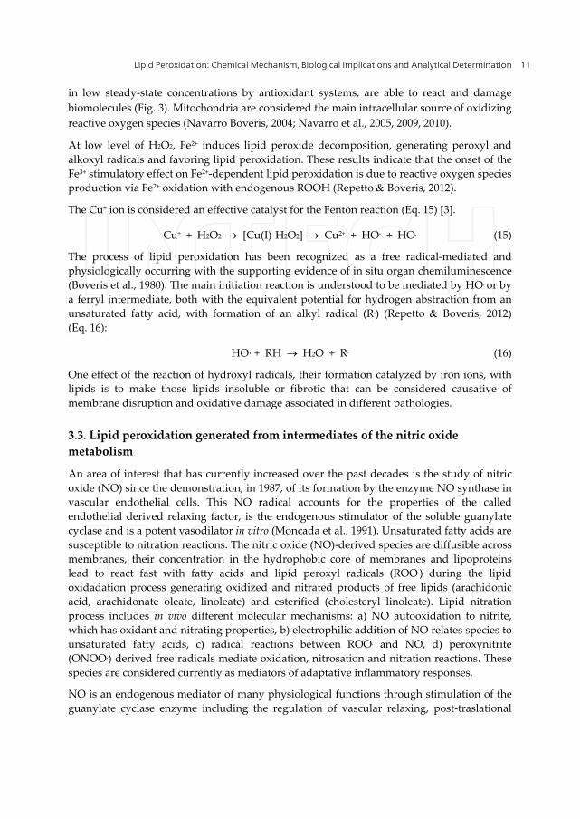
The Cu+ ion is considered an effective catalyst for the Fenton reaction (Eq. 15) [3].
Cu+ + H2O2 [Cu(I)-H2O2] Cu2+ + HO- + HO. (15)
The process of lipid peroxidation has been recognized as a free radical-mediated and
physiologically occurring with the supporting evidence of in situ organ chemiluminescence
(Boveris et al., 1980). The main initiation reaction is understood to be mediated by HO. or by
a ferryl intermediate, both with the equivalent potential for hydrogen abstraction from an
unsaturated fatty acid, with formation of an alkyl radical (R.) (Repetto Boveris, 2012)
(Eq. 16):
HO· + RH H2O + R. (16)
One effect of the reaction of hydroxyl radicals, their formation catalyzed by iron ions, with
lipids is to make those lipids insoluble or fibrotic that can be considered causative of
membrane disruption and oxidative damage associated in different pathologies.
3.3. Lipid peroxidation generated from intermediates of the nitric oxide
metabolism
An area of interest that has currently increased over the past decades is the study of nitric
oxide (NO) since the demonstration, in 1987, of its formation by the enzyme NO synthase in
vascular endothelial cells. This NO radical accounts for the properties of the called
endothelial derived relaxing factor, is the endogenous stimulator of the soluble guanylate
cyclase and is a potent vasodilator in vitro (Moncada et al., 1991). Unsaturated fatty acids are
susceptible to nitration reactions. The nitric oxide (NO)-derived species are diffusible across
membranes, their concentration in the hydrophobic core of membranes and lipoproteins
lead to react fast with fatty acids and lipid peroxyl radicals (ROO.) during the lipid
oxidadation process generating oxidized and nitrated products of free lipids (arachidonic
acid, arachidonate oleate, linoleate) and esterified (cholesteryl linoleate). Lipid nitration
process includes in vivo different molecular mechanisms: a) NO autooxidation to nitrite,
which has oxidant and nitrating properties, b) electrophilic addition of NO relates species to
unsaturated fatty acids, c) radical reactions between ROO. and NO, d) peroxynitrite
(ONOO.) derived free radicals mediate oxidation, nitrosation and nitration reactions. These
species are considered currently as mediators of adaptative inflammatory responses.
NO is an endogenous mediator of many physiological functions through stimulation of the
guanylate cyclase enzyme including the regulation of vascular relaxing, post-traslational
Lipid Peroxidation 12
protein changes, gene expression and inflammatory cell function (Moncada et al., 1991). Free
and esterified fatty acids as arachidonic and linoleic acids are important components of
lipoproteins and membranes that may be oxidized for different compounds. The NO and
NO-derived radicals react with fatty acids generating oxidized and nitrated species as
nitroalkenes and consequently, nitroalcohols. At low oxygen concentrations the most
important biological NO derivatives is ONOO.. The nitroalkylation process occurs in vitro
and in vivo, is involved in redox processes and cell signaling through the reversible covalent
bound and post-traslational modifications responsible for structure, function and
subcellular distribution of proteins (Valdez et al., 2011) and regulating the pro-inflammatory
effect of oxidant exposure (Nair et al., 2007).
A novel mechanism for hydroxyl radical production, which is not dependent on the
presence of transition metals, has recently been proposed. This involves the production of
peroxynitrite (Beckman et al., 1990, 1994; Rachmilewitz et al., 1993) which has
proinflammatory effects in vitro (Moncada et al., 1991), from the reaction of NO with O2.-
(Eqs. 17 to 20):
NO + O2.- ONOO- (17)
ONOO- + H+ ONOOH (18)
ONOOH HO. + NO2 (19)
2 H+ + O2.- + O2.- H2O2 + O2.- (20)
In pathological situations, macrophages and neutrophils, recruited to a site of injury, are
activated to produce NO as part of the inflammatory response. Furthermore, SOD activity
rapidly scavenges O2.- and also prolongs the vaso-relaxant effects of NO (Murphy Sies,
1991; Hogg et al., 1992; Rachmilewitz et al., 1993).
3.4. Modifications of lipid membrane structure
The presence of cholesterol in cell surface membranes influences their susceptibility to
peroxidation, probably both by intercepting some of the radicals present and by affecting
the internal structure of the membrane by interaction of its large hydrophobic ring structure
with fatty acid-side-chains. As lipid peroxidation precedes in any membrane, several of the
products produced have a detergent-like activity, specially released fatty acids or
phospholipids with one of their fatty-acid side-chains removed. This will contribute to
increased membrane disruption and further peroxidation.
The onset of lipid peroxidation within biological membranes is associated with changes in
their physicochemical properties and with alteration of biological function of lipids and
proteins. Polyunsaturated fatty acids and their metabolites play physiological roles: energy
provision, membrane structure, fluidity, flexibility and selective permeability of cellular
membranes, and cell signaling and regulation of gene expression (Catala, 2006). The
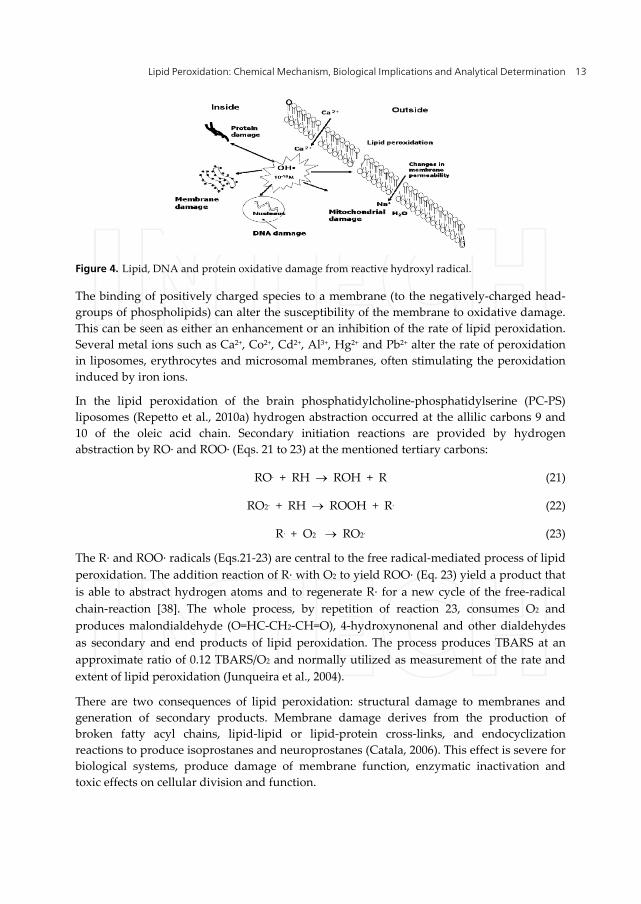
hydroxyl radical generated as a consequence of the Fenton reaction, oxidizes the cellular
components of biological membranes (Fig. 4).
Lipid Peroxidation: Chemical Mechanism, Biological Implications and Analytical Determination 13
Figure 4. Lipid, DNA and protein oxidative damage from reactive hydroxyl radical.
The binding of positively charged species to a membrane (to the negatively-charged head-
groups of phospholipids) can alter the susceptibility of the membrane to oxidative damage.
This can be seen as either an enhancement or an inhibition of the rate of lipid peroxidation.
Several metal ions such as Ca2+, Co2+, Cd2+, Al3+, Hg2+ and Pb2+ alter the rate of peroxidation
in liposomes, erythrocytes and microsomal membranes, often stimulating the peroxidation
induced by iron ions.
In the lipid peroxidation of the brain phosphatidylcholine-phosphatidylserine (PC-PS)
liposomes (Repetto et al., 2010a) hydrogen abstraction occurred at the allilic carbons 9 and
10 of the oleic acid chain. Secondary initiation reactions are provided by hydrogen
abstraction by RO· and ROO· (Eqs. 21 to 23) at the mentioned tertiary carbons:
RO. + RH ROH + R (21)
RO2. + RH ROOH + R. (22)
R. + O2 RO2. (23)
The R· and ROO· radicals (Eqs.21-23) are central to the free radical-mediated process of lipid
peroxidation. The addition reaction of R· with O2 to yield ROO· (Eq. 23) yield a product that
is able to abstract hydrogen atoms and to regenerate R· for a new cycle of the free-radical
chain-reaction [38]. The whole process, by repetition of reaction 23, consumes O2 and
produces malondialdehyde (O=HC-CH2-CH=O), 4-hydroxynonenal and other dialdehydes
as secondary and end products of lipid peroxidation. The process produces TBARS at an
approximate ratio of 0.12 TBARS/O2 and normally utilized as measurement of the rate and
extent of lipid peroxidation (Junqueira et al., 2004).
There are two consequences of lipid peroxidation: structural damage to membranes and
generation of secondary products. Membrane damage derives from the production of
broken fatty acyl chains, lipid-lipid or lipid-protein cross-links, and endocyclization
reactions to produce isoprostanes and neuroprostanes (Catala, 2006). This effect is severe for
biological systems, produce damage of membrane function, enzymatic inactivation and
toxic effects on cellular division and function.
Lipid Peroxidation 14
4. Role of transition metal on lipid peroxidation process
Studies in the past two decades have shown that redox active metals undergo redox cycling
reactions and possess the ability to produce reactive radicals such as superoxide anion
radical and nitric oxide in biological systems. Disruption of metal ion homeostasis leads to
oxidative stress, a state with increased formation of reactive oxygen species that
overwhelms antioxidant protection and subsequently induces DNA damage, lipid
peroxidation, protein modification and other effects, all symptomatic for numerous diseases,
involving cancer, cardiovascular disease, diabetes, atherosclerosis, neurological disorders,
and chronic inflammation.
The mechanism of lipid peroxidation in biological systems caused by free radicals has been
the focus of scientific interest for many years (Chance et al., 1976; Fridovich Porter, 1981;
Fraga et al., 1988; Gonzalez-Flecha et al., 1991a, 1991b; Famulari et al., 1996; Fiszman et al.,
2003; Junqueira et al., 2004; Catala, 2006; Boveris et al., 2008; Dianzani Barrera, 2008;
Dominguez et al., 2008; Repetto, 2008; Repetto et al., 2010b). Currently, it is known that the
OH. radical, is formed mainly by the Haber-Weiss reaction, and it is responsible for the
biological damage (Repetto et al., 2010a; Repetto et al., 2010b) (Eq.24):
O2- + H2O2 O2 + HO· + HO- (24)
However, this reaction would not proceed significantly in vivo because the rate constant for
the reaction is lower than that of the dismutation reaction. Nevertheless, a modification of
the Haber-Weiss reaction, the Fenton reaction and Fenton-like reactions, utilizes the redox
cycling ability of iron to increase the rate of reaction, is more feasible in vivo (Chance et al.,
1979; Boveris et al., 1980; Gonzalez Flecha et al., 1991b), and is frequently used to explain the
toxic effects of redox-active metals where M(n)+ is usually a transition metal ion.
As a transition metal that can exist in several valences and that can bind up to six ligands,
iron is an important component of industrial catalysts in the chemical industry especially for
redox reactions (Repetto et al., 2010a; Repetto Boveris, 2012).
There are several reports on the role of transition metals in lipid peroxidation process
associated with cellular toxicities, because once they enter our physiological systems, these
metals play a role in oxidative adverse effects. Some transition metals including iron,
chromium, lead, and cadmium generate lipid peroxidation in vitro e in vivo: fatty acids, cod
liver oil, biological membranes, tissues and organs, suggesting that metals contribute to the
oxidative effects of lipid peroxidation observed in various diseases (Repetto et al., 2010a;
Repetto Boveris, 2012).
The Fenton reaction occurs in vivo at a very low rate, and hence cannot account for any
substantial production of OH. radicals in biology. On the other hand, when catalysed by
transition metal ions, OH. radicals can be formed through reactions 25 and 26:
M(n) + + O2- M(n-1)+ + O2 (25)
M(n-1) + + H2O2 M(n)+ + HO· + HO- (26)
Lipid Peroxidation: Chemical Mechanism, Biological Implications and Analytical Determination 15
The concentration of intracellular redox active transition metals is either low or negligible:
free Fe2+ is 0.2-0.5 M and the pool of free Cu2+ is about a single ion per cell. However, trace
(nM) levels of cellular and circulating active transition metal ions seem enough for the
catalysis of a slow Fenton reaction in vivo at the physiological levels of hydrogen peroxide
(H2O2, 0.1-1.0 M) (Repetto et al., 2010a; Repetto Boveris, 2012).
It is well known that iron serves as a catalyst for the formation of the highly reactive hydroxyl
radical via Fenton reaction. In addition to ferrous ion, many metal ions including Cu (I), Cr (II),
and Co (II) were found to have the oxidative features of the Fenton reagent. Therefore, the
mixtures of these metal compounds with H2O2 were named ‘‘Fenton like reagents”. In actual
in vivo systems, once organic peroxides (ROOH) are formed by the action of ROS, heat, and/or
photo-irradiation, ROOH can be substituted for HO., where ROOH reacts with metal ions to
form alkoxyl radicals. Subsequently, a chain reaction of lipid peroxidation occurs.
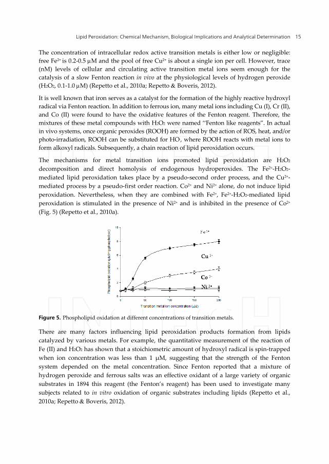
The mechanisms for metal transition ions promoted lipid peroxidation are H2O2
decomposition and direct homolysis of endogenous hydroperoxides. The Fe2+-H2O2-
mediated lipid peroxidation takes place by a pseudo-second order process, and the Cu2+-
mediated process by a pseudo-first order reaction. Co2+ and Ni2+ alone, do not induce lipid
peroxidation. Nevertheless, when they are combined with Fe2+, Fe2+-H2O2-mediated lipid
peroxidation is stimulated in the presence of Ni2+ and is inhibited in the presence of Co2+
(Fig. 5) (Repetto et al., 2010a).
Figure 5. Phospholipid oxidation at different concentrations of transition metals.
There are many factors influencing lipid peroxidation products formation from lipids
catalyzed by various metals. For example, the quantitative measurement of the reaction of
Fe (II) and H2O2 has shown that a stoichiometric amount of hydroxyl radical is spin-trapped
when ion concentration was less than 1 μM, suggesting that the strength of the Fenton
system depended on the metal concentration. Since Fenton reported that a mixture of
hydrogen peroxide and ferrous salts was an effective oxidant of a large variety of organic
substrates in 1894 this reagent (the Fenton’s reagent) has been used to investigate many
subjects related to in vitro oxidation of organic substrates including lipids (Repetto et al.,
2010a; Repetto Boveris, 2012).
Lipid Peroxidation 16
In the in vitro model of phosphatidylcholine/phosphatidyserine (60:40) liposomes and
hydrogen peroxide (H2O2), Fe and Cu promote lipid peroxidation, interpreted as the
consequence of the homolytic scission of H2O2 and of endogenous hydroperoxides (ROOH)
and of the generation of hydroxyl (HO•) and alcoxyl (RO•) radicals (Cadenas, 1989)
depending strictly on the participation of Fe and Cu as redox-reactive metals . However,
Co2+ and Ni2+ alone, do not induce lipid peroxidation. Nevertheless, when they are
combined with Fe2+, Fe2+-H2O2-mediated lipid peroxidation is stimulated in the presence of
Ni2+ and inhibited in the presence of Co2+ (Repetto et al., 2010a; Repetto Boveris, 2012).
Cr(III) occurs in nature and is an essential trace element utilized in the regulation of blood
glucose levels. Cr(III) reacts with superoxide, subsequently Cr(II) yields hydroxyl radical
via Fenton-like reaction with H2O2 to initiate lipid peroxidation.
Cadmium intoxication was shown to increase lipid peroxidation in rat liver, kidney and
heart. However, the mechanisms of cadmium toxicity are not fully understood. Cadmium
indirectly affects the generation of various radicals including superoxide and hydroxyl
radical. The generation of hydrogen peroxide by cadmium ion may become a source of
radicals in the Fenton system (Jomova Valko, 2011).
5. Toxic effects of secondary products of lipid peroxidation
Many aldehydes are produced during the peroxidative decomposition of unsaturated fatty
acids. Compared with free radicals, aldehydes are highly stable and diffuse out from the cell
and attack targets far from the site of their production. About 32 aldehydes were identified
as products of lipid peroxidation: a) saturated aldehydes (propanal, butanal, hexanal,
octanal, being the decanal the most important); b) 2,3-trans-unsaturated-aldehydes (hexenal,
octenal, nonenal, decenal and undecenal); c) a series of 4-hydroxylated,2,3-trans-unsaturated
aldehydes: 4-hydroxyundecenal, being 4-hydroxinonenal (HNE) the most important
quantitatively. Malonyldialdehyde (MDA) was considered for a long time as the most
important lipid peroxidation metabolite. However, MDA is practically no toxic.
Recent studies have demonstrated that the most effective product of lipid peroxidation
causing cellular damage is HNE. HNE produces different effects: acts as an intracellular
signal able to modulate gene expression, cell proliferation, differentiation and apoptosis.
The hydroxyl-group close to a carbonyl group present in HNE chemical structure is related
to its high reactivity with different targets (thiol and amine groups). HNE is easily diffusible
specie, but its biological effect depends on the molecule target and behavior as a signal to
produce the damage.
Oxidative stress is a well known mechanism of cellular injury that occurs with increased
lipoperoxidation of cell phospholipids and that has been implicated in various cell
dysfunctions (Sies, 1991a,b; Catala, 2006). Aldehydes exhibit high reactivity with bio-
molecules, such as proteins, DNA and phospholipids generating intra and intermolecular
adducts.
The physiological concentrations of these products are low; however, higher concentrations
correspond to pathological situations. Therefore, DNA damage caused by lipid peroxidation
Lipid Peroxidation: Chemical Mechanism, Biological Implications and Analytical Determination 17
end products could provide promising markers for risk prediction and targets for
preventive measures. DNA-reactive aldehydes can damage DNA either by reacting directly
with DNA bases or by generating more reactive bifunctional intermediates, which form
exocyclic DNA adducts. Of these, HNE and MDA, acrolein, and crotonaldehyde have been
shown to modify DNA bases, yielding promutagenic lesions and to contribute to the
mutagenic and carcinogenic effects associated with oxidative stress-induced lipid
peroxidation and HNE and MDA implicated carcinogenesis.
The end-products of lipid peroxidation (HNE and MDA) cause protein damage by addition
reactions with lysine amino groups, cysteine sulfhydryl groups, and histidine imidazole
groups (Esterbauer et al., 1991; Esterbauer, 1996). Modifications of protein by aldehyde
products of lipid peroxidation contribute to neurodegenerative disorders, activation of
kinases (Uchida et al., 1999; Uchida, 2003) and inhibition of the nuclear transcription factor
(Camandola et al., 2000).
6. Lipid peroxidation of subcellular fragments
6.1. Microsomes
Microsomes isolated from liver have been shown to catalyze an NADPH-dependent
peroxidation of endogenous unsaturated fatty acids in the presence of ferric ions and metal
chelators, such as ADP or pyrophosphates. Microsomal membranes are particularly
susceptible to lipid peroxidation owing to the presence of high concentrations of
polyunsaturated fatty acids (Poyer McCay, 1971). The mechanism involved in the
initiation of peroxidation in the NADPH-dependent microsomal system do not appear to
involve neither superoxide nor hydrogen peroxide, since neither superoxide dismutase nor
catalase cause inhibition of peroxidation. Nevertheless, reduced iron plays an important role
in both the initiation and propagation of NADPH-dependent microsomal lipid peroxidation
(Shires, 1975).
Microsomal membrane lipids, particularly the polyunsaturated fatty acids, undergo
degradation during NADPH-dependent lipid peroxidation. The degradation of membrane
lipids during lipid peroxidation has been observed to result in the production of singlet
oxygen, which is detected as chemiluminescence (Boveris et al., 1980).
Nonenzymatic peroxidation of microsomal membranes also occurs and is probably
mediated in part by endogenous hemoproteins and transition metals. High concentrations
of transition metals (50 M) promote auto-oxidation of phospholipids (Repetto et al., 2010a).
6.2. Mitochondria
It is currently accepted that mitochondrial complex I is particularly sensitive to inactivation
by oxygen free radicals and reactive nitrogen species. This special characteristic is frequently
referred as complex I syndrome, with the symptoms of reduced mitochondrial respiration
with malate-glutamate and ADP and of reduced complex I activity. This complex I
syndrome has been observed in aging (Navarro et al., 2005; Navarro Boveris, 2004, 2008),
Lipid Peroxidation 18
in ischemia-reperfusion (Gonzalez-Flecha et al., 1993), in Parkinson’s disease, and in other
neurodegenerative diseases (Schapira et al., 1990a, 1990b; Sayre et al., 1999; Carreras et al.,
2004; Schapira, 2008; Navarro et al., 2009), and in this study, with the addition of the
increased rates of production of O2.- and H2O2 by complex I mediated reactions, reactions
with the free radicals intermediates of the lipid peroxidation process (mainly ROO·), and
amine-aldehyde adduction reactions. It is now understood that the three processes above
mentioned alter the native non-covalent polypeptide interactions of complex I and promote
synergistically protein damage and inactivation by shifting the noncovalent bonding to
covalent cross linking (Navarro et al., 2005). Complex I oxidative protein damage has also
been considered the result of protein modification by reaction with malonaldehyde and 4-
HO-nonenal (Sayre et al., 1999). It was hypothesized that protein damage in the subunits of
complexes I and IV follows to free radical-mediated cross-linking and inactivation. The
subunits that are normally held together by noncovalent forces are shifted to covalent cross-
linking after reaction with the hydroperoxyl radicals (ROO·) and the stable aldehydes
produced during the lipid peroxidation process.
The hypothesis that cumulative free radical-mediated protein damage is the chemical basis
of respiratory complexes I and IV inactivation (Berlett Stadtman, 1997) offers the
experimental approach of the chronic use of vitamin E, as an antioxidant for the lipid phase
of the inner mitochondrial membrane and for the prevention of the mitochondrial /damage
associated with aging. The adduction reactions of malonaldehyde and 4-HO-nonenal with
protein evolve to stable advanced lipid peroxidation products (Sayre et al., 1999) and
protein carbonyls (Nair et al., 2007; Navarro et al., 2008). The molecular mechanism
involved in the inactivation of complex I is likely accounted for by ROO. and ONOO-. Upon
aging, frontal cortex and hippocampal mitochondria show a decreased rate of respiration,
especially marked with NAD-dependent substrates, and decreased enzymatic activities of
complexes I and IV associated with an increase in the content of oxidation products (TBARS
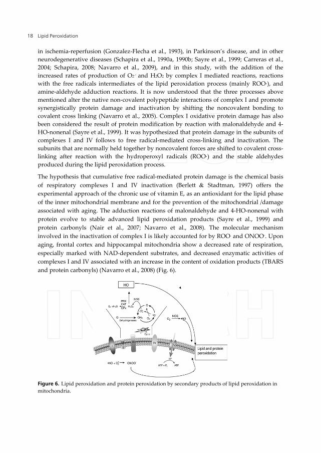
and protein carbonyls) (Navarro et al., 2008) (Fig. 6).
Figure 6. Lipid peroxidation and protein peroxidation by secondary products of lipid peroxidation in
mitochondria.
Lipid Peroxidation: Chemical Mechanism, Biological Implications and Analytical Determination 19
7. Lipid peroxidation and human pathologies
The organism must confront and control the balance of both pro-oxidants and antioxidants
continuously. The balance between these is tightly regulated and extremely important for
maintaining vital cellular and biochemical functions. This balance often referred to as the
redox potential, is specific for each organelle and biological site, and any interference of the
balance in any direction might be deleterious for the cell and organism. Changing the
balance towards an increase in the pro-oxidant over the capacity of the antioxidant is
defined as oxidative stress and might lead to oxidative damage. Changing the balance
towards an increase in the reducing power, or the antioxidant, might also cause damage and
can be defined as reductive stress.
Oxidative stress and damage have been implicated in numerous disease processes,
including inflammation, degenerative diseases, and tumor formation and involved in
physiological phenomena, such as aging and embryonic development. The dual nature of
these species with their beneficial and deleterious characteristics implies the complexities of
their effects at a biological site.
Lipid peroxidation has been pointed out as a key chemical event in the oxidative stress
associated with several inborn and acquired pathologies. Disruption of organelle and cell
membranes together with calcium homeostasis alterations are the main supramolecular
events linked to lipid peroxidation. However, it is not clear if lipid peroxidation process is a
cause, triggering step of the clinical manifestations of the disease, or a consequence of toxic
effects of lipid peroxidation products.
In pathological situations the reactive oxygen species are generated and as a consequence
lipid peroxidation occurs with -tocopherol deficiency. In addition to containing high
concentrations of polyunsaturated fatty acids and transitional metals, red blood cells are
constantly being subjected to various types of oxidative stress. Red blood cells however are
protected by a variety of antioxidant systems which are capable of preventing most of the
adverse effects under normal conditions. Among the antioxidant systems in the red cells, -
tocopherol possesses an important and unique role. -tocopherol may protect the red cells
from oxidative damage via a free radical scavenging mechanism and as a structural
component of the cell membrane (Chitra Shyamaladevi, 2011).
Levels of Met-Hb. are regarded as an index of intracellular damage to the red cell and it is
increased when -tocopherol is consumed and the rate of lipid peroxidation is increased.
Scavenging of free radicals by -tocopherol is the first and the most critical step in
defending against oxidative damage to the red cells. When -tocopherol is adequate, GSH
and ascorbic acid may complement the antioxidant functions of -tocopherol by providing
reducing equivalents necessary for its recycling/regeneration.
On the other hand, when -tocopherol is absent, GSH and ascorbic acid release transitional
metals from the bound forms and/or maintain metal ions in a catalytic state. Free radical
generation catalysed by transition metal ions in turn initiates oxidative damage to cell
Lipid Peroxidation 20
membranes. Membrane damage can lead to release of heme compounds from erythrocytes.
The heme compounds released may further promote oxidative damage especially when
reducing compounds are present (Boveris et al., 2008).
8. Lipid peroxidation and aging
Aging is a process directly related to systemic oxidative stress. Two components of the
oxidative stress situation have been recognized in human aging: a decrease in availability of
nutritional molecular antioxidants and an accumulation of products derived from the
oxidation of biological structures. Oxidation of biomolecules is related to susceptibility to
diseases, such as cancer and heart disease, as well as associated with the process of aging
(Navarro et al., 2005; Navarro Boveris, 2007, 2008).
The products derived from lipid peroxidation, measured in plasma by Junqueira et al.,
(2004) as fluorescent products, were higher in elderly than younger human subjects and
even higher in disabled octogenarians and nonagenarians. This increase in lipid
peroxidation products was directly correlated with age, and was associated with decreases
in vitamin E and C.
9. Analytical determination of lipid peroxidation
Since the acceptation of the oxidative stress concept, scientists and physicians have been
searching for a simple assay or a small group of determination that would result useful for
the assessment of oxidative stress and lipid peroxidation in clinical situations. The
determinations of marker metabolites are usually performed in blood, red blood cells or
plasma. The markers for systemic oxidative stress are normally present in healthy humans
and the assays for systemic oxidative stress are comparative, which makes necessary to have
reference values from normal individuals.
At present, the plasma levels of oxidation products derived from free-radical mediated
reactions and of antioxidants are used as indicators of systemic oxidative stress in humans
and experimental animals. The more utilized determination of an oxidation product is
MDA, which is determined with low specificity but with great efficiency by the simple and
useful assay of TBARS with measurements made by spectrophotometry or
spectrofluorometry. The normal plasma levels of TBARS are 2-3 M (Junqueira et al., 2004).
Oxidative damage is characterized by increases in the levels of the oxidation products of
macromolecules, such as thiobarbituric acid reactive substances (TBARS), and protein
carbonyls. Many of these products can be found in biological fluids, as well as addition-
derivatives of these reactive end-products. As a result of lipid peroxidation a great variety of
aldehydes can be produced, including hexanal, malondialdehyde (MDA) and 4-
hydroxynonenal (Catala, 2006).
Oxidation of an endogenous antioxidant reflects an oxidative stress that is evaluated by
measuring the decrease in the total level of the antioxidant or the increase in the oxidative

Lipid Peroxidation: Chemical Mechanism, Biological Implications and Analytical Determination 21
form. The only way not to be influenced by nutritional status is to measure the ratio
between oxidized and reduced antioxidants present in blood. The published literature
provides compelling evidence that a) MDA represents a side product of enzymatic PUFA-
oxygenation and a secondary end product of no enzymatic (autoxidative) fatty peroxide
formation and decomposition and b) sensitive analytical methods exist for the
unambiguous isolation and direct quantification of MDA. Conceptually, these two facts
indicate that MDA is an excellent index of lipid peroxidation. However, this conclusion is
limited in practice by several important consideration: a) MDA yield as a result of lipid
peroxidation varies with the nature of the PUFA peroxidised (specially its degree of
instauration) and the peroxidation stimulus, b) only certain lipid oxidation products
decompose yield MDA, c) MDA is only one of several end product of fatty peroxide
formation and decomposition, d) the peroxidation environment influences both the
formation of lipid-derived precursors and their decomposition to MDA, e) MDA itself is a
reactive substance which can be oxidative and metabolically degraded, f) oxidative injury
to no lipid biomolecules has the potential to generate MDA. With biological materials, it
appears prudent to consider the TBARS test more than an empirical indicator of the
potential occurrence of peroxidative lipid damage and not as a measure of lipid
peroxidation (Repetto, 2008). The thiobarbituric acid test (TBARS) has been employed to a
uniquely great degree over the last five decades to detect and quantify lipid peroxidation
in a variety of chemical as well as biological material. Two underlying assumptions are
implicit from the widespread use of the TBARS test to assess lipid peroxidation: a) an
operative and quantitative relationship exists between lipid peroxidation and MDA, b)
product formation during the TBARS test is diagnostic of the presence and amount of
fatty peroxides.
Lipid peroxidation proceeds by a free-radical mediated chain reaction that includes
initiation, propagation and termination reactions. The chain reaction is initiated by the
abstraction of a hydrogen atom from a methylene group of an unsaturated fatty acid.
Propagation is cycled through rounds of lipid peroxyl radical abstraction of the bis-
methylene hydrogen atoms of a polyunsaturated fatty acyl chain to generate new radicals,
after O2 addition, resulting in the conversion of alkyl radical in hydroperoxyl radical.
Termination involves the reaction of two hydroperoxyl radicals to form non-radical
products. This reaction is particularly interesting since it is accompanied, although at low
yield, by emission of light or chemiluminiscence. Some lipid peroxidation products are
light-emitting species and their luminescence is used as an internal marker of oxidative
stress (Chance et al., 1979; Boveris et al., 1980, Gonzalez-Flecha et al., 1991b; Sies, 1991a;
Repetto, 2008). The measurement of light emission derived from 1O2 and excited triplet
carbonyl compounds, which are the most important chemiluminiscent species in the lipid
peroxidation of biological systems, is directly related to the rate of lipid peroxidation and
allows an indirect assay of the content of lipophilic antioxidants in the sample (Gonzalez-
Flecha et al., 1991a). Lipophilic antioxidants react with lipid peroxyl radicals and lower
antioxidant content is associated with higher chemiluminescence (Repetto, 2008).
Lipid Peroxidation 22
The low-level chemiluminescence which accompanies the peroxidation of polyunsaturated
fatty acids has been used as a tool in kinetic and mechanistic studies of biological samples to
estimate the extent of the reactions and even to indicate tissue damage promoted by
oxidants. Triplet carbonyls and singlet oxygen formed in the annihilation of intermediate
peroxyl radicals (ROO.) have been identified as the chemiluminescence emitters.
Chemiluminescence is a very interesting way to evaluate an oxidative stress and lipid
peroxidation in biological samples and living systems. The emission of light has been
observed during stress in different experimental models. Chemiluminescence is very
sensitive and thus can be applied to measure free radical production in human tissues.
Chemiluminescent systems may be classified in two classes based on the origin of the
emitting molecule. In the first class, the emitter is a product of the chemical reaction (direct
chemiluminescence). In the second class, there is energy transfer between an electronically
excited product molecule and a second substance which then becomes the emitter
(sensitized chemiluminescence) (Boveris et al., 1980; Gonzalez-Flecha et al., 1991b; Repetto,
2008).
The chemical mechanism responsible for spontaneous organ light emission is provided by
the Russell’s reaction in which two secondary or tertiary peroxyl radicals (ROO•) yield 1O2
and excited carbonyl groups (=CO*) as products. In turn, two 1O2, through dimol emission,
lead to photoemission at 640 and 670 nm, whereas =CO* yields photons at the 460-470 nm
band (Boveris et al., 1980). The main sources of the chemiluminescence detected in the direct
and sensitized chemiluminescence is the dimol emission of ¹O2 (reaction 27) and the photon
emission from excited carbonyl groups (reaction 28) (Boveris et al., 1980).
2 ¹O2 2 O2 + h (634-703 nm) (27)
RO* RO + h (380-460 nm) (28)
These reactions are accompanied by chemiluminescence whose intensity may serve as an
indirect measure of peroxide free radical and -tocopherol concentration in the sample.
Lipid peroxidation has been recognized as free radical-mediated and physiologically
occurring (Navarro Boveris, 2004, Navarro et al., 2010; Repetto Boveris, 2012) with the
supporting evidence of in situ organ chemiluminescence (Repetto, 2008). Spontaneous
chemiluminescence of in situ organs directly reports the intracellular formation of singlet
oxygen (1O2) (Boveris et al., 1980) and represents an issue of direct chemiluminescence. The
generation of 1O2 implies the collision of two peroxyl radicals (ROO·) with formation of
excited species, 1O2 itself and excited carbonyls, followed by photoemission. Light emission
from in situ organs is a physiological phenomenon that provides a determination of the
steady state concentration of singlet oxygen and indirectly of the rate of oxidative free
radical reactions (Boveris et al., 1980). In situ liver chemiluminescence has been recognized
as a reliable indicator of oxidative stress and damage in rat liver upon hydroperoxide
infusion (Gonzalez-Flecha et al., 1991b), ischemia-reperfusion (Gonzalez-Flecha et al., 1993),
Lipid Peroxidation: Chemical Mechanism, Biological Implications and Analytical Determination 23
and chronic and acute alcohol intoxication (Videla et al., 1983). The increases in
photoemission observed were parallel to increased contents of indicators of lipid
peroxidation (malonaldehyde and 4-HO-nonenal) but with a higher experimental/control
ratio in organ chemiluminescence (Boveris et al., 1980).
Tert-butyl hydroperoxide initiated chemiluminescence is an example of sensitized
chemiluminescence, and it has been used to enhance the chemiluminescence accompanying
lipid peroxidation and the -tocopherol content of tissues. This method has been
successfully utilized to detect the existence of oxidative damage associated to experimental
or pathological situations in tissue homogenates, subcellular fractions, and in human heart,
liver and muscle biopsies (Gonzalez-Flecha et al., 1991b).
Tissue homogenates or blood samples are subjected to in vitro oxidative damage by
supplementation with tert-butyl hydroperoxide. It reacts with hemoproteins and Fe2+
producing peroxyl and alcoxyl free radicals, which enter to the propagation phase of the
lipid peroxidation radical chain reaction. The termination steps of the chain reaction
generate compounds in an excited state: singlet oxygen and carbonyl groups. This assay is
useful to evaluate the integral level of the non-enzymatic antioxidant defenses of a tissue
(Gonzalez-Flecha et al., 1991a, 1993).
The increase of tert-butyl hydroperoxide-initiated chemiluminescence is indicative that -
tocopherol is the antioxidant consumed in erythrocytes and suggest that reactive oxygen
species and lipid peroxidation catalyzed by reduced transition metals may be responsible
for the onset of oxidative damage and the occurrence of systemic oxidative stress in patients
suffering oxidative damage associated to neurological pathologies as Parkinson (Famulari et
al., 1996, Dominguez et al., 2008), Alzheimer disease (Famulari et al., 1996; Repetto et al.,
1999; Dominguez et al., 2008; Serra et al., 2009), and vascular dementia (Famulari et al.,
1996, Dominguez et al., 2008; Serra et al., 2009); immunological diseases as HIV infection
and AIDS (Repetto et al., 1996), hyperthyroidism and hypothyroidism (Abalovich et al.,
2003). These methods were used to evaluate lipid peroxidation and oxidative damage in
experimental models of oxidative stress in rats (Repetto et al., 2003, 2010; Ossani et al., 2007;
Repetto Ossani, 2008; Repetto Boveris, 2010).
A common question of the researchers in the field is which the method of choice is. The
answer is: none of them, and all of them. Each assay measures something different. Diene
conjugation tells one about the early stages of peroxidation, as a direct measurement of lipid
peroxides. In the absence of metal ions to decompose lipid peroxides there will be little
formation of hydrocarbon gases, carbonyl compounds, or their fluorescent complexes, which
does not necessarily mean therefore that nothing is happening. Even if peroxides do not
decompose, the TBARS test can still detect them because of decomposition of peroxides.
Changes in the mechanism of peroxide decomposition might alter the amount generated
without any change in the overall rate of lipid peroxidation. Whatever method is chosen, one
should think clearly what is being measured and how it relates to the overall lipid
peroxidation process. Whatever possible, two or more different assay methods should be used.
Lipid Peroxidation 24
10. Conclusion
Lipid peroxidation is a physiological process that takes place in all aerobic cells.
Unsaturated fatty acids which are structural part of cell membranes are subjected to lipid
peroxidation by a non enzymatic and free-radical mediated reaction chain. The molecular
mechanisms of the lipid peroxidation process are known and it can be estimated that about
1 % of the total oxygen uptake of cells, organs and bodies in taken up by the reactions of
lipid peroxidation. The initiation reactions are provided by the transition-metal catalyzed
hemolytic scission of H2O2 and ROOH. In turn, H2O2 is mainly generated from the
mitochondrial dismutation of superoxide radical (O2.-). The products and by-products of
lipid peroxidation are cytotoxic and lead in successive steps to oxidative stress, oxidative
damage and apoptosis. In a long series of physiological and pathophysiological processes,
including aging and neurodegenerative diseases, the rates of mitochondrial O2.- and H2O2
are increased with a parallel increase in the rate of the lipid peroxidation process. It is
expected that supplementation with adequate antioxidants, as for instance, α-tocopherol,
will keep sensitive cells and organs in healthy conditions and increase lifespan.
Author details
Marisa Repetto, Jimena Semprine and Alberto Boveris
University of Buenos Aires, School of Pharmacy and Biochemistry,
General and Inorganic Chemistry,
Institute of Biochemistry and Molecular Medicine (IBIMOL-UBA-CONICET), Argentina
Acknowledgement
We thank to Dr. Jorge Serra for helping in the revision of this version.
11. References
Abalovich, M.; Llesuy, S.; Gutierrez, S. Repetto, M. (2003) Peripheral markers of oxidative
stress in Graves´ disease. The effects of methimazole and 131 Iodine treatments. Clinical
Endocrinology. Vol. 59, pp. 321-327, ISSN: 1365-2265
Beckman, J.; Beckman, T.; Chen, J.; Marshall, P. Freeman, B. (1990) Apparent hydroxyl
radical production from peroxynitrite. Implications for endothelial injury from nitric
oxide oxide and superoxide. Proceeding of the National Academy of Sciences of the United
States. Vol. 87, pp. 1620-1624, ISSN: 0027-8424
Beckman, J.; Chen, J.; Ischiropulos, H. Crow, J. (1994) Oxidative chemistry of
peroxynitrite. Methods in Enzymology. Vol. 233, pp. 229-240, ISSN: 0076-6879
Berlett, B.S. Stadtman, E.R. (1997) Protein oxidation in aging, disease, and oxidative stress.
The Journal of Biological Chemistry. Vol. 272, pp. 20313–20316, ISSN: 0021-9258
Lipid Peroxidation: Chemical Mechanism, Biological Implications and Analytical Determination 25
Boveris, A.; Cadenas, E.; Reiter, R.; Filipkowski, M.; Nakase, Y. & Chance, B. (1980) Organ
chemiluminescence: noninvasive assay for oxidative radical reactions Proceeding of the
National Academy of Sciences of the United States. Vol. 177, pp. 347-351, ISSN: 0027-8424
Boveris, A.; Fraga, C.; Varsavsky, A. & Koch, O. (1983) Increased chemiluminescence and
superoxide production in the liver of chronically ethanol-treated rats. Archives of
Biochemistry and Biophysics. Vol. 227, pp. 534-541, ISSN: 0003-9861
Boveris, A. & Navarro, A. (2008) Brain mitochondrial dysfunction in aging. Life, Vol. 60,
No.5, pp. 308-314, ISSN: 1521-6543
Boveris, A.; Repetto, M.G.; Bustamante, J.; Boveris, A.D. & Valdez, L.B. (2008). The concept
of oxidative stress in pathology. In: Álvarez, S.; Evelson, P. (ed.), Free Radical
Pathophysiology, pp. 1-17, Transworld Research Network: Kerala, India, ISBN: 978-81-
7895-311-3
Cadenas, E. (1989) Biochemistry of oxygen toxicity. Annual Review of Biochemistry. Vol. 58,
pp. 79-110, ISSN:0066-4154
Camandola, S.; Poli, G. & Mattson, M. (2000) The lipid peroxidation product 4-hydroxy-2,3-
nonenal inhibits constitutive and inducible activity of nuclear factor-bin neurons.
Molecular Brain Research. Vol. 85, pp. 53–60, ISSN: 0021-9258
Carreras, M.C.; Franco, M.C.; Peralta, J.G. & Poderoso, J.J. (2004) Nitric oxide, complex I, and
the modulation of mitochondrial reactive species in biology and disease. Molecular
Aspects of Medicine. Vol. 25, pp. 125–139, ISSN: 0098-2997
Catala, A. (2006) An overview of lipid peroxidation with emphasis in outer segments of
photoreceptors and the chemiluminescence assay. The International Journal of
Biochemistry and Cell Biology. Vol. 38, pp. 1482-1495, ISSN: 1357-2725
Chance, B.; Sies, H. & Boveris, A. (1979) Hydroperoxide metabolism in mammalian organs.
Physiological Reviews. Vol. 59, pp. 527-605, ISSN:�0031-9333
Cutrin, JC.; Cantino, D.; Biasi, F.; Chiarpotto, E.; Salizzoni, M.; Andorno, E.; Massano, G.;
Lanfranco, G.; Rizetto, M.; Boveris, A. & Poli, G. (1996) Reperfusion damage to the bile
canaliculi in transplanted human liver. Hepatology. Vol. 24, pp. 1053-1057, ISSN: 1527-
3350
Dianzani, M. & Barrera, G. (2008) Pathology and physiology of lipid peroxidation and its
carbonyl products. In: Álvarez, S.; Evelson, P. (ed.), Free Radical Pathophysiology, pp. 19-
38, Transworld Research Network: Kerala, India, ISBN: 978-81-7895-311-3
Domínguez, R.O.; Marschoff, E.R.; Guareschi, E.M.; Repetto, M.G.; Famulari, A.L.; Pagano,
M.A. & Serra, J.A. (2008). Insulin, glucose and glycated haemoglobin in Alzheimer’s
and vascular dementia with and without superimposed Type II diabetes mellitus
condition. Journal of Neural Transmission, Vol. 115, pp. 77-84, ISSN: 0300-9564.
Esterbauer, H.; Schaur, J. & Zollner, H. (1991) Chemistry and biochemistry of 4-
hydroxynonenal, malondialdehyde and related aldehydes. Free Radical in Biology &
Medicine. Vol. 11, pp. 81–128, ISSN: 0891-5849
Esterbauer, H. (1996) Estimation of peroxidative damage. A critical review. Pathologie
Biologie. Vol. 44, pp. 25–28, ISSN: 0031-3009

Lipid Peroxidation 26
Famulari, A.; Marschoff, E.; Llesuy, S.; Kohan, S.; Serra, J.; Domínguez, R.; Repetto, M.G.;
Reides, C. & Lustig, E.S. de (1996). Antioxidant enzymatic blood profiles associated
with risk factors in Alzheimer’s and vascular diseases. A predictive assay to
differentiate demented subjects and controls. Journal of the Neurological Sciences, Vol. 141,
pp. 69-78, ISSN: 0022-510X
Farooqui, T. & Farooqui, A. (2011) Lipid-mediated oxidative stress and inflammation in the
pathogenesis of Parkinson´s disease. Parkinson´s disease. DOI: 10.4061/2011/247467
Fiszman, M.; D´Eigidio, M.; Ricart, K.; Repetto, M.G.; Llesuy, S.; Borodinsky, L.; Trigo, R.;
Riedstra, S.; Costa, P.; Saizar, R.; Villa, A. & Sica, R. (2003). Evidences of oxidative stress
in Familial Amyloidotic Polyneuropathy Type 1. Archives of Neurology, Vol. 60, pp. 593-
597, ISSN 0003-9942
Fraga, C.; Leibovitz, B. & Tappel, A. (1988). Lipid peroxidation measured as thiobarbituric
acid-reactive substances in tissue slices: characterization and comparison with
homogenates and microsomes. Free Radicals in Biology and Medicine, Vol. 4, pp. 155-161,
ISSN: 0891-5849
Fridovich, I. (1978) Superoxide radicals, superoxide dismutases and the aerobic lifestyle.
Photochemistry and Photobiology. Vol. 28, pp. 733-741, ISSN: 1010-6030
Fridovich, S. & Porter, N. (1981) Oxidation of arachidonic acid in micelles by superoxide
and hydrogen peroxide. The Journal of Biological Chemistry. Vol. 256, pp. 260-265, ISSN:
0021-9258
Gatto, E.; Carreras, M.C.; Pargament, G.; Reides, C.; Repetto, M.G.; Llesuy, S.; Fernández
Pardal, M. & Poderoso, J. (1996). Neutrophil function nitric oxide and blood oxidative
stress in Parkinson’s disease. Movement Disorders, Vol. 11, pp. 261-267, ISSN: 0885-3185
Gatto, E.; Carreras, C.; Pargament, G.; Riobó, N.; Reides, C.; Repetto, M.; Fernández Pardal,
N.; Llesuy, S. & Poderoso, J. (1997). Neutrophyl function nitric oxide and blood
oxidative stress in Parkinson´s Disease. Focus Parkinson´s Disease, Vol. 9, pp. 12-14
Gonzalez Flecha, B., Repetto, M.; Evelson, P. & Boveris, A. (1991a) Inhibition of microsomal
lipid peroxidation by -tocopherol and -tocopherol acetate. Xenobiotica. 21: 1013–
1022, ISSN: 0049-8254
González Flecha, B.; Llesuy, S. & Boveris, A. (1991b). Hydroperoxide-initiated
chemiluminescence: assay for oxidative stress in biopsies of heart, liver and muscle.
Free Radicals in Biology and Medicine, Vol. 10, pp. 93-100, ISSN: 0891-5849
Gonzalez-Flecha, B.; Cutrin, J.C. & Boveris, A. (1993) Time course and mechanism of
oxidative stress and tissue damage in rat liver subjected to in vivo ischemia-reperfusion.
Journal of Clinical Investigation. Vol. 91, pp. 456–464, ISSN:�0021-9738
Halliwell, B. & Gutteridge, J.M.C. (1984). Oxygen toxicity, oxygen radicals, transition metals
and disease. Biochemical Journal, Vol. 218, pp. 1-14, ISSN: 0264-6021
Hogg, N.; Darley-Usmar, V.; Wilson, M. & Moncada, S. (1992) Production of hydroxyl
radicals from the simultaneous generation of superoxide and nitric oxide. Biochemical
Journal. Vol. 281, pp. 419-424, ISSN: 0264-6021
Lipid Peroxidation: Chemical Mechanism, Biological Implications and Analytical Determination 27
Jomova, K. & Valko, M. (2011) Advances in metal-induced oxidative stress and human
disease. Toxicology. Vol. 283, pp. 65-87, ISSN: 0300-483X.
Junqueira, V.; Barros, S.; Chan, S.; Rodríguez, L.; Giavarotti, L.; Abud, R. & Deucher, G.
(2004) Aging and oxidative stress. Molecular Aspects of Medicine. Vol. 25, pp. 5–16, ISSN:
0098-2997
Liu, Q.; Raina, A.K.; Smith, M.A.; Sayre,. LM. & Perry, G. (2003) Hydroxynonenal, toxic
carbonyls, and Alzheimer disease. Molecular Aspects of Medicine. Vol. 24, pp. 305–313,
ISSN: 0098-2997
Moncada, S.; Palmer, R. & Higgs, E. (1991) Nitric oxide: Physiology, patophysiology and
pharmacology. Pharmaceutical Reviews. Vol. 43, pp. 109-141, ISSN:�1918-5561
Murphy, M. & Sies, H. (1991) Reversible conversion of nitroxyl anion to oxide by superoxide
dismutase. Proceeding of the National Academy of Sciences of the United States. Vol. 88, pp.
10860-10864, ISSN: 0027-8424
Nair, U.; Barstsch, H. & Nair, J. (2007) Lipid peroxidation-induced DNA damage in cancer-
prone inflammatory diseases: a review of published adduct types and levels in humans.
Free Radical in Biology & Medicine. Vol. 43, pp. 1109-1120, ISSN: 0891-5849
Navarro, A. & Boveris, A. (2004). Rat brain and liver mitochondria develop oxidative stress
and lose enzymatic activities on aging. American Journal of Physiology - Regulatory,
Integrative and Comparative Physiology, Vol. 287, pp. 1244-1249, ISSN: 0363-6119
Navarro, A.; Gomez, C.; Sanchez-Pino, MJ.; Gonzalez, H.;, Bandez, MJ.; Boveris, AD.;
Boveris, A. (2005) Vitamin E at high doses improves survival, neurological
performance, and brain mitochondrial function in aging male mice. American Journal of
Physiology - Regulatory, Integrative and Comparative Physiology, Vol. 289, pp. 1392–1399,
ISSN: 0363-6119
Navarro, A.; Boveris, A. (2007) The mitochondrial energy transduction system and the aging
process. American Journal of Physiology - Regulatory, Integrative and Comparative
Physiology, Vol. 292, pp. 670-686, ISSN: 0363-6119
Navarro, A.; Lopez-Cepero, JM.; Bandez, MJ.; Sanchez-Pino, MJ.; Gomez, C.; Cadenas, E.;
Boveris, A. (2008) Hippocampal mitochondrial dysfunction in rat aging. American
Journal of Physiology - Regulatory, Integrative and Comparative Physiology, Vol. 294, pp. 501-
509, ISSN: 0363-6119
Navarro, A. & Boveris, A. (2009). Brain mitochondrial dysfunction and oxidative damage in
Parkinson's disease. Journal of Bioenergetics and Biomembranes, Vol. 41, pp. 517-521, ISSN:
0145-479X
Navarro, A.; Boveris, A.; Bández, M.J.; Sánchez-Pino, M.J.; Gómez, C.; Muntane, G. &
Ferrer, I. (2009). Human brain cortex: mitochondrial oxidative damage and adaptive
response in Parkinson’s disease and in dementia with Lewy bodies. Free Radicals in
Biology and Medicine, Vol. 46, pp. 1574-1580, ISSN: 0891-5849
Navarro, A.; Bández, M.; Gómez, C.; Sánchez-Pino, M.; Repetto, M.G. & Boveris, A. (2010).
Effects of rotenone and pyridaben on complex I electron transfer and on mitochondrial
Lipid Peroxidation 28
nitric oxide synthase functional activity. Journal of Bioenergetics and Biomembranes, Vol.
42, pp. 405-412, ISSN: 0145-479X
Ossani, G.; Dalghi, M. & Repetto, M. (2007) Oxidative damage and lipid peroxidation in the
kidney of choline-defficient rats. Frontiers in Bioscience. Vol. 12, pp. 1174-1183,
ISSN:1093-9946
Poyer, J. & McCay, P. (1971) Reduced triphosphopyridine nucleotide oxidase-catalyzed
alterations of membrane phospholipids. Dependence on Fe3+. The Journal of Biological
Chemistry. Vol. 246, pp. 263-269, ISSN: 0021-9258
Rachmilewitz, D.; Stamler, J.; Karmeli, F.; Mollins, M.; Singel, D.; Loscalo, J.; Xavier, R. &
Podolsky, D. (1993) Peroxynitrite induced rat colitis-a new model of colonic
inflammation. Gastroenterology. Vol. 105. pp. 1681-1688, ISSN: 0016-5085
Repetto, M.; Reides, C.; Gomez Carretero, M.; Costa, M.; Griemberg, G., & Llesuy S. (1996)
Oxidative Stress in Erythrocytes of HIV infected patients. Clinica Chimica Acta. Vol. 255,
pp. 107-117, ISSN: 0009-8981
Repetto, M.G.; Reides, C.; Evelson, P.; Kohan, S.; Lustig, E.S. de & Llesuy, S. (1999).
Peripheral markers of oxidative stress in probable Alzheimer patients. European Journal
of Clinical Investigation, Vol. 29, pp. 643-649, ISSN: 0014-2972
Repetto, M.; María, A.; Giordano, O.; Guzmán,J.; Guerreiro, E. & Llesuy, S. (2003) Protective
effect of Artemisia douglasiana Besser extracts on ethanol induced oxidative stress in
gastric mucosal injury. Journal of Pharmacy and Pharmacology. Vol. 55, pp. 551-557, ISSN:
0022-3573
Repetto, M.G. (2008). Clinical use of chemiluminescence assays for the determination of
systemic oxidative stress. In: Popov, I.; Lewin, G. (ed.), Handbook of chemiluminescent
methods in oxidative stress assessment. Transworld Research Network: Kerala, India; pp.
163-194, ISBN: 978-81-7895-334-2
Repetto, M.G. & Ossani, G. (2008) Sequential histopathological and oxidative damage in
different organs in choline deficient rats. In: Álvarez, S.; Evelson P. (ed.), Free Radical
Pathophysiology. Transworld Research Network: Kerala, India; pp. 433-450, ISBN: 978-
81-7895-311-3
Repetto, M.G.; Ferrarotti, N.F. & Boveris, A. (2010a) The involvement of transition metal
ions on iron- dependent lipid peroxidation. Archives of Toxicology. Vol. 84, pp. 255-262,
ISSN: 0340-5761
Repetto, M.; Ossani, G.; Monserrat, A. & Boveris, A. (2010b) Oxidative damage: The
biochemical mechanism of cellular injury and necrosis in choline deficiency.
Experimental and Molecular Pathology. Vol. 88, pp. 143-149. ISSN: 0014-4800.
Repetto, M. & Boveris, A. (2010) Bioactivity of sesquiterpenes: novel compounds that
protect from alcohol-induced gastric mucosal lesions and oxidative damage. Mini
Reviews in Medicinal Chemistry. Vol. 10, pp. 615-623. ISSN: 1389-5575
Repetto, M.G. & Boveris A. (2012). Transition metals: bioinorganic and redox reactions in
biological systems. In: Transition metals: uses and characteristics. Nova Science Publishers
Inc (ed.): New York, USA. pp. 349-370., ISBN: 978-1-61761-110-0
Lipid Peroxidation: Chemical Mechanism, Biological Implications and Analytical Determination 29
Sayre, L.M.; Perry, G. & Smith, M.A. (1999) In situ methods for detection and localization of
markers of oxidative stress: application in neurodegenerative disorders. Methods in
Enzymology. Vol. 309, pp. 133–152, ISSN: 0076-6879
Sayre, L.M.; Sha, W.; Xu, G.; Kaur, K.; Nadkarni, D.; Subbanagounder, G. & Salomon, R.G.
(1996) Immunochemical evidence supporting 2-pentylpyrrole formation on proteins
exposed to 4-hydroxy-2-nonenal. Chemical Research in Toxicology. Vol. 9, pp. 1194–1201,
ISSN: 0893-228X
Schapira, A.H. (2008) Mitochondria in the aetiology and pathogenesis of Parkinson’s
disease. The Lancet Neurology. Vol. 7, pp. 97–109, ISSN:�1474-4422
Schapira, A.H.; Cooper, J.M.; Dexter, D.; Clark, J.B.; Jenner, P. & Marsden, C.D. (1990)
Mitochondrial complex I deficiency in Parkinson’s disease. Journal of Neurochemistry.
Vol. 54, pp. 823–827, ISSN: 0022-3042
Schapira, AH.; Mann, V.M.; Cooper,. JM.; Dexter, D.; Daniel, S.E.; Jenner, P.; Clark, J.B. &
Marsden, C.D. (1990) Anatomic and disease specificity of NADH CoQ1 reductase
(complex I) deficiency in Parkinson’s disease. Journal of Neurochemistry. Vol. 55, pp.
2142–2145, ISSN: 0022-3042
Serra, J.A.; Domínguez, R.O.; Marschoff, E.R.; Guareschi E.M.; Famulari, A.L. & Boveris, A.
(2009) Systemic oxidative stress associated with the neurological diseases of aging.
Neurochemical Research, Vol. 34, pp. 2122–2132, ISSN/ISBN: 03643190
Shires, T. (1975) Inhibition by lipoperoxidation of amino acid incorporation by rough
microsomal membranes in vitro and its partial reversibility. Archives of Biochemistry and
Biobiophys. Vol. 171, pp. 695-707. ISSN: 0003-9861
Sies, H. (1991a) Oxidative stress: from basic research to clinical application. American Journal
of Medicine. Vol. 91, pp. 31-38, ISSN: 0002-9343
Sies, H. (1991b). Role of reactive oxygen species in biological processes. Wiener Klinische
Wochenschrift, Vol. 69, pp. 965–968, ISSN: 1613-7671
Valdez, L.B.; Zaobornij, T.; Bombicino, S.; Iglesias, D.E.; Boveris, A.; Donato, M.;
D’Annunzio, V.; Buchholz, B. & Gelpi, R.A. (2011) Complex I syndrome in myocardial
stunning and the effect of adenosine. Free Radical in Biology & Medicine. Vol. 51, pp.
1203-1212, ISSN: 0891-5849
Verstraeten, S.; Nogueira, L., Schreier, S. & Oteiza, P. (1997) Effect of trivalent metal ions on
phase separation and membrane lipid packing: role in lipid peroxidation. Archives of
Biochemistry and Biobiophys. Vol. 338, pp. 121-127, ISSN: 0003-9861
Videla, L.; Fraga, C.; Koch, O. & Boveris, A. (1983) Chemiluminescence of the in situ rat
liver alter acute ethanol intoxication-effect of (+)-cyanidanol-3. Biochemical Pharmacology.
Vol. 32, pp. 2822-2825,�ISSN: 0006-2952
Uchida, K.; Shiraishi, M.; Naito, Y.; Tori, Y.; Nakamura, Y. & Osawa, T. (1999) Activation of
stress signaling pathways by the end product of lipid peroxidation, 4-hydroxy-2-
nonenal is a potential inducer of intracellular peroxide production. Journal of Biological
Chemistry. Vol. 274, pp. 2234–2242. ISSN: 0021-9258

Lipid Peroxidation 30
Uchida, K. (2003) 4-Hydroxy-2-nonenal: A product and mediator of oxidative stress. Progress
in Lipid Research. Vol. 42, pp. 318–343. ISSN: 0163-7827
Yin, H.; Xu, L. & Porter, N.A. (2011) Free radical lipid peroxidation: mechanisms and
analysis. Chemical Reviews. 111: 5944-5972. ISSN: 0009-2665