lihs holiday get-together - long island ... annual lihs holiday get-together rapidly approaches (...
TRANSCRIPT

HERPETOFAUNA Journal of the
Long Island Herpetological Society
October/November 2009 Volume 19, Issue 6
LIHS HOLIDAY GET-TOGETHER DECEMBER 20th, 2009
( Members and their families only ) RESPONSE REQUIRED
DETAILS on page 6
Don’t Mess with My Per - page 5
20th Annual LIHS Reptile & Amphibian Show Winners - page 8
Field Trip to FROGS: A CHORUS OF COLORS - page 20
AMERICAN MUSEUM OF NATURAL HISTORY
PRESENTS: FROGS: A CHORUS OF COLORS pages 29 & 30 / for additional information
Mycoplasmosis and Upper Respiratory Tract Diseases of Tortoises - page 31
Herp Marketplace – pages 61 - 65
Meeting Dates & Information – page 66
Support the LIHS JOIN
or RENEW NOW
Membership $25.00

LIHS Herpetofauna Journal ~ October/November 2009 ~ Volume 19, Issue 6 ~ www.LIHS.org Page 2
LIHS PRESIDENT MESSAGE Fall 2009
all is upon us & a lot has been going on in the Herpetological industry. Our organization ( The LIHS ) was started back in the eighties by a group of enthusiast ( Myself included & many of the current board members ) that had a passion for Reptiles & wanted to share their ideas & pas-
sions with other like minded people. Their motto back then was “Fight for your Herpetological Rights “. This motto came about simply due to ignorance from the general public’s attitude towards our odd ball pets ( reptiles ). But times have changed and reptiles have become quite main stream. So much so that APPMA ( American Pet Products Manufacturers Association ) reports that there are over 4.4 million Pet Reptiles in the United States as of 2006.
So why am I mentioning all of this?
Right now, our Industry is facing a big issue in Congress & the House of Representatives. Two bills are currently in place to ban the ownership of all Pythons & boas. These bills are HR669 & S2811. Both are looking to place Pythons & Boas on the Lacey Act out of claims that they are to be considered “Injurious Wildlife “. All of this is based on the recent USGS Risk Assessment findings.
Now I am personally working closely with two organizations to combat these issues ( USARK & PIJAC ) and we are currently working with other scientists & Herpetologists to discredit this Risk Assessment because of some scientific flaws ( Gray Science ) & lack of peer review.
Meanwhile, both organizations are working with the founders of these bills to try & amend them to exclude Captive Bred & Born Pythons & Boas. It seems that it is the massive amounts of im-ported Burmese Pythons & Boa constrictors that concern the Politicians most. But yet they are not aware of the captive breeding part of our industry & we are trying to relay that to them now with economical scenarios and benefits to captive breeding. So what can you, the members of our society do? You can help by contacting ( in Writing – not email ) your Congressman and letting them know that you appose these Bills. You can also log onto the internet ( a few places to go will be published in this issue of our Newsletter ) and simply give your support by signing petitions of logging onto such sites. Therefore – it’s now time to go back to our roots & “Fight for Our Herpetological Rights”. These matters are quite serious & will simply not go away without intervention on the parts of all of us. I will keep you all posted to our progress & setbacks in future messages. Vin Russo President LIHS
F

LIHS Herpetofauna Journal ~ October/November 2009 ~ Volume 19, Issue 6 ~ www.LIHS.org Page 3
Finding your NY Government Representatives
If you go to the New York State Board of Elections website: NY State Board of Elections or http://nymap.elections.state.ny.us/nysboe/ you will be able to find the following information: New York Officials:
David A. Paterson, Governor
Richard Ravitch, Lt. Governor
U.S. Senators:
Kirsten E. Gillibrand
Charles Schumer
U.S. Congress Representative
State Senate Senator
State Assembly Assembly member CLICKING on the LINK of each listed party will provide the contact information (“SNAIL MAIL” address ) that will be necessary to “SNAIL MAIL” a letter to the chosen party. ”SNAIL MAIL” is much more effec-tive ( so, if you feel the need, “SNAIL MAIL” first, then Email ).
NOT SURE what DISTRICT you are located in, there are boxes to input your home address, which will then locate the appropriate district and parties ( when different ).
2009 / 2010 LIHS Executive Board
The following LIHS Members ran for and were appointed ( by cast ballot – November 15th, 2009 ) to the LIHS Executive Board Offices as follows:
President: Vin Russo
Vice-President: John Heiser
2nd Vice-President: Kirk Peters
Treasurer: Rich Hume
Sergeant-at-Arms: Mike Russo
Secretary: Ed Bennett
Programs Coordinator: Rich Meyer, Jr
LIHS Executive Board 2008 / 2009
President: Vin Russo Vice-President: John Heiser 2nd Vice-President: Kirk Peters Secretary: Ed Bennett Sergeant-at-Arms: Mike Russo Treasurer: Rich Hume Programs Coordinator: Rich Meyer, Jr. Herpetofauna Editor: Rich Meyer, Jr.
Contact the LIHS
Web: www.LIHS.org E-mail: [email protected] Tel: ( 631 ) 884-5447 Mail: 476 North Ontario Avenue Lindenhurst, New York 11757-3909

LIHS Herpetofauna Journal ~ October/November 2009 ~ Volume 19, Issue 6 ~ www.LIHS.org Page 4
Gut-Loading
Hi All, A couple of quick items… Another show has come and gone.. Hope you all enjoyed it.. A lot of work goes into the show, so please THANK those who helped put the show together and kept it running. Please join the new PIJAC Program; “Don’t Mess with MY PET” ( see next page ). It is in your best in-terest if you hope to own ANY pet ( other than a dog or cat – and maybe not even th8ose ) in the fu-ture. The Annual LIHS Holiday Get-Together rapidly approaches ( see page 6 for info ). C’mon down and kick back for a few hours ( who can’t use a break during the “HOLI-DAZE FRENZEEEE”? Hope everyone had a Happy and Gut-busting THANKSGIVING…, and wish you all a cheerful, healthy, and joyous Holiday Season. Happy Hanukah, Merry Christmas and Happy New Year to one and all.
Rich Meyer, Jr. LIHS Editor
JOINING the LIHS or RENEWING an LIHS Membership
You can JOIN the LIHS or RENEW an LIHS Membership in several manners. Join or Renew at a meet-
ing or LIHS Event or MAIL your completed LIHS MEMBERSHIP APPLICATION with $25.00 payment (
cash [ meetings only ], check or money order ) made to the LIHS. Not sure if your RENEWAL is due??
Email me at < [email protected] >
Print out an LIHS membership application from our website at:
http://www.lihs.org/files/member.htm or “CLICK” on LIHS MEMBERSHIP APPLICATION .
Fill it out and bring to a meeting or mail it to:
LIHS 476 North Ontario Avenue Lindenhurst, New York 11757-3909
As ALL LIHS JOURNALS will now be sent ELECTRONICALLY, so, PLEASE make sure to in-clude an EMAIL ADDRESS with your LIHS Membership / Renewal Application.

LIHS Herpetofauna Journal ~ October/November 2009 ~ Volume 19, Issue 6 ~ www.LIHS.org Page 5
Don’t Mess with
My Pet™ Protect responsible pet ownership
Since 1970, the Pet Industry Joint Advisory
Council (PIJAC) has protected pets and the pet indus-try – promoting responsible pet ownership and ani-mal welfare, fostering environmental stewardship,
and ensuring the availability of pets. PIJAC members include retailers, companion animal suppliers, manufacturers, wholesale distributors, manufacturers’ representatives, pet hobbyist groups, and other trade organizations. Through the combined voice of these people, PIJAC serves the best interests of the entire pet industry. For more information, please visit www.pijac.org.
You love your pet. You take good care of your pet. How would you feel if the government tried to limit your ability to have pets in your life? ...Angry? ...Scared? ...Betrayed? ...Well, it’s happening.
Each year, local, state and federal governments propose thousands of laws that would restrict your ability to own pets – to include pets as vital members of your family. While we believe that some regulations are warranted to ensure that pets are well care for, we also believe that responsible pet ownership should be applauded, supported, and encouraged, not limited!
Unfortunately, many government officials don’t understand how their bills might affect you, the responsible pet owner. Furthermore, they may be basing their decisions on propaganda that, while it seems to call for better pet care, is actually part of an agenda to end pet ownership.
Help us protect responsible pet ownership by defending you and your pets against misdirected and poorly crafted legislation.
Support the “Don’t Mess with My Pet™” campaign by becoming a Member of the Pet Industry Joint Advisory Council (PIJAC). Your $25 contribution you will help us guard responsible pet owner-ship…on behalf of you and your non-human family members. As a thank you from us, you’ll receive a free campaign t-shirt. Visit these links:
"Don't Mess With My Pet" Video
"Don't Mess With My Pet" Website or http://www.dontmesswithmypet.org/
PIJAC Government Affairs or http://www.pijac.org/governmentaffairs/

LIHS Herpetofauna Journal ~ October/November 2009 ~ Volume 19, Issue 6 ~ www.LIHS.org Page 6
LIHS ANNUAL HOLIDAY GET-TOGETHER Sunday, December 20th, 2009 - 1:00 – 3:00 PM
SUNY-Farmingdale Campus – Conference Center ( Members and their family ONLY – RESPONSE REQUIRED PLEASE )
DIRECTIONS to SUNY Farmingdale: http://www.lihs.org/files/meetingplace.htm SUNY Farmingdale College Campus Map: http://www.lihs.org/files/FSUNY_MAP.jpg Come back and take a break with us during the busy “Holi-DAZE” Season. There will NOT be a speaker or presentation at this meeting. The LIHS will provide heros, beverages, paper goods and plastic-ware. As we have access to electricity this year we will have COFFEE ( maybe TEA ), and we can keep HOT FOOD warm ( if you bring in a crock pot or warming tray ) We ask each member/family to bring a snack ( potato chips, pretzels, Doritos, etc. ) or a dessert ( cakes, cookies, brownies, etc [ HOME-MADE when possible – hint, hint – and once again – I LUV GINGERBREAD ] to the Get-Together. If you will be attending, OR if you are bring children ( 13 And under ), please let me know how many adults/children by December 10th. Email me ( Rich Meyer ) at < [email protected] >. Anyone who would like to bring a GRAB BAG gift for the KIDS GRAB BAG, is more than welcome to do so. It need not be wrapped ( actually best, unless a unisex gift )..
Ho! Ho! Ho! ~ Frog gets in the Christmas spirit, gets illuminated
James Snyder took this striking photo of a frog
that ate a small light bulb. It was featured in National Geographic's "Daily Dozen."
This is a Cuban tree frog on a tree in my backyard in southern Florida.
How and why he ate this light is a mystery. It should be noted that at the time I was taking this photo, I thought this frog was dead having cooked himself from the inside. I’m happy to say I was wrong.
After a few shots he adjusted his position. So after I was finished shooting him, I pulled the light out of his mouth and he was fine. Actually, I might be crazy but I don’t think he was very happy when I took his light away.
Posted by Mark Frauenfelder, April 22, 2009 http://boingboing.net/2009/04/22/frog-eats-christmas.html

LIHS Herpetofauna Journal ~ October/November 2009 ~ Volume 19, Issue 6 ~ www.LIHS.org Page 7
Herp Holiday Gift Ideas
The holiday season is upon us. Looking for that unique gift for a loved one? Go no Further. Any of the following gifts will surely make you a hit with any herpetoculturist. If you
need to hint to someone what you would like, leave this list lying around ( checked off - of course ).
Please avoid giving “LIVE” gifts at the holidays MAGAZINES and BOOKS: ( perhaps the best gifts you can give )
Reptiles Magazine
Various Herp Books
“The Complete Boa Constrictor” http://www.cuttingedgeherp.com/
CAGING
Glass Terrariums
Screen Caging
Resin Cages
Aquariums
Screen Covers
Rack Systems
Rubbermaid Containers HEATING EQUIPMENT
Ceramic Heat Emitters
Dome Reflectors with Porcelain Fixtures
Heat Tape & Clips
Under Tank Heat (UTH) Pads
Dimmer Switches
Thermometers
Stick on "High Range" Liquid Crystal
Stick on "High Range" Analog
Electronic LED/LCD - Radio Shack
Submersible Heaters
Power Strips LIGHTING EQUIPMENT
Fluorescent FULL SPECTRUM UVB Bulbs:
( must state UVB EMITTING )
Incandescent “Night” bulbs
Incandescent “Basking” bulbs
Electronic Timers
Dimmer Switches ENVIRONMENTAL EQUIPMENT
Humidity Indicators
Foggers
Water Filters o Submersible Filters o External Canister Filters
Cleaning Filters: Diatom (Vortex)
Waterfall Kits FOOD & WATER PRODUCTS
Supermarket Gifts cards ( herbivore owner )
PET SHOP Gifts certificates
Water bowls & Low sided crocks
Forceps ( for temperamental feeders )
Mineral & Vitamin Supplements
Cricket Food ( for the lizard owner )
Spray Misting Bottles
Python ( water changing ) Cleaning System MISCELLANEOUS
Hand Sanitizer
Snake sexing probes
Plastic/Silk Plants (Check for color fastness)
Dog/Cat Nail Clippers ( turtles/tortoises )
Heat Packs ( Sports Authority) COMPLETELY UNIQUE GIFTS
LIHS MEMBERSHIP - $25 per year.
Diamondback Trading Cards
How about giving someone a trip to Africa, South America, Australia, or Madagascar.

2009 LIHS REPTILE & AMPHIBIAN SHOW RIBBON WINNERS
SNAKES
1st ~ Colubrids - Hypo Lavender Blood Red Corn snake – Rich Hume 2nd ~ Colubrids – Goldust Blood Red Corn snake – Rich Hume 3rd ~ Colubrids – California King snake – Ed Vega
1st ~ Tri / Bi-color Colubrids – Scarlet King snake – Ed Vega 2nd ~ Tri / Bi-color Colubrids – Honduran Milksnake – Ed Vega
1st ~ Boas & Pythons – Killer Bee Ball Python – Sabrina Gomez
LIZARDS
1st ~ Skinks - Blue Tongued Skink – John Heiser
1st ~ Agamids - Ornate Uromastyx – Ed Bennett

LIHS Herpetofauna Journal ~ October/November 2009 ~ Volume 19, Issue 6 ~ www.LIHS.org Page 9
TURTLES / TORTOISES
1st ~ Indian Star Tortoise – Ed Bennett
AMPHIBIANS
1st ~ Frogs & Toads – White’s Tree Frog – Rich Meyer, Jr 2nd ~ Frogs & Toads – Albino African Clawed Frog – Rich Meyer, Jr 3rd ~ Frogs & Toads – Dwarf African Clawed Frog – Rich Meyer, Jr 1st ~ Salamanders, Newts, Etc. – Blue-tailed Newt ( male ) – Glenn Bartley 2nd ~ Salamanders, Newts, Etc. – Blue-tailed Newt ( female ) – Glenn Bartley 3rd ~ Salamanders, Newts, Etc. – Blue-tailed Newt ( female ) – Glenn Bartley

LIHS Herpetofauna Journal ~ October/November 2009 ~ Volume 19, Issue 6 ~ www.LIHS.org Page 10
BEST of SHOW Trophy Winners
BEST SNAKE Hypo Lavender Blood Corn snake
Rich Hume
BEST LIZARD
Blue Tongued Skink John Heiser

LIHS Herpetofauna Journal ~ October/November 2009 ~ Volume 19, Issue 6 ~ www.LIHS.org Page 11
BEST TURTLES / TORTOISES Indian Star Tortoise
Ed Bennett
BEST AMPHIBIAN Blue-tailed Newt ( male )
Glenn Bartley
LIHS Herpetofauna Journal ~ October/November 2009 ~ Volume 19, Issue 6 ~ www.LIHS.org Page 12
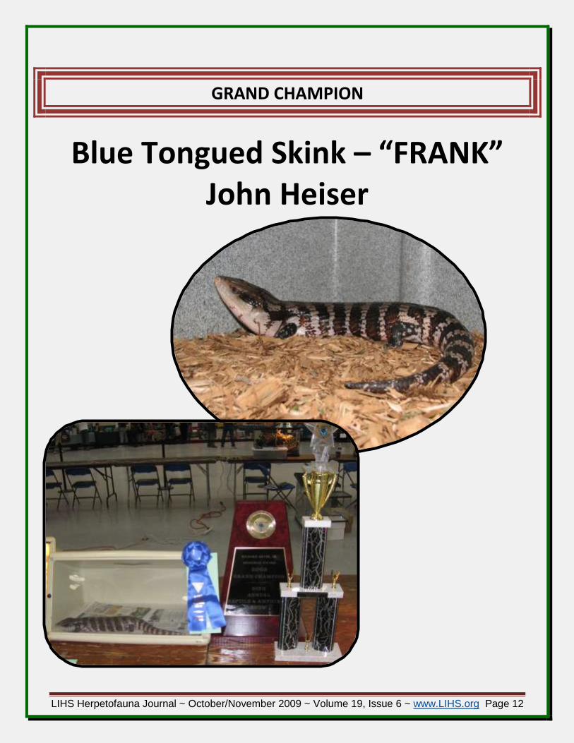
GRAND CHAMPION
Blue Tongued Skink – “FRANK” John Heiser

LIHS Herpetofauna Journal ~ October/November 2009 ~ Volume 19, Issue 6 ~ www.LIHS.org Page 13
Kids Ribbon Winners
SNAKES
1st ~ Colubrids - Corn snake– Clara Dunlop 2nd ~ Colubrids - Corn snake– Clara Dunlop 3rd ~ Colubrids - Corn snake– Clara Dunlop
1st ~ Boas & Pythons – Red-tail Boa Constrictor – Christopher Cusumano 2nd ~ Boas & Pythons – Dakota Kennedy
1st ~ Miscellaneous– Western Hognose – Clara Dunlop
LIZARDS
1st ~ Agamids– Bearded Dragon – Clara Dunlop
1st ~ Geckos – Viper Gecko – Clara Dunlop 2nd ~ Geckos – Leopard Gecko – Eric Brennan 3rd ~ Geckos – Crested Gecko – Clara Dunlop
TURTLES / TORTOISES
1st ~ Semi-Aquatic Turtle – Red-eared Slider – Eric Brennan 2nd ~ Semi-Aquatic Turtle – Red-eared Slider – Eric Brennan
AMPHIBIANS
1st ~ Frogs & Toads – Fire Bellied Toad – Clara Dunlop 2nd ~ Frogs & Toads – Fire Bellied Toad – Clara Dunlop
1st ~ Salamanders, Newts, Etc. – Fire Bellied Newt – Clara Dunlop

LIHS Herpetofauna Journal ~ October/November 2009 ~ Volume 19, Issue 6 ~ www.LIHS.org Page 14
Kids OPEN CLASS Trophy Winners
1st Place Red-tail Boa Constrictor
Christopher Cusumano ( RIGHT )
2nd Place Fire Bellied Toad
Clara Dunlop ( LEFT ) with brother Will
3rd Place Viper Gecko
Clara Dunlop ( RIGHT )

LIHS Herpetofauna Journal ~ October/November 2009 ~ Volume 19, Issue 6 ~ www.LIHS.org Page 15
Snapshots from the 20TH Annual LIHS Reptile & Amphibian Show

LIHS Herpetofauna Journal ~ October/November 2009 ~ Volume 19, Issue 6 ~ www.LIHS.org Page 16

LIHS Herpetofauna Journal ~ October/November 2009 ~ Volume 19, Issue 6 ~ www.LIHS.org Page 17

LIHS Herpetofauna Journal ~ October/November 2009 ~ Volume 19, Issue 6 ~ www.LIHS.org Page 18

LIHS Herpetofauna Journal ~ October/November 2009 ~ Volume 19, Issue 6 ~ www.LIHS.org Page 19
THANKS to our SHOW VOLUNTEERS/HELPERS
THANKS to ALL of YOU WHO HELPED with the show. We couldn’t have done it without you.
Glenn Bartley, Ed Bennett, Tony Carrozzo, Daniel Collins, Scott Collins, Jamie Dunlop, Noelle Dunlop, Clara Dunlop, Will Dunlop, Andrew Eichorn, Adam Forman, John Heiser, Rich Hume, Rich Hume, Mike Kelly, Bridgett Kelly, Ben Kelly, Tom Kennedy, Dakota Kennedy, Ann Ott, Kirk Peters, Vin Russo, Mike Russo, Christian Scheidt, Jr., and Barbara Slater.
Thanks to Rich Hume, for going above and beyond the “Call Of Duty”, as usual.
Thanks to our SHOW JUDGES, John Heiser and Tom Kennedy… Excellent job…
Special Thanks to Dr. Cindy Meyer and John Collins who helped defray the cost of the trophies.
THANK YOU to Glenn Bartley for adopting out the aquatic turtles, and his dedication to the LIHS
SPECIAL THANKS to ZooMed & The following Non-profit Educational organizations
ZooMed – THANK YOU to Andy, who drove all the way from New Hampshire and back to represent ZooMed at the Show The following organizations helped to make our show a success, and for that we THANK them.
Organization Website
Atlantis Marine World Aquarium http://www.atlantismarineworld.com/
Caleb Smith State Park http://nysparks.state.ny.us/parks/124/details.aspx
Center For Science Teaching And Learning ( CSTL )
http://www.cstl.org/
Coastal Research And Education Society Of Long Island ( CRESLI )
http://www.cresli.org/
Northeast Partners In Amphibian And Reptile Conservation ( NEPARC )
http://www.pwrc.usgs.gov/neparc/
Riverhead Foundation For Marine Research And Preservation
http://www.riverheadfoundation.org/
Theodore Roosevelt Sanctuary at Jones Beach State Park
http://www.www.nysparks.state.ny.us/environment/nature-centers/4/details.aspx

LIHS Herpetofauna Journal ~ October/November 2009 ~ Volume 19, Issue 6 ~ www.LIHS.org Page 20
PICTURED: Scott Collins, Brandon Slater ( my ne-phews ), Ben Kelly ( a neighbor ), and Ann Ott.
Field Trip to the American Museum of Natural History “Frogs: A Chorus of Colors”
( see pages 29 & 30 for information on the exhibition ) By Rich Meyer, Jr
n Saturday, November, 14th, 2009, several of us traveled to the American Museum of Natural History ( NYC ) to see one of the
current limited Special Exhibits, “Frogs: A Chorus of Colors”. This is a return engagement for this exhibit, and its current run started in May 2009, and ends on January 3rd, 2010. So, you still have time to visit the exhibition.
Would I travel just to see the exhibition? NO…. BUT, when you go to the AMNH, you are visit-ing the exhibition as a “side to the main meal”. If the AMNH is a “10”, then, “Frogs: A Chorus of Col-ors”, raises it to an “11”, so the exhibit is well worth taking in, and a must see if you visit the AMNH.
The exhibition is located on the 4th floor of the museum. There is a “timed entrance ( every 30 minutes )”, but the usher at the door let us in prior to our 12:00 pm chosen entrance time. Once inside, we stayed as long as we cared to. Each and every exhibit was extremely clean, and well decorated ( sans the hand prints from children, and “ignorant” adults * who, I found myself telling, “Don’t tap on the glass” + ). The interior of the room was dark, while the exhibits were well lit, provid-ing great contrast, and highlighting the ani-mals and their habitats. Each frog exhibit had informative and educational signs adjacent to each exhibit.
Since, I have been in the hobby for many years, I have seen many of the frogs in the exhibit, but there were equally as many that I had only seen in books, magazines or on-line. To see these animals in the stunning displays, made it a worth-while trip. Actually made me envious, wishing I could have some ( if not all ) of these displays.
O

LIHS Herpetofauna Journal ~ October/November 2009 ~ Volume 19, Issue 6 ~ www.LIHS.org Page 21
ABOVE: Central “POISON DART FROG” display.
Note mini-video camera ( in clear box )
LEFT: Brandon and Ben operate video camera
from remote station
BELOW: Brandon at one of the interactive con-
soles, “Froguts ( frog guts )”
BELOW LEFT: Smooth Sided Frogs
The animals all appeared to be in excellent health, and despite their public viewing ( and “tap-ping” on the glass ) did not appear to be stressed, nor camera shy. Some of the frogs actually seemed to pose for our pictures.
A highlight of the exhibit was the central “POISON DART FROG” display ( RIGHT ), with its concave glass walls. This exhibit could be viewed from all sides. Po-sitioned at floor level ( of the display unit ) were several min-video cameras. These cameras sent a feed to several stations around the display, where individuals could operate the cameras to search for the poison dart frogs ( though they were readily visible, as there were numerous frogs in the unit ). The cameras allowed users to get a “close-up” view of the frogs.
There were numerous frog/toad interactive exhibits
set-up around the room, further highlighting the educa-tional as well as the entertainment experience of the exhi-bition.

LIHS Herpetofauna Journal ~ October/November 2009 ~ Volume 19, Issue 6 ~ www.LIHS.org Page 22
RIGHT: Borneo Eared Frog ( Polypedates otilophus )
ABOVE: Skeletal frame of the African Goliath Frog ( Conraua goliath )
RIGHT: The Amazon Milk Frog ( Trachycephalus resinifictrix )
More Photos from FROGS: A Chorus of Colors
( mind you, my photos do not do the exhibit nor the animals’ justice )

LIHS Herpetofauna Journal ~ October/November 2009 ~ Volume 19, Issue 6 ~ www.LIHS.org Page 23
BELOW: perhaps my favorite frog of the entire exhibition, the Long-nosed Horned Frog, also known as the Malayan Horned Frog ( Megophrys nasuta ). This guy was huge, almost 5-inches.
RIGHT: Suriname Smooth-sided Toad ( Bufo guttatus )

LIHS Herpetofauna Journal ~ October/November 2009 ~ Volume 19, Issue 6 ~ www.LIHS.org Page 24
BELOW: Waxy Monkey Frogs ( Phyllomedusa sauvagii ): these guys seemed to
pose for your picture taking.

LIHS Herpetofauna Journal ~ October/November 2009 ~ Volume 19, Issue 6 ~ www.LIHS.org Page 25
ABOVE: Signage adjacent to the Waxy Monkey Frogs exhibit

LIHS Herpetofauna Journal ~ October/November 2009 ~ Volume 19, Issue 6 ~ www.LIHS.org Page 26
BELOW: Several Poison Dart Frogs from the central display exhibit

LIHS Herpetofauna Journal ~ October/November 2009 ~ Volume 19, Issue 6 ~ www.LIHS.org Page 27
LEFT / BELOW /
NEXT PAGE: Edu-
cational signs and
Interactive Dis-
plays were placed
throughout the
exhibition.

LIHS Herpetofauna Journal ~ October/November 2009 ~ Volume 19, Issue 6 ~ www.LIHS.org Page 28
All in all it was well worth the trip to see this fascinating exhibit.
A Few Notes:
We parked at the museum parking lot ( below the museum ). Parking for our stay, ran $41.00, but, considering the cost to travel by train and subway ( for 5 of us ), it was cheaper. Public transportation might be best for the remainder of this year ( holiday grid-lock, etc. ). Or go very early, and stay cool in traffic when you leave.
The museum food court closes at about 4:30/4:45 pm.. We were dying for a drink and bite to eat.. Next time, we will pack some sodas and food in the trunk..
I forgot how big the museum was. I haven’t been there since grade school.. Wear comfortable shoes, and don’t intend to do the museum in a day. Print out the on-line map, and plan ahead. My legs were aching by the end of the day. Take your time, take small breaks, and enjoy.
I wish to THANK Bernadette Dirr who arranged the passes for us to visit the museum. Much ap-preciated, and much enjoyed..

LIHS Herpetofauna Journal ~ October/November 2009 ~ Volume 19, Issue 6 ~ www.LIHS.org Page 29
Ornate Horned Frog ( Ceratophrys ornate )
Joe McDonald, Clyde Peeling's Reptiland
AMERICAN MUSEUM OF NATURAL HISTORY Presents
A captivating exhibition showcasing more than 200 live frogs from around the world
Web: http://www.amnh.org/exhibitions/frogs/?src=h_h Admission***:
Adults: $24.00
Children (2-12): $14.00
Senior/Student with ID: $18.00 Timed entrance to Frogs is available every thirty minutes from 10:30 am to 4:30 pm daily. The last timed-entry to the exhibition is at 4:30 pm. An engaging, fact-filled exhibition at the American Museum of Natural History that features more than 200 live frogs, including 9 species of colorful dart-poison frogs. On view from May 30, 2009, through January 3, 2010, the exhibition explores the colorful and diverse world of these complex amphibians by introducing visitors to their biology and evolution, their importance to ecosystems, and the threats they face in the wild. New to the exhibition this year are Amazon milk frogs. the females lay eggs in foam nests, created by beating a frothy secretion into foam with their hind legs, attached to branches overhanging the water; and long-nosed horned frogs, which are camouf-laged to mimic leaves. The centerpiece of the exhibition - a 110-cubic-foot dart-poison frog vivarium - showcases more than 70 dart-poison frogs. A soundscape featuring the calls of more than 20 species fills this area with some of the most unusual and bizarre vocalizations made 6+by these amphibians.

LIHS Herpetofauna Journal ~ October/November 2009 ~ Volume 19, Issue 6 ~ www.LIHS.org Page 30
Photos: ( Left ) Smokey Jungle Frog ( Leptodactylus pentadactylus )
Dave Northcott; ( Middle ) African Clawed Frog ( Xenopus laevis )
Courtesy of Clyde Peeling's Reptiland; ( Right ) Golden Mantella Frog (
Mantella aurantiaca ) John Netherton, Clyde Peeling's Reptiland
Frogs also features a diverse array of species from around the world, including American and African bullfrogs, Chinese gliding frogs, ornate horned frogs, African clawed frogs, and fire-bellied toads (see full list below). Species Featured in Frogs: A Chorus of Colors African bullfrogs (Pyxicephalus adspersus)
African clawed frogs (Xenopus laevis)
Amazon milk frogs (Trachycephalus resinifictrix)
American bullfrogs and tadpoles (Lathobates cates-beianus)
Borneo eared frogs (Polypedates otilophus)
Chinese gliding frogs (Rhacophorus dennysi)
Dart poison frogs ( 12 species represented )
Long-nosed horned frogs (Megophrys nasuta)
Fire-bellied toads (Bombina orientalis)
Ornate horned frogs (Ceratophrys ornata)
Smokey jungle frogs (Leptodactylus pentadactylus)
Smooth-sided toads (Rhaebo guttatus)
Vietnamese mossy frogs (Theloderma corticale)
Waxy monkey frogs (Phyllomedusa sauvagii)
Hours: The Museum is open daily, 10 am –5:45 pm ( closed Thanksgiving and Christmas )
Admission*** : Suggested general admission, which supports the Museum’s scientific and educa-tional endeavors and includes 46 Museum halls and the Rose Center for Earth and Space, is $15 ( adults ) suggested, $11 ( students/seniors ) suggested, $8.50 ( children ) suggested. All prices are sub-ject to change. The Museum offers discounted combination ticket prices that include suggested gen-eral admission plus special exhibitions, IMAX films, and Space Shows.
Information: call 212-769-5100 or visit the Museum’s website at www.amnh.org or got to http://www.amnh.org/museum/welcome/
Visitors can also explore the Frogs Shop located on the first floor of the Main Shop, just outside the exit to Frogs. The Shop features a wide selection of whimsical frog-themed merchandise.

LIHS Herpetofauna Journal ~ October/November 2009 ~ Volume 19, Issue 6 ~ www.LIHS.org Page 31
Mycoplasmosis and Upper Respiratory Tract Disease of Tortoises
Upper Respiratory Tract Disease (URTD): Anatomic Components, Definition, and Possible Causes: The major component of the upper respiratory tract of tortoises is a large nasal cavity located cranial to the eye (Jacobson et al. 1991; Figure 1; Figure 2; Figure 3). The external nares open into a short vestibule that is continuous with a recess ventrally and with olfactory chambers dorsally.
The ventral recess continues into the nasal passageway that opens into the roof of the mouth as the choanae. By defini-tion, disease is “any deviation from or interruption of the normal structure or function of any part, organ, or system (or combination thereof) of the body that is manifested by characteristic set of symptoms and signs and whose etiology,
pathology, and prognosis may be known or unknown” (Dorland’s Illustrated Medical Dic-tionary 1985). While hypovitaminosis A was considered a predisposing problem in certain tortoises, Fowler (1980) discussed the differences be-tween URTD and hypovitaminosis A. Several agents have been hypothesized to cause res-piratory tract disease in tortoises, including viruses (Jackson and Needham 1983),

LIHS Herpetofauna Journal ~ October/November 2009 ~ Volume 19, Issue 6 ~ www.LIHS.org Page 32
lasma spp. (Fowler 1980; Lawrence and Needham 1985), and Pasteurella testudinis (Snipes et al. 1980; Snipes and Biberstein 1995; Snipes et al. 1995). In Europe, Sendai virus was considered a possible cause of rhinitis in captive spur-thighed ( Testudo graeca ) and Hermann's tortoises ( T. hermanni ) ( Jackson and Needham 1983 ), but a later study ( Lawrence and Needham 1985 ) found no increase in antibody titers against Sendai virus over a 3-month period. Mycoplasmosis of Desert and Gopher Tortoises: Rhinitis and chronic URTD have been reported in a variety of species of wild and captive tortoises in the United States (Jacobson et al. 1991) and Europe (Jackson 1980). In the 1980s, major declines (33-76% over 10 yr) of desert tortoises (Gopherus agassi-zii) were documented at several sites in the western Mojave Desert of California, USA, and at one site in the eastern Mojave (Corn 1994). Tortoises with clinical signs of URTD were observed among affected populations at several sites (Knowles 1989; Berry 1990). As a result of the declines, desert tortoises in the Mojave Desert north and west of the Colorado River were declared threatened (U.S. Fish and Wild-life Service 1990). Beginning in 1989, efforts were undertaken to determine the etiology of URTD in desert tortoises (Jacobson et al, 1991). By electron microscopy, pleomorphic organisms resembling Mycoplasma sp. were seen on cell surfaces and tightly adhered to cell membranes of ill tortoises
(Figure 4; Figure 5). The Mycoplasma was isolated and de-termined to be a new species, named Mycoplasma agassi-zii (Brown et al. 1995). They are quite pleomorphic, ing from 300 to 900 nm (Figure 6). A monoclonal antibody was produced against desert tortoise light chain of IgY and IgM. An enzyme-linked immunosorbent assay (ELISA) to detect antibodies against the Mycoplasma in plasma and serum samples was developed (Schumacher et al. 1993), and experiments were undertaken to determine if M. agassizii caused URTD. The most stringent requirement for
LIHS Herpetofauna Journal ~ October/November 2009 ~ Volume 19, Issue 6 ~ www.LIHS.org Page 33
definitive proof of a causative relationship between an infectious agent and a disease is the fulfillment of the Henle-Koch-Evans postulates (Evans 1976a, 1976b). Thus, three controlled experimental infec-tion cohort studies were performed to fulfill the Henle-Koch-Evans postulates and additionally, to de-termine sensitivity, specificity, and predictive values of diagnostic assays for mycoplasmal infections of tortoises (Brown et al. 1994; Brown et al. 1999; McLaughlin 1997; and summarized in Brown et al. 2002). The disease was induced by inoculation of tortoises with pure cultures of the Mycoplasma, but not Pasteurella testudinus (Brown et al. 1994). Histologically, the lesions were consistent with those seen in the previously examined naturally infected tortoises. A polymerase chain reaction (PCR) test was developed to detect nucleotide sequences of the 16s rRNA gene of the bacteria in nasal flush and swab samples (Brown et al. 1995). Although URTD has been seen in captive gopher tortoises (ERJ, un-published data), the first documentation of the disease in wild gopher tortoises was in 1989, when an epizootic of URTD was documented on Sanibel Island, Lee County, Florida, USA (G.S. McLaughlin and M.S. Elie, unpublished data), during the course of an ecological study (McLaughlin 1990). When tested by ELISA, >80% of the adult tortoises from Sanibel Island were seropositive for antibodies against M. agassizii (Beyer 1993). Due to the 1979 listing by the then Florida Game and Fresh Water Fish Commission of the gopher tor-toise as a species of special concern, and the subsequent permitting of over 450 relocations involving more than 8000 tortoises (J.E. Berish, personal communication), particular attention was focused on the dynamics and persistence of both natural and relocated populations during the late 1980’s (Cox 1989). The observation of URTD on Sanibel Island and the association of mycoplasmosis with the de-cline of certain desert tortoise populations in the Mojave Desert elicited concerns regarding declining and isolated populations of gopher tortoises. Because an understanding of the effects of URTD on both individuals and populations is essential for proper management of remaining populations, a study was begun in 1993 at the University of Florida on the etiology, pathology, and diagnosis of URTD in gopher tortoises. The gopher tortoise is currently listed as a Threatened species (since 2007) by the Florida Fish and Wildlife Conservation Commission, and estimates of legally relocated tortoises over the last several decades exceed 70,000 (J.E. Berish, personal communication). Contrary to what was recently reported elsewhere (Sandmeier et al., 2009), only two mycoplasmas have been isolated and named from the desert tortoise. An original isolate from a desert tortoise with URTD was named Mycoplasma agassizii (Brown et al. 1994, 1995, 2001). Experimental transmission studies have confirmed this Mycoplasma as a cause of URTD in desert and gopher tortoises (Brown et al. 1994; Brown et al. 1999b). A second, genetically distinct Mycoplasma has also been isolated from desert tortoises and from gopher tortoises in northeastern Florida and has been named M. testudi-neum (Brown et al, 2002). Transmission studies with this Mycoplasma in gopher tortoises are ongoing (Wendland, personal communication). Contrary to what was presented in a review of URTD in desert tortoises (Sandmeier et al, 2009), a third Mycoplasma , M. testudinis, was not isolated from a desert tortoise, but instead was isolated from the cloaca of a Greek tortoise in England (Hill, 1985). M. testu-dinis has not been associated with URTD. Other mycoplasmas from tortoises will probably be described in the future. Hosts for Chelonian Mycoplasmosis: To date, mycoplasmas have been recovered by culture or de-tected by PCR in the following species of chelonians: desert tortoise (Gopherus agassizii), gopher tor-

LIHS Herpetofauna Journal ~ October/November 2009 ~ Volume 19, Issue 6 ~ www.LIHS.org Page 34
toise (G. polyphemus), Texas tortoise (G. berlandieri), Chaco (Argentine) tortoise (Geochelone chilen-sis), leopard tortoise (G. pardalis), Indian star tortoise (G. elegans), African spurred tortoise (G. sulca-ta), radiated tortoise (G. radiata), red footed tortoise (G. carbonaria), Travancore tortoise (Indotestudo forstenii), spider tortoise (Pyxis arachnoides), flat-tailed tortoise (P. planicauda), spurred-thighed tor-toise (Testudo graeca), marginated tortoise (T. marginata), Egyptian tortoise (T. kleinmanni), Russian tortoise (T. horsfieldii), Hermann's tortoise (T. hermanni), and eastern box turtles (Terrapene carolina carolina) in the United States (Brown et al. 2002; Wendland et al. 2006) and captive Testudo sp. in the United Kingdom (Soares et al. 2004). Distribution in the Wild: Known to occur in wild gopher tortoises in Florida, USA, and in wild desert tortoises in California, Nevada, Utah, Arizona, USA and Testudo graeca in France. Ages Affected: Primarily seen in adult tortoises in the wild, but under experimental conditions all age groups are susceptible. Clinical Signs vs. Symptoms; Clinical vs. Subclinical Infection: Symptoms are often confused with clini-cal signs in non-medical journals (see Sandmeier et al. 2009). Symptoms are sensations experienced by a human. Clinical signs are abnormalities we observe in an animal. Regarding URTD, major clinical signs include palpebral edema, conjunctivitis, and rhinitis. Clear serous to tenacious mucous may be seen bubbling from nares (Figure 7). Mycoplasmosis can occur as a subclinical in-fection (Jacobson et al. 1995). A subclinical infection is one in which lesions may be present in a tissue or organ without the an-imal manifesting any clinical signs of dis-ease. In other words, the animal, while in-fected, may appear clinically normal. Special diagnostic tests may be needed to distin-guish between a healthy animal and one with subclinical disease. We have seen an annual cycle of convalescence and recru-descence of clinical signs in some captive desert and gopher tortoises. Mycoplasmal diseases in other hosts also can exist as chronic, subclinical infections, with recurrence of clinical signs and increases in transmission potential when the host is stressed (Simecka et al. 1992). Thus, this pat-tern is not unique to mycoplasmosis in tortoises. Mycoplasmosis and Modern Concepts of Infectious Disease: Mycoplasmosis refers to any disease caused by infection with Mycoplasma spp. Previous concepts of microbial pathogenicity do not take in-to account that both the microorganism and the host contribute to microbial pathogenesis. Recently, a damage-response framework of microbial pathogenesis has been proposed as a new theoretical ap-proach to understand microbial pathogenesis (Casadevall and Pirofski, 2003). The three tenets of this framework are: “1. Microbial pathogenesis is an outcome of an interaction between a host and a mi-

LIHS Herpetofauna Journal ~ October/November 2009 ~ Volume 19, Issue 6 ~ www.LIHS.org Page 35
croorganism. 2. The host-relevant outcome of the host-microorganism interaction is determined by the amount of damage to the host; and 3) Host damage can result from microbial factors and/or the host response”. In chelonian mycoplasmosis, the host response to pathogenic Mycoplasma accounts for much of the pathological change seen in the nasal cavity of the host. Damage is enhanced by strong immune responses of the host. Also, as discussed below, predisposing and contributing factors are probably involved in epizootics of mycoplasmosis in tortoises and other animals. Predisposing and Contributing Factors: Although data are lacking, we suspect that various extrinsic and predisposing factors are involved in outbreaks of URTD (Jacobson et al, 1991; Brown et al, 2002; Sandmeier et al, 2009). Clinically silent infections may become exacerbated by environmental stress (Brown et al, 2002). Environmental pertuberations may influence the periodicity of outbreaks in popu-lations of tortoises known to have mycoplasmosis. Annual fluctuations in temperature, rainfall, and fo-rage availability may be sufficient to cause detectable outbreaks in an infected population. Increased morbidity and mortality may occur in times of unusually severe environmental stress, such as pro-longed drought, hurricanes, excessive rainfall with flooding of burrows, or very cold winters. The data available for desert tortoises indicate that drought is a natural part of the desert tortoise’s environ-ment, but when combined with disease or habitat loss, may contribute to additional disease problems and mortality (Peterson 1996). Clinical signs of URTD, lower leukocyte counts, positive nasal cultures of M. agassizii, and mild to moderate azotemia were more commonly seen in 1993–94, a year of below-normal annual and winter precipitation (Christopher et al 2003). Heteropenia has been associated with drought and starvation (Berry et al. 2002). Tortoises entering hibernation in a drought year may be physiologically compromised, because clinical signs of URTD and heteropenia were noted at the time of emergence from hibernation in 1990–91 and 1994–95, years following a period of drought (Christo-pher et al, 2003). Most dehydrated tortoises and most deaths at the Desert Tortoise Natural Area and Ivanpah also occurred in 1990–91 and 1994–95, years following dry winters. Human impacts on tor-toises and their habitat, whether through disruption of normal behavior patterns, degradation of habi-tat through agriculture, silviculture, mining or development operations, or pollution, may cause suffi-cient physiological stress to trigger proliferation of the Mycoplasma and recurrence of signs. Capturing and transporting of tortoises during relocation, restocking and repatriation efforts also may be signifi-cant sources of stress that result in overt disease. The release of ill captive tortoises may be a signifi-cant factor accounting for the presence of URTD in certain populations. For instance, the release onto Sanibel Island of gopher tortoises originating in northern Florida and southern Georgia following tor-toise races has been documented. Many of these tortoises were kept under very poor husbandry con-ditions that would have allowed transmission of various pathogens (Dietlein and Smith, 1979). A Bro-chure with photographs with clinical signs has been developed for the gopher tortoise and a link to this document will be available in the near future. Use Acrobat Reader to view this brochure. Pathologic Diagnosis: On a light microscopic level, the ventral recess consists of a mucous and ciliated epithelial mucosa (Figure 8), while the olfactory chambers consist of a multilayered olfactory epithe-lium (Figure 9). Disease is a change in structure and/or function of an organ and in tortoises with URTD, there is focal to diffuse, minimal to severe, inflammatory changes in the nasal cavity (Figure 10; Figure 11). Basal cell hyperplasia, infiltrates of heterophils and histiocytes, and lymphoid hyperplasia in the submucosa all may be seen. By electron microscopy, mycoplasmas can be seen closely associated with

LIHS Herpetofauna Journal ~ October/November 2009 ~ Volume 19, Issue 6 ~ www.LIHS.org Page 36
the nasal cavity mucosa. Mycoplasmas are more readily seen in the ventral recess compared to other areas of the upper respiratory tract. The lower respiratory tract consists of the glottis, trachea, trachea, bronchi, and paired lungs. Rarely is the lower respiratory tract affected in tortoises with URTD.
ELISA Diagnostics: An ELISA was developed at the University of Florida to measure plasma/serum anti-bodies that are specific for Mycoplasma (Schumacher et al, 1993). Monoclonal antibodies (MAbs) spe-cific for desert tortoise immunoglobulins were developed to ensure the long-term availability of highly specific secondary antibodies for testing of plasma from desert tortoise populations by ELISA. Figure 1 in Schumacher et al (1993) shows the Western blot reactivity on desert tortoise plasma of the IgY(L)-specific MAb HL673 and the IgY(H)-specific MAbHL665. Lanes 2, 4, and 6 were negative controls in which PBS/A was substituted for primary antibody. The blot (lane 7) shows that MAb HL673 reacted with a single band at approximately 27,000 Da corresponding to desert tortoise IgY(L). Mab HL665

LIHS Herpetofauna Journal ~ October/November 2009 ~ Volume 19, Issue 6 ~ www.LIHS.org Page 37
reacted with a single band at approximately 65,000 Da (lane 5) corresponding to desert tortoise IgY(H) (1). Polyclonal anti-Testudo horsfieldii IgY(H) and polyclonal anti-T. horsfieldii IgM(H) were tested on the same immunoblot for their cross-reactivities with desert tortoise plasma to determine whether these antibodies, although available in very limited supply, could serve as polyclonal reference reagents for the newly developed MAbs. Polyclonal anti-T. horsfieldii IgM(H) (lane 1) reacted with one major dark band at approximately 74,000 Da which cor-responds to desert tortoise IgM(H). Polyclonal anti-T. horsfieldii IgY(H) (lane 3) reacted with a single band of approximately 65,000 Da which corres-ponds to desert tortoise IgY(H) (1). MAb HL673 also reacted with desert tortoise IgM(L), as determined by ELISA on IgM-rich fractions of desert tortoise
immunoglobulins (Schumacher et al, 1993). The monoclonal based ELISA was validated using experi-mental transmission studies (see below) with Mycoplasma agassizii in desert tortoises (Gopherus agas-sizii, Brown et al, 1994) and gopher tortoises (G. polyphemus; Brown et al, 1999b). Gold standards for confirmation of mycoplasmosis used in these studies were clinical signs, Mycoplasma culture, Mycop-lasma PCR, and histopathology (Brown et al, 2002). In a recent publication (Sandmeier et al, 2009) the authors claim that past infection studies have selected tortoises with the lowest ‘‘background” levels of antibodies as the ‘‘best” negative control specimens (Schumacher et al., 1993; Brown et al., 1994), which may have introduced bias into the research design”. This assessment is incorrect since in Schumacher et al (1993), pre-challenge plasma samples were used to verify post-challenge seroconversion, and in Brown et al (1994), the control group ( see Figure 3 in Brown et al, 1994 ) did not have the lowest “background” le-vels.) Since its first development at the University of Florida, the ELISA has been refined, drawing on the accumulation of an immense database of ELISA results from more than 20,000 plasma/serum samples (Wendland et al, 2007). Results of the original ELISA were reported as an enzyme immunoassay ratio (EIA) ratio. An EIA ratio >3 was considered to be a positive result. The ELISA was refined to include more strin-gent quality control measures and has been converted to a clinically more meaningful titer reporting system, consistent with other diagnostic serologic tests. The ELISA results for 5,954 desert and gopher tortoises were plotted, and a subset of these serum samples (n
LIHS Herpetofauna Journal ~ October/November 2009 ~ Volume 19, Issue 6 ~ www.LIHS.org Page 38
= 90) was used to determine end-point titers, to establish an optimum serum dilution for analyzing samples, and to construct a standard curve. The relationship between titer and A405was validated us-ing 77 serum samples from known positive (n = 48) and negative (n =29) control tortoises from prior transmission studies. The Youden index, J, and the optimal cut point, c, were estimated using ELISA re-sults from the 77 control sera. Based on this evaluation, the refinement has substantially improved the performance of the assay (sensitivity of 0.98, specificity of 0.99, and J of 0.98), thus providing a clinical-ly more reliable diagnostic test for this important infection of tortoises. PCR Diagnostics: Limitations associated with detection of tortoise mycoplasmas and a theoretical low limit of detection by culture necessitated the development of an alternative diagnostic test for the presence of Mycoplasma in nasal lavage samples from tortoises. The principle of the PCR test is to syn-thesize an easily detectable number of DNA copies of a segment of the mycoplasmal chromosome by using Mycoplasma-specific primers. The segment of the M. agassizii chromosome selected for analysis, the 16S ribosomal RNA gene, contains conserved (genus-specific) DNA sequences and intervening vari-able (species-specific) DNA sequences. Thus a primer pair consisting of 1 genus-specific primer and 1 species-specific primer can very reliably distinguish among organisms. Alternatively, the presence of other Mycoplasma species can be determined by using generic genus-specific PCR primers. However, species identification for such samples usually requires further nucleotide sequence analyses. Nasal la-vage samples or cultures can be analyzed for presence of Mycoplasma by PCR. The sensitivity of the as-say can be increased by culturing nasal flush samples before analysis. Contamination by other sources of DNA does not interfere with PCR. However, blood, calcium alginate swabs and other unidentified agents can inhibit the reaction. The advantages of diagnosing infections by PCR are noninvasive sam-pling, direct proof of infection at the time the sample was taken, the reaction is not inhibited by sam-ple contamination with other microbes, the potential short sample turnaround time if the sample is not cultured before analysis, accurate identification of M. agassizii and other species of Mycoplasma, and a theoretical low limit of detection. Disadvantages of PCR include the need for specialized labora-tory equipment and sophisticated and meticulous technique, the high cost of special reagents, the po-tential for false positive results caused by cross-contamination, uninformative samples caused by inhi-bitory substances in the reaction, and consumption of a portion of the sample during the reaction. Additional Diagnostic Tests: Recently a polyclonal ELISA was developed for determining exposure of desert tortoise to Mycoplasma agassizii (Hunter et al, 2008). However, in this report, no transmission studies were performed to demonstrate that seroconversion could be detected and no indices of per-formance such as sensitivity and specificity were provided. Natural Antibody and Western Blots: In a recent publication (Hunter et al, 2008), natural antibody was reported to occur in desert tortoises. The desert tortoises used in this study originated from a universi-ty colony that purportedly was free of Mycoplasma infection. However, little information was pre-sented on the history of this colony, specifically, the Mycoplasma serological status of this colony and the culture and PCR status of nasal lavage specimens from this colony. It is unclear if necropsies were ever performed to assess the histological status of the upper respiratory tract to confirm that the tor-toises in the colony were free of lesions in the nasal cavities. While natural antibodies probably exist in chelonians, the well-established polyreactive nature of such antibodies for multiple microbial epitopes seriously confounds the interpretation of the Western Blot data described in Hunter et al (2008). Hunt-

LIHS Herpetofauna Journal ~ October/November 2009 ~ Volume 19, Issue 6 ~ www.LIHS.org Page 39
er et al (2008 did not demonstrate that natural antibodies were specific for M. agassizii. It is impossible to distinguish IgM natural antibody in an individual (theoretically developed in the absence of specific extrinsic antigen exposure) from IgM antibodies produced very early in the immune response following specific antigen exposure. We have monitored the IgM antibody response to M .agassizii using IgM specific monoclonal antibodies. Mycoplasma infection of tortoises elicits an IgM antibody response ap-proximately 4 weeks after exposure and shifts to a long-lasting, predominantly IgY-like antigen-specific antibody response approximately 10 weeks after exposure. Natural antibody is polyreactive, produced by B-1 cells and reacts with many epitopes on multiple, mostly unrelated antigens that are often found on multiple potential pathogenic microbes. Thus, such “natural” antibodies in desert tortoises are not M. agassizii-specific. In Sandmeier et al (2009), the authors state: “a Western blot may be used as a confirmatory test to an ELISA”. The authors appear to be unaware of the limitations of Western blots. Extensive literature supports the need for multiple Mycoplasma strains as antigens in Western blot analysis of naturally infected animals. Most mycoplasmas exhibit extensive intraspecies genotypic and phenotypic variability that can be manifested as antigenic variation in the contexts of immune recogni-tion. Further, this occurs even in individual animals infected with a defined isolate. This heterogeneity can confound analysis of mycoplasmal immunogen recognition, especially in Western blots, when only a single isolate is used as antigen (Kittelberger et al, 2006). Hunter et al (2008) hypothesized that dif-ferences observed in Western blot patterns were a result of natural antibodies. Unfortunately, lack of experience with mycoplasmas and unfamiliarity with the published literature on mycoplasmal diagnos-tics has likely led to well-intentioned, but erroneous conclusions. The data presented in Hunter et al. (2008) does not show that any of the bands revealed on Western Blots are indeed due to M. agassizii specific antibodies. The reliance of Hunter et al (2008) on a single strain as antigen in the Western blot is likely to result in false negatives. Importantly, detection of specific antibodies by ELISA is strain-independent, whereas other assays such as Western blot, metabolic inhibition, and complement fixa-tion assays are documented to be strain-dependent. These differences are explained by the location of the antigens (surface exposed, membrane or cytosolic), degree of surface variation, biofunctional as-says, and in vivo expression of antigens. Individual variation in the immune response among animals, even to the same strain of M. agassizii, is common in Western blots. An individual tortoise may pro-duce antibody that recognizes all or only a few of the proteins of a strain, and yet another animal may have a very different profile to the same antigen preparation. This observation also is consistent with mycoplasmal infections in other species. Monoclonal vs. Polyclonal Antibodies: The decision regarding whether to use polyclonal antibodies (PAb) or a monoclonal antibody (MAb) depends on a number of factors, the most important of which are its intended use and whether the antibody is readily available from commercial suppliers or re-searchers (Lipman et al, 2005). Hunter et al. (2008) indicated that “PAbs can be generated much more rapidly, at less expense, and with less technical skill than is required to produce MAbs. One can rea-sonably expect to obtain PAbs within several months of initiating immunizations, whereas the genera-tion of hybridomas and subsequent production of MAbs is more costly and can take up to a year or longer, to produce”. However, the principal advantages of MAbs are their homogeneity and consisten-cy and, once the desired hybridoma has been generated, MAbs can be produced as a constant and re-newable resource for the ELISA. In contrast, PAbs generated to the same antigen using multiple ani-mals will differ among immunized animals, and their avidity may change as they are harvested over time. The quantity of PAbs obtained is limited by the size of the animal and its lifespan. Importantly,

LIHS Herpetofauna Journal ~ October/November 2009 ~ Volume 19, Issue 6 ~ www.LIHS.org Page 40
the concentration and levels of specific antibody are higher in MAbs. The concentration of specific an-tibody in polyclonal sera is typically 50 to 200 µg/mL, and the range of total Ig concentration in sera is between 5 and 20 mg/mL. In comparison, MAbs generated as ascites or in specialized cell culture ves-sels are frequently 10-fold higher in concentration and of much higher purity. The HL-673 monoclonal (Mab) antibody-based ELISA, developed at the University of Florida, has been validated over the past 17 years using rigorous statistical methods and comparisons with highly purified polyclonal anti-tortoise antibodies as well as selective diagnostic necropsies to confirm the absence of histopathologi-cal lesions, all for the development of reliable negative and positive control reagents (Shumacher et al., 1993, Brown et al., 1994, Brown et al., 1999, Wendland et al., 2007). A principal advantage of using this highly specific and potent Mab is its unlimited supply that has insured high year-to-year assay reliabili-ty. This is not the case for polyclonal antibodies. The HL673 monoclonal antibody has never failed to bind to the light chains of any desert or gopher tortoise immunoglobulins during more than 10,000 as-says and titrations on control wells coated with dilutions of individual tortoise’s plasma. In addition we have shown (Wendland, et al, submitted for publication) that Mab HL673 reacts strongly with the im-munoglobulins of 28 other Chelonian species, including Chelonia mydas and Caretta caretta. This com-pares exceptionally well with our most highly reactive affinity purified polyclonal which reacted with the immunoglobulins of all 30 tested Chelonian species (Wendland, et al, submitted for publication). In contrast, Hunter et al (2008) developed a polyclonal antibody-based ELISA test , but did not disclose any details concerning the methods used to validate their “new ELISA” (Sandmeir et al. 2009). As a re-sult it is impossible to compare results in these recent reports with the well-established and validated ELISA developed by the University of Florida. Pathogen Transmission: The results of the experimental challenge studies support the hypothesis that M. agassizii was horizontally transmitted between adult tortoises, and between adult and hatchling tortoises(Brown et al. 1994; Brown et al, 1999b; Brown et al, 2002). Preliminary observations indicate that seronegative hatchlings are at least as susceptible to infection as adults, and that the disease progresses more rapidly, with high morbidity in the first 6 months post infection (McLaughlin, 1997). Direct contact is the most likely route of transmission between tortoises and can easily occur during combat or courtship. If aerosol transmission occurs, it is probably over short distances (<0.5 m). Senti-nel and control animals housed in pen sections adjacent to ill tortoises did not become ill or serocon-vert, which indicated that aerosolized bacteria did not travel even relatively short distances over low (0.7 m) barriers. Based on the environmental transmission study, fomites are unlikely to play a major role in Mycoplasma transmission. The hypothesis that transmission was more likely when the infected tortoise was symptomatic was supported. However, tortoises without clinical signs may be infected and able to transmit the pathogen under appropriate conditions (Jacobson et al, 1995). After rains-torms, it is not uncommon to find two or more tortoises drinking from the same puddle. When tortois-es drink, they often get water in their noses, which they then blow or "sneeze" out. An asymptomatic tortoise harboring bacteria may shed enough in this manner to infect a nearby conspecific via the aerosol route. “The current evidence supports horizontal transmission of Mycoplasma in desert and gopher tortoises (Brown et al. 2002)., At this time there is no evidence of vertical transmission (McLaughlin, 1997). Eggs from gopher tortoises with mycoplasmosis were collected and no maternal cloacal swab samples, or egg yolk, albumin, membrane, or chorioallantoic/amniotic fluid samples were positive by culture or PCR for Mycoplasma. None of 200 hatchling gopher tortoises hatched from eggs collected from females with mycoplasmosis showed evidence of infection with M. agassizii. Therefore,

LIHS Herpetofauna Journal ~ October/November 2009 ~ Volume 19, Issue 6 ~ www.LIHS.org Page 41
tortoises with clinical signs of URTD can produce M. agassizii-free eggs. However, the sample size in this study was small and vertical transmission cannot be ruled out (Brown et al, 2002). Further, mater-nal antibodies were transferred via the egg yolk, and were detected by Effects of repeated exposure of clinically healthy seropositive adult gopher tortoises in experimental studies indicated that the clinical response of these tortoises was more rapid and severe than naive tortoises, suggesting no protection was conferred by previous exposure to the organism (McLaughlin, 1997). This is consistent with some other mycoplasmal diseases in which the immune response confers limited or no protection (Brown et al, 2002), or contributes to disease pathogenesis, such as arthritis in fowl caused by M. synoviae, con-junctivitis in cattle caused by M. bovoculi, and pneumonia in humans caused by M. pneumoniae. In ad-dition, some mycoplasmal surface proteins share sequence and structural homologies with vertebrate proteins; these may play a role in eliciting autoimmune responses. Repeated exposure to mycoplasmal proteins that resemble a host's proteins may sensitize the host and induce an autoimmune response, leading to chronic manifestations of disease even if the primary etiologic agent is cleared. Consequences of Infection: The effects of mycoplasmosis on the long-term health and viability of af-fected chelonian populations is poorly understood. Based on data collected at Sanibel Island, Florida (McLaughlin 1990), at least 30% and possibly up to 50% of the adults on one site died with signs of URTD. Given the low recruitment rate of gopher tortoise populations (Cox 1989), it is unknown if the population on that site will recover to its previous levels without intensive management. While M. agassizii was associated with declines in certain population of desert tortoises in the Mojave Desert, the exact causes of death of those tortoises were not determined. However, changes in the hormonal profiles of some infected desert tortoises (Rostal et al, 1996) could lead to altered foraging and repro-ductive behavior and decreased reproductive potential. In nutritional studies with captive desert tor-toises, deaths of juvenile tortoises were attributed to URTD (Oftedal et al, 1996). The chronic inflam-matory changes seen in the nasal cavity tissues of affected tortoises could have adverse effects on fo-raging and reproductive behavior due to disruption of olfactory function. Both direct and indirect pa-thogenic mechanisms need to be studied to better predict the effects of URTD on tortoise populations. Maternal Antibody: Desert tortoise females transfer specific IgG and IgM antibodies to their offspring that are still detectable after 1 year (Schumacher et al, 1999). The IgG and IgM antibodies were trans-ferred. but M agassizii-specific antibodies were of the IgG class. Yolk and hatchling plasma had signifi-cantly lower amounts of specific antibodies than did plasma from adult females. Hatchlings were not infected with mycoplasmas. Offspring of sick females had significantly higher specific antibody titers than did offspring of healthy females. Titers were still significantly different in 1-year-old hatchlings. Since maternal antibody in neonates potentially confounds interpreting the ELISA test in tortoises of this age group, infection with M agassizii may be misdiagnosed in hatchlings with persistent maternal antibodies. Control and Treatment: Ill tortoises need to be isolated from healthy tortoises. Do not release ill tor-toises back to the wild. Healthy tortoises need to be thoroughly evaluated if they are to be released to the wild. In a seroprevalence study of captive desert tortoises from the greater Barstow area, Mojave Desert, USA, of 179 tortoises sampled, anti-mycoplasmal antibodies were present in 82.7 % of the tor-toises. What was also interesting was the presence of anti-herpesvirus antibodies in 26.6 % of the sampled tortoises. Herpesvirus is known to infect the desert tortoise (Johnson et al, 2005) and the

LIHS Herpetofauna Journal ~ October/November 2009 ~ Volume 19, Issue 6 ~ www.LIHS.org Page 42
source of this virus is unknown. Herpesvirus infections have been reported in a variety of exotic tor-toises (Jacobson et al., 1985; Heldstab and Bestetti, 1989; Muro et al., 1998; Drury et al., 1998) and 15.5 % of the owners of desert tortoises in the greater Barstow area also had exotic chelonians in their collection. Thus desert tortoise potentially can become infected with an exotic herpesvirus and escape or release of these animals into wild populations may result in a new emerging disease. In affected tor-toises, mycoplasmosis is a typically a chronic upper respiratory disease. While various antibiotics have been utilized for treatment of mycoplasmosis, it is unknown if the organism will be completely elimi-nated. Clinical signs of illness may abate following treatment, but relapses commonly occur. Consensus Statement: This consensus statement reviews the pertinent accumulated information on URTD in gopher tortoises as studied by the University of Florida group and attempts to provide a scien-tifically sound perspective on the known effects of the disease. It is certain that:
Mycoplasma agassizii (strains PS6 and 723) is a cause of URTD in desert and gopher tortoises. The pathology of mycoplasmosis involves hyperplastic and dysplastic lesions in the upper respi-ratory tract and eyes. Clinical signs of URTD vary in onset, duration, and severity Mycoplasmosis is chronic and may be clinically silent (subclinical) in adult tortoises. Infection with Mycoplasma agassizii elicits specific antibody responses that can be detected by ELISA.
The antibody responses to Mycoplasma agassizii are reliably detectable by ELISA beginning 8 weeks after experimental infection. Under experimental conditions, gopher tortoises become ill quicker after repeated exposure to Mycoplasma agassizii. Colonization of the upper respiratory tract with Mycoplasma agassizii may be detected by culture and PCR, but assay sensitivity is not as high as the ELISA.
Mycoplasmosis is a horizontally transmissible disease. It is probable, but not clearly established, that:
Pathogenic and nonpathogenic tortoise mycoplasmas exist. There is variation among strains of Mycoplasma agassizii in their ability to cause URTD. Other species of Mycoplasma (such as M. testudineum) also may cause URTD.
Mycoplasma can be transmitted by some forms of indirect contact. Areas of uncertainty:
If vertical egg transmission of M. agassizii occurs.
The effect of mycoplasmosis on survival and reproduction of individual tortoises.
The effect of mycoplasmosis on desert and gopher tortoise population dynamics and viability.
The relationship among infection rates, transmission rates, population size, and clinical disease expression.
The probability of transmission via burrows, fomites or vectors.

LIHS Herpetofauna Journal ~ October/November 2009 ~ Volume 19, Issue 6 ~ www.LIHS.org Page 43
If tortoises can clear M. agassizii and develop protective immunity.
Potential systemic effects of mycoplasmosis and the association between M. agassizii infection and hemosiderosis in the liver.
M. agassizii ELISA and euthanasia of tortoises. ELISA testing has been used as a convenient method for determining the ultimate disposition of gopher and desert tortoises in certain parts of their range. It is important to note that the Jacobson et al. (1995) paper did not recommend indiscriminate eutha-nasia of M. agassizii ELISA-positive desert tortoises that were otherwise clinically healthy. The policy of euthanasia of such tortoises can be found in the 1996 “Protocol for the Prevention of the Transmis-sion of Disease among Desert Tortoises (Gopherus agassizii) at the Desert Tortoise Conservation Center and Transfer and Holding Facility”, which was developed by Southern Nevada Environmental Incorporated and the Bureau of Land Management. This policy was only adopted in Nevada and not in any other state where desert tortoises are found. In Brown et al. (2002) the following is stated: “There are inadequate scientific data to provide definitive guidelines for the disposition of seropositive tor-toises.” This paper was partially responsible for termination of the ELISA-based euthanasia program in Nevada in June 2007 (Roy Averill-Murray, USFWS, personal communication). Despite continued re-search over the past two decades, that statement still holds true. Euthanasia of seropositive tortoises eliminates animals that might otherwise have provided valuable reproductive and genetic contribu-tions to wild populations. However, relocation of seropositive tortoises may result in spread of mycop-lasmosis to susceptible animals and could have detrimental impacts on recipient populations. Thus, when making management decisions on the basis of the M. agassizii ELISA, it is critical to establish clear goals for the tortoise population of interest, to determine a necessary sample size to meet the goals for detection, and finally, upon receipt of results, to consider the predictive values of the test be-fore implementing any policy. The establishment of clear goals will greatly facilitate the decision-making process. It is important to stress that the M. agassizii ELISA alone should not be used as the sole means for eva-luating the health of an individual animal; however, it is one of a very small battery of non-subjective tools available that can be used as part of a comprehensive health assessment. Other causes of URTD: In 1985 a herpesvirus-like infection was identified in a die-off of Argentine tor-toises (Geochelone chilensis) imported into south Florida that had rhinitis, and anorexia (Jacobson et al, 1985). Herpesvirus infections were first described in tortoises in the early 1980s (Harper et al., 1982). They have since been reported in many species of tortoises, including the desert tortoise, with varying clinical signs and degrees of severity (Heldstab and Bestetti, 1989; Drury et al., 1998; Muro et al., 1998). Infection is most often associated with the oral cavity and respiratory tract, with necrotizing stomatitis, glossitis, tracheitis, pharyngitis, and rhinitis having been repeatedly described (Jacobson et al., 1985; Muro et al., 1998; Drury et al., 1998; Johnson et al, 2005). Encephalitis (Heldstab and Bestet-ti, 1989) and hepatitis (Herva´s et al., 2002) have also been observed. An iridovirus (Ranavirus) was identified in a gopher tortoise in Florida with clinical signs of URTD (Westhouse et al, 1996), and then several years later five instances of Ranavirus infection were docu-mented in chelonians between 2003 and 2005 in Georgia, Florida, New York, and Pennsylvania, USA (Johnson et al, 2008). Affected species included captive Burmese star tortoises (Geochelone platynota),

LIHS Herpetofauna Journal ~ October/November 2009 ~ Volume 19, Issue 6 ~ www.LIHS.org Page 44
a free-ranging gopher tortoise (Gopherus polyphemus), free-ranging eastern box turtles (Terrapene carolina carolina), and a Florida box turtle (Terrapene carolina bauri). Results suggested that certain amphibians and chelonians are infected with a similar virus and that different viruses exist among dif-ferent chelonians. Amphibians may serve as a reservoir host for susceptible chelonians. This paper also demonstrated that significant disease associated with Ranavirus infections are likely more widespread in chelonians than previously suspected. Collaborators: The work on Mycoplasmosis of tortoises has been a multidisciplinary effort at the Uni-versity of Florida and elsewhere involving faculty, graduate students, research scientists and laboratory technicians. The following present and former UF personnel were involved in this project:
Elliott R. Jacobson, D.V.M, PhD, Professor Mary B. Brown, Ph.D., Professor Paul A. Klein, Ph.D., Professor Lori Wendland, D.V.M., Ph.D., Research Assistant Professor Daniel R. Brown Ph.D., Associate Professor Isabella M. Schumacher, D.V.M., (Research scientist, BEECS Program) Bruce L. Homer, D.V.M, PhD, D.A.C.Z.M. Catherine McKenna (Biological scientist) Grace S. McLaughlin, Ph.D. (Research coordinator) Barbara C. Crenshaw (Laboratory manager) Linda Green, Scientific Director, UF Hybridoma Core Laboratory Diane Duke Laboratory technician, UF Hybridoma Core Laboratory John Hutchison (Technical assistant) Alyssa Whitemarsh (Technical assistant) Michael Lao (Technical assistant) David Bunger (Technical assistant) Sylvia J. Tucker (Senior laboratory technician) Noelle T. Chmielewski, D.V.M. (Ophthalmology resident) Nicole Gottdenker, D.V.M. (Graduate assistant)
The following non-UFL scientists and faculty were also involved:
Kristin H. Berry, Ph.D., USGS, Riverside, CA. Mary M. Christopher, D.V.M., Ph.D., University of California at Davis Joan Berish, MS, FWS Gainesville, Florida
Complete Reference List Contact: Dr. Mary Brown Department of Pathobiology Box 110926 College of Veterinary Medicine University of Florida Gainesville, FL 32610

LIHS Herpetofauna Journal ~ October/November 2009 ~ Volume 19, Issue 6 ~ www.LIHS.org Page 45
PHOTO 2
PHOTO 1
PHOTO 1 – SCOTT COLLINS – LI PET EXPO- 1990 ( He’s the one on the RIGHT )
PHOTO 2 – Forrest Bennett ( Ed’s grandson ) in a TURTLE COSTUME for Halloween 2009
E-Mail: [email protected] For General Information Contact: Dr. Elliott Jacobson Box 100126 Department of Small Animal Clinical Sciences College of Veterinary Medicine University of Florida Gainesville, FL 32610-0126 E-Mail: [email protected]
Reprinted from the University of Florida, College of Veterinarian Medical webpage http://www.vetmed.ufl.edu/college/departments/sacs/research/MycoplasmosisofTortoises.html
Updated November 4, 2009 Downloaded and Accessed November 9, 2009
Can you guess who these Current / Future LIHS Members are ??? ( ANSWER below )

LIHS Herpetofauna Journal ~ October/November 2009 ~ Volume 19, Issue 6 ~ www.LIHS.org Page 46
Male South African dwarf chameleons signal their dominance with conspi-cuous colors, emphasized during dis-plays. The photo shows a Knysna dwarf chameleon ( Bradypodion da-maranum ) displaying a combination of visible greens and ultraviolet greens, which appear similar to the human eye but very different to cha-meleons. ( Credit: Photograph by A. Moussalli, Museum Victoria, Austral-ia, and D. Stuart-Fox, University of Melbourne, Australia )
Conspicuous Social Signaling Drives Evolution of Chameleon Color Change
What drove the evolution of color change in chameleons? Chameleons can use color change to camouflage and to sig-nal to other chameleons, but a new paper shows that the need to rapidly signal to other chameleons, and not the need to camouflage from predators, has driven the evolution of this characteristic trait.
The research, conducted by Devi Stuart-Fox and Ad-nan Moussalli, shows that the dramatic color changes of chameleons are tailored to aggressively display to conspecif-ic competitors and to seduce potential mates. Because these signals are quick--chameleons can change color in a matter of milliseconds--the animal can afford to make it obvious, as the risk that a predator will notice is limited.
This finding means that the evolution of color change serves to make chameleons more noticeable, the complete opposite of the camouflage hypothesis. The amount of color change possible varies between species, and the authors cle-verly capitalize on this in their experiments.
Stuart-Fox and Moussalli measured color change by setting up chameleon "duels": sitting two males on a branch opposite each other and measuring the color variation.
By comparing species that can change color dramati-cally to those that only change slightly, and considering the evolutionary interrelationships of the species, the research-ers showed that dramatic color change is consistently asso-ciated with the use of color change as a social signal to other chameleons. The degree of change is not predicted by the amount of color variation in the chameleons' habitat, as would be expected if chameleons had evolved such remarkable color changing abilities in order to ca-mouflage. Citation: Stuart-Fox D, Moussalli A (2008) Selection for social signaling drives the evolution of cha-meleon colour change. PLoS Biol 6(1): e25. doi:10.1371/journal.pbio. 0060025 Adapted from materials provided by PLoS Biology, via EurekAlert!, a service of AAAS
Reprinted from ScienceDaily http://www.sciencedaily.com/releases/2008/01/080129125524.htm
Posted February3, 2008, Accessed July 28, 2009 Copyright © 1995-2009 ScienceDaily LLC

LIHS Herpetofauna Journal ~ October/November 2009 ~ Volume 19, Issue 6 ~ www.LIHS.org Page 47
Catfish on the hunt follow invisible tracks that lead directly to their prey. The organ that makes this possible is the lateral-line system, which registers changes in currents and even smaller disturbances. ( Credit: iStockphoto /Dmitry Demidovich )
Scientists Explore The Fundamental Basis Of The Lateral-Line Sensory System Basic discoveries suggest robotics applications
Fish and some amphibians possess a unique sensory capability in the so-called lateral-line sys-
tem. It allows them, in effect, to "touch" objects in their surroundings without direct physical contact or to "see" in the dark. Professor Leo van Hermmen and his team in the physics department of the Technische Universitaet Muenchen are exploring the fundamental basis for this sensory system. What they discover might one day, through biomimetic engineering, better equip robots to orient them-
selves in their environments. With our senses we take in only a small fraction of
the information that surrounds us. Infrared light, elec-tromagnetic waves, and ultrasound are just a few exam-ples of the external influences that we humans can grasp only with the help of technological measuring devices - whereas some other animals use special sense organs, their own biological equipment, for the purpose. One such system found in fish and some amphibians is under investigation by the research team of Professor Leo van Hemmen, chair of theoretical biophysics at TUM, the Technische Universitaet Muenchen.
Even in murky waters hardly penetrated by light, pike and pickerel can feel out their prey before making contact. The blind Mexican cave fish can perceive struc-tures in its surroundings and can effortlessly avoid ob-stacles. Catfish on the hunt follow invisible tracks that lead directly to their prey. The organ that makes this
possible is the lateral-line system, which registers changes in currents and even smaller disturbances, providing backup support for the sense of sight particularly in dark or muddy waters.
This remote sensing system, at first glance mysterious, rests on measurement of the pressure distribution and velocity field in the surrounding water. The lateral-line organs responsible for this are aligned along the left and right sides of the fish's body and also surround the eyes and mouth. They consist of gelatinous, flexible, flag-like units about a tenth of a millimeter long. These so-called neuro-masts - which sit either directly on the animal's skin or just underneath, in channels that water can permeate through pores - are sensitive to the slightest motion of the water. Coupled to them are hair cells similar to the acoustic pressure sensors in the human inner ear. Nerves deliver signals from the hair cells for processing in the brain, which localizes and identifies possible sources of the changes de-tected in the water's motion.
These changes can arise from various sources: A fish swimming by produces vibrations or waves that are directly conveyed to the lateral-line organ. Schooling fishes can recognize a nearby attacker and synchronize their swimming motion so that they resemble a single large animal. The Mexican cave fish pushes a bow wave ahead of itself, which is reflected from obstacles. The catfish takes advantage of the fact that a swimming fish that beats its tail fin leaves a trail of eddies behind. This so-called "vor-tex street" persists for more than a minute and can betray the prey.

LIHS Herpetofauna Journal ~ October/November 2009 ~ Volume 19, Issue 6 ~ www.LIHS.org Page 48
For the past five years, Leo van Hemmen and his team have been investigating the capabilities of the lateral-line system and assessing the potential to translate it into technology. How broad is the operating range of such a sense organ, and what details can it reveal about moving objects? Which stimuli does the lateral-line system receive from the eddy trail of another fish, and how are these sti-muli processed? To get to the bottom of these questions, the scientists develop mathematical models and compare these with experimentally observed electrical nerve signals called action potentials. The biophysicists acquire the experimental data - measurements of lateral-line organ activity in clawed frogs and cave fish - through collaboration with biologists. "Biological systems follow their own laws," van Hemmen says, "but laws that are universally valid within biology and can be described mathemati-cally - once you find the right biophysical or biological concepts, and the right formula."
The models yield surprisingly intuitive-sounding conclusions: Fish can reliably fix the positions of other fish in terms of a distance corresponding to their own body length. Each fish broadcasts defi-nite and distinguishing information about itself into the field of currents. So if, for example, a prey fish discloses its size and form to a possible predator within the radius of its body length, the latter can de-cide if a pursuit is worth the effort. This is a key finding of van Hemmen's research team.
The TUM researchers have discovered another interesting formula. With this one, the angle be-tween a fish's axis and a vortex street can be computed from the signals that a lateral-line system ac-quires. The peak capability of this computation matches the best that a fish's nervous system can do. The computed values for nerve signals from an animal's sensory organ agree astonishingly well with the actual measured electrical impulses from the discharge of nerve cells. "The lateral-line sense fasci-nated me from the start because it's fundamentally different from other senses such as vision or hear-ing, not just at first glance but also the second," van Hemmen says. "It's not just that it describes a dif-ferent quality of reality, but also that in place of just two eyes or ears this sense is fed by many discrete lateral-line organs - from 180 in the clawed frog to several thousand in a fish, each of which in turn is composed of several neuromasts. The integration behind it is a tour de force."
The neuronal processing and integration of diverse sense impressions into a unified mapping of reality is a major focus for van Hemmen's group. They are pursuing this same fundamental investation through the study of desert snakes' infrared perception, vibration sensors in scorpions' feet, and barn owls' hearing.
"Technology has overtaken nature in some domains," van Hemmen says, "but lags far behind in the cognitive processing of received sense impressions. My dream is to endow robots with multiple sensory modalities. Instead of always building in more cameras, we should also along the way give them additional sensors for sound and touch." With a sense modeled on the lateral-line system, but which would function as well in air as under water, robots might for example move safely among crowds of people. But such a system also offers many promising applications in the water. Underwater robots could use it to orient themselves during the exploration of inaccessible cave systems and deep-sea volcanoes. Autonomous submarines could also locate obstacles in turbid water. Such an underwa-ter vehicle is currently being developed within the framework of the EU project CILIA, in collaboration with the TUM chair for guidance and control technology.
Further research includes collaborations with the excellence cluster CoTeSys ( Cognition for Technical Systems ) and the newly created Leonardo da Vinci Center for Bionics at TUM, as well as with the chair for humanoid robots and the Bernstein Center for Computational Neuroscience.
Reprinted from The Medical News

LIHS Herpetofauna Journal ~ October/November 2009 ~ Volume 19, Issue 6 ~ www.LIHS.org Page 49
http://www.news-medical.net/news/20090829/Scientists-explore-the-fundamental-basis-of-the-lateral-line-sensory-system.aspx
August 29, 2009
Bangladesh awards farmer who killed 83,000 rats By Julhas Alam
Bangladesh on Wednesday awarded a farmer who killed more
than 83,000 rats and launched a month long campaign nationwide to kill millions more, to protect crops and reduce the need for food im-ports.
Mokhairul Islam, 40, won a first prize of a color television for killing some 83,450 rats in the past nine months in Gazipur district near the South Asian country's capital, Dhaka. He collected their tails for proof.
"I am so happy to get this honor," Islam told The Associated Press after receiving a 14-inch television and a certificate amid cheers at an official ceremony packed with 500 farmers and officials. "I had no idea that the government gives prizes for this."
"This is an exciting moment. I will continue to kill them," he vowed.
Officials say the impoverished nation imports some 3 million tons of food annually, while the Ministry of Agriculture estimates that rodents annually destroy 1.5 mil-lion to 2 million tons of food.
"We can cut the import of food by at least half if we can succeed in this year's campaign," said Wais Kabir, executive chairman of the Bangladesh Agriculture Research Council.
He asked everyone, especially farmers, to take on the killing mission as a sport. The government has said it will train mainly farmers and students for this year's campaign.
"Killing rats is not that easy, it needs training," Kabir said. Islam said he mainly used poison to kill the rats at his poultry farm, and that the cull has paid off
as the rodents now scavenged less. "Previously I needed 33 sacks of poultry feed per week, now I need less than 30," he said. Fakhrul Haque Akanda, a farmer from northern Bangladesh and the second-prize winner, killed
some 37,450 rats mostly with traps, some he invented. "These bloody rats are my enemy, they destroy my gardens," Akanda said. "Please pray for me so that I can continue my mission, and teach and motivate others to join
me," he told the audience before taking his prize, also a television. Last year, the U.N. World Food Program launched a month’s long food aid project in the coun-
try's southeast after a plague of rats devoured rice crops.
Atlanta Journal-Constitution / AP http://www.ajc.com/news/nation-world/bangladesh-awards-farmer-who-150749.html
September 30, 2009
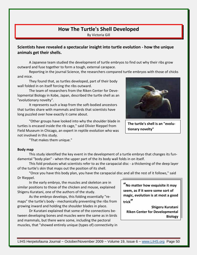
LIHS Herpetofauna Journal ~ October/November 2009 ~ Volume 19, Issue 6 ~ www.LIHS.org Page 50
“No matter how exquisite it may
seem, as if it were some sort of magic, evolution is at most a good
trick” Shigeru Kuratani
Riken Center for Developmental Biology
The turtle's shell is an "evolu-
tionary novelty"
How The Turtle's Shell Developed By Victoria Gill
Scientists have revealed a spectacular insight into turtle evolution - how the unique animals get their shells.
A Japanese team studied the development of turtle embryos to find out why their ribs grow outward and fuse together to form a tough, external carapace.
Reporting in the journal Science, the researchers compared turtle embryos with those of chicks and mice.
They found that, as turtles developed, part of their body wall folded in on itself forcing the ribs outward.
The team of researchers from the Riken Center for Deve-lopmental Biology in Kobe, Japan, described the turtle shell as an "evolutionary novelty".
It represents such a leap from the soft-bodied ancestors that turtles share with mammals and birds that scientists have long puzzled over how exactly it came about.
"Other groups have looked into why the shoulder blade in turtles is encased inside the rib cage," said Olivier Rieppel from Field Museum in Chicago, an expert in reptile evolution who was not involved in this study.
"That makes them unique." Body map
This study identified the key event in the development of a turtle embryo that changes its fun-damental "body plan" - when the upper part of the its body wall folds in on itself.
This fold produces what scientists refer to as the carapacial disc - a thickening of the deep layer of the turtle's skin that maps out the position of its shell.
"Once you have this body plan, you have the carapacial disc and all the rest of it follows," said Dr Rieppel.
In the early embryo, the muscles and skeleton are in similar positions to those of the chicken and mouse, explained Shigeru Kuratani, one of the authors of the study.
As the embryo develops, this folding essentially "re-maps" the turtle's body - mechanically preventing the ribs from growing inward and holding the shoulder blades in place.
Dr Kuratani explained that some of the connections be-tween developing bones and muscles were the same as in birds and mammals, but there were some, including the pectoral muscles, that "showed entirely unique (types of) connectivity in

LIHS Herpetofauna Journal ~ October/November 2009 ~ Volume 19, Issue 6 ~ www.LIHS.org Page 51
The fossil captured an "inter-
mediate step in the evolution of
turtles"
turtles". The discovery helps define a position in evolutionary histo-
ry for a 220-million-year-old turtle fossil discovered last year in China, which had an incomplete shell that only covered its under-side.
"The developmental stage of the modern turtle, when the ribs have not encapsulated the shoulder blade yet, resembles the (body) of this fossil species," said Dr Kuratani. Dr Rieppel, who examined the Chinese fossil when it was discov-ered late in 2008, said this study illustrated that the ancient turtle was "basically an intermediate step in the animals' evolution".
The scientists do not yet know what causes the folding. "That belongs to a future project," said Dr Kuratani.
Stressing the importance of developmental research to evolutionary biology, Dr Kuratani said: "Developmental changes in evolution give rise to an enormous diversity of animal forms."
"No matter how exquisite it may seem, as if it were some sort of magic, evolution is at most a good trick... and there is a way to make it work.
"In case of turtle evolution, a major part of the trick was found to be (this) embryonic folding."
Story reprinted from BBC NEWS: http://news.bbc.co.uk/go/pr/fr/-/2/hi/science/nature/8142664.stm
Published: July 10, 2009, Accessed July 12, 2009 Submitted by LIHS Member Deb Hoppe
MOSQUITOES ADAPT AND EXPAND DIET, THREATENING GALAPAGOS ISLANDS' REPTILES
By Katherine Harmon in 60-Second Science Blog
The most widespread mosquito on the Galápagos Islands appears to have developed a taste for
reptile blood. Biologists fear that the newly discovered behavior may leave many of the islands' rare reptiles at risk for mosquito-borne diseases, such as West Nile virus, that they haven't adapted to cope with.
A study published online today in the Pro-ceedings of the National Academy of Sciences USA reports that the black salt marsh mosquito ( Aedes taeniorhynchus ), which feeds on birds and mam-mals and is common along the coasts of North and South America, is now also feasting on the blood of marine iguanas and Galápagos tortoises.

LIHS Herpetofauna Journal ~ October/November 2009 ~ Volume 19, Issue 6 ~ www.LIHS.org Page 52
Vietnamese Soft-shell turtle ( Rafetus Leloiiis )
Rather than arriving with humans in recent years—like the other two mosquito species on the Galápagos—the A. taeniorhynchus seems to have been on the islands for about 200,000 years, accord-ing to the new study. This stretch of time would have given the species a chance to adapt to the novel environment—which was dominated by reptiles before the arrival of humans—where it also breeds at higher and drier conditions than its continental counterparts.
"The genetic differences of the Galápagos mosquitoes from their mainland relatives are as large as those between different species," Arnaud Bataille, a graduate researcher at the University of Leeds and lead study author, said in a statement, "suggesting that the mosquito in Galápagos may be in the process of evolving into a new species."
Sampling of blood-full black salt marsh mosquitoes from 18 sites across the archipelago's isl-ands revealed that 58 percent of the insects had been slurping reptile blood. Only one of the 105 meals sampled came from a bird—a surprising figure because birds were more common than reptiles in some sampled areas, "suggesting that reptile blood may have become a preferential choice for this mosqui-to," the authors wrote.
The researchers recommend keeping a better eye on mosquito populations to look out for new pathogens, as well as stringent spraying standards for planes and boats. "With tourism growing so ra-pidly the chance of a disease-carrying mosquito hitching a ride from the mainland on a plane is also in-creasing," study co-author Andrew Cunningham, a wildlife epidemiologist at the Zoological Society of London, said in the statement.
Downloaded from ScientificAmerican.com http://www.scientificamerican.com/blog/60-second-science/post.cfm?id=mosquitoes-adapt-and-
expand-diet-th-2009-06-01 June 1, 2009
Image of a tortoise in the Galapagos courtesy of Marilyn Cruz/Galapagos National Park
Legendary Lake in Vietnam Being Gently Cleaned For Giant Turtle
Researchers have started testing "SediTurtles," a de-
vice they say will defend a renowned Vietnamese turtle while at the same time clean the lake where it lives.
Experts debuted the sediment-consuming machines as part of a cleanup program on Hoan Kiem Lake, located at Vietnam's capital.
The Lake of the Returned Sword houses a mysterious turtle that symbolizes Vietnam's exertion for independence.
The children’s story of the 15th century rebel leader Le Loi says that he used a sword to fight off Chinese invaders. Af-ter Emperor Le Loi went boating on the lake one day, a turtle swam up, took his sword and swam to the bottom, safeguard-ing the weapon for the next time freedom needed a hero.

LIHS Herpetofauna Journal ~ October/November 2009 ~ Volume 19, Issue 6 ~ www.LIHS.org Page 53
Sightings of the turtle are considered lucky, espe-cially when they land on national events. The lake’s his-torical value therefore needs a special cleanup plan, and the teams of Vietnamese and German experts have plot-ted cleaning the turtle's home with smallest possible risk to it.
Leonhard Fechter, of Berlin's Herbst Umwelttech-nik GmbH, knows that people worry about the turtle, so the SediTurtle was made with "soft" technology as not to hurt the animal.
"We are sure we won't touch the turtle," he said. The device has a hose that floats on the water attached to a metal box. A dredging device, hid-
den below the surface, removes sediment from the bottom by sending it to a different machine that removes sludge from the water.
"That device is moving very slowly. That big turtle can easily escape," said Celia Hahn, the project manager at the Dresden University of Technology.
Over time, the sediment in the Hoan Kiem Lake has increased and the water level has declined, specifically in urban areas. The experts note that the lake is only five feet deep, but the majority is sludge created by industrial pollutants.
Draining the lake cannot be done because it would harm the water body's ecosystem, ex-perts insist.
"The big turtle is living from crabs or small fish," Werner said, noting that sediment removal would occur slowly over time.
Christian Richter, from FUGRO-HGN GmbH, said his firm has already reviewed the lake's geolo-gy. They will map parts of the lake where sediment can be extracted without harming the lake.
"Even if they start immediately, they would need at least one or two years for the removal," Richter said.
Reprinted from REDORBIT NEWS http://www.redorbit.com/news/science/1705002/legendary_lake_in_vietnam_being_gently_cleaned_
for_giant_turtle/index.html?source=r_science# Published: June 14, 2009, Accessed June 16, 2009

LIHS Herpetofauna Journal ~ October/November 2009 ~ Volume 19, Issue 6 ~ www.LIHS.org Page 54
Amphibians Synchronize Mating Under the Full Moon
When it comes to mating, timing is everything for amphibians.
Researchers have found that the mating activity of amphibians is synchronized by the full Moon.
The fascinating fact that frogs, toads and newts across the globe seem to enjoy mating by moonlight has never before been noticed.
It appears that in order to make sure that a suffi-cient number of males and females join up at the same time, they use the lunar cycle to co-ordinate their ga-therings.
This proves to be an ingenious tactic. By taking advantage of this very specific time, the animals are able to maximize success in procreation and reduce their odds of being eaten by predators.
A full detailed report of this discovery has been published in the journal Animal Behavior. It all started in 2005 when biologist Rachel Grant of the Open University was studying sala-
manders near a lake in central Italy for her PhD. That is when she noticed toads all over the road, under a full Moon.
"Although this might have been a coincidence, the following month I went along the same route every day at dusk and found that the numbers of toads on the road increased as the Moon waxed to a peak at full Moon and then declined again," she says.
After reviewing the scientific literature and finding little to no mention of any similar records, Grant returned to the same site for two subsequent years to again survey the amphibians more care-fully.
Grant was then able to collate her data with a 10-year analysis of the mating habits of frogs and toads at a pond near Oxford, UK, collected by her supervisor Tim Halliday as well as with data on toads and newts in Wales collected by her colleague Elizabeth Chadwick from Cardiff University, UK.
"We analyzed the data, and found a lunar effect at all three sites," Grant says. The common toad ( Bufo bufo ), for example, would arrive at all its breeding spots, and then
mate and spawn under the full Moon. Also, the common frog ( Rana temporaria ) reportedly spawns around the time of the full Moon.
"Newts also seem to be affected by the lunar cycle but the results are less clear," says Grant. Newt ( Lissotriton vulgaris, L. helveticus and Triturus cristatus ) arrivals are at their highest during both the full and new moons.
However, "newts appear to avoid arriving at the breeding site when the Moon is in its third quarter. This could be because the Earth's magnetic field is highest at that time. More research is needed to clarify this," said Grant.
After looking at historical data collected in Java on the Javanese toad ( Bufo melanostictus ), the researchers found that their mating patterns also correlate with the lunar cycle, with females ovulating on or near the full Moon.

LIHS Herpetofauna Journal ~ October/November 2009 ~ Volume 19, Issue 6 ~ www.LIHS.org Page 55
"We now have evidence of lunar cycles affecting amphibians in widespread locations. We defi-nitely think that Moon phase has been an overlooked factor in most studies of amphibian reproductive timing," says Grant.
"We think this may be a worldwide phenomenon. However, differences between species in ecology and reproductive strategy may mean that not all amphibians are affected in the same way. This is something we would like to investigate further."
Now, Grant and her colleagues are hoping to develop a statistical model that would take into account weather factors and environmental variables such as geomagnetism along with the lunar cycle.
The ability to accurately predict the movements of amphibians is a crucial part of conservation, according to Grant. If mating times were predicted, they could close roads at that given time in order to prevent cars killing thousands of the frogs and toads mating there.
"Given the current global crisis among amphibian populations, further understanding of [their] breeding behavior is extremely important," she says.
Reprinted from REDORBIT NEWS: http://www.redorbit.com/news/display/?id=1719692 http://www.redorbit.com/news/science/1719692/amphibians_synchronize_mating_under_the_full
_moon/index.html?source=r_science# Published July 13, 2009/07/13, Accessed July 25, 2009
Ancient Fossil Find: This Snake Could Eat a Cow! By THE ASSOCIATED PRESS
NEW YORK (AP) -- Never mind the 40-foot snake that menaced Jennifer Lopez in the 1997 movie ''Anaconda.'' Not even Hollywood could match a new discovery from the ancient world. Fossils from northeastern Colombia reveal the biggest snake ever discovered: a behemoth that stretched 42 to 45 feet long, reaching more than 2,500 pounds.
''This thing weighs more than a bison and is longer than a city bus,'' enthused snake expert Jack Conrad of the American Museum of Natural History in New York, who was familiar with the find.
''It could easily eat something the size of a cow. A human would just be toast immediately.'' ''If it tried to enter my office to eat me, it would have a hard time squeezing through the door,''
reckoned paleontologist Jason Head of the University of Toronto Missisauga. Actually, the beast probably munched on ancient relatives of crocodiles in its rainforest home
some 58 million to 60 million years ago, he said. Head is senior author of a report on the find in Thursday's issue of the journal Nature.
( The same issue carries another significant report from the distant past. Scientists said they'd found the oldest known evidence of animal life, remnants of steroids produced by sponges more than 635 million years ago in Oman. )
The discoverers of the snake named it Titanoboa cerrejonensis (''ty-TAN-o-BO-ah sare-ah-HONE-en-siss''). That means ''titanic boa from Cerrejon,'' the region where it was found.

LIHS Herpetofauna Journal ~ October/November 2009 ~ Volume 19, Issue 6 ~ www.LIHS.org Page 56
ABOVE: Artist rendering of Titanboa ( Titanoboa cerrejonensis ) – Jason Bourque
A photo released by Nature magazine shows a Precloacal vertebra of an adult Green Ana-conda ( Eunectes murinus ), lighter colored vertebra dwarfed by a vertebra of the giant boid snake they named Titanoboa cerrejonensis, mean-ing ``titanic boa from Cerrejon,'' the region where it was found. Fossils from NE Colombia reveal the biggest snake ever discov-ered: a behemoth that was 42 feet or longer, reaching an es-timated 1.27 tons.
While related to modern boa constrictors, it behaved more like an anaconda and spent almost all its time in the water, Head said. It could slither on land as well as swim.
Conrad, who wasn't involved in the discovery, called the find ''just unbelievable.... It mocks your preconceptions about how big a snake can get.''
Titanoboa breaks the record for snake length by about 11 feet, surpassing a creature that lived about 40 million years ago in Egypt, Head said. Among living snake species, the record holder is an individual python measured at about 30 feet long, which is some 12 to 15 feet shorter than typical Titanoboas, said study co-author Jonathan Bloch.
The beast was revealed in early 2007 at the University of Florida's Florida Museum of Natural History in Gainesville. Bones collected at a huge open-pit coal mine in Colombia were being un-packed, said Bloch, the museum's curator of vertebrate paleontol-ogy.
Graduate students unwrapping the fossils ''realized they were looking at the bones of a snake. Not only a snake, but a really big snake.''
So they quickly consulted the skeleton of a 17-foot anacon-da for comparison. A backbone from that creature is about the size of a silver dollar, Bloch said, while a backbone from Titanoboa is ''the size of a large Florida grapefruit.''
So far the scientists have found about 180 fossils of back-

LIHS Herpetofauna Journal ~ October/November 2009 ~ Volume 19, Issue 6 ~ www.LIHS.org Page 57
bone and ribs that came from about two dozen individual snakes, and now they hope to go back to Co-lombia to find parts of the skull, Bloch said.
Titanoboa's size gives clues about its environment. A snake's size is related to how warm its en-vironment is. The fossils suggest equatorial temperatures in its day were significantly warmer than they are now, during a time when the world as a whole was warmer. So equatorial temperatures ap-parently rose along with the global levels, in contrast to the competing hypothesis that they would not go up much, Head noted.
''It's a leap'' to apply the conditions of the past to modern climate change, Head said. But given that, the finding still has ''some potentially scary implications for what we're doing to the climate to-day,'' he said.
The findings suggest the equatorial regions will warm up along with the planet, he said. ''We won't have giant snakes, however, because we are removing most of their habitats by de-
velopment and deforestation'' in equatorial regions, he said. On the Net:
Nature: http://www.nature.com/nature
Reprinted from OregonLive.com http://www.oregonlive.com/news/index.ssf/2009/02/ancient_fossil_find_this_snake.html
Posted and Accessed February 4, 2009
Prof Finds New Cause for Warped Frogs By Denise Richardson
ONEONTA - After more than 20 years studying amphibians, Hartwick College Professor Stanley Sessions has revealed another discovery about how tadpoles turn into frogs with deformities.
His early studies were about deformities caused by parasites. He responded to arguments by researchers and environmentalists about the effects of chemical pollutants. His most recent collabora-tion has revealed that legless frogs are the result of dragonfly nymphs taking bites of tadpoles.
Sessions and Brandon Ballengee, of the University of Plymouth in the United Kingdom, submit-ted their findings last year in a paper to the Journal of Experimental Zoology. Their article, ``Explana-tion for Missing Limbs in Deformed Amphibians,'' appeared online June 8 pending publication in its next issue, according to the journal's website.
In 1990, the same journal published Sessions' research results about frogs with extra limbs. The findings about parasites and predatory nymphs make ``bookends'' to his work studying frogs, Ses-sions said during an interview last week in his office at Hartwick College.
Shelves in his office are heavy with books, photographs on the wall feature amphibians and a horse skull sits among other natural artifacts on his desk. He illustrates with his hands how a dragonfly nymph finds a meal.
Around the world, frogs have been found with missing or deformed limbs, Sessions said. Re-searchers studied the possible causes, and the search for answers sparked controversy and produced books on the issues, he said.

LIHS Herpetofauna Journal ~ October/November 2009 ~ Volume 19, Issue 6 ~ www.LIHS.org Page 58
For a decade, Ballengee and Sessions have collaborated on art and science projects that show details inside amphibians' bodies. The BBC's Earth News has reported the recent findings on the dra-gonfly nymph. In a news release from Hartwick College, he describes the results:
"What we've found is that these predators grab tadpoles and almost surgically remove their tender hind limbs with their mandibles. They then let the tadpole swim away, and it can metamor-phose into a frog that's missing a hind limb or part of hind limb. We call this phenomenon A Selective predation' since the predators consume only selected parts of the prey. It seems pretty obvious in hind sight, but this solves the whole rest of the problem."
For years, researchers have debated the roles of parasites, predators, chemical pollution or UV-B radiation caused by the thinning of the ozone layer, was triggering the deformations, the BBC report said.
"Deformed frogs became one of the most contentious environmental issues of all time, with the parasite researchers on one side, and the A chemical company' as I call them, on the other," Ses-sions said in the recent news report. "There was a veritable media firestorm, with millions of dollars of grant money at stake."
Sessions, 60, who joined Hartwick in 1989, teaches animal development, evolution, natural his-
tory of vertebrates, and developmental genetics plus an off-campus course on the natural history of Costa Rica.
Hartwick is a private, liberal arts college with about 1,480 undergraduates. Hartwick students were co-authors on about half of the dozen papers Sessions has published
about deformed amphibians, college officials said. He started his research on deformities during his postdoctoral work at the University of California's Berkeley and Irvine campuses.
Sessions published his research indicating that extra limbs on frogs were caused by parasitic flatworms known as trematodes. But what about frogs without legs?
"People said, A-Okay, you explained the frogs with extra limbs, but what about these?'" Ses-sions said. "We thought it couldn't be predation because predators eat frogs, usually killing them in the process, and these frogs were otherwise healthy and intact. It turned out the missing limbs were much more difficult to explain than extra limbs, but I think we've finally cracked it!"
Ballengee and Sessions ran tests to see if stickleback fish, diving beetles, water scorpions, dra-gonfly nymphs or other suspected predators attacked tadpoles, the BBC said. Only three species of dragonfly nymph did.
Adult amphibians with one hind limb appear able to live for quite a long time, explaining why so many deformed frogs and toads are discovered, he told the BBC.
Sessions said he doesn't completely rule out chemicals as the cause of some missing limbs, but a ‘selective predation’ by dragonfly nymphs is the leading explanation.
"Are parasites sufficient to cause extra limbs?," he said in a prepared statement. "Yes. Is selec-tive predation by dragonfly nymphs sufficient to cause loss or reduction of limbs. Yes. Are chemical pol-lutants necessary to understand either of these phenomena? No."
Reprinted from The Daily Star http://www.thedailystar.com/local/local_story_187074533.html
Posted July 06, 2009, Accessed July 9, 2009

LIHS Herpetofauna Journal ~ October/November 2009 ~ Volume 19, Issue 6 ~ www.LIHS.org Page 59
Endangered Arakan Forest Turtle Spotted in the Wild by Mariella Moon
The Arakan forest turtle was once thought to be extinct until some specimens were found ped-
dled as food in China. According to LiveScience, it was 1908 since a live specimen – just a single one – was last collected by an Army officer from Britain. But in an elephant sanctuary in Myanmar, a team of Wildlife Conservation Society ( WCS ) personnel discovered five endangered turtle species, one of which is the Arakan forest turtle. The animal is small, with adults measuring less than a foot in length, and is characterized by its brown coloring and block mottling. Like many other turtle species in Asia, the Arakan forest turtle’s existence has been threatened by poaching.
“Throughout Asia, turtles are being wiped out by poachers for the illegal wildlife trade,” said Colin Poole, WCS
Director of Asia programs. “We are delighted and asto-nished that this extremely rare species is alive and well in Myanmar. Now we must do what we can to protect the remaining population.”
Due to the discovery of the Arakan forest turtles among other endangered turtle species in the elephant
sanctuary, preparatory steps for the proper care of the animals are proposed. These include training the staff, taking measures to pre-
vent poaching, and collecting further data on the species.
Reprinted from Ecoworldly.com http://ecoworldly.com/2009/09/17/endangered-arakan-forest-turtle-spotted-in-the-wild/
Posted September 17th, 2009 / Accessed and downloaded September 25, 2009
Facts about the Arakan Forest Turtle
SCIENTIFIC NAME: Heosemys depressa
CONSERVATION STATUS: Critically endangered (once thought extinct until 1994)
SIZE: 7-10 pounds, 11 inches in length
RANGE: Arakan hills of western Burma
HABITAT: Dense bamboo forests
LIFESTYLE: Mostly solitary diurnal turtles tend to lie dormant most of the time, hiding in leaves and debris unless they are eating or foraging for food.
Myanmar jungle

LIHS Herpetofauna Journal ~ October/November 2009 ~ Volume 19, Issue 6 ~ www.LIHS.org Page 60
These are rather calm animals that can be found in streams and ponds in their forest, where they will rest in hiding under vegetation for hours on end. Rain seems to bring them to life, stimulating them to forage for food. They are rather aggressive when it comes to eating insects, worms and fish.
FOOD: Arakan forest turtles are omnivores; diet consists of equal parts animals and plants. They are big fans of fruit that fall to the forest floor. They are aggressive in eating insects, crustaceans, worms, fish or anything they can get their mouths on.
LIFE CYCLE: Arakan forest turtles lay two to six eggs, usually once a year, that incubate for 120 to 130 days at 80 to 84 degrees F. Little is known about their life cycle. Successful breeding started about five years ago, and those young are not sexually mature yet. Eggs take approximately six weeks to be pro-duced.
POPULATION STATUS AND THREATS: Arakan forest turtles are critically endangered, and natural popu-lations are ever decreasing due to habitat loss and collection for Asian food markets. Once thought ex-tinct in the wild, the Arakan forest turtle was rediscovered in an Asian food market in 1994. These tur-tles are collected for food and for their mystical medicinal cures. Habitat loss is due to agricultural ex-pansion, logging and bamboo harvesting.

LIHS Herpetofauna Journal ~ October/November 2009 ~ Volume 19, Issue 6 ~ www.LIHS.org Page 61
DECORATOR HABITATS CLASSIC GLASS HABITATS NATURAL WOOD HABITATS Furniture Style Vivariums Black Frame & Glass Terrariums Wood Frame & Glass Terrariums
The Herp Marketplace
TERRARIUM AND VIVARIUM HABITATS FROM REPTOLOGY. Introducing three new lines of
Reptology™ Habitat Enclosures for Reptiles and Amphibians. Designed to meet the essential require-ments for herp care. Every unit is constructed of quality materials, tempered glass and detailed with numerous features that encourage natural and healthy living environments.
REPTOLOGY’S TRADITIONAL LINE offers all of the features you need to create the ideal environment for both reptiles and amphibians. A wide range of sizes is available to meet your herp’s housing re-quirements. Fully assembled habitats with tempered glass • Front sliding doors with locking tabs • In-tegrated front vents • Deep fixed front window allows for aquatic section and substrate • Removable top screen - larger units include support bars to hold weight of light fixtures • Inlet access for tubes and wires • (7) Seven sizes available • Complimenting matching stands are strong and durable - shelving is

LIHS Herpetofauna Journal ~ October/November 2009 ~ Volume 19, Issue 6 ~ www.LIHS.org Page 62
provided for storage space (Stands sold separately - available for all but the 2 smallest sizes) • Stands are designed as Knock down flat packaging- Easy to assemble. NATURAL WOOD HABITATS - WOOD FRAME & GLASS TERRARIUMS & STANDS - Similar in design and essential elements to the Classic Glass Habitats with additional unique features. Natural beech color wood frame - These habitats add a touch of sophistication to exhibit herps. Unique Feature for larger units! - Two larger units engineered into a knock down design - flat package. Easy to assemble or disas-semble. Beautiful natural wood frames with tempered glass • Front sliding doors with locking tabs • Integrated front vents • Two smaller units include fixed front window for aquatic section or substrate • Two larger units include bottom tray for shallow water section or substrate • Removable top screen with support bars to hold weight of light fixtures • Inlet access for tubes and wires • Matching stands provide shelving for storage space (Stands sold separately) • Stands are designed as Knock down flat packaging- Easy to assemble. DECORATOR HABITATS VIVARIUMS & STANDS - Offering you the perfect combination - An environ-ment created for healthy reptile care and designed as a beautiful piece of furniture to showcase them in! Available in Beech, Black and Cherry. Ideal for lizards, snakes and other reptiles - create a desert landscape • Easily customized to meet your heating, lighting and ventilation requirements. Panels may be drilled to install fixtures and accessories. • Includes ventilation and cord ports • Enclosed walls pro-vide secure environment, encouraging natural and healthy behavior • Locking sliding tempered glass doors for added security • High Quality E0 grade Medium Density Fiberboard - water resistant, solid construction, drillable and easy to clean • Great insulator - helps to reduce energy cost • Compliment-ing Stands are designed with closing cabinet doors with quality hinges • Habitats & Stands designed as Knock down flat packaging - Easy to assemble - Easier to transport.
http://pennplax.com/Welcome.html
EXO TERRA TURTLE HEATER maintains an ideal water temperature ( 78°F )
for aquatic turtles and most other aquatic reptiles and amphibians. The high-impact plastic casing and the stainless steel power cord protector keeps aquatic reptiles and amphibians safe. Included is a mounting bracket with two oversized suction cups for easy mounting in any aquatic habitat setup. Can heat up to 30 gallons.
Keeps terrarium water around or 78°F
Preset submersible heater
Stainless steel power cord protector
Rugged construction for safety
Compact design with suction cup mounts
Submersible Aquatic Terrarium heater for up to 30 Gallons
Turtle Heater - 25W - PT3700
Turtle Heater - 50W - PT3702

LIHS Herpetofauna Journal ~ October/November 2009 ~ Volume 19, Issue 6 ~ www.LIHS.org Page 63
www.exo-terra.com

LIHS Herpetofauna Journal ~ October/November 2009 ~ Volume 19, Issue 6 ~ www.LIHS.org Page 64
DIAMONDBACK TRADING CARDS - Diamondback
Trading Cards manufactures unique trading cards for the natural history enthusiast of all ages. Watch their video: http://www.youtube.com/watch?v=j8wVCrNsnoI to find out more about us or click around to browse current stock and upcoming releases.
http://www.diamondbacktradingcards.com/Home
Natural Cricket Care with added vitamins and minerals is formulated to
provide a nutrient-filled gut-load for the optimum health of crickets and the herps that eat them. The product comes in 1 3/4 - and 10-oz. sizes, and its par-ticles come finely ground for all sizes of crickets, according to the company. Zoo Med Laboratories Inc.
www.zoomed.com
THE NATURE ZONE HUMIDI-MAT helps raise
the humidity level in your enclosure by controlled evaporation of water. They are made from a tough simulated leather fabric combined with a special osmotic fabric which allows the release of fine water vapor. Soak in water 12-24 hours to activate, then place in terrarium. The nontoxic gel inside pouch slowly releases water vapor without saturating the terrarium. Remove and reactivate when dry.
An easy and hassle-free way to create a humid microclimate for your reptile. Convenient mat provides essential moisture to your reptile's terrarium.
A great way to offer moisture to insects without drowning them.
The mats have heat fused seams making them extremely durable and allow them to be used over and over again. Easily activated by simply soaking in water.
ideal for tropical animals such as Rainbow Boas, Ball Pythons, Dart Frogs, Chameleons, Skinks, Salamanders or any other humidity loving reptile or animal.
Small: 7.25" x 3". Medium: 12” x 3.5”
www.NatureZonePet.com

LIHS Herpetofauna Journal ~ October/November 2009 ~ Volume 19, Issue 6 ~ www.LIHS.org Page 65
ECOZONE VIVARIUM CONTROLLER is de-
signed to give herps total naturalistic environ-mental simulation. It features unique lighting in-tensities, mist intervals, on/off times and tem-peratures programmed over a 12-month calen-dar, according to the company, which also states that the product’s four 450W proportional con-trolled temperature-probe inputs and AC outlets provide more than 1600W of AC power control. With its network connectivity, the product can al-so have advanced functionality through software updates. According to the manufacturer, the controller can allows the user to program Day, Night, and Seasonal variations – A Vivarium First! Control your herps environment on a natural seasonal cycle.
Four preprogrammed EcoZones ( Desert, Rainforest, Woodlands, or Savannah ) for fast, easy setup, or fully customize the settings to your herps needs.
Four proportional controlled outputs - Up to 450W per output each output. Any output can control a heater/temperature element, dimmable lighting, moon phase lighting, misters, and more. Four Temperature Sensor inputs – EcoZone Vivarium controllers support up to 4 temper-ature sensors per controller!
Leading edge Power Control technology - EcoZone controllers utilize advanced dimming out-puts which minimize electrical noise and interference.
www.ecozonevivarium.com
EXO TERRA’S SWAMP GLO LAMP is a broad-spectrum day-
light spot lamp designed to resist splashes, is mist resistant and stands up to high humidity conditions. Ideal for aquatic or ex-tremely humid terrarium set-ups and herp habitats. The product has an outer sleeve made from extra strong neodymium glass, the manufacturer states. It is available in 50W, 75W and 100W.
Splash and mist resistant basking lamp
Broad-spectrum daylight spot lamp
Ideal for aquatic or humid terrariums
Made from extra-strong neodymium glass
Enhances animal coloration
R20 / 50W - PT3780 R20 / 75W - PT3781 R25 / 100W - PT3782
www.exo-terra.com

LIHS Herpetofauna Journal ~ October/November 2009 ~ Volume 19, Issue 6 ~ www.LIHS.org Page 66
PET-TECH MAGNETIC TERRARIUM JUNGLE VINE CLUSTER ( Size: up to 24" long ), JUNGLE
VINE BRIDGE ( size: 34" ), and HANGING JUNGLE ( sizes: 9" and 12’ ) are unique reptile habitat accessories that fea-ture a MEGA STRONG magnet so you can place them an-ywhere in your reptile's environment. Place these plants in a new place every time you clean the habitat to change the look of your reptile environment over and over again. Life-like silk foliage accessories stay put through glass up to 1/4" thick and will complement any reptile enclosure.
Reptile habitat ac-cessories feature MEGA STRONG magnets so you can place them anywhere
Easy-to-clean artificial vines look like the real thing
Attractive silk foliage looks appealing in any terrarium setting
www.pettech.com/
PetTech MAGNATURAL HIDEAWAYS AND LEDGES ( regular and planter ) are locked against terra-
rium glass with Mega Magnetic Power! These Hideaways are perfect for all reptiles and amphibians. Place the Hi-deaway high in the terrarium for tree dwellers or rock climbers or place it low in the terrarium for terrestrial animals. Hideaways offer the perfect retreat for all ter-rarium animals. Key Features
Install or relocate in seconds. NO glue or Velcro.
100% non-toxic materials.
Magnetic force guaranteed for life.
Easy to clean.
Various sizes. Create exciting terrariums with multiple units... then recreate them at will!
Not only are MagNaturals original and innovative, they are also made in the U.S.A. We are dedicated to developing new products that will enhance the lives of terrarium pets as well as creating more natural and beautiful environments for you to enjoy.
www.pettech.com/

LIHS Herpetofauna Journal ~ October/November 2009 ~ Volume 19, Issue 6 ~ www.LIHS.org Page 67
MEETING DATES & INFORMATION
Meeting Dates Speaker / Topic
December 20, 2009 HOLIDAY GET TOGETHER - MEMBERS ONLY
January 10, 2010 Terrarium Basics - includes a demo build followed by a raffle or auc-
tion of the Penn Plax donated terrarium used in the demo John Heiser
February 21, 2010 TBD
March 21, 2010 The role of reptiles in human health" ~ Dr. Russell Burke
April 11, 2010 TBD
May 16, 2010 TBD
June 13, 2010 2nd Annual LIHS Auction
ALL Meetings ( unless otherwise noted ):
are OPEN and FREE to the PUBLIC ( UNLESS NOTED )… Bring your friends and family.
will start at 1:00 PM. They may end earlier than the 4:00 end time, so please be on time.
will be held at the Farmingdale State College Conference Center on the SUNY Farmingdale College Campus.
DIRECTIONS to SUNY Farmingdale: http://www.lihs.org/files/meetingplace.htm
SUNY Farmingdale College Campus Map: http://www.lihs.org/files/FSUNY_MAP.jpg Speakers will be updated as they are scheduled. You will receive meeting updates via email, the Herpetofauna Journal, REMINDER POSTCARDS, or for the most CURRENT INFORMATION check the LIHS Website:
In case of inclement weather check your e-mail and the LIHS Website:
www.LIHS.org