leukocyte differential pentra 120 sps smears and … leukocytes were counted by ... with two slides...
TRANSCRIPT

18 English Edition No.8
Guest ForumGuest ForumGuest ForumGuest ForumGuest ForumLeukocyte DifferentialComparison betweenPentra 120 SPS smearsand Manual SlideReview
An evaluation was conducted to compare theleukocyte differential on smears made by theSPS to a manual slide review with 200-celldifferential count (reference method).
Introduction
The study was designed to accomplish two aims:• Determine the differential accuracy between the
leukocyte differential on the SPS slide (test instrument)and the manual slide review (reference) over a clinicalrange, including normal and morphologically abnormalsamples.
• Test the functionality of the SPS for laboratory work.
Materials and Methods
The NCCLS *1 recommendations for evaluating ahematology analyzer were followed.
*1: Global Consensus Standardization for HealthTechnologiesThe acronym used to stand for “National Committee forClinical Laboratory Standards,” but NCCLS is now a globalorganization and develops consensus documents foradditional audiences beyond the clinical laboratorycommunity. Therefore, the organization should be referred toby the acronym, “NCCLS.” (from http://www.nccls.org/)
Sample
131 unselected routine normal and pathological sampleswere analyzed.These samples were collected in K3EDTA*2, maintainedat room temperature and analyzed within six hours.Peripheral blood smears were prepared by the wedgemethod on all samples.
*2: Tripotassium EthleneDiamine Tetra-Acetate
Dr. JouHospital Clinic, hematology laboratory, Barcelona
19
Technical Reports
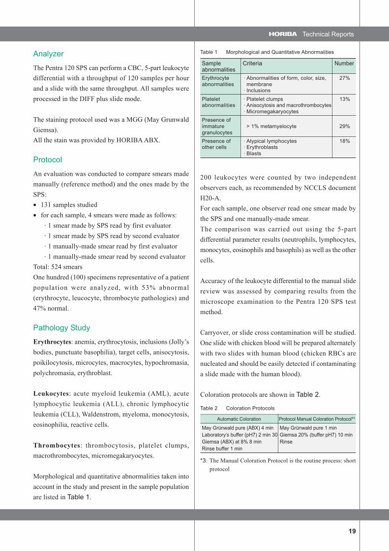
Analyzer
The Pentra 120 SPS can perform a CBC, 5-part leukocytedifferential with a throughput of 120 samples per hourand a slide with the same throughput. All samples wereprocessed in the DIFF plus slide mode.
The staining protocol used was a MGG (May GrunwaldGiemsa).All the stain was provided by HORIBA ABX.
Protocol
An evaluation was conducted to compare smears mademanually (reference method) and the ones made by theSPS:• 131 samples studied• for each sample, 4 smears were made as follows:
· 1 smear made by SPS read by first evaluator· 1 smear made by SPS read by second evaluator· 1 manually-made smear read by first evaluator· 1 manually-made smear read by second evaluator
Total: 524 smearsOne hundred (100) specimens representative of a patientpopulation were analyzed, with 53% abnormal(erythrocyte, leucocyte, thrombocyte pathologies) and47% normal.
Pathology Study
Erythrocytes: anemia, erythrocytosis, inclusions (Jolly’sbodies, punctuate basophilia), target cells, anisocytosis,poikilocytosis, microcytes, macrocytes, hypochromasia,polychromasia, erythroblast.
Leukocytes: acute myeloid leukemia (AML), acutelymphocytic leukemia (ALL), chronic lymphocyticleukemia (CLL), Waldenstrom, myeloma, monocytosis,eosinophilia, reactive cells.
Thrombocytes: thrombocytosis, platelet clumps,macrothrombocytes, micromegakaryocytes.
Morphological and quantitative abnormalities taken intoaccount in the study and present in the sample populationare listed in Table 1.
Table 1 Morphological and Quantitative Abnormalities
Sample Criteria NumberabnormalitiesErythrocyte · Abnormalities of form, color, size, 27%abnormalities membrane
· InclusionsPlatelet · Platelet clumps 13%abnormalities · Anisocytosis and macrothrombocytes
· MicromegakaryocytesPresence ofimmature > 1% metamyelocyte 29%granulocytesPresence of · Atypical lymphocytes 18%other cells · Erythroblasts
· Blasts
200 leukocytes were counted by two independentobservers each, as recommended by NCCLS documentH20-A.For each sample, one observer read one smear made bythe SPS and one manually-made smear.The comparison was carried out using the 5-partdifferential parameter results (neutrophils, lymphocytes,monocytes, eosinophils and basophils) as well as the othercells.
Accuracy of the leukocyte differential to the manual slidereview was assessed by comparing results from themicroscope examination to the Pentra 120 SPS testmethod.
Carryover, or slide cross contamination will be studied.One slide with chicken blood will be prepared alternatelywith two slides with human blood (chicken RBCs arenucleated and should be easily detected if contaminatinga slide made with the human blood).
Coloration protocols are shown in Table 2.
Table 2 Coloration Protocols
Automatic Coloration Protocol Manual Coloration Protocol*3
May Grünwald pure (ABX) 4 min May Grünwald pure 1 minLaboratory’s buffer (pH7) 2 min 30 Giemsa 20% (buffer pH7) 10 minGiemsa (ABX) at 8% 8 min RinseRinse buffer 1 min
*3: The Manual Coloration Protocol is the routine process: shortprotocol
20 English Edition No.8
Guest ForumGuest ForumGuest ForumGuest ForumGuest Forum Leukocyte Differential Comparison between Pentra 120 SPS smears and Manual Slide Review
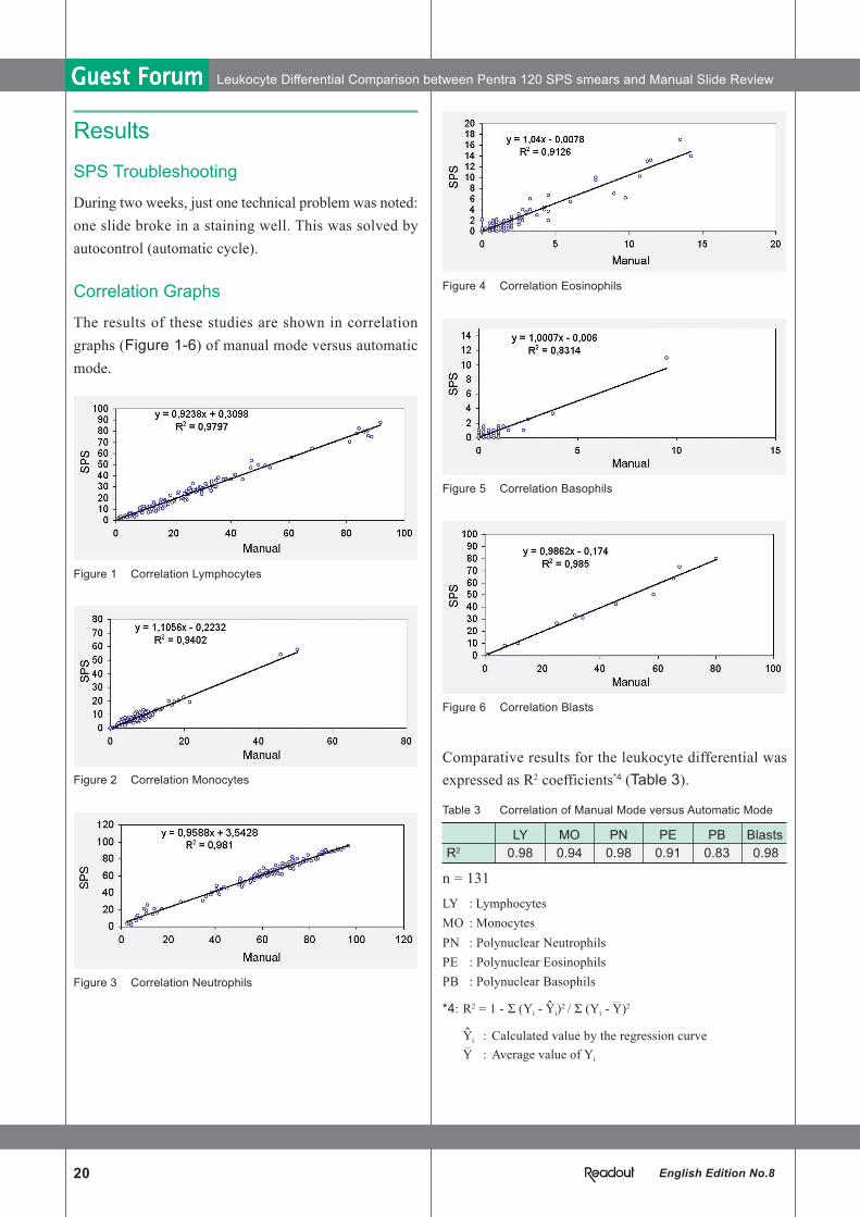
Figure 4 Correlation Eosinophils
Figure 5 Correlation Basophils
Figure 6 Correlation Blasts
Comparative results for the leukocyte differential wasexpressed as R2 coefficients*4 (Table 3).
Table 3 Correlation of Manual Mode versus Automatic Mode
LY MO PN PE PB BlastsR2 0.98 0.94 0.98 0.91 0.83 0.98
n = 131LY : LymphocytesMO : MonocytesPN : Polynuclear NeutrophilsPE : Polynuclear EosinophilsPB : Polynuclear Basophils
*4: R2 = 1 - Σ (Yi - Yi)2 / Σ (Yi - Y)2
Yi : Calculated value by the regression curveY : Average value of Yi
Results
SPS Troubleshooting
During two weeks, just one technical problem was noted:one slide broke in a staining well. This was solved byautocontrol (automatic cycle).
Correlation Graphs
The results of these studies are shown in correlationgraphs (Figure 1-6) of manual mode versus automaticmode.
Figure 1 Correlation Lymphocytes
Figure 2 Correlation Monocytes
Figure 3 Correlation Neutrophils
ˆ
ˆ

21
Technical Reports
Conclusion
Practicability
• The Pentra 120 SPS is easy to use as it is fully automatic• Flags are given for stain reagent levels• Maintenance is completely automatic
Carry Over
The results from the carryover protocol are consideredacceptable if the chicken NRBCs do notcontaminate any of the human blood samples.No NRBCs were observed in human samples.
Accuracy
Correlation studies between the SPS slides and the manualslides display excellent coefficients.The percentage of cell analysis showed excellent resultsexcept for CLL (increase of neutrophils in SPS smearsbecause some lymphocytes were broken).
Quality of the Slides
The reading area of the SPS smear is larger than that ofthe manual smear.No cell concentration was observed on the smear edge.The staining quality with MGG is very good.
For laboratory work, the SPS gave excellent qualitativeand quantitative results compared to the manual mode.Its throughput allows for quick, regular and reliable work.