lesson assignment lesson 13 the special senses. · md0007 13-2 lesson 13 the special senses section...
TRANSCRIPT

MD0007 13-1
LESSON ASSIGNMENT
LESSON 13 The Special Senses.
LESSON ASSIGNMENT Paragraphs 13-1 through 13-24.
LESSON OBJECTIVES After completing this lesson, you should be able to:
13-1. Identify functions of structures related to thespecial senses.
13-2. Given a list of statements about the physiologyof the special senses, identify the falsestatement.
SUGGESTION After completing the assignment, complete theexercises at the end of this lesson. These exerciseswill help you to achieve the lesson objectives.
MD0007 13-2
LESSON 13
THE SPECIAL SENSES
Section I. INTRODUCTION
13-1. GENERAL VERSUS SPECIAL SENSES
a. The human body is continuously bombarded by all kinds of stimuli. Certainof these stimuli are received by sense organs distributed throughout the entire body.These are referred to as the general senses.
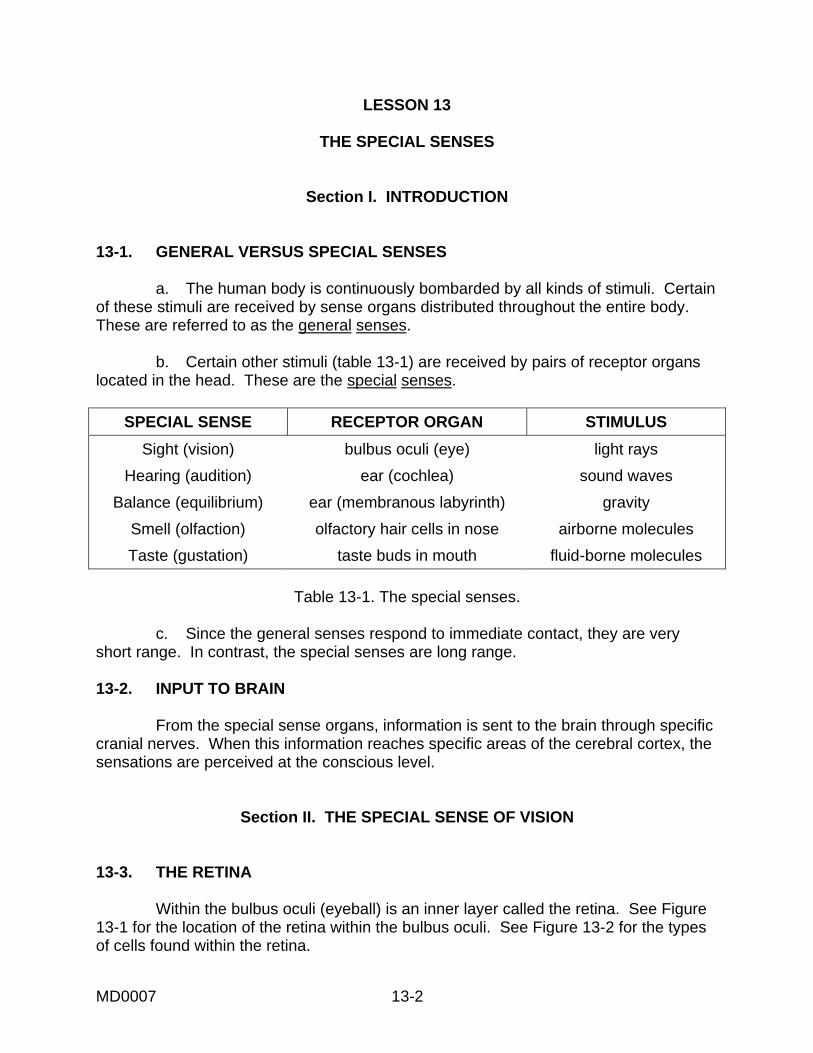
b. Certain other stimuli (table 13-1) are received by pairs of receptor organslocated in the head. These are the special senses.
SPECIAL SENSE RECEPTOR ORGAN STIMULUS
Sight (vision) bulbus oculi (eye) light rays
Hearing (audition) ear (cochlea) sound waves
Balance (equilibrium) ear (membranous labyrinth) gravity
Smell (olfaction) olfactory hair cells in nose airborne molecules
Taste (gustation) taste buds in mouth fluid-borne molecules
Table 13-1. The special senses.
c. Since the general senses respond to immediate contact, they are veryshort range. In contrast, the special senses are long range.
13-2. INPUT TO BRAIN
From the special sense organs, information is sent to the brain through specificcranial nerves. When this information reaches specific areas of the cerebral cortex, thesensations are perceived at the conscious level.
Section II. THE SPECIAL SENSE OF VISION
13-3. THE RETINA
Within the bulbus oculi (eyeball) is an inner layer called the retina. See Figure13-1 for the location of the retina within the bulbus oculi. See Figure 13-2 for the typesof cells found within the retina.

MD0007 13-3
Figure 13-1. A focal-axis section of the bulbus oculi.

MD0007 13-4
Figure 13-2. Cellular detail of the retina.
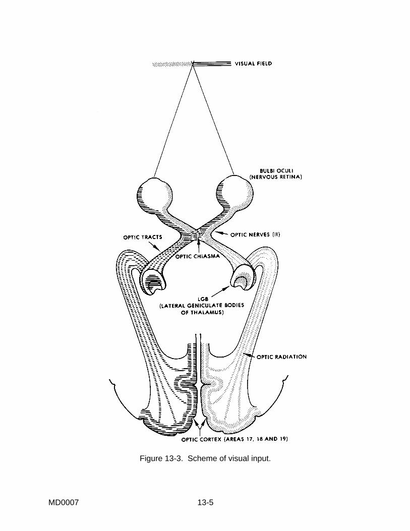
a. Visual Fields (Figure 13-3). When a human looks at an object, light fromthe right half of the visual field goes to the left half of each eye. Likewise, light from theleft half of the visual field goes to the right half of each eye. Later, in paragraph 13-4,we will see how the information from both eyes about a given half of the visual field isbrought together by the nervous system.
b. Photoreception and Signal Transmission. The cells of the retina includespecial photoreceptor cells in the form of cones and rods. The light ray stimuluschemically changes the visual chemical of the cones and rods. This produces areceptor potential which passes through the bodies of the rods and cones and whichacts at the synapses to induce a signal in the bipolar cells. This signal is thentransmitted to the ganglion cells.
MD0007 13-5
Figure 13-3. Scheme of visual input.

MD0007 13-6
(1) Cones. The cones of the retina are for acute vision and also receivecolor information. The cones tend to be concentrated at the rear of the eyeball. Thegreatest concentration is within the macula lutea at the inner end of the focal axis(Figure 13-1).
(2) Rods. Light received by the rods is perceived in terms of black andwhite. The rods are sensitive to less intensive light than the cones. The rods areconcentrated to the sides of the eyeball.
(3) Signal transmission. The stimulus from the photoreceptors (conesand rods) is transferred to the bipolar cells. In turn, the stimulus is transferred to theganglion cells, the cells of the innermost layer of the retina. The axons of the ganglioncells converge to the back side of the eyeball. The axons leave the eyeball to becomethe optic nerve, surrounded by a dense FCT sheath. There are no photoreceptors inthe circular area where the axons of the ganglion cells exit the eyeball; thus, this area iscalled the blind spot.
13-4. NERVOUS PATHWAYS FROM THE RETINAS
a. The two optic nerves enter the cranial cavity and join in a structure knownas the optic chiasma. Leading from the optic chiasma on either side of the brainstem isthe optic tract. In the optic chiasma, the axons from the nasal (medial) halves of theretinas cross to the opposite sides. Thus, the left optic tract contains all of theinformation from the left halves of the retinas (right visual field), and the right optic tractcontains all of the information from the right halves of the retinas (left visual field).
b. The optic tracts carry this information to the LGB (lateral geniculate body)of the thalamus. From here, information is carried to the posterior medial portions(occipital lobes) of the cerebral cortex, where the information is perceived as consciousvision. Note that the right visual field is perceived within the left hemisphere, and theleft visual field is perceived within the right hemisphere.
c. The LGB also sends information into the midbrainstem. This information isused to activate various visual reflexes.
13-5. FOCUSING OF THE LIGHT RAYS
a. The light rays, which enter the eyeball from the visual field, are focused toensure acute vision. The majority of this focusing is accomplished by the permanentlyrounded cornea.
b. Fine adjustments of focusing, for acuteness of vision, are provided by thecrystalline lens (biconvex lens). See Figure 13-4. This is particularly important whenchanging one's gaze between far and near objects.

MD0007 13-7
Figure 13-4. Bending of the light rays by a biconvex lens.
13-6. ACCOMMODATION
The additional focusing provided by the crystalline lens, mentioned above, isone of the processes involved in accommodation. Accommodation refers to the variousadjustments made by the eye to see better at different distances.
a. The crystalline lens is kept in a flattened condition by the tension of thezonular fibers (zonule ligaments; fibers of the ciliary zonule) around its equator, ormargin. Contraction of the ciliary muscle of the eyeball releases this tension and allowsthe elastic lens to become more rounded. Since the elasticity of the crystalline lensdecreases with age, old people may find it very difficult to look at close objects.
b. A second process in accommodation is the constriction of the pupils. Thediameter of the pupil (the hole in the middle of the iris) controls the amount of light thatenters the eyeball. As a light source comes closer and closer, the intensity of the lightincreases greatly. Therefore, the pupils must be constricted to control the amount oflight entering the eyeball as an object under view comes close to the individual.
c. A third process in accommodation is the convergence of the axes of thetwo eyeballs toward the midline. Since both eyes tend to focus on the same object(binocular vision), there is an angle between the two axes. As an object draws closer,the angle increases to enable the axes to still intersect the object.
13-7. EYE MOVEMENTS
a. Convergent and Conjugate Eye Movements. In a conjugate eyemovement, both eyeballs move through an equal angle in one direction, such as right orleft. In a convergent eye movement, both eyeballs turn toward the midline to focusupon a nearby object. In both cases, the movement of the left and right eyeballs is

MD0007 13-8
highly coordinated so that an object may be viewed by both eyes. Therefore, the objectcan be perceived within both cerebral hemispheres in a binocular fashion.
b. "Searching" and "Following" Eye Movements. "Searching" and"following" movements of the eyeball are also called, respectively, voluntary fixationmovements and involuntary fixation movements. For the first type of movement, theeyeballs move in a searching pattern, without focusing on a particular object until it islocated. Once an object is located, the eyeballs will continually fix on that object in afollowing-type motion.
c. Eye Movements During Reading. During reading of printed or writtenmaterial, the eyeball demonstrates several physical characteristics. The amount ofmaterial that can be recognized at a given glance occupies a given width of a writtenline. Each glance is referred to as a fixation. During a fixation, the eyeball is essentiallynot moving, and each eyeball is oriented so that the image falls upon the macula lutea(the maximum receptive area). Reading is a series of motions in which the eyeballsfixate on a portion of the written line and then move very rapidly to the next portion.
d. Compensation for Head Movements (Vestibular Control of Eye Move-ments). Since the human body cannot be held absolutely still, the eyeballs must movein order to remain fixed upon an object. For this purpose, the eyeballs must be movedin the opposite direction and at the opposite speed of the movement of the head. Thisis accomplished by a delicate and complicated mechanism. This mechanism includesthe motor neurons of the muscles of the eyeball and the vestibular nuclei of thehindbrain (responsible for balance and spatial orientation).
13-8. VISUAL REFLEXES
In the sense of vision, one consciously perceives the various objects beinglooked upon. In addition to this, there are a number of protective reactions to visualinput--the visual reflexes.
a. When an unexpected visual stimulus occurs within the visual field, theindividual's response will often include movement and other types of reaction. This is apart of the startle reflex.
b. When there is a change in the amount of light entering the eyeball, the sizeof the pupil will change. This is the pupillary reflex. The muscles of the irisautomatically constrict or dilate to control the amount of light entering the eyeball.
c. In the blink reflex, the eyelids automatically move over the exterior surfaceof the eyeball. This reflex results in the automatic washing of the exterior surface of theeyeball with the lacrimal fluids. It also helps to keep the surface moist.

MD0007 13-9
13-9. LACRIMAL APPARATUS
The eyeball is suspended in the orbit and faces outward. Helping to fill the orbitare a number of structures associated with the eyeball; these are the adnexa. Amongthese other structures is the lacrimal apparatus.
a. The lacrimal gland is located in the upper outer corner in front. Via smallducts, it secretes the lacrimal fluid into the space between the external surface of theeyeball and the upper eyelid.
b. The inner surface of the eyelids and the outer surface of the eyeball arecovered by a continuous membrane known as the conjunctiva. The lacrimal fluid keepsthe conjunctiva transparent. Also, with the blink reflex, the lacrimal fluid washes awayany foreign particles that may be on the surface of the conjunctiva.
c. The free margins of the upper and lower eyelids have special oil glands.The oily secretion of these glands helps prevent the lacrimal fluid from escaping.
d. With the movement of the eyeball and the eyelids, the lacrimal fluid isgradually moved across the exterior surface of the eyeball to the medial inferior corner.Here, the lacrimal fluid is collected into a lacrimal sac, which drains into the nasalchamber by way of the nasolacrimal duct. Thus, the continuous production of lacrimalfluid is conserved by being recycled within the body.
Section III. THE SPECIAL SENSE OF HEARING (AUDITORY SENSE)
13-10. INTRODUCTION
If a medium is set into vibration within certain frequency limits (averagebetween 25 cycles per second and 18,000 cycles per second), we have what is called asound stimulus (Figure 13-5). The sensation of sound, of course, occurs only whenthese vibrations are interpreted by the cerebral cortex of the brain at the consciouslevel.
a. The human ear is the special sensory receptor for the sound stimulus. Asthe stimulus passes from the external medium (air, water, or a solid conductor of sound)to the actual receptor cells in the head, the vibrations are in the form of (1) airbornewaves, (2) mechanical oscillations, and (3) fluid-borne pulses.

MD0007 13-10
Figure 13-5. Characteristics of sound.
b. The ear (Figure 13-6) is organized in three major parts: external ear,middle ear, and internal (inner) ear. Each part aids in the transmission of the stimulusto the receptor cells.

MD0007 13-11
Figure 13-6. A frontal section of the human ear.
13-11. THE EXTERNAL EAR
The external ear begins with a funnel-like auricle. This auricle serves as acollector of the airborne waves and directs them into the external auditory meatus. Atthe inner end of this passage, the waves act upon the tympanic membrane (eardrum).The external auditory meatus is protected by a special substance called earwax(cerumen).
13-12. THE MIDDLE EAR
a. Tympanic Membrane. The tympanic membrane separates the middle andexternal ears. It is set into mechanical oscillation by the airborne waves from theoutside.
b. Middle Ear Cavity. Within the petrous bone of the skull is the air-filledmiddle ear cavity.
(1) Function of the auditory tube. Due to the auditory tube, the air of themiddle ear cavity is continuous with the air of the surrounding environment. Theauditory tube opens into the lateral wall of the nasopharynx. Thus, the auditory tube

MD0007 13-12
serves to equalize the air pressures on the two sides of the tympanic membrane. Ifthese two pressures become moderately unequal, there is greater tension upon thetympanic membrane; this reduces (dampens) mechanical oscillations of the membrane.Extreme pressure differences cause severe pain. The passage of the auditory tube intothe nasopharynx opens when one swallows; therefore, the pressure differences arecontrolled somewhat by the swallowing reflex.
(2) Associated spaces. The middle ear cavity extends into the mastoidbone as the mastoid air cells. The relatively thin roof of the middle ear cavity separatesthe middle ear cavity from the middle cranial fossa.
c. Auditory Ossicles. There is a series of three small bones, the auditoryossicles, which traverse the space of the middle ear cavity from the external ear to theinternal ear. The auditory ossicles function as a unit.
(1) The first ossicle, the malleus, has a long arm embedded in thetympanic membrane. Therefore, when the tympanic membrane is set into mechanicaloscillation, the malleus is also set into mechanical oscillation.
(2) The second ossicle is the incus. Its relationship to the malleusproduces a leverage system which amplifies the mechanical oscillations receivedthrough the malleus.
(3) The third ossicle, the stapes, articulates with the end of the arm of theincus. The foot plate of the stapes fills the oval (vestibular) window.
d. Auditory Muscles. The auditory muscles are a pair of muscles associatedwith the auditory ossicles. They are named the tensor tympani muscle and thestapedius muscle. The auditory muscles help to control the intensity of the mechanicaloscillations within the ossicles.
13-13. THE INTERNAL EAR
a. Transmission of the Sound Stimulus. The foot plate of the stapes fillsthe oval (vestibular) window, which opens to the vestibule of the internal ear (Figure13-7A). As the ossicles oscillate mechanically, the stapes acts like a plunger againstthe oval window. The vestibule is filled with a fluid, the perilymph. These mechanical,plunger-like actions of the stapes impart pressure pulses to the perilymph.

MD0007 13-13
Figure 13-7. Diagram of the scalae.

MD0007 13-14
b. Organization of the Internal Ear. The internal ear is essentially amembranous labyrinth suspended within the cavity of the bony (osseous) labyrinth ofthe petrous bone (Figure 13-8). The membranous labyrinth is filled with a fluid, theendolymph. Between the membranous labyrinth and the bony labyrinth is theperilymph.
Figure 13-8. The labyrinths of the internal ear.
c. The Cochlea. The cochlea is a spiral structure associated with hearing.Its outer boundaries are formed by the snail-shaped portion of the bony labyrinth. Theextensions of the bony labyrinth into the cochlea are called the scala vestibuli and thescala tympani (Figure 13-7B). These extensions are filled with perilymph.
(1) Basilar membrane (Figure 13-7B). The basilar membrane forms thefloor of the cochlear duct, the spiral portion of the membranous labyrinth. The basilarmembrane is made up of transverse fibers. Each fiber is of a different length, and thelengths increase from one end to the other. Thus, the basilar membrane is constructedsimilarly to a harp or piano. Acting like the strings of the instrument, the individual fibersmechanically vibrate in response to specific frequencies of pulses in the perilymph.Thus, each vibration frequency of the sound stimulus affects a specific location of thebasilar membrane.
MD0007 13-15
(2) Organ of Corti. Located upon the basilar membrane is the organ ofCorti. The organ of Corti is made up of hair cells. When the basilar membrane vibrates,the hair cells are mechanically deformed so that the associated neuron is stimulated.
13-14. NERVOUS PATHWAYS FOR HEARING
The neuron (associated with the hair cells of the organ of Corti) then carries thesound stimulus to the hindbrainstem. Via a special series of connections, the signalultimately reaches Brodmann's area number 41, on the upper surface of the temporallobe (see para 12-36). Here, the stimulus is perceived as the special sense of sound. Itis interesting to note that speech in humans is primarily localized in the left cerebralhemisphere, while musical (rhythmic) sounds tend to be located in the right cerebralhemisphere.
Section IV. THE SPECIAL SENSE OF EQUILIBRIUM, THE GENERAL BODYSENSE, AND POSTURAL REFLEXES
13-15. INTRODUCTION
a. The human body is composed of a series of linkages, block on top of block.These blocks can be arranged in a multitude of patterns called postures. In order toproduce and control these postures, the human brain utilizes a great number ofcontinuous inputs telling the brain the instantaneous condition of the body posture.Overall, we refer to this process as the general body sense.
b. The internal ear provides one of the input systems for the general bodysense. The internal ear responds to gravitational forces, of which there are twokinds--static and kinetic (in motion). Of the kinetic stimuli, the motion may be in astraight line (linear) or angular (curvilinear).
13-16. THE MACULAE
The membranous labyrinth of the internal ear has two sac-like parts--thesacculus and the utriculus. On the wall of each of these sacs is a collection of hair cellsknown as the macula (plural: maculae). The hairs of these hair cells move in responseto gravitational forces, both static and linear kinetic. The maculae are particularlysensitive to small changes in the orientation of the head from an upright position. Thus,the maculae are very important in maintaining a standing or upright position.
13-17. THE SEMICIRCULAR DUCTS
a. In addition, three tubular structures are associated with the utriculus. Thecircle of each of these semicircular ducts is completed by the cavity of the utriculus. At

MD0007 13-16
one end of each semicircular duct is a crista, a ridge of hair cells across the axis of theduct.
b. When a jet takes off, a passenger tends to remain in place at first and canfeel the resulting pressure of the seat against his back. Also, when the jet is no longeraccelerating, the passenger can feel that the pressure of the seat against his back hasreturned to normal.
c. Likewise, in the appropriate semicircular duct, the endolymph ("passeng-er") tends to remain in place early during an acceleration. Because the duct ("seat")itself is moving with the body ("jet"), the hairs of the crista are affected by the change inmovement. Later, when acceleration stops, the effect upon the hairs of the crista is alsoregistered.
d. However, the cristae of the semicircular ducts detect rotation of the head(angular acceleration and angular velocity). Linear acceleration, as with our example ofthe passenger and the jet, is detected primarily by the maculae, discussed above.
13-18. RESULTING INPUTS FOR THE SPECIAL SENSE OF EQUILIBRIUM
The combined inputs from the maculae of the sacs and the cristae of thesemicircular ducts provide continuous, instantaneous information about the specificlocation and posture of the head in relationship to the center of gravity of the earth.These inputs are transmitted by the vestibular neurons to the hindbrainstem.
13-19. INPUTS FOR THE GENERAL BODY SENSE
In addition to the inputs from the membranous labyrinth, various other inputsare used to continuously monitor the second-to-second posture of the human body.
a. We have already examined the proprioceptive sense, which monitors thecondition of the muscles of the body.
b. Various other receptors are associated with the joint capsules, theintegument, etc. They indicate the precise degree of bending present in the body.
c. A very important body sense is vision. Even when other inputs are lacking,if an individual can see his feet, he may still be able to stand and move.
13-20. POSTURAL REFLEXES
To automatically control the posture, the human nervous system has a numberof special reflexes. These reflexes are coordinated through the cerebellum.
a. The head and neck tonic reflexes orient the upper torso in relationship tothe head.

MD0007 13-17
b. Another set of reflexes does likewise for the body in general. The rightingreflexes come into play when the body falls out of balance or equilibrium.
c. A special set of reflexes connects the vestibular apparatus to theextraocular muscles of the eyeball. This was discussed earlier in the section on thespecial sense of the vision.
Section V. THE SPECIAL SENSE OF SMELL (OLFACTION)
13-21. SENSORY RECEPTORS
Molecules of various materials are dispersed (spread) throughout the air webreathe. A special olfactory epithelium is located in the upper recesses of the nasalchambers in the head. Special hair cells in the olfactory epithelium are calledchemoreceptors, because they receive these molecules in the air.
13-22. OLFACTORY SENSORY PATHWAY
The information received by the olfactory hair cells is transmitted by way of theolfactory nerves (cranial nerves I). It passes through these nerves to the olfactory bulbsand then into the opposite cerebral hemisphere. Here, the information becomes thesensation of smell.
Section VI. THE SPECIAL SENSE OF TASTE (GUSTATION)
13-23. SENSORY RECEPTORS
Molecules of various materials are also dispersed or dissolved in the fluids(saliva) of the mouth. These molecules are from the food ingested (taken in). Organsknown as taste buds are scattered over the tongue and the rear of the mouth. Specialhair cells in the taste buds are chemoreceptors to react to these molecules.
13-24. SENSORY PATHWAY
The information received by the hair cells of the taste buds is transmitted to theopposite side of the brain by way of three cranial nerves (VII, IX, and X). Thisinformation is interpreted by the cerebral hemispheres as the sensation of taste.
Continue with Exercises

MD0007 13-18
EXERCISES, LESSON 13
REQUIREMENT. The following exercises are to be answered by completing theincomplete statements.
After you have completed all the exercises, turn to "Solutions to Exercises" at the end ofthe lesson, and check your answers.
1. Please complete the table below.
SPECIAL SENSE RECEPTOR ORGAN STIMULUS
Sight
Hearing
Balance
Smell
Taste
2. When you look at an object, light from the right half of the visual field goes to the t half of each eye. Light from the left half of the visual field goes to the t half ofeach eye.
The light ray stimulus chemically changes the visual chemical found in the esand ds. The cones of the retina are for te vision and also receive rinformation. Light received by the rods is perceived in terms of and e. Thestimulus from the cones and rods is transferred to the b r cells and then to the n cells. The axons of the ganglion cells leave the eyeball to become the cnerve. Since the circular area where these axons exit contains neither cones nor rods,this area is called the spot.
3. The axons from the nasal (medial) halves of the retinas cross to the oppositesides at the optic sma. Thus, if an object is in your right visual field, theinformation is carried by your t optic tract. If an object is in your left visual field,the information is carried by your t optic tract. For conscious perception of vision,the information enters the al lobes of the cerebral cortex. Note that the rightvisual field is perceived within the t cerebral hemisphere, and the left visual field isperceived within the t cerebral hemisphere.

MD0007 13-19
4. The majority of focusing of light rays is accomplished by the a. Fineadjustments of focusing are provided by the crystalline s.
5. The additional focusing provided by the crystalline lens is one of the processesinvolved in n. Accommodation refers to the various adjustments madeby the eye to see better at different s.
The crystalline lens is kept in a flattened condition by the tension of the zonular s. This tension is released by contraction of the y muscle, resulting ingreater r ing of the lens.
6. When both eyeballs move through an equal angle in the same direction, it iscalled a con e eye movement. When both eyeballs turn toward the midline tofocus upon a nearby object, the result is a con t eye movement.
During a "searching" eye movement, the eyeballs do not focus on a particularobject until it is l d. During a "following" eye movement, the eyeballs continually x on an object.
Vestibular control of eye movements is necessary in order to compensate for_______ movements.
7. The sudden movement of an individual in response to an unexpected visualstimulus is part of the tle reflex.
Changes in the size of the pupil with changes in the amount of light are producedby the y reflex.
Automatic movement of the eyelids over the exterior surface of the eyeball iscalled the reflex.
8. The lacrimal fluid keeps the conjunctiva ent. Also, with the blink reflex,the lacrimal fluid w s away foreign particles.
The secretion of the special oil glands of the upper and lower eyelids helpsprevent the fluid from escaping.
9. The auricle serves as a collector of airborne w s. At the inner end of theexternal auditory meatus, the waves act upon the c membrane.

MD0007 13-20
10. The tympanic membrane separates the external ear from the ear. Thetympanic membrane mechanically oscillates in response to e s from theoutside.
The air of the middle ear cavity is continuous with the air of the surroundingenvironment, due to the y tube. The auditory tube serves to equalize the airpressures on the two sides of the c membrane. Extreme pressure differencescause severe n. The passage of the auditory tube into the nasopharynx openswhen one s.
Mechanical oscillations are transmitted from the tympanic membrane to the ovalwindow by way of the y cles. The intensity of these mechanicaloscillations is somewhat controlled by the auditory s.
11. The mechanical, plunger-like actions of the stapes impart pressure pulses to the ph.
The basilar membrane is made up of transverse fibers. Acting like the strings ofan instrument, the individual fibers mechanically v e in response to specific cies of pulses in the perilymph.
When the basilar membrane vibrates, the cells of the organ of Corti aremechanically d ed so that the associated neuron is stimulated.
12. The "blocks" of the human body can be arranged in many patterns called s. The input systems by which the brain monitors these patterns aretogether known as the l y sense.
13. The maculae are particularly sensitive to small changes in the orientation of thehead from an t position. Thus, the maculae help us maintain a ding or t position.
14. The cristae detect n of the head, that is, r acceleration and r y.
15. Additional inputs for the general body sense include the proprioceptive sense,which monitors the s of the body, and various other receptors associated withstructures such as the joint s and the t. A very important bodysense is n.

MD0007 13-21
16. Postural reflexes are coordinated in the m. Orienting the upper torsoin relationship to the head are the head and neck c reflexes. Important when thebody falls out of balance are the ing reflexes.
17. The sensory receptors for the special sense of smell are special cells inthe y epithelium. They detect molecules in the .
18. The specialized structures for the special sense of taste are the taste s. Thereceptors in these organs are special cells. They detect molecules dispersed ordissolved in the a.
Check Your Answers on Next Page

MD0007 13-22
SOLUTIONS TO EXERCISES, LESSON 13
1. Please check your entries in the table with table 13-1 of this lesson.
2. When you look at an object, light from the right half of the visual field goes to theleft half of each eye. Light from the left half of the visual field goes to the righthalf of each eye.
The light ray stimulus chemically changes the visual chemical found in the conesand rods. The cones of the retina are for acute vision and also receive colorinformation. Light received by the rods is perceived in terms of black and white.The stimulus from the cones and rods is transferred to the bipolar cells and thento the ganglion cells. The axons of the ganglion cells leave the eyeball tobecome the optic nerve. Since the circular area where these axons exit containsneither cones nor rods, this area is called the blind spot. (para 13-3)
3. The axons from the nasal (medial) halves of the retinas cross to the oppositesides at the optic chiasma. Thus, if an object is in your right visual field, theinformation is carried by your left optic tract. If an object is in your left visual field,the information is carried by your right optic tract. For conscious perception ofvision, the information enters the occipital lobes of the cerebral cortex. Note thatthe right visual field is perceived within the left cerebral hemisphere, and the leftvisual field is perceived within the right cerebral hemisphere.(para 13-4)
4. The majority of focusing of light rays is accomplished by the cornea. Fineadjustments of focusing are provided by the crystalline lens. (para 13-5)
5. The additional focusing provided by the crystalline lens is one of the processesinvolved in accommodation. Accommodation refers to the various adjustmentsmade by the eye to see better at different distances.
The crystalline lens is kept in a flattened condition by the tension of the zonularfibers. This tension is released by contraction of the ciliary muscle, resulting ingreater rounding of the lens. (para 13-6)
6. When both eyeballs move through an equal angle in the same direction, it iscalled a conjugate eye movement. When both eyeballs turn toward the midlineto focus upon a nearby object, the result is a convergent eye movement.
During a "searching" eye movement, the eyeballs do not focus on a particularobject until it is located. During a "following" eye movement, the eyeballscontinually fix on an object.
Vestibular control of eye movements is necessary in order to compensate forhead movements. (para 13-7)

MD0007 13-23
7. The sudden movement of an individual in response to an unexpected visualstimulus is part of the startle reflex.
Changes in the size of the pupil with changes in the amount of light are producedby the pupillary reflex.
Automatic movement of the eyelids over the exterior surface of the eyeball iscalled the blink reflex. (para 13-8)
8. The lacrimal fluid keeps the conjunctiva transparent. Also, with the blink reflex,the lacrimal fluid washes away foreign particles.
The secretion of the special oil glands of the upper and lower eyelids helpsprevent the lacrimal fluid from escaping.(para 13-9)
9. The auricle serves as a collector of airborne waves. At the inner end of theexternal auditory meatus, the waves act upon the tympanic membrane.(para 13-11)
10. The tympanic membrane separates the external ear from the middle ear. Thetympanic membrane mechanically oscillates in response to airborne waves fromthe outside.
The air of the middle ear cavity is continuous with the air of the surroundingenvironment, due to the auditory tube. The auditory tube serves to equalize theair pressures on the two sides of the tympanic membrane. Extreme pressuredifferences cause severe pain. The passage of the auditory tube into thenasopharynx opens when one swallows.
Mechanical oscillations are transmitted from the tympanic membrane to the ovalwindow by way of the auditory ossicles. The intensity of these mechanicaloscillations is somewhat controlled by the auditory muscles. (para 13-12)
11. The mechanical, plunger-like actions of the stapes impart pressure pulses to theperilymph.
The basilar membrane is made up of transverse fibers. Acting like the strings ofan instrument, the individual fibers mechanically vibrate in response to specificfrequencies of pulses in the perilymph.
When the basilar membrane vibrates, the hair cells of the organ of Corti aremechanically deformed so that the associated neuron is stimulated. (para 13-13)

MD0007 13-24
12. The "blocks" of the human body can be arranged in many patterns calledpostures. The input systems by which the brain monitors these patterns aretogether known as the general body sense. (para 13-15a)
13. The maculae are particularly sensitive to small changes in the orientation of thehead from an upright position. Thus, the maculae help us maintain a standing orupright position.(para 13-16)
14. The cristae detect rotation of the head, that is, angular acceleration and angularvelocity. (para 13-17)
15. Additional inputs for the general body sense include the proprioceptive sense,which monitors the muscles of the body, and various other receptors associatedwith structures such as the joint capsules and the integument. A very importantbody sense is vision. (para 13-19)
16. Postural reflexes are coordinated in the cerebellum. Orienting the upper torso inrelationship to the head are the head and neck tonic reflexes. Important whenthe body falls out of balance are the righting reflexes. (para 13-20)
17. The sensory receptors for the special sense of smell are special hair cells in theolfactory epithelium. They detect molecules in the air. (para 13-21)
18. The specialized structures for the special sense of taste are the taste buds. Thereceptors in these organs are special hair cells. They detect moleculesdispersed or dissolved in the saliva.(para 13-23)
End of Lesson 13