lesson assigmment lesson 5 text assignment · pdf filelesson assigmment lesson 5 manual cell...
TRANSCRIPT

MD0853 5-1
LESSON ASSIGMMENT LESSON 5 Manual Cell Counts. TEXT ASSIGNMENT Paragraphs 5-1 through 5-6. LESSON OBJECTIVES After completing this lesson, you should be able to: 5-1. Select the statement that best describes what
specimen and methods used for cell counts. 5-2. Select the statement that best describes the materials, procedures, and calculations needed to perform manual counts for blood specimens (WBC, total eosinophil, and reticulocyte cell counts). 5-3. Select the statement which best describes the
materials and procedures to perform a total eosinophil count.
5-4 Select the statement which best describes the materials, procedures, and calculations needed to perform a reticulocyte count. 5-5 Select the statement which best describes the materials, procedures, and calculations needed to perform manual counts for other body fluids (cerebrospinal fluid). 5-6 Select the statement which best describes the materials and procedures to perform a semen analysis. SUGGESTION After completing the assignment, complete the exercises of this lesson. These exercises will help you to achieve the lesson objectives.

MD0853 5-2
LESSON 5
MANUAL CELL COUNTS
Section I. MANUAL COUNTS, BLOOD SPECIMENS 5-1. INTRODUCTION a. Blood cells are subject to quantitative variations as well as the qualitative variations described in Lesson 4. Some diseases stimulate the production of blood cells while others prevent or diminish the production of blood cells. For this reason a cell count gives valuable information to the physician concerning his patient's condition. Furthermore, in the case of the leukocyte count, the total count is necessary to calculate absolute counts for each type of leukocyte. This is done by multiplying the total count by the percentage of the particular cell type. b. Cell counts can be performed by a variety of methods. Erythrocytes and leukocytes are counted by manual methods or automated methods. Other cell counts are performed only by manual methods. It is important when performing a cell count to maintain good quality control. Great care should be taken when performing any cell count. c. The following paragraphs outline procedures for white blood cell (WBC) count, total eosinophil count, and reticulocyte count. The WBC counts are routinely done; they are performed either by the hemacytometer method (manually) or by automated methods. Total eosinophil counts are performed by a hemacytometer method (manually) using special diluting fluids to accentuate these cells. Reticulocytes are demonstrated by using a supravital stain. Semen analysis and cerebrospinal fluid (CSF) counts use a hemacytometer to perform the procedure as well. They are included in the next section. 5-2. WHITE BLOOD CELL COUNT Unopette 1: 20 or 1:100 dilution. a. Principle - Whole blood is mixed with a weak acid solution to dilute the blood and hemolyze the red blood cells. Then loaded into a Hemacytometer and counted. b. Specimen - Whole blood may be obtained from a venous EDTA sample or a free flowing capillary puncture and diluted 1:100 with a Unopette.
MD0853 5-3
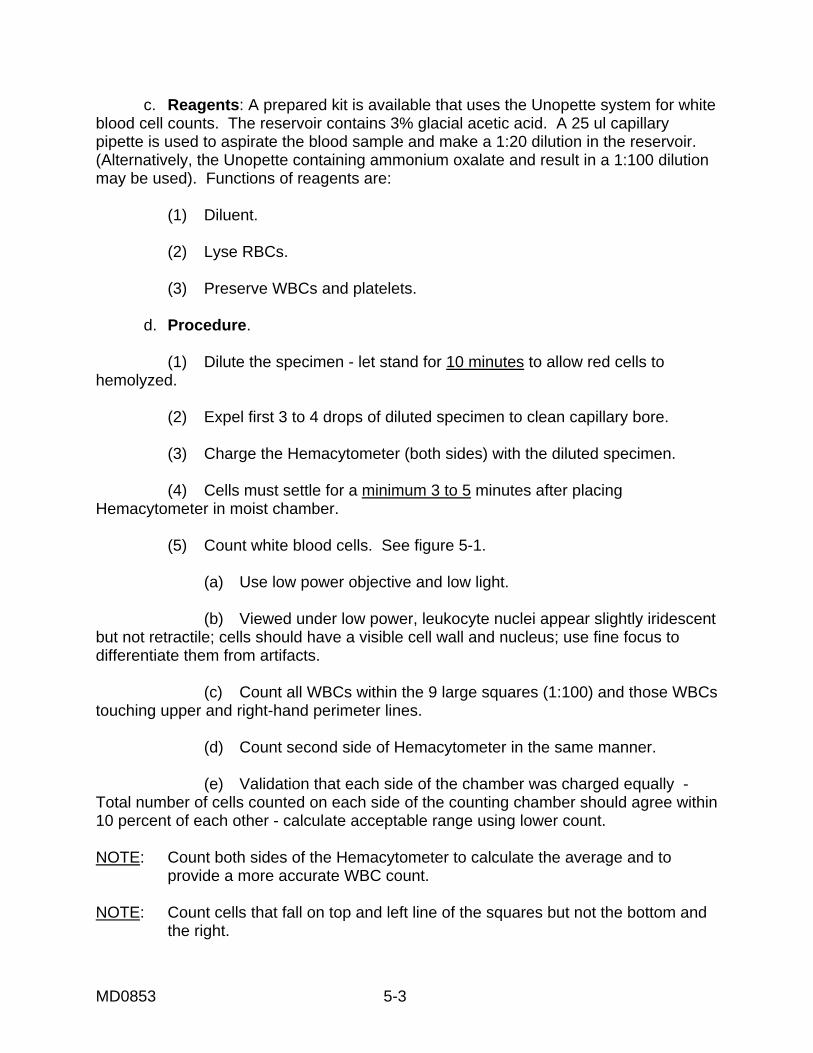
c. Reagents: A prepared kit is available that uses the Unopette system for white blood cell counts. The reservoir contains 3% glacial acetic acid. A 25 ul capillary pipette is used to aspirate the blood sample and make a 1:20 dilution in the reservoir. (Alternatively, the Unopette containing ammonium oxalate and result in a 1:100 dilution may be used). Functions of reagents are: (1) Diluent. (2) Lyse RBCs. (3) Preserve WBCs and platelets. d. Procedure. (1) Dilute the specimen - let stand for 10 minutes to allow red cells to hemolyzed. (2) Expel first 3 to 4 drops of diluted specimen to clean capillary bore. (3) Charge the Hemacytometer (both sides) with the diluted specimen. (4) Cells must settle for a minimum 3 to 5 minutes after placing Hemacytometer in moist chamber. (5) Count white blood cells. See figure 5-1. (a) Use low power objective and low light. (b) Viewed under low power, leukocyte nuclei appear slightly iridescent but not retractile; cells should have a visible cell wall and nucleus; use fine focus to differentiate them from artifacts. (c) Count all WBCs within the 9 large squares (1:100) and those WBCs touching upper and right-hand perimeter lines. (d) Count second side of Hemacytometer in the same manner. (e) Validation that each side of the chamber was charged equally - Total number of cells counted on each side of the counting chamber should agree within 10 percent of each other - calculate acceptable range using lower count. NOTE: Count both sides of the Hemacytometer to calculate the average and to provide a more accurate WBC count. NOTE: Count cells that fall on top and left line of the squares but not the bottom and the right.
MD0853 5-4
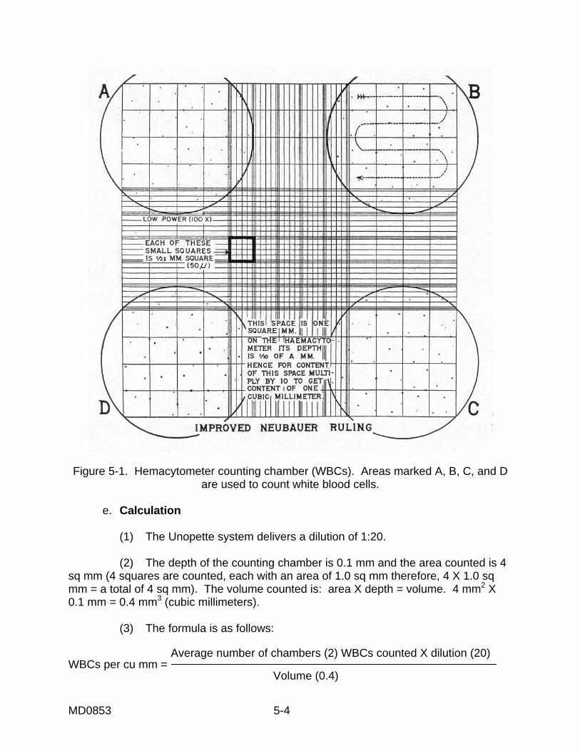
Figure 5-1. Hemacytometer counting chamber (WBCs). Areas marked A, B, C, and D are used to count white blood cells.
e. Calculation (1) The Unopette system delivers a dilution of 1:20. (2) The depth of the counting chamber is 0.1 mm and the area counted is 4 sq mm (4 squares are counted, each with an area of 1.0 sq mm therefore, 4 X 1.0 sq mm = a total of 4 sq mm). The volume counted is: area X depth = volume. 4 mm2 X 0.1 mm = 0.4 mm3 (cubic millimeters). (3) The formula is as follows: Average number of chambers (2) WBCs counted X dilution (20) WBCs per cu mm = Volume (0.4)

MD0853 5-5
(4) For example: First Chamber Second Chamber Cells counted in Cells counted each square in each square 35 45 40 37 44 36 39 44 158 WBCs counted 162 WBCs counted Average of the 2 chamber counts. Total the amount and divide by 2: 158 320 - = 160 WBCs +162 2 320 Then: 60 X 20 =8,000/mm3 or 8.0 X 10⁹ WBCs/ L 0.4 f. Sources of Error. (1) Improper collection of blood specimens causes variable results. (2) Wet or dirty pipets. (3) Not allowing cells to settle for an adequate amount of time. (4) Poor pipetting technique causes high or low counts. Poor pipetting technique includes: (a) Undershooting Unopette with blood. (b) Overfilling Unopette with blood. (c) Air bubbles in the shaft. (d) Not mixing the blood specimen thoroughly. (5) Failure to expel 3 or 4 drops in the pipet tips before charging the Hemacytometer. (6) Overfilling the chamber of the hemacytometer, which causes erroneously high counts.
MD0853 5-6
(7) Not mixing the diluted specimen prior to filling the Hemacytometer. (8) Uneven distribution of cells in the counting chamber causes erroneous results. (9) Counting artifacts. (10) Dirty or scratched Hemacytometer. (11) Failure to mix anticoagulated blood thoroughly before use. g. Discussion. (1) The counting chamber must be scrupulously clean and free of debris that might be mistaken for cells. (2) The minimum blood sample recommended for performing routine white blood cell counts is that obtained using one pipet and counting two chambers as previously outlined. (3) If nucleated erythrocytes are present, the count is corrected by the following formula: observed count X 100 corrected count = 100 + percent nucleated erythrocytes (4) The percent nucleated erythrocyte is obtained from the differential count, which is discussed in another subcourse. h. Normal Values (1) Adults (both sexes): 4,500 to 11,500 WBCs per cu mm or 4.5-11.5 X 10⁹ WBCs/L. (2) Childhood: 6,000 to 14,000 WBCs per cu mm or 6.0-14.0 X 10⁹ WBCs/L. (3) Birth: 9,000 to 30,000 WBCs per cu mm or 9.0-34.0 X 10⁹ WBCs / L. 5-3. TOTAL EOSINOPHIL COUNT a. Principle. A sample of blood is diluted with a solution that selectively stains the eosinophils and eliminates all other leukocytes and erythrocytes from view. Following mixing, the specimen is introduced into the counting chamber and the number of eosinophils in a known volume of blood is counted.

MD0853 5-7
b. Reagent. A prepared kit is available that uses the Unopette system for absolute eosinophil counts. The reservoir contains phloxine B solution in propylene glycol and distilled water. A 25 ul capillary pipette is used to aspirate the blood sample and make a 1:32 dilution in the reservoir. Alternatively, stains including Pilot’s solution or Randolph’s stain, may be prepared by the laboratory as described elsewhere. c. Procedure. (1) Add the sample for the Unopette pipette to the reservoir. (2) Mix by gently shaking the pipets for 30 seconds. Prolonged and harsh shaking will tend to cause rupturing of the eosinophils. (3) Let stand for 10 minutes to allow red cells to hemolyze. (4) Expel first 3 to 4 drops of diluted specimen to clean capillary bore. (5) Using one pipet, charge both chambers of a hemacytometer and with the other pipet charge both chambers of the second Hemacytometer. (6) Allow both hemacytometers to stand for 15 minutes to permit staining of the eosinophils. To prevent evaporation, the hemacytometers are placed on a damp towel and covered with Petri dish covers. (7) Under low-power magnification, count the red-stained eosinophils in the entire ruled area (9 sq mm) each of the four chambers (a total area of 36 sq mm). The chamber has a depth of 0.1 mm so the total vo1ume is 3.6 cu mm. d. Calculations. Number of eosinophils counted X dilution (10) Number of eosinophils = Per cu mm volume (3.6) e. Source of Error. See paragraph 5-2e. f. Discussion. (1) Fuchs-Rosenthal or Levy Hemacytometer is preferable to a standard counting chamber since its greater volume (4.0 X 4.0 X 0.2 mm) allows for counting of more cells, thereby reducing the statistical error. Counting two of these chambers is equivalent in accuracy to seven standard chamber counts. (2) In eosinopenia, it is necessary to set up more chambers to provide an optional number of cells to be counted.
MD0853 5-8
(3) The eosinacetone diluting fluids are unsatisfactory and should not be used. (4) Estimation of eosinophils on a stained blood smear is too inaccurate for use because of poor cellular distribution. (5) The propylene glycol in Pilot's solution renders the erythrocytes invisible, and the sodium carbonate causes lysis of all the leukocytes except the eosinophils. The phloxine stains the eosinophils. (6) In the Thorn test an eosinophil count must be made prior to the initiation of the test proper. This establishes the patient's total eosinophil count, to which the response of the adrenal cortex to adrenocorticotropic hormone (ACTH) can be judged. The ACTH is then injected and, at an interval of 4 hours, another eosinophil count is made. The interpretation of this test is as follows: (a) Normal--approximately a 50 percent drop in eosinophils. (b) Cushing's disease (hyperadrenalism)--0-30 eosinophils per cu mm. (c) Addison's disease (hypoadrenalism) no change in eosinophil count. (7) Nasal smears are also submitted for eosinophil evaluation. These smears are stained with Wright's stain and examined for the presence of eosinophils. g. Normal Value. Normal value: 150-300 eosinophils per cu mm. 5-4. RETICULOCYTE COUNT a. Principle. Nonnucleated immature erythrocytes contain nuclear remnants of RNA and the cell is known as a reticulocyte. To detect the presence of this RNA, the red cells must be stained while they are still living. This process is called supravital staining. With supravital staining, the RNA appears as a reticulum within the red cell. b. Reagent. (1) New Methylene Blue Solution. Dissolve 0.5 grams of new methylene blue, 1.4 grams of potassium oxalate, and 0.8 grams of sodium chloride in distilled water. Dilute to 100 ml. Filter before use. (2) Brilliant Cresyl Blue Solution. Dissolve 1.0 grams of brilliant cresyl blue in 99 ml of .85 per cent sodium chloride. Filter before use .

MD0853 5-9
c. Procedure. (1) Mix equal amounts of blood and new methylene blue stain (2-3 drops or 50 uL each) and allow to incubate at room temperature for 3-6 min. (2) This allows the reticulocytes adequate time to take up the stain. (3) At the end of 15 minutes, mix the contents of the tube well. (4) Place a small drop of the mixture on two clean glass slides and prepare a thin smear (prepare two smears). (5) Counter stain with Wright's stain, if desired. (6) Allow smear to air-dry. (7) Place the slide on the microscope stage and, using the low power objective, locate the thin portion of the smear in which the red cells are evenly distributed and are not touching each other. (8) Switch to oil immersion magnification and count the number of reticulocytes in five fields of 200 RBCs. d. Calculation. Number of reticulocytes counted = % reticulocytes 10 e. Sources of Error. (1) Equal volumes of blood and stain give optimum staining conditions. An excess of blood causes the reticulum to understain. An excess of stain usually obscures the reticulum. (2) Crenated erythrocytes and rouleaux formation make an accurate count difficult to perform. (3) Stain precipitated on erythrocytes causes them to appear as reticulocytes. (4) Dirty slides cause uneven spreading. (5) The dye solution should have adequate time to penetrate the cell and stain the reticulum.

MD0853 5-10
f. Discussion. (1) Reticulocytes are nonnucleated erythrocytes that exhibit blue reticulum strands within their cytoplasm when stained supravitally. When stained only with Wright's stain, they are buff-pink in color and larger and darker than erythrocytes. (2) Reticulocytes serve as an index of the activity of the bone marrow in blood regeneration. As such, these counts are of value in following anti-anemia therapy. Satisfactory response to therapy is evidenced by an increase of reticulocytes in the peripheral blood. Increased reticulocyte counts also occur whenever there is rapid bone marrow activity as in leukemia or blood regeneration associated with hemorrhage or hemolysis. Decreased reticulocyte counts occur in conditions in which the bone marrow is not producing adequate red blood cells, such as aplastic anemia. (3) Several methods for staining and counting reticulocytes are in common use. Compared to the use of alcoholic solutions of dye, methods employing saline solutions of new methylene blue can give slightly higher values for reticulocytes. For comparative studies, the same method should be used throughout the work. (4) Precipitated stain is often confused with reticulum but can be recognized by its presence throughout the smear and apart from the red cells. Precipitation can be eliminated as a source of error by frequently filtering the stain. (5) An alternate method of counting reticulocytes utilizes the Miller disk that is placed inside the microscope eyepiece. This disc consists of 2 squares as shown below in figure 5-2. The area of the smaller square (B) is a tenth that of square A. Therefore, if there are 40 red cells in square A, there should be four red cells present in square B. When employing this method to count reticulocytes, the red cells in square B are counted in successive fields on the slide, until a total of 500 red cells have been counted. At the same time, the reticulocytes in square A are enumerated. At the completion of the count, theoretically, the reticulocytes obtained in this way are divided by 50, in order to obtain the percent reticulocytes present in the blood.
Figure 5-2. Miller disc.

MD0853 5-11
g. Normal Values. (1) Birth to 1 day. Two and one-half to 6.0 percent. (2) 1 day to 2 weeks. 0.30 to 1.5 percent. (3) 2 weeks to adult. 0.50 to 2.20 percent.
Section II. MANUAL COUNTS, OTHER BODY FLUIDS 5-5. CEREBROSPINAL FLUID CELL COUNTS a. Principle. Cerebrospinal fluid is delivered to a counting chamber and examined microscopically for blood cells. Normally, spinal fluid is clear. If it is cloudy, dilute before charging the counting chamber. b. Reagent. Normal saline. c. Procedure. NOTE: Set up cell counts on spinal fluids within 30 minutes after withdrawal of the specimen. (1) Clear spinal fluid is set up as follows: (a) With a transfer pipet introduce a drop of well-mixed spinal fluid into both counting chamber of a Hemacytometer. CAUTION: Avoid contamination by careful handling of spinal fluid. (b) Examine the entire ruled area for the presence of cellular elements. If both leukocytes and erythrocytes are observed, note the condition of the red cells (fresh or crenated). (c) Count all cells in the entire ruled area (0.9 cu mm). (2) Turbid sample – Perform dilution using normal saline. (a) Slightly hazy – 1:10 dilution. (b) Hazy – 1:20 dilution. (c) Mix the specimen well. (d) Discard the fluid in the capillary portion of the pipet.

MD0853 5-12
(e) Charge the counting chamber and allow the cells to settle for five minutes. (f) Under low-power magnification count all cells in the entire ruled area (0.9 cu mm). (g) Switch to high-power and perform a rough differential count on prepared smear. d. Calculations. (1) Clear spinal fluid: Number of cells counted x dilution factor x area factor x depth factor = total cells / ul (2) Turbid spinal fluid: Number of cells counted X dilution (10) x area factor x depth factor = total cells / ul (3) Very clouded spinal fluid: Number of cells counted X dilution (20) x area factor x depth factor = total cells / ul e. Source of Error. See paragraph 5-2e. f. Discussion. (1) If more than 100 leukocytes per cu mm are present, centrifuge the undiluted specimen, make a smear, and stain with modified Wright's stain. Perform a routine differential count and also estimate the ratio of erythrocytes to leukocytes. NOTE: It may be necessary to use egg albumin or cell-free serum to make the sediment adhere to the slide. (2) Normally the spinal fluid is water clear. It can be turbid if cell count is 500 or more cells per cu mm. If there is fresh blood with spontaneous clotting, the indications are those of a bloody tap. Xanthochromia develops after subarachnoid hemorrhage has been present for a few hours and is due to disintegration of blood pigments. Xanthochromia may also develop from tumors, abscesses, and inflammation.

MD0853 5-13
(3) Cell counts above ten are considered to be evidence of intracranial disease. The predominant cell in most viral infections, syphilis, and tuberculous meningitis is the lymphocyte. Bacterial infections due to meningococcus, pneumococcus, and so forth, usually result in a predominance of the neutrophil. Cerebral and extradural abscesses as well as subdural hemorrhages produce a neutrophilic response although bacteria are not demonstrated. (4) Biochemical, bacteriological, virological, serological, and hematological ands are all necessary to reflect the true condition of the cerebrospinal fluid. The current laboratory standing operating procedures should give guidance for the most efficient method to accomplish all the necessary ands. g. Normal Value: Zero to five cells per cu mm (chiefly lymphocytes). 5-6. SEMEN ANALYSIS a. Principle. Semen analysis involves gross examination (volume, color, turbidity, viscosity, and pH) and microscopic examination (motility and spermatozoa count). b. Reagent. Tap water. c. Collection Instructions. A physician will usually give the instructions; however, the patient should be reminded of several critical points. (1) The patient may be required to abstain from intercourse for 48 to 72 hours. (2) The specimen is collected in a clean container that has been pre-warmed to body temperature. (3) The specimen should be delivered to the laboratory within 1 hour. (4) The specimen must be kept at body temperature (37oC) and not subjected to extremes of heat or cold. d. Gross Examination. (1) Record. the time of collection and receipt of the specimen. (2) Measure and record the volume. (3) Observe and record the color (white, gray, yellow, and so forth), turbidity (clear, opalescent, opaque, and so forth), and viscosity (viscid, gelatin, liquid). (4) Determine the pH with a pH reagent strip and record this.

MD0853 5-14
e. Motility Examination. (1) When the specimen becomes fluid (within 15 to 30 minutes after collection, the semen liquifies by the action of fibrinolysin), place one drop on a slide (pre-warmed to 37ºC) and place a cover slip on it. (2) Under high dry power, count motile and nonmotile spermatozoa in two or more areas until a total of at least 200 spermatozoa have been observed. It is necessary to focus through the entire depth of a given field so as to include nonmotile spermatozoa that may have settled to the bottom of the slide. Only those that move forward actively are considered motile. Record the percent of motile spermatozoa seen. (3) Repeat this procedure in three hours and six hours, using a new drop from the original specimen each time. f. Spermatozoa Count. (1) Make a 1:20 dilution of seminal fluid with diluent (tap water). (2) Mix sample thoroughly and charge a Hemacytometer. (3) Count the spermatozoa in the same manner as you would count white blood cells. (4) After counting the sperm, examine the morphology and report the percent of abnormal forms. Morphologically normal sperm are quite uniform in appearance. Any sperm with rounded, enlarged, small, or bilobed heads are abnormal. Abnormal tails are enlarged, small, irregular in length, absent, or multiple. See figure 5-3 for morphology of spermatozoa.
Figure 5-3. Morphology of spermatozoa.
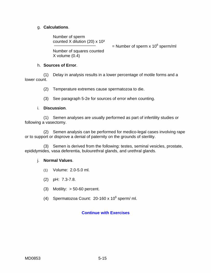
MD0853 5-15
g. Calculations. Number of sperm counted X dilution (20) x 10³ = Number of sperm x 106 sperm/ml Number of squares counted X volume (0.4) h. Sources of Error. (1) Delay in analysis results in a lower percentage of motile forms and a lower count. (2) Temperature extremes cause spermatozoa to die. (3) See paragraph 5-2e for sources of error when counting. i. Discussion. (1) Semen analyses are usually performed as part of infertility studies or following a vasectomy. (2) Semen analysis can be performed for medico-legal cases involving rape or to support or disprove a denial of paternity on the grounds of sterility. (3) Semen is derived from the following: testes, seminal vesicles, prostate, epididymides, vasa deferentia, bulourethral glands, and urethral glands. j. Normal Values. (1) Volume: 2.0-5.0 ml. (2) pH: 7.3-7.8. (3) Motility: > 50-60 percent. (4) Spermatozoa Count: 20-160 x 106 sperm/ ml.
Continue with Exercises

MD0853 5-16
EXERCISES, LESSON 5 INSTRUCTIONS: Answer the following exercises by marking the lettered responses that best answer the exercise, by completing the incomplete statement, or by writing the answer in the space provided at the end of the exercise. After you have completed all of the exercises, turn to "Solutions to Exercises" at the end of the lesson and check your answers. For each exercise answered incorrectly, reread the material referenced with the solution. 1. For a WBC count, after drawing blood into the diluting pipet, it is necessary to expel first 3 to 4 drops of diluted specimen in order to: a. Clean capillary bore. b. Dirtying the pipet. c. Dirtying the hemacytometer cover glass. d. Waste cells. 2. When doing a WBC count, to what mark should the diluting fluid be drawn? a. 3. b. 7. c. 11. d. 13. 3. When performing a WBC count, which reagents may be used as diluants? a. Acetic acid or Ammonium Oxalate. b. Acetic acid or citric acid. c. Acetic acid or hydrochloric acid. d. Acetic acid or trisphosphoric acid.

MD0853 5-17
4. For the WBC count, immediately after the contents of the pipet have been mixed for about three minutes, it is necessary to: a. Use a mechanical mixer. b. Expel three to four drops. c. Observe for even distribution of cells. d. Refill both chambers of the Hemacytometer. 5. After the WBCs have settled for about three minutes during a manual WBC count, which powered magnification and lighting arrangements are used to focus on the ruled area to observe for even distribution of WBC? a. Low-power (10X); reduced light. b. High-power (43X); bright light. c. Oil immersion (97X); bright light. d. High-power (43X); reduced light. 6. When counting WBCs, a variation of more than ___________ cells between any of the four areas counted or a variation of more than _____________cells between sides of the Hemacytometer indicate uneven distribution and require that the procedure be repeated. a. 6; 12. b. 7; 9. c. 10; 20. d. 10; 18.

MD0853 5-18
7. How long do you let the WBC diluent sit to allow RBCs to lyse? a. 10 min. b. 20 min. c. 15 min. d. 5 min. 8. Which WBCs are counted? a. Those touching the inner left-hand bottom lines. b. All WBCs outside the squares. c. All WBCs within the square and those touching the upper and right hand center lines. d. All WBCs within the square and those touching the upper and left- hand center lines. 9. How many 1-sq-mm comer areas and chambers are used to count WBCs? a. 3; 4. b. 4; 2. c. 2; 6. 10. Which chemical is mixed with whole blood when obtaining a WBC count? a. Sodium chloride. b. Weak acid. c. Weak base. d. Calcium carbonate.
MD0853 5-19
11. Let diluted blood stand for ______ minutes and allow cells to settle for _____ minutes after placing in the hemacytometer. a. 45, 5-10. b. 15, 1-2. c. 10, 3-5. d. 1, 6-8. 12. The usual blood dilution for the manual WBC count is: a. 1:10. b. 1:20. c. 1:100. d. 1:200. 13. The volume is the: a. Area X width. b. Width X length. c. Width X depth. d. Area X depth.

MD0853 5-20
14. Using the Hemacytometer counting chamber, the formula for calculating the WBC count is: a. Average number of WBCs counted X Dilution = WBCs per cu mm Volume b. Average number of WBCs counted X Dilution = WBCs per sq in Volume c. Average number of WBCs counted X Volume = WBCs per cu mm Dilution d. Average number of WBCs per cu mm X WBCs counted = Dilution Volume 15. If blood is drawn in Unopette with a dilution of 1:20, what is the patient if the average of two chamber counts is 163? a. 3,260 WBCs per cu mm. b. 8,530 WBCs per cu mm. c. 8,150 WBCs per cu mm. d. 10,320 WBCs per cu mm. 16. If blood is drawn in Unopette with a dilution of 1:20, what is the patient if the average of two chamber counts is 214? a. 4,280 WBCs per cu mm. b. 8,350 WBCs per cu mm. b. 9,984 WBCs per cu mm. c. 10,700 WBCs per cu mm.

MD0853 5-21
17. If blood is drawn in Unopette with a dilution of 1:20, what is the patient if the average of two chamber counts is 198? a. 3,960 WBCs per cu mm. b. 9,900 WBCs per cu mm. c. 9,984 WBCs per cu mm. d. 10,540 WBCs per cu mm. 18. What are common sources of error when performing a manual Hemacytometer WBC count? a. Wet or dirty pipets. b. Poor pipetting techniques. c. Improper collection of blood specimen. d. All of the above. 19. Overfilling the chamber of the Hemacytometer can cause: a. Erroneous low counts. b. Erroneous high counts. c. Erroneous equal counts. d. No errors to the counts. 20. If the WBC count is 10,210 and the differential indicates there are 19 nucleated RBCs per 100 WBCs, What is the corrected WBC count? a. 8,650. b. 8,580. c. 9,580. d. 1,021,000.

MD0853 5-22
21. If the WBC count is 9,640 and the differential indicates there are 14 nucleated RBCs per 100 WBCs, What is the corrected WBC count? a. 7,390. b. 8,256. c. 8,456. d. 946,000. 22. What is the normal range of the WBC count in adults? a. 4,500-11,500 WBCs per cu mm. b. 6,000-14,000 WBCs per cu mm. c. 9,000-30,000 WBCs per cu mm. d. 4.2-5.4 million WBCs per cu mm. 23. If 88 eosinophils are counted in a 36-sq mm area of a Hemacytometer using a 1:10 dilution, what is the eosinophil count? a. 20 eosinophil per cu mm. b. 150 eosinophil per cu mm. c. 244 eosinophil per cu mm. d. 300 eosinophil per cu mm. 24. An unchanged eosinophil count 4 hours after the injection of ACTH is indicative of: a. Addison’s disease. b. Hyperadrenalism. c. Cushing’s disease. d. Normal adrenocortical function.
MD0853 5-23
25. Which stain is used to evaluate eosinophil nasal smears? a. Pink. b. Orange. c. Supravital. d. Wright's. 26. The reticulocyte is an immature: a. Rubriblast. b. Erythroyte. c. Prorubricyte. d. Metarubricyte 27. If 15 reticulocytes are counted in a total of 1,000 erythrocytes, what percentage of reticulocytes should be reported? a. 0.015 percent. b. 1.15 percent. c. 2.5 percent. d. 1.5 percent. 28. If 86 reticulocytes are counted in a total of 1,000 erythrocytes, what percentage of reticulocytes should be reported? a. 8.6 percent. b. 4.6 percent. c. 66 percent. d. 86 percent.

MD0853 5-24
29. In tuberculosis meningitis, the predominant WBC type usually found in the spinal fluid is the: a. Monocyte. b. Eosinophil. c. Lymphocyte. d. All of the above. 30. If 198 cells are counted in an undiluted spinal fluid, what is the cell count? a. 2.2 per cu mm. b. 22 per cu mm. c. 220 per cu mm. d. 2,200 per cu mm. 31. If 47 cells are counted in a spinal fluid diluted 1:10, what is the cell count? a. 4.7 per cu mm. b. 52.2 per cu mm. c. 470 per cu mm. d. 522 per cu mm. 32. White blood cell counts on spinal fluid that are above ___________________ are usually considered indicative of some type of intracranial disease. a. 10 per cu mm. b. 15 per cu mm. c. 20 per cu mm. d. 25 per cu Im1.

MD0853 5-25
33. In most viral infections, the predominant cell usually found in the spinal fluid is the: a. Neutrophil. b. Basophil. c. Eosinophil. d. Lymphocyte. 34. In subdural hemorrhages, the predominant cell type found in spinal fluid is usually the: a. Lymphocyte. b. Neutrophil. c. Monocyte. d. Segmented lymphocyte. 35. The neutrophil cell is predominant in which disease or infection? a. Tuberculous meningitis. b. Syphilis. c. Bacterial infections. d. Hodgkin’s disease. 36. Except for the diluting fluid used, the spermatozoa count is almost identical in procedure to the: a. RBC count. b. WBC count. c. Reticulocyte count. d. Total eosinophil count.

MD0853 5-26
37. What is the patient required to abstain from prior to having a Semen analysis collected? a. Water intake. b. Using a pre-warmed container. c. Intercourse. d. Using the Miller disk. 38. What three factors should be observed and recorded during gross and of the semen specimen? a. Color, viscosity, and temperature. b. Color, amount of blood, and viscosity. c. Mucus dissolved, temperature, and color. d. Viscosity, color, and turbidity. 39. During a motility and of spermatozoa, which cells are considered to be motile? a. The entire mixture. b. The entire depth of the field. c. All active ones moving forward. d. Only those that are floating. 40. When should the motility procedure be repeated when examining spermatoza specimens? a. Every 15 minutes. b. In 3 hours and 6 hours. c. Within 30 minutes of collection.

MD0853 5-27
41. Fibrinolysin causes what type of change to the semen? a. It solidifies. b. It is modified with Wright's stain. c. It causes it to liquefy. d. Nothing. 42. Semen analysis can be performed for ____________________ cases involving rape or in support or denial of paternity on the grounds of __________________. a. Medico-legal; sterility. b. Medico-vasa deferentia; sterility. c. Abnormal; epididymides. 43. From which of the following male body parts is semen derived? a. Testes, seminal vesicles, prostate, epididymides, vasa deferentia, reticulocytes, and urethral glands. b. Testes, seminal vesicles, prostate, erythrocytes, vasa deferentia, bulbourethral glands, and urethral glands. c. Testes, motility spermatozoa, seminal vesicles, prostate, epididymides, vasa deferentia, bulbourethral glands, and urethral glands. d. Testes, seminal vesicles, prostate, epididymides, deferentia, bulbourethral glands, and urethral glands. 44. Which of the following is a correct normal value of spermatoza? a. Volume: 0.5-5.0 ml. b. pH: 7.4-7.6. c. Motility: greater than 50 percent. d. Spermatozoa Count: 25 to 50 million per ml.

MD0853 5-28
45. Which of the following is a correct normal value of spermatoza? a. Volume: 1.5-5.2 ml. b. pH: 7.3-7.8. c. Motility: 60 to 94 percent. d. Spermatozoa Count: 60-170 million per ml.
Check Your Answers on Next Page

MD0853 5-29
SOLUTIONS TO EXERCISES, LESSON 5 1. a (para 5-2d(2)) 2. c (para 5-2c(3)) 3. c (para 5-2b) 4. b (para 5-2d(2)) 5. a (para 5-2d(5a)) 6. c (para 5-2d(5e)) 7. d (para 5-2c(1)) 8. c (para 5-2d(5c)) 9. b (paras 5-2d(5c, d)) 10. b (para 5-2a) 11. c (paras 5-2d(1), (4)) 12. b (para 5-2c) 13. d (para 5-2e(2)) 14. a (paras 5-2e(3), (4)) 15. c (paras 5-2e(3), (4)) 16. d (paras 5-2e(3), (4)) 17. b (paras 5-2e(3), (4)) 18. c (paras 5-2f(1), (2), (4)) 19. b (para 5-2f(6)) 20. b (para 5-2g(3)) 21. c (para 5-2g(3)) 22. a (para 5-2h(1))
MD0853 5-30
23. c (para 5-3d) 24. a (para 5-3f(6)) 25. d (para 5-3f(7)) 26. b (para 5-4a) 27. d (para 5-4d) 28. a (para 5-4d) 29. d (para 5-5f(3)) 30. c (para 5-5d(1)) 31. d (para 5-5d(2)) 32. a (para 5-5f(3)) 33. d (para 5-5f(3)) 34. b (para 5-5f(3)) 35. c (para 5-5f(3)) 36. b (para 5-6f(5)) 37. c (para 5-6c(1)) 38. d (para 5-6d(3)) 39. c (para 5-6e(2)) 40. b (para 5-6e(3)) 41. c (para 5-6e(1)) 42. a (para 5-6i(2)) 43. d (para 5-6i(3)) 44. c (para 5-6j(3)) 45. b (para 5-6j(2))
End of Lesson 5