leprosy of the hard palate and the premaxillary...
TRANSCRIPT
1286 Journal of Clinical and Diagnostic Research. 2011 November (Suppl-1), Vol-5(6): 1286-1288Journal of Clinical and Diagnostic Research. 2011 November (Suppl-1), Vol-5(6): 1286-12881286 1287Journal of Clinical and Diagnostic Research. 2011 November (Suppl-1), Vol-5(6): 1286-1288Journal of Clinical and Diagnostic Research. 2011 November (Suppl-1), Vol-5(6): 1286-12881286 1287
Leprosy of The Hard Palate and The Premaxillary Gingiva: A Case Report
Key Words: oral lesions, leprosy, premaxillary gingiva, hard palate
ABSTRACTLeprosy was first described in the ancient Indian texts from
the 6th century BC, as a non fatal, chronic infectious disease
which was caused by Mycobacterium leprae, whose clinical
manifestation was largely confined to the skin, the peripheral
nervous system, the upper respiratory tract, the eyes, and the
testis. Oral lesions are rare, but when they are present, they occur
in the lepromatous form. This article describes the clinical and
the microscopic findings of a case of lepromatous leprosy with
oral manifestations. The diagnosis was based on the clinical and
histopathological findings, the multidrug therapy for multibacillary
leprosy was started and continued for 24 months and the patient
completed the treatment. We describe here, a case of a 64 yrs
old female who presented to us with a large, left premaxillary
growth. Clinically, a large, well defined, lobulated mass over the
left premaxillary region and the adjoining gingiva was observed.
Sanjay P. KiShve, PuruShottam a. Giri, Kiran j. Shinde
en
tCase Report
InTRoduCTIonLeprotic oral lesions which are more common in the lepromatous form of leprosy, indicate a late manifestation, and have a great epidemiological importance as a source of infection. The global burden of leprosy has declined dramatically, from 5.2 million cases in 1985 to 204,800 cases at the end of 2009, having a prevalent rate which is <1 per 10,000 [1]. In India, after the introduction of MDT, the leprosy case load came down from 57.6 cases per 10,000 population in 1985 to less than one case per 10, 000 population in 2005 [1]. The two polar ends of the disease spectrum include lepromatous and tuberculoid leprosy. The tuberculoid form represents the strongest response, whereas a relatively anergic state is reflected by the lepromatous form [2].Cell mediated immunity is considered to be the crucial defence against the disease and the magnitude of this immunity defines the extent of the disease [2]. Oral mucosal lesions are seen in about 20-60% cases of lepromatous leprosy, while they are quite rare in the tuberculoid and the borderline forms [2].The lesions are proportional to the duration of the disease, indicating that these are late manifestations [3,4]. The propensity of the disease, when untreated, results in characteristic deformities and the recognition in most of the cultures, that the disease is communicable from person to person, has resulted historically in a profound social stigma. With the institution of appropriate and effective antimicrobial therapy, the patients can lead productive lives in the community, and deformities and other visible manifestations can largely be prevented. The authors emphasize here, the importance of the evaluation of the oral mucosa by a medical health professional during patient care, since the oral lesions may act as a source of infection.
CASe RepoRT:A 64 yrs old female presented with complaints of a progressively growing mass at the left upper alveolar region, of 3 yrs duration. The growing mass was about the size of a peanut over the palatal side of the upper alveolar region, which progressively increased to
the present size. Approximately, 1 yr ago, a similar mass appeared over the buccal surface, involving the same alveolar margin. She did complain of some difficulty in mastication and phonetic articulation, but gave no complaints of the loss of sensation of the hard palate or the oral mucosa. Her husband was also diagnosed for lepromatous leprosy and he had received treatment for it 10 years ago.
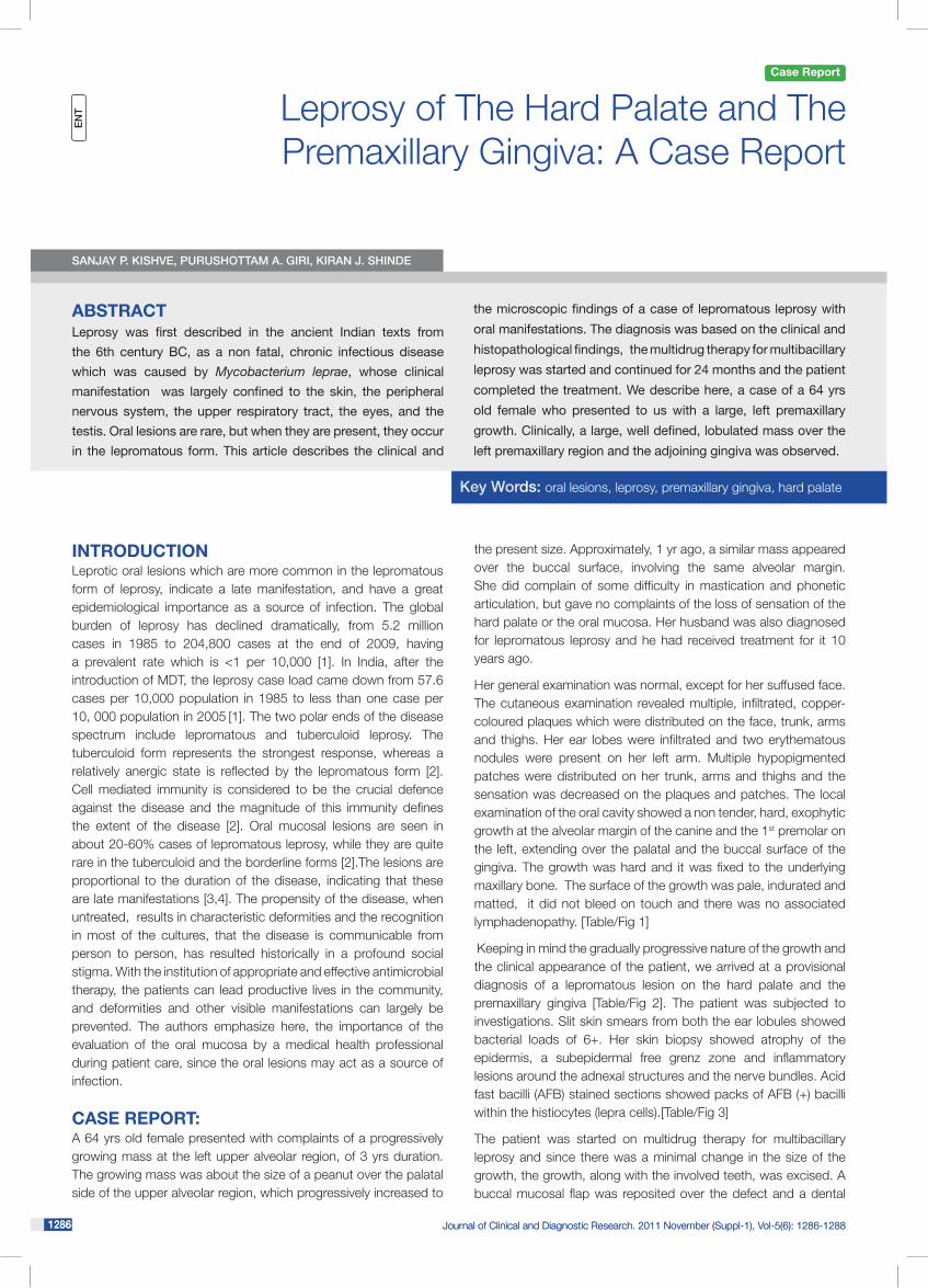
Her general examination was normal, except for her suffused face. The cutaneous examination revealed multiple, infiltrated, copper-coloured plaques which were distributed on the face, trunk, arms and thighs. Her ear lobes were infiltrated and two erythematous nodules were present on her left arm. Multiple hypopigmented patches were distributed on her trunk, arms and thighs and the sensation was decreased on the plaques and patches. The local examination of the oral cavity showed a non tender, hard, exophytic growth at the alveolar margin of the canine and the 1st premolar on the left, extending over the palatal and the buccal surface of the gingiva. The growth was hard and it was fixed to the underlying maxillary bone. The surface of the growth was pale, indurated and matted, it did not bleed on touch and there was no associated lymphadenopathy. [Table/Fig 1]
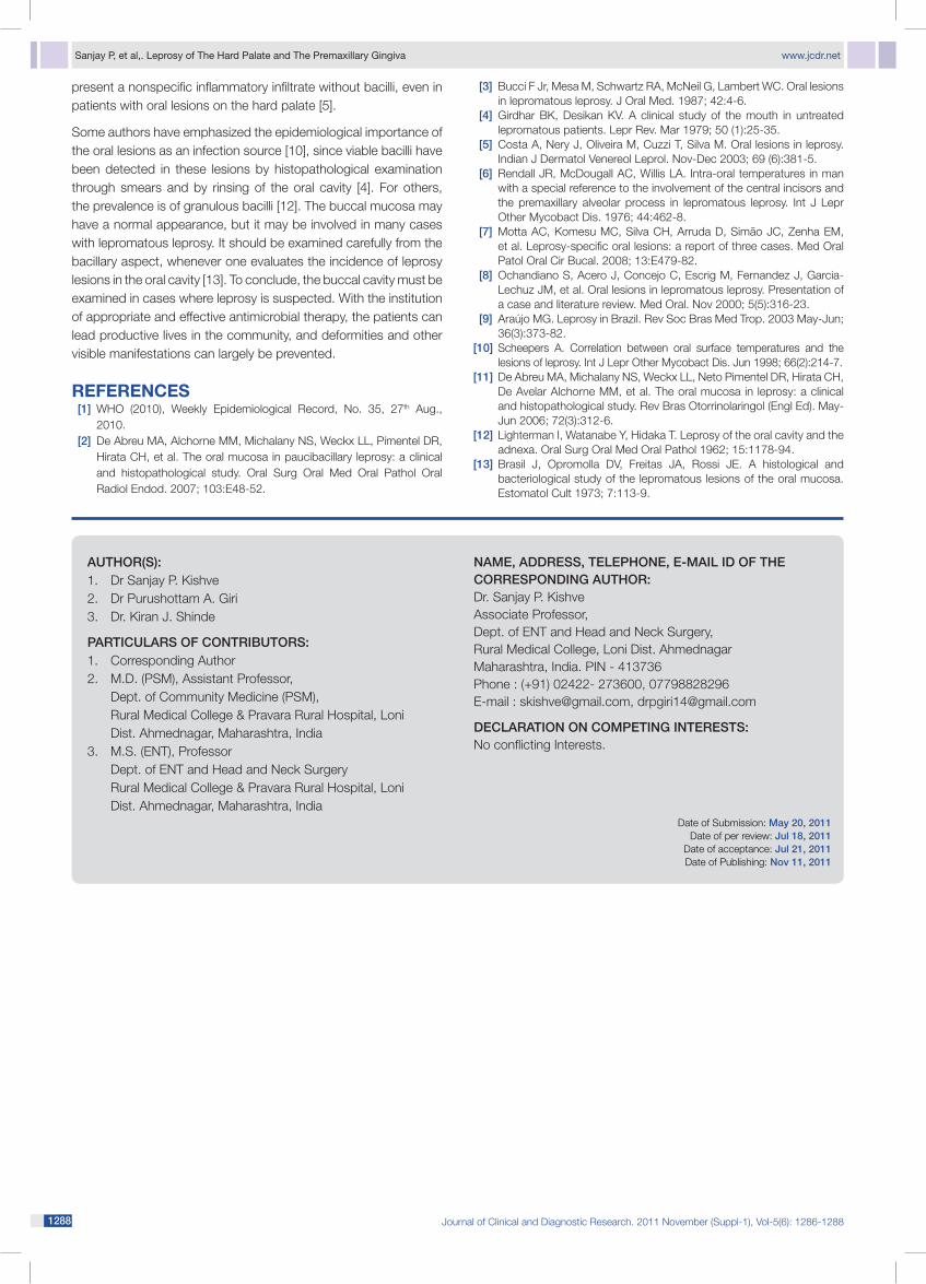
Keeping in mind the gradually progressive nature of the growth and the clinical appearance of the patient, we arrived at a provisional diagnosis of a lepromatous lesion on the hard palate and the premaxillary gingiva [Table/Fig 2]. The patient was subjected to investigations. Slit skin smears from both the ear lobules showed bacterial loads of 6+. Her skin biopsy showed atrophy of the epidermis, a subepidermal free grenz zone and inflammatory lesions around the adnexal structures and the nerve bundles. Acid fast bacilli (AFB) stained sections showed packs of AFB (+) bacilli within the histiocytes (lepra cells).[Table/Fig 3]
The patient was started on multidrug therapy for multibacillary leprosy and since there was a minimal change in the size of the growth, the growth, along with the involved teeth, was excised. A buccal mucosal flap was reposited over the defect and a dental

Journal of Clinical and Diagnostic Research. 2011 November (Suppl-1), Vol-5(6): 1286-1288Journal of Clinical and Diagnostic Research. 2011 November (Suppl-1), Vol-5(6): 1286-12881286 1287
www.jcdr.net Sanjay P, et al,. Leprosy of The Hard Palate and The Premaxillary Gingiva
1287Journal of Clinical and Diagnostic Research. 2011 November (Suppl-1), Vol-5(6): 1286-1288Journal of Clinical and Diagnostic Research. 2011 November (Suppl-1), Vol-5(6): 1286-12881286 1287
implant was advised. On follow up visits, the lesion over the palate was found to be completely resolved. [Table/Fig 4]
Histopathological findings: Sections of the excised mass showed lining stratified squamous epithelial hyperplasia with occasional pleomorphism and hyperchromatism. The epithelial stroma showed dense, fibrocollagenous stroma with congested and dilated blood vessels and infiltration by chronic inflammatory cells, mainly lymphocytes. No evidence of malignancy was noted.
dISCuSSIonThe upper airway is the main point of entry for the bacillus and a route for bacillary elimination in leprosy [4,9,10]. For this reason, the control of the mucosal lesions is very important. The mucosal involvement is particularly outstanding in the nose, probably due to the preference of M. leprae for cooler sites [9,10,11]. The oral lesions of leprosy occur more frequently in areas of the mouth which have a lower surface temperature [6]. The oral lesions usually appear as ulcerations on the hard or soft palates [3,5,8] as was observed in our case. The main oral cavity sites of leprosy include the gingiva in the anterior portion of the maxilla, the hard and soft palates, the uvula and the tongue [3]. In advanced leprosy, the mouth can acquire the characteristics of a reservoir of bacilli, and it may thus act as an important risk factor for the transmission of the illness [7].
M. leprae favours temperatures which are a little below the body temperature, for its multiplication [6,10]. Based on this fact, a pathophysiological mechanism has been postulated for the oral involvement: a nasal lesion with obstruction of the air flow leads to oral breathing (mouth breathing), which is very common in lepromatous leprosy. This causes a decrease in the intra-oral temperature, mainly in sites near the air intake and in the anterior areas, thus facilitating the harbouring of the bacillus [6,8,10].
In a study which was conducted in Brazil, which included 26 patients with leprosy, whose oral lesions were evaluated and biopsied, 11 were found to have the lepromatous form, 14 had borderline leprosy and one had tuberculoid leprosy. Only in two lepromatous patients, solid staining bacilli were found on histopathological examination. Biopsies of the buccal mucosa did not show any changes or
[Table/Fig-1]: Preoperative picture of the gingival and premaxillary growth
[Table/Fig-2]: Intra-operative picture after excision of the growth
[Table/Fig-3]: Fast stain of the ear lobule split skin smear (Stained with Hematoxylin-Eosin (H & E) and Wade (AFB) stain)
[Table/Fig-4]: Excised specimen
Sanjay P, et al,. Leprosy of The Hard Palate and The Premaxillary Gingiva www.jcdr.net
1288 Journal of Clinical and Diagnostic Research. 2011 November (Suppl-1), Vol-5(6): 1286-1288Journal of Clinical and Diagnostic Research. 2011 November (Suppl-1), Vol-5(6): 1286-12881288 PB
author(S):1. Dr Sanjay P. Kishve2. Dr Purushottam A. Giri3. Dr. Kiran J. Shinde
PartiCuLarS oF ContriButorS:1. Corresponding Author2. M.D. (PSM), Assistant Professor, Dept. of Community Medicine (PSM), Rural Medical College & Pravara Rural Hospital, Loni Dist. Ahmednagar, Maharashtra, India 3. M.S. (ENT), Professor Dept. of ENT and Head and Neck Surgery Rural Medical College & Pravara Rural Hospital, Loni Dist. Ahmednagar, Maharashtra, India
name, addreSS, teLePhone, e-maiL id oF the CorreSPondinG author:Dr. Sanjay P. Kishve Associate Professor, Dept. of ENT and Head and Neck Surgery, Rural Medical College, Loni Dist. Ahmednagar Maharashtra, India. PIN - 413736Phone : (+91) 02422- 273600, 07798828296E-mail : [email protected], [email protected]
deCLaration on ComPetinG intereStS: No conflicting Interests.
Date of Submission: may 20, 2011 Date of per review: jul 18, 2011
Date of acceptance: jul 21, 2011Date of Publishing: nov 11, 2011
present a nonspecific inflammatory infiltrate without bacilli, even in patients with oral lesions on the hard palate [5].
Some authors have emphasized the epidemiological importance of the oral lesions as an infection source [10], since viable bacilli have been detected in these lesions by histopathological examination through smears and by rinsing of the oral cavity [4]. For others, the prevalence is of granulous bacilli [12]. The buccal mucosa may have a normal appearance, but it may be involved in many cases with lepromatous leprosy. It should be examined carefully from the bacillary aspect, whenever one evaluates the incidence of leprosy lesions in the oral cavity [13]. To conclude, the buccal cavity must be examined in cases where leprosy is suspected. With the institution of appropriate and effective antimicrobial therapy, the patients can lead productive lives in the community, and deformities and other visible manifestations can largely be prevented.
ReFeRenCeS [1] WHO (2010), Weekly Epidemiological Record, No. 35, 27th Aug.,
2010. [2] De Abreu MA, Alchorne MM, Michalany NS, Weckx LL, Pimentel DR,
Hirata CH, et al. The oral mucosa in paucibacillary leprosy: a clinical and histopathological study. Oral Surg Oral Med Oral Pathol Oral Radiol Endod. 2007; 103:E48-52.
[3] Bucci F Jr, Mesa M, Schwartz RA, McNeil G, Lambert WC. Oral lesions in lepromatous leprosy. J Oral Med. 1987; 42:4-6.
[4] Girdhar BK, Desikan KV. A clinical study of the mouth in untreated lepromatous patients. Lepr Rev. Mar 1979; 50 (1):25-35.
[5] Costa A, Nery J, Oliveira M, Cuzzi T, Silva M. Oral lesions in leprosy. Indian J Dermatol Venereol Leprol. Nov-Dec 2003; 69 (6):381-5.
[6] Rendall JR, McDougall AC, Willis LA. Intra-oral temperatures in man with a special reference to the involvement of the central incisors and the premaxillary alveolar process in lepromatous leprosy. Int J Lepr Other Mycobact Dis. 1976; 44:462-8.
[7] Motta AC, Komesu MC, Silva CH, Arruda D, Simão JC, Zenha EM, et al. Leprosy-specific oral lesions: a report of three cases. Med Oral Patol Oral Cir Bucal. 2008; 13:E479-82.
[8] Ochandiano S, Acero J, Concejo C, Escrig M, Fernandez J, Garcia-Lechuz JM, et al. Oral lesions in lepromatous leprosy. Presentation of a case and literature review. Med Oral. Nov 2000; 5(5):316-23.
[9] Araújo MG. Leprosy in Brazil. Rev Soc Bras Med Trop. 2003 May-Jun; 36(3):373-82.
[10] Scheepers A. Correlation between oral surface temperatures and the lesions of leprosy. Int J Lepr Other Mycobact Dis. Jun 1998; 66(2):214-7.
[11] De Abreu MA, Michalany NS, Weckx LL, Neto Pimentel DR, Hirata CH, De Avelar Alchorne MM, et al. The oral mucosa in leprosy: a clinical and histopathological study. Rev Bras Otorrinolaringol (Engl Ed). May-Jun 2006; 72(3):312-6.
[12] Lighterman I, Watanabe Y, Hidaka T. Leprosy of the oral cavity and the adnexa. Oral Surg Oral Med Oral Pathol 1962; 15:1178-94.
[13] Brasil J, Opromolla DV, Freitas JA, Rossi JE. A histological and bacteriological study of the lepromatous lesions of the oral mucosa. Estomatol Cult 1973; 7:113-9.