latium clinical seminar · 2017-07-18 · method of arterial stiffness for: •its relative ease in...
TRANSCRIPT

LATIUM CLINICAL SEMINARRoma Policlinico Umberto I
21 aprile 2012
Il danno vascolare: la rigidità arteriosa
Vascular damage: arterial stiffness
Giuseppe Germanò

2007 ESH/ESC Hypertension Guidelines
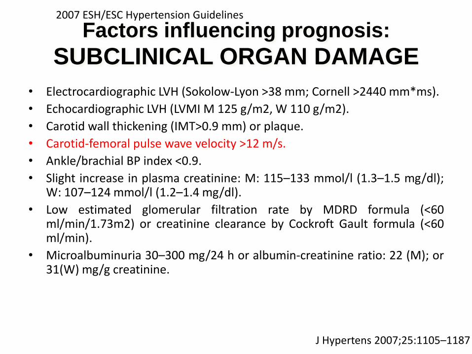
• Electrocardiographic LVH (Sokolow-Lyon >38 mm; Cornell >2440 mm*ms).
• Echocardiographic LVH (LVMI M 125 g/m2, W 110 g/m2).
• Carotid wall thickening (IMT>0.9 mm) or plaque.
• Carotid-femoral pulse wave velocity >12 m/s.
• Ankle/brachial BP index <0.9.
• Slight increase in plasma creatinine: M: 115–133 mmol/l (1.3–1.5 mg/dl);W: 107–124 mmol/l (1.2–1.4 mg/dl).
• Low estimated glomerular filtration rate by MDRD formula (<60ml/min/1.73m2) or creatinine clearance by Cockroft Gault formula (<60ml/min).
• Microalbuminuria 30–300 mg/24 h or albumin-creatinine ratio: 22 (M); or31(W) mg/g creatinine.
Factors influencing prognosis:
SUBCLINICAL ORGAN DAMAGE
J Hypertens 2007;25:1105–1187


• Central Aortic Pressure
• Pulse Wave Velocity (PWV)
• Augmentation Index (AIx)
Measures of Arterial Stiffness

7.6 m/sec605 mm690 mm85 mm80 msec135 msec55 msec
Aortic PWV
(distance/tim
e)
distance
Notch-
femoral
Notch-
carotid
timeQRS-
femoral
QRS-
carotid
85 mm
690 mm
EKG-QRS
CAROTID
55 msec 135 msec
FEMORAL
EKG-QRS
How PWV is measured...
Velocity = Distance/Time

APWV measurement (cont.)

• Pulse Wave Velocity ( PWV )
• Complior ( Carotid-Femoral PWV )• SphygmoCor ( Applanation Tonometry)• Tensiomed Arteriograph
• Augmentation Index ( Aix)
• SphygmoCor ( Applanation Tonometry)• Tensiomed Arteriograph
• Central Blood pressure ( SBP, PP )
• SphygmoCor ( Applanation Tonometry)• Tensiomed Arteriograph
“…studies clearly demonstrate that PWV and the augmentation index are associated with the structural changes of atherosclerosis”
Davies, J.I., J Hypertens 21:463-472; 2003


PULSE WAVE VELOCITYThe Complior® device

PULSE WAVE VELOCITY
Augmentation Index ( Aix)The SphygmoCor device
Probe
Bone
Artery


THE AUGMENTATION INDEX (Aix)Augmentation index (Aix %) = (P2 – P1 / PP) x 100
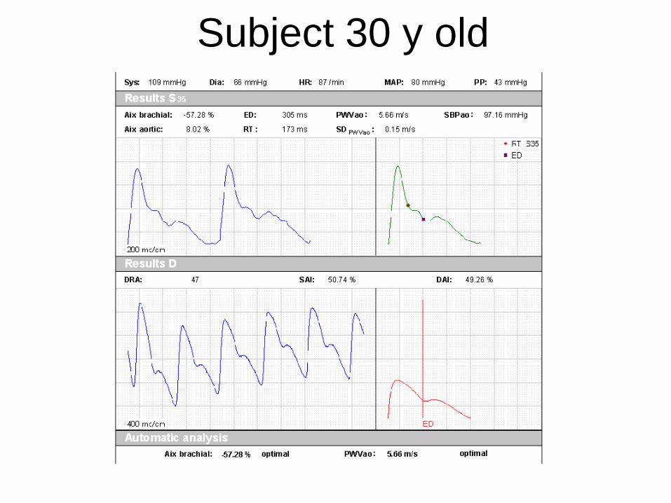
Subject 30 y old

Subject 60 year old

Subject 90 y old

QAS
,.


Systole
Aortic Stiffening and Early Wave Reflection
Diastole
Systole
Young compliant arteries : Normal PW velocity (8 m/sec)
Elderly stiff arteries with ISH : Increased PW velocity (12 m/sec)
(1) Ventricular-Vascular coupling
(2) coronary blood flow
(1) Ventricular-vascular mismatch
(2) The reflected wave increases or “augments” central SBP during late systole:

THE AUGMENTATION INDEX (Aix)E’DEFINITO COME LA DIFFERENZA TRA IL SECONDO (ONDA RIFLESSA) ED IL PRIMO
PICCO (ONDA INCIDENTE) ED ESPRESSO COME PERCENTUALE DELLA PRESSIONE DIFFERENZIALE.
Augmentation index (Aix %) = (P2 – P1 / PP) x 100
AIX È UN INDICE INDIRETTO DELLA RIGIDITA AORTICA, RAPPRESENTA L’EFFETTODELL’ONDA RIFLESSA SULL’ONDA INCIDENTE A LIVELLO DELL’AORTA ASCENDENTE.È UNA MISURA DEL CARICO ADDIZIONALE A CUI IL Vsx È SOTTOPOSTO A CAUSADELL’ONDA DI RIFLESSIONE.

SphygmoCor
• Arterial stiffness measures– CBP (central BP)
– AIX (Augmentation Index)
– PVW(Pulse wave velocity)
• ? Evidence to change management?
• Does depend on accurate peripheral blood pressure measurement eg: BPtru / manual BP
• How to incorporate it with out interfering with the work flow?


First author
(year; country)
Events Follow-up
(years)
Type of patient
(number)
Mean age
at entry
(years)
Aortic PWV – YES
Blacher (1999;Fr) CV mortality 6,0 ESRD (241) 51
Laurent (2001;Fr) CV mortality 9,3 Hypertension (1980) 50
Meaume (2001;Fr) CV mortality 2,5 Elderly (>70) (141) 87
Shoji (2001;Jp) CV mortality 5,2 ESRD (265) 55
Boutouyrie (2002;Fr) CHD events 5,7 Hypertension (1045) 51
Cruickshank (2002;GB) All cause M. 10,7 Diabetes and MS (571) 51
Laurent (2003;Fr) Fatal strokes 7,9 Hypertension (1715) 51
Sutton-Tyrrell (2005;USA) CV events 4,6 Elderly (2488) 74
By Laurent S
First author
(year; country)
Events Follow-up
(years)
Type of patient
(number)
Mean age at
entry
(years)
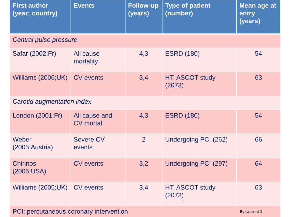
Central pulse pressure
Safar (2002;Fr) All cause
mortality
4,3 ESRD (180) 54
Williams (2006;UK) CV events 3,4 HT, ASCOT study
(2073)
63
Carotid augmentation index
London (2001;Fr) All cause and
CV mortal
4,3 ESRD (180) 54
Weber
(2005;Austria)
Severe CV
events
2 Undergoing PCI (262) 66
Chirinos
(2005;USA)
CV events 3,2 Undergoing PCI (297) 64
Williams (2005;UK) CV events 3,4 HT, ASCOT study
(2073)
63
PCI: percutaneous coronary intervention By Laurent S

-Arterial wave reflections and survival in end
stage renal failure.
London GM et al Hypertension 2001;38:434-
438.
- Central pulse pressure and mortality in end
stage renal failure.
Safar ME et al Hypertension 2002;39:735-738.
- Increased arterial wave reflections predict
severe cardiovascular events in patient
undergoing percutanous coronary
interventations
Eur Heart J 2005;26:2657-2663
- Central pressure more strongly relates to
vascular disease and outcome than does
brachial pressure: the Strong Heart Study.
Roman MJ et al Hypertension 2007;50:197-
203.
•


Aix PWV
Ottimale <- 30% <7m/s
Normale da -30% a < -10% da 7m/s a < 9.7m/s
Aumentato da -10% a <10% da 9.7m/s a <12m/s
Anormale > 10% >12m/s
Arterial Stiffness
PULSE WAVE VELOCITY (PWV)AUGMENTATION INDEX (Aix)

Carotid to femoral pulse wave velocity
(PWV) has emerged as the gold standard
method of arterial stiffness for:
• its relative ease in determination,
• its perceived reliability
and because of :
• its association with incident CV disease
independently of traditional risk factors
THE PROS
Boutouyre P et al Eur Heart J 2010
doi:1093/eurheartj/ehq165

• The fixed threshold value (12 m/s) proposed in
the 2007 ESH/ESC hypertension guidelines was
based on published epidemiological studies but
could not take into account the multiple factors
influencing PWV
• it has been proven that important differences in
absolute PWV values exist between
methodologies and/or between populations.
• its strong dependence on age and BP•
THE CONS
Boutouyre P et al Eur Heart J 2010
doi:1093/eurheartj/ehq165
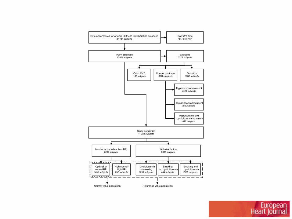
Flowchart describing the selection and categorization of subjects from the reference value database for the present analysis.
Eur Heart J 2010;eurheartj.ehq165

Normal values for pulse wave velocity: average according to age (1455 subjects).
Eur Heart J 2010;eurheartj.ehq165

(Top) Pulse wave velocity (PWV) vs age and MBP.
Eur Heart J 2010;eurheartj.ehq165

Reference values for pulse wave velocity (PWV): mean values according to age and blood pressure (BP) categories (11 092 subjects).
Eur Heart J 2010;eurheartj.ehq165
.org

RESULTS
• The establishment of normal and reference
values for PWV based on an extensive data set
obtained from 13 centres distributed across
Europe
• to take into account different methodological
approaches for the determination of PWV
• to apply previously established conversion
equations for path lengths and transit times.
• to present reference values per age decade
and BP category.
Boutouyre P et al Eur Heart J 2010
doi:1093/eurheartj/ehq165

TOP: Brachial (solid symbols) and derived central aortic (open symbols) systolic
blood pressure with time (mean, 95% CI) for patients randomized to receive
atenolol ± thiazide- or amlodipine ±perindopril-based therapy.
BOTTOM: Systolic blood pressure difference (brachial minus central aortic; mean, 95% CI) with time. For calculation
of AUC, see the Data Supplement. Numbers below abscissa represent the number of patients seen at each time
point. Time represents the duration from randomization into ASCOT to patient
follow-up visit at which tonometry measurement was made in the CAFE
study.PP indicates pulse pressure.
CAFE Investigators, for ASCOT Investigators. Circulation
2006;113:1213.

CAFÉ Study Results