kynr, a lrp/asnc-type transcriptional regulator, directly
TRANSCRIPT
JOURNAL OF BACTERIOLOGY, Dec. 2011, p. 6567–6575 Vol. 193, No. 230021-9193/11/$12.00 doi:10.1128/JB.05803-11Copyright © 2011, American Society for Microbiology. All Rights Reserved.
KynR, a Lrp/AsnC-Type Transcriptional Regulator, Directly Controlsthe Kynurenine Pathway in Pseudomonas aeruginosa�
Claire A. Knoten, L. Lynn Hudson, James P. Coleman, John M. Farrow III, and Everett C. Pesci*Department of Microbiology and Immunology, The Brody School of Medicine at
East Carolina University, Greenville, North Carolina 27834
Received 12 July 2011/Accepted 23 September 2011
The opportunistic pathogen Pseudomonas aeruginosa can utilize a variety of carbon sources and producesmany secondary metabolites to help survive harsh environments. P. aeruginosa is part of a small group ofbacteria that use the kynurenine pathway to catabolize tryptophan. Through the kynurenine pathway, trypto-phan is broken down into anthranilate, which is further degraded into tricarboxylic acid cycle intermediatesor utilized to make numerous aromatic compounds, including the Pseudomonas quinolone signal (PQS). Wehave previously shown that the kynurenine pathway is a critical source of anthranilate for PQS synthesis andthat the kynurenine pathway genes (kynA and kynBU) are upregulated in the presence of kynurenine. A putativeLrp/AsnC-type transcriptional regulator (gene PA2082, here called kynR), is divergently transcribed from thekynBU operon and is highly conserved in Gram-negative bacteria that harbor the kynurenine pathway. Weshow that a mutation in kynR renders P. aeruginosa unable to utilize L-tryptophan as a sole carbon source anddecreases PQS production. In addition, we found that the increase of kynA and kynB transcriptional activityin response to kynurenine was completely abolished in a kynR mutant, further indicating that KynR mediatesthe kynurenine-dependent expression of the kynurenine pathway genes. Finally, we found that purified KynRspecifically bound the kynA promoter in the presence of kynurenine and bound the kynB promoter in theabsence or presence of kynurenine. Taken together, our data show that KynR directly regulates the kynureninepathway genes.
Pseudomonas aeruginosa is a Gram-negative bacillus that isubiquitous throughout nature and infects a variety of hosts. Itis a common nosocomial pathogen known to cause seriousopportunistic infections in immunocompromised individuals(25). P. aeruginosa also causes a chronic infection in cysticfibrosis (CF) patients that is difficult, if not impossible, toeradicate and ultimately leads to increased morbidity and mor-tality in this population (4, 37). In order to survive during suchinfections and in many other harsh environments, P. aeruginosautilizes numerous different carbon sources and produces awide range of secondary metabolites (21, 24, 28, 35, 38). Onepotential nutrient, tryptophan, can be used by some bacteria toprovide building blocks for many secondary metabolites, someof which function as siderophores, signaling molecules, andprotective compounds (1, 11, 28, 44). Similar to eukaryotes, P.aeruginosa catabolizes tryptophan through the kynureninepathway (11, 22). Kurnasov et al. utilized comparative geneticsto identify several bacteria with putative kynurenine pathways,including Bacillus anthracis, P. aeruginosa, and Bordetella per-tussis (22). The kynurenine pathway contrasts significantlyfrom the major tryptophan catabolic pathway of Escherichiacoli (and many other species of bacteria), which catabolizesL-tryptophan anaerobically into indole, pyruvate, and ammoniavia a pyridoxal phosphate-dependent tryptophanase (41, 50).Nevertheless, with either catabolic pathway the ability to utilize
both tryptophan and tryptophan breakdown products as a car-bon and nitrogen source, and as precursors for many secondarymetabolites, provides a unique advantage for survival withinnutrient-limited and harsh environments.
The conversion of L-tryptophan into quinolinate via thekynurenine pathway has been demonstrated in multiple bacte-rial species (22). However, P. aeruginosa only encodes thegenes for the anthranilate branch of the kynurenine pathway(23). This branch catabolizes L-tryptophan into anthranilate viaa three-step enzymatic pathway (22). The three enzymes areencoded by kynA (which encodes a tryptophan-2,3-dioxyge-nase), kynB (kynurenine formamidase), and kynU (kynureni-nase) (see Fig. 7 for pathways) (11, 23). The kynA gene islocated separately on the P. aeruginosa chromosome, whilekynB and kynU are encoded in a putative operon. The kynure-nine pathway was linked to P. aeruginosa virulence when it wasshown that radiolabeled tryptophan was incorporated into thePseudomonas quinolone signal (PQS) (11), which is importantfor virulence in multiple models of infection (10, 13, 27, 34,49). These data also showed that despite the presence of twoalternative pathways for anthranilate production, the kynure-nine pathway is the main source of anthranilate for PQS pro-duction when P. aeruginosa is grown in the presence of tryp-tophan or tryptophan breakdown metabolites (11, 31).
Due to both the importance of the kynurenine pathway inthe production of PQS and because tryptophan is a costlyamino acid to synthesize (11, 52), it would be advantageous forP. aeruginosa to strictly regulate the catabolism of tryptophan.Multiple transcriptome arrays have shown that quorum sensingmay regulate the kynurenine pathway (36, 43), and the quo-rum-sensing regulator LasR was predicted through chromatin
* Corresponding author. Mailing address: Department of Microbi-ology and Immunology, East Carolina University School of Medicine,BT 132, 600 Moye Blvd., Greenville, NC 27834. Phone: (252) 744-2351.Fax: (252) 744-3535. E-mail: [email protected].
� Published ahead of print on 30 September 2011.
6567
on April 10, 2019 by guest
http://jb.asm.org/
Dow
nloaded from
immunoprecipitation-chip analysis to bind to the promoterregion of kynB (15). Our laboratory has also shown that thetranscription of both kynA and kynB was significantly increasedin the presence of kynurenine (11). With these data in mind,we began to search for a transcriptional regulator that couldspecifically regulate kynA and kynB in the presence of kynure-nine. We identified a putative transcriptional regulator en-coded by gene PA2082, which is divergently transcribed fromkynB and is homologous to the Lrp/AsnC family of transcrip-tional regulators. Lrp/AsnC-type regulators are known to con-trol amino acid metabolism, and though Lrp has a global reg-ulatory role in E. coli, many other regulators within the familyhave more specific regulons (3, 51). We demonstrate here thatthe protein encoded by gene PA2082 directly binds to andregulates the kynurenine pathway genes, and we thereforepropose that this protein be named the kynurenine pathwayregulator (KynR).
MATERIALS AND METHODS
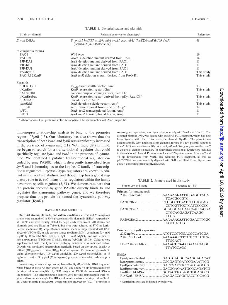
Bacterial strains, plasmids, and culture conditions. E. coli and P. aeruginosastrains were maintained in 30% glycerol and 10% skim milk (Difco), respectively,at �80°C and were freshly plated to begin each experiment. All strains andplasmids used are listed in Table 1. Bacteria were cultured at 37°C in Luria-Bertani medium (LB), Vogel Bonner minimal medium supplemented with 0.5%glycerol (VBG) (42), or sole carbon source medium (SCM); containing 73.4 mMK2HPO4, 16.76 mM NaNH5PO4 � 4H2O, 0.8 mM MgSO4, and with either 10mM L-tryptophan (TSCM) or 10 mM L-alanine (ASCM) (pH 7.0). Cultures weresupplemented with the kynurenine pathway metabolites as indicated below.Growth was monitored spectrophotometrically based on the optical density at600 nm (OD600) for E. coli or OD660 for P. aeruginosa. To maintain plasmids, 30�g/ml chloramphenicol, 100 �g/ml ampicillin, 200 �g/ml carbenicillin, or 15�g/ml (E. coli) or 30 �g/ml (P. aeruginosa) gentamicin was added when appro-priate.
In order to generate an expression plasmid for KynR, a 568-bp DNA fragment,which began at the kynR start codon (ATG) and ended 46 bp downstream fromthe stop codon, was amplified by PCR using strain PAO1 chromosomal DNA asthe template. The oligonucleotide primers used for this amplification were en-gineered to contain a single HindIII site downstream from the stop codon (Table2). Vector plasmid pHERD30T, which contains an araBAD (PBAD) promoter to
control gene expression, was digested sequentially with SmaI and HindIII. Thedigested plasmid DNA was ligated with the kynR PCR fragment, which had alsobeen digested with HindIII, to create the plasmid pKynRex. This plasmid wasused to amplify kynR and regulatory elements for use in a two-plasmid system inE. coli. PCR was used to amplify both the kynR and divergently transcribed araCto ensure all elements necessary for controlled expression of KynR were includedin the subcloned plasmid. Primers were located 52 bp downstream from araC and46 bp downstream from kynR. The resulting PCR fragment, as well aspACYC184, were sequentially digested with SalI and HindIII and ligated to-gether, generating plasmid pKynRsubex.
TABLE 1. Bacterial strains and plasmids
Strain or plasmid Relevant genotype or phenotypea Reference
E. coli DH5� F� endA1 hsdR17 supE44 thi-1 recA1 gyrA relA1 (lacZYA-argF)U169 deoR��80dlac�(lacZ)M15recA1�
48
P. aeruginosa strainsPAO1 Wild type 19PAO-R1 lasR::Tc deletion mutant derived from PAO1 14PJF-KA1 kynA deletion mutant derived from PAO1 11PJF-KB1 kynB deletion mutant derived from PAO1 11PJF-KU1 kynU deletion mutant derived from PAO1 11PA�KynR kynR deletion mutant derived from PAO1 This studyPAO-R1�KynR kynR lasR deletion mutant derived from PAO-R1 This study
PlasmidspHERD30T PBAD-based shuttle vector, Gmr 33pKynRex KynR expression vector, Gmr This studypACYC184 General purpose cloning vector, Tetr Chlr 5pKynRsubex KynR expression vector derived from pKynRex, Chlr This studypEX18Ap Suicide vector, Ampr 17pkynRdel kynR deletion suicide vector, Ampr This studypLP170 lacZ transcriptional fusion vector, Ampr 32pJF01 kynB�-lacZ transcriptional fusion, Ampr 11pJF03 kynA�-lacZ transcriptional fusion, Ampr 11
a Abbreviations: Gm, gentamicin; Tet, tetracycline; Chl, chloramphenicol; Amp, ampicillin.
TABLE 2. Primers used in this study
Primer use and name Sequence (5�–3�)a
Primers for mutagenesis2082-F1-remake..........................AAAAAAGAATTCGAGGTAGA
TCACGCCGTCPA2082Rev1 ...............................CCGGCCTTGATCTCCTGCAGT
CCTGGTTGCTCATCCGCCCPA2082Fwd2...............................GGGCGGATGAGCAACCAGGA
CTGCAGGAGATCAAGGCCGG
PA2082Rev2 ...............................AAAAAAGAATTCGAACTTGGCGGCGATGG
Primers for KynR expression2082atgfwd ..................................ATGTCCCTGGACGCCATCGA2082 Rev Herd ...........................AAAAAAGCTTCGTCCCTCTCA
TTGCACTHerd2082exsubRev ....................AAAAGTCGACCGAAGCAGGG
TTATGCAGC
EMSAkynApromoterfwd ......................GAGTGAGGGCAAGGACACATkynApromoterrev .......................CGCGAGTGATCCGAAATTCGkynBpromoterfwd.......................GACTGATGTCCCAGTAGCGGkynBpromoterrev........................GACGGAGAATGCGCAGATCGFwdKynU-EMSA .......................GGTACTTGTAGGTGCAGCCGRevKynU-EMSA........................CAAGACCGGCTACCTGCACG
a Restriction sites are indicated by bold type.
6568 KNOTEN ET AL. J. BACTERIOL.
on April 10, 2019 by guest
http://jb.asm.org/
Dow
nloaded from
Generation of kynR mutants. A splicing-by-overlap extension protocol wasused to generate mutant alleles (47). Alleles were constructed to contain in-frame deletions in the coding DNA sequences corresponding to amino acids 25to 140 for KynR (73% of the protein sequence). Primers were designed tocontain approximately 1 kb of DNA both upstream and downstream from thesplice junction, and each primer added an EcoRI restriction site to both ends ofthe PCR product. Both the kynRdel PCR product and pEX18Ap (suicide vector)were digested with EcoRI and ligated together. The resulting plasmid, pkynRdel,was transformed into strains PAO1 and PAO-R1 by electroporation (6). Mutantswere selected by plating transformants on medium containing carbenicillin andthen on medium containing 6% sucrose to remove the vector sequence (17).PCR was used to screen colonies, and DNA sequencing of PCR products wasused to confirm mutants.
PQS production. Washed cells from overnight cultures were used to inoculate10-ml cultures of LB or VBG supplemented with either water, 1 mM L-trypto-phan, 1 mM L-kynurenine, or 1 mM anthranilate (final concentrations) to anOD660 of 0.05. After 6 and 24 h of growth, 300-�l samples of each culture wereextracted with 900 �l of acidified ethyl acetate as previously described (8).One-half of the resulting organic phase was evaporated to dryness at 37°C, and50 �l of 1:1 acidified ethyl acetate-acetonitrile was used to reconstitute theextract. Samples were analyzed by thin-layer chromatography (TLC), visualizedby long-wave UV light, and photographed (8).
�-Gal assays for P. aeruginosa. Cells from overnight cultures of P. aeruginosagrown in VBG were washed and resuspended in fresh medium to an OD660 of0.05 and incubated at 37°C with shaking at �180 rpm for 6 h. At that time, eitherwater or 1 mM L-kynurenine was added, and cultures continued to grow at 37°Cwith shaking at �180 rpm for an additional 18 h. The cells were collected bycentrifugation at 12,000 g for 2 min, resuspended in fresh VBG, and assayedfor -Galactosidase (-Gal) activity in duplicate. Data are presented in Millerunits as the mean � standard deviation [�(n � 1)] of at least 3 separate experi-ments.
�-Gal assays for E. coli. Overnight cultures of E. coli carrying the appropriateplasmids were grown in LB and used to inoculate 10-ml cultures of fresh mediumto an OD600 of 0.08. The subcultures were incubated at 37°C with shaking at�180 rpm until an OD600 of 0.3 was reached. Then, 2-ml aliquots were trans-ferred to tubes that contained 0.1% L-arabinose to induce kynR expression, andeither water, 1 mM L-kynurenine, 1 mM L-tryptophan, or 1 mM anthranilic acid(final concentrations) was added. The cells were incubated for 2 h at 37°C withshaking at �180 rpm. After incubation, the -Gal activity produced by eachculture was assayed, and data are reported as the mean � �(n � 1) of at least 3independent experiments.
Purification of KynR. The method used for purification of KynR is a modifi-cation of those described by Madhusudhan et al. for the purification of BkdRfrom Pseudomonas putida (26). A 10-ml overnight culture of E. coli strainDH5�(pKynRex) was used to inoculate 250 ml of LB and was incubated at 37°Cwith shaking at �180 rpm until an OD600 of 0.7 was reached. At this point, 1%L-arabinose (Sigma-Aldrich) was added to induce kynR expression, and theculture was grown for an additional 3 h at 37°C with shaking at �180 rpm. Cellswere harvested by centrifugation at 6,000 g for 10 min and then resuspendedin 20 mM Tris-HCl (pH 7.4) and 1 mM dithiothreitol (DTT) (buffer A). The cellswere lysed by sonication, and the lysate was cleared by centrifugation at 37,000 g for 1 h at 4°C (Beckman Coulter Optima L-100 XP ultracentrifuge). KynRpurification was performed at room temperature, and all samples were kept onice or at 4°C until purification was complete. The soluble fraction (90 mg of totalprotein) was applied to a HiTrap DEAE Sepharose Fast Flow column (1 ml; GEHealthcare) in buffer A at a flow rate of 0.5 ml/min. The column was washed withbuffer A until the A280 returned to 0 ( 50 ml). Bound proteins were eluted witha 30-ml linear gradient of 0 to 0.3 M NaCl at a flow rate of 1 ml/min, and 3-mlfractions were collected. Samples from the collected fractions were precipitatedusing deoxycholic acid and tricholoracetate acid (DCA/TCA) and then separatedby 15% SDS-PAGE to identify fractions containing KynR. These fractions werecollected and dialyzed against a buffer containing 20 mM Tris-HCl (pH 7.4), 100mM NaCl, 5 mM MgCl2, and 1 mM DTT (buffer B) overnight at 4°C. Thedialyzed fractions were then applied to a HiTrap heparin column (5-ml; GEHealthcare) equilibrated with buffer B. The column was washed with buffer Buntil the A280 returned to 0 ( 24 ml). Bound proteins were eluted with a 50-mllinear gradient of 0.1 to 0.4 M NaCl in buffer B at a flow rate of 1 ml/min, and3-ml fractions were collected. Again, proteins from the collected fractions wereprecipitated with DCA/TCA and were separated on a 15% SDS-polyacrylamidegel to identify the fractions containing KynR. All fractions containing KynR werepooled and dialyzed against 20 mM Tris-HCl (pH 7.8) and 1 mM DTT (buffer C)overnight at 4°C. The dialyzed fraction was then added to a HiTrap DEAESepharose Fast Flow column (1 ml; GE Healthcare) equilibrated with buffer C.
The column was washed with buffer C until the A280 returned to 0 ( 12 ml),bound proteins were eluted with a 30-ml linear gradient of 0 to 0.3 M NaCl at aflow rate of 1 ml/min, and 3-ml fractions were collected. Samples from collectedfractions were precipitated with DCA/TCA and separated on a 15% SDS-PAGEto identify the fractions that contained KynR. The fractions containing KynRresulted in 95% purity of KynR as judged by SDS-PAGE (data not shown).Glycerol was added to the pooled KynR fractions to a final concentration of15%, and the protein was stored at �80°C.
Column fractions during purification were monitored by absorbance measure-ments at 280 and 260 nm with a NanoDrop ND-1000 spectrophotometer. Afterpurification, the final protein concentration was 4.34 mg/ml (244 mol) of protein,as determined by a Bradford assay using Bio-Rad reagents. In addition, proteinconcentrations for electrophoretic mobility shift assays (EMSA) were deter-mined by the Bradford assay using Bio-Rad reagents.
EMSA. PCR was used to generate DNA fragments containing the kynA (228bp, �208 bp to �20 bp relative to ATG) and kynB (200 bp, �169 bp to �31 bprelative to the ATG) promoter regions. An internal fragment of kynU (157 bp,�527 bp to �685 bp relative to the ATG) was also generated by PCR as anegative control. DNA fragments were labeled with [�-32P]ATP (Perkin-Elmer,Wellesley, MA) by using T4 polynucleotide kinase (Invitrogen, Carlsbad, CA).The binding assays were carried out in buffer containing 20 mM Tris-HCl (pH7.4), 50 mM NaCl2, 5 mM MgCl2, 1 mM dithiothreitol, and 10% glycerol. Eachreaction mixture contained 0.3 �g of salmon sperm DNA, approximately 1.5 104 cpm radiolabeled DNA, and 0 to 488 pmol of protein. Binding reactions wereperformed in both the presence and absence of 0.1 mM L-kynurenine or 0.1 mML-tryptophan. Reaction mixtures were incubated at room temperature for 20 minand separated by electrophoresis at 4°C on a native 10% polyacrylamide gel (29:1acrylamide–bis-acrylamide, 1 Tris-borate buffer [89 mM Tris and 89 mM bo-rate, pH 8.0], and 2.5% glycerol) for 5 min at 200 V and then for 4 h at 100 V.After electrophoresis, the gels were dried and visualized by autoradiography.
RESULTS
Identification of a putative regulator of the kynureninepathway. To begin our search for potential regulators of thekynurenine pathway, we first analyzed the predicted functionof annotated genes located near the kynurenine pathway oper-ons in P. aeruginosa (The Pseudomonas Genome Database).The kynurenine pathway genes are located separately on the P.aeruginosa chromosome, with kynB and kynU located in a pu-tative polycistronic operon and kynA located on a distant pu-tative monocistronic operon (Fig. 1A). A potential transcrip-tional regulator encoded by gene PA2082 was divergentlytranscribed from kynB in P. aeruginosa and appeared to be amember of the Lrp/AsnC family of transcriptional regulators(Fig. 1B). These regulators are known to be important forcontrolling amino acid metabolism and are often encoded ad-jacent to pathway genes that they regulate (3). We used acomparative genomics approach and began to examine otherbacteria that harbor the kynurenine pathway genes to deter-mine if they also possessed similar regulator proteins. Both theBurkholderiaceae and Pseudomonadaceae families encode aPA2082 homolog divergently oriented from either kynB orkynU in the Burkholderia, Ralstonia, Cupavarius, Bordetella,and Pseudomonas genera (Fig. 1B). P. aeruginosa only containsthe anthranilate branch of the kynurenine pathway, while someof the Burkholderia species and Pseudomonas fluorescens ap-parently contain the pathway to catabolize tryptophan intoquinolinate or the siderophore quinolobactin (22, 28). Also,Bacillus cereus and Bacillus anthracis contain the kynureninepathway genes (23), but these species have a TetR-like regu-lator divergently oriented from kynU that may be responsiblefor an alternative regulatory scheme. Overall, these findingssuggest that the putative transcriptional regulator encoded byPA2082, which we hereby designate KynR (kynurenine path-
VOL. 193, 2011 KynR CONTROLS THE KYNURENINE PATHWAY 6569
on April 10, 2019 by guest
http://jb.asm.org/
Dow
nloaded from

way regulator), might play a role in regulating tryptophancatabolism in P. aeruginosa.
In order to examine the role of KynR in tryptophan catab-olism, we assessed the impact of KynR on kynurenine pathway-dependent phenotypes. The kynurenine pathway is the onlyenzymatic pathway identified in P. aeruginosa capable of de-grading tryptophan (22). To confirm this, we grew mutantscontaining deletions of the kynurenine pathway genes kynA(PJF-KA1), kynB (PJF-KB1), and kynU (PJF-KU1) in a min-imal salts medium with tryptophan as the only carbon source(TSCM) (Fig. 2A). The wild-type strain PAO1 utilized trypto-phan as a sole carbon source and grew to an OD660 of over 0.5(Fig. 2A). However, the kynurenine pathway mutants wereunable to grow with only tryptophan as a carbon source (Fig.2A). As a control, the strains were also grown on alanine as asole carbon source (ASCM), and the kynurenine pathwaysmutants were able to grow to wild-type levels (data not shown).These results confirmed that the kynurenine pathway is re-quired for tryptophan catabolism in P. aeruginosa. Next, wetested whether the kynR mutant strain PA�KynR was able togrow on tryptophan as a sole carbon source (Fig. 2B). Theresults indicated that, like the kynurenine pathway mutants,PA�KynR was unable to grow on tryptophan alone. In orderto determine if the inhibition of growth was due to the loss ofkynR, strains PA�KynR and PAO1 were transformed with aplasmid harboring wild-type kynR under an arabinose-induc-ible promoter (pKynRex). Expression of KynR in strainPA�KynR caused growth on tryptophan to be similar to that ofthe parent strain PAO1 (Fig. 2B). Since P. aeruginosa canutilize arabinose as a carbon source, we also included controlcultures to ensure that the arabinose that was added for KynRexpression was not affecting the growth phenotypes, and weobserved that the effect was negligible (Fig. 2B). Overall, thesedata indicated that a kynR mutant was unable to grow ontryptophan as a sole carbon source and further supported the
notion that KynR has a role in the regulation of the kynureninepathway.
PQS production is affected in a kynR mutant. Previous stud-ies showed that the kynurenine pathway mutants producedlittle to no detectable PQS and suggested that the kynureninepathway is the main source of anthranilate for PQS productionwhen tryptophan or its breakdown metabolites are present(11). Therefore, we were interested in testing whether a mu-tation in kynR would also have an effect on PQS production.Strains PAO1 and PA�KynR were grown in LB and VBGsupplemented with tryptophan, kynurenine, anthranilic acid,or water (as a control). The cultures were grown for either 6 or24 h and then were extracted with acidified ethyl acetate. Theextracts were analyzed by TLC to assay both PQS and anthra-nilate production. After 6 and 24 h of growth in LB or LBsupplemented with tryptophan, strain PA�KynR produced less
FIG. 1. The kynR gene is conserved in other Gram-negative bacte-ria that have the kynurenine pathway genes. (A) The two loci of thekynurenine pathway genes in P. aeruginosa. (B) A schematic to showthe conservation of kynR. The shading on each gene corresponds to thehomologous genes in panel A. *, kynB and kynA are located separatelyon the chromosome in a putative operon.
FIG. 2. Utilization of L-tryptophan as a sole carbon source.(A) Bacterial strains PAO1 (circles), PJF-KA1 (X), PJF-KB1 (dia-monds), and PJF-KU1 (asterisks) were grown in TSCM, and theOD660 was measured. (B) Bacterial strains PAO1 (open circles), PAO1with 0.1% arabinose (open squares), PAO1(pKynRex) with 0.1% ar-abinose (open triangles), PA�KynR (closed circles), PA�KynR with0.1% arabinose (closed squares), and PA�KynR(pKynRex) with 0.1%arabinose (closed triangles) were grown in TSCM, and the OD660 wasmeasured. Values depict the mean � �(n � 1) from at least threeseparate experiments.
6570 KNOTEN ET AL. J. BACTERIOL.
on April 10, 2019 by guest
http://jb.asm.org/
Dow
nloaded from
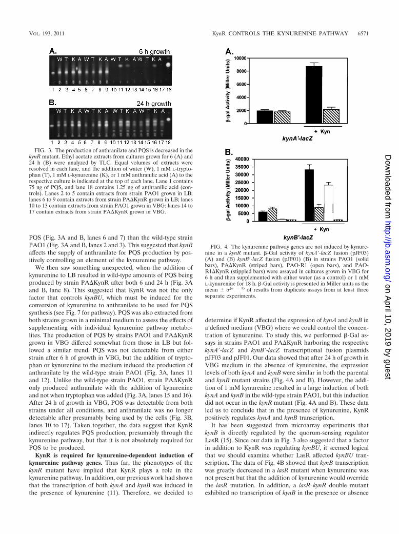
PQS (Fig. 3A and B, lanes 6 and 7) than the wild-type strainPAO1 (Fig. 3A and B, lanes 2 and 3). This suggested that kynRaffects the supply of anthranilate for PQS production by pos-itively controlling an element of the kynurenine pathway.
We then saw something unexpected, when the addition ofkynurenine to LB resulted in wild-type amounts of PQS beingproduced by strain PA�KynR after both 6 and 24 h (Fig. 3Aand B, lane 8). This suggested that KynR was not the onlyfactor that controls kynBU, which must be induced for theconversion of kynurenine to anthranilate to be used for PQSsynthesis (see Fig. 7 for pathway). PQS was also extracted fromboth strains grown in a minimal medium to assess the effects ofsupplementing with individual kynurenine pathway metabo-lites. The production of PQS by strains PAO1 and PA�KynRgrown in VBG differed somewhat from those in LB but fol-lowed a similar trend. PQS was not detectable from eitherstrain after 6 h of growth in VBG, but the addition of trypto-phan or kynurenine to the medium induced the production ofanthranilate by the wild-type strain PAO1 (Fig. 3A, lanes 11and 12). Unlike the wild-type strain PAO1, strain PA�KynRonly produced anthranilate with the addition of kynurenineand not when tryptophan was added (Fig. 3A, lanes 15 and 16).After 24 h of growth in VBG, PQS was detectable from bothstrains under all conditions, and anthranilate was no longerdetectable after presumably being used by the cells (Fig. 3B,lanes 10 to 17). Taken together, the data suggest that KynRindirectly regulates PQS production, presumably through thekynurenine pathway, but that it is not absolutely required forPQS to be produced.
KynR is required for kynurenine-dependent induction ofkynurenine pathway genes. Thus far, the phenotypes of thekynR mutant have implied that KynR plays a role in thekynurenine pathway. In addition, our previous work had shownthat the transcription of both kynA and kynB was induced inthe presence of kynurenine (11). Therefore, we decided to
determine if KynR affected the expression of kynA and kynB ina defined medium (VBG) where we could control the concen-tration of kynurenine. To study this, we performed -Gal as-says in strains PAO1 and PA�KynR harboring the respectivekynA�-lacZ and kynB�-lacZ transcriptional fusion plasmidspJF03 and pJF01. Our data showed that after 24 h of growth inVBG medium in the absence of kynurenine, the expressionlevels of both kynA and kynB were similar in both the parentaland kynR mutant strains (Fig. 4A and B). However, the addi-tion of 1 mM kynurenine resulted in a large induction of bothkynA and kynB in the wild-type strain PAO1, but this inductiondid not occur in the kynR mutant (Fig. 4A and B). These dataled us to conclude that in the presence of kynurenine, KynRpositively regulates kynA and kynB transcription.
It has been suggested from microarray experiments thatkynB is directly regulated by the quorum-sensing regulatorLasR (15). Since our data in Fig. 3 also suggested that a factorin addition to KynR was regulating kynBU, it seemed logicalthat we should examine whether LasR affected kynBU tran-scription. The data of Fig. 4B showed that kynB transcriptionwas greatly decreased in a lasR mutant when kynurenine wasnot present but that the addition of kynurenine would overridethe lasR mutation. In addition, a lasR kynR double mutantexhibited no transcription of kynB in the presence or absence
FIG. 3. The production of anthranilate and PQS is decreased in thekynR mutant. Ethyl acetate extracts from cultures grown for 6 (A) and24 h (B) were analyzed by TLC. Equal volumes of extracts wereresolved in each lane, and the addition of water (W), 1 mM L-trypto-phan (T), 1 mM L-kynurenine (K), or 1 mM anthranilic acid (A) to therespective culture is indicated at the top of each lane. Lane 1 contains75 ng of PQS, and lane 18 contains 1.25 ng of anthranilic acid (con-trols). Lanes 2 to 5 contain extracts from strain PAO1 grown in LB;lanes 6 to 9 contain extracts from strain PA�KynR grown in LB; lanes10 to 13 contain extracts from strain PAO1 grown in VBG; lanes 14 to17 contain extracts from strain PA�KynR grown in VBG.
FIG. 4. The kynurenine pathway genes are not induced by kynure-nine in a kynR mutant. -Gal activity of kynA�-lacZ fusion (pJF03)(A) and (B) kynB�-lacZ fusion (pJF01) (B) in strains PAO1 (solidbars), PA�KynR (striped bars), PAO-R1 (open bars), and PAO-R1�KynR (stippled bars) were assayed in cultures grown in VBG for6 h and then supplemented with either water (as a control) or 1 mML-kynurenine for 18 h. -Gal activity is presented in Miller units as themean � �(n � 1) of results from duplicate assays from at least threeseparate experiments.
VOL. 193, 2011 KynR CONTROLS THE KYNURENINE PATHWAY 6571
on April 10, 2019 by guest
http://jb.asm.org/
Dow
nloaded from
of kynurenine (Fig. 4B). Taken together, the data of Fig. 4 leadus to conclude that kynBU is controlled by LasR when kynure-nine is absent and by KynR when kynurenine is available.
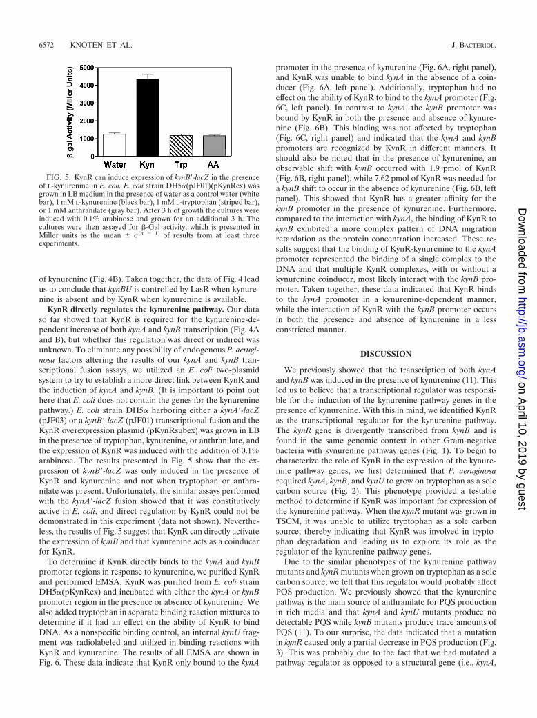
KynR directly regulates the kynurenine pathway. Our dataso far showed that KynR is required for the kynurenine-de-pendent increase of both kynA and kynB transcription (Fig. 4Aand B), but whether this regulation was direct or indirect wasunknown. To eliminate any possibility of endogenous P. aerugi-nosa factors altering the results of our kynA and kynB tran-scriptional fusion assays, we utilized an E. coli two-plasmidsystem to try to establish a more direct link between KynR andthe induction of kynA and kynB. (It is important to point outhere that E. coli does not contain the genes for the kynureninepathway.) E. coli strain DH5� harboring either a kynA�-lacZ(pJF03) or a kynB�-lacZ (pJF01) transcriptional fusion and theKynR overexpression plasmid (pKynRsubex) was grown in LBin the presence of tryptophan, kynurenine, or anthranilate, andthe expression of KynR was induced with the addition of 0.1%arabinose. The results presented in Fig. 5 show that the ex-pression of kynB�-lacZ was only induced in the presence ofKynR and kynurenine and not when tryptophan or anthra-nilate was present. Unfortunately, the similar assays performedwith the kynA�-lacZ fusion showed that it was constitutivelyactive in E. coli, and direct regulation by KynR could not bedemonstrated in this experiment (data not shown). Neverthe-less, the results of Fig. 5 suggest that KynR can directly activatethe expression of kynB and that kynurenine acts as a coinducerfor KynR.
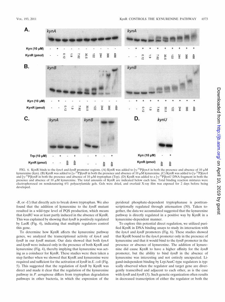
To determine if KynR directly binds to the kynA and kynBpromoter regions in response to kynurenine, we purified KynRand performed EMSA. KynR was purified from E. coli strainDH5�(pKynRex) and incubated with either the kynA or kynBpromoter region in the presence or absence of kynurenine. Wealso added tryptophan in separate binding reaction mixtures todetermine if it had an effect on the ability of KynR to bindDNA. As a nonspecific binding control, an internal kynU frag-ment was radiolabeled and utilized in binding reactions withKynR and kynurenine. The results of all EMSA are shown inFig. 6. These data indicate that KynR only bound to the kynA
promoter in the presence of kynurenine (Fig. 6A, right panel),and KynR was unable to bind kynA in the absence of a coin-ducer (Fig. 6A, left panel). Additionally, tryptophan had noeffect on the ability of KynR to bind to the kynA promoter (Fig.6C, left panel). In contrast to kynA, the kynB promoter wasbound by KynR in both the presence and absence of kynure-nine (Fig. 6B). This binding was not affected by tryptophan(Fig. 6C, right panel) and indicated that the kynA and kynBpromoters are recognized by KynR in different manners. Itshould also be noted that in the presence of kynurenine, anobservable shift with kynB occurred with 1.9 pmol of KynR(Fig. 6B, right panel), while 7.62 pmol of KynR was needed fora kynB shift to occur in the absence of kynurenine (Fig. 6B, leftpanel). This showed that KynR has a greater affinity for thekynB promoter in the presence of kynurenine. Furthermore,compared to the interaction with kynA, the binding of KynR tokynB exhibited a more complex pattern of DNA migrationretardation as the protein concentration increased. These re-sults suggest that the binding of KynR-kynurenine to the kynApromoter represented the binding of a single complex to theDNA and that multiple KynR complexes, with or without akynurenine coinducer, most likely interact with the kynB pro-moter. Taken together, these data indicated that KynR bindsto the kynA promoter in a kynurenine-dependent manner,while the interaction of KynR with the kynB promoter occursin both the presence and absence of kynurenine in a lessconstricted manner.
DISCUSSION
We previously showed that the transcription of both kynAand kynB was induced in the presence of kynurenine (11). Thisled us to believe that a transcriptional regulator was responsi-ble for the induction of the kynurenine pathway genes in thepresence of kynurenine. With this in mind, we identified KynRas the transcriptional regulator for the kynurenine pathway.The kynR gene is divergently transcribed from kynB and isfound in the same genomic context in other Gram-negativebacteria with kynurenine pathway genes (Fig. 1). To begin tocharacterize the role of KynR in the expression of the kynure-nine pathway genes, we first determined that P. aeruginosarequired kynA, kynB, and kynU to grow on tryptophan as a solecarbon source (Fig. 2). This phenotype provided a testablemethod to determine if KynR was important for expression ofthe kynurenine pathway. When the kynR mutant was grown inTSCM, it was unable to utilize tryptophan as a sole carbonsource, thereby indicating that KynR was involved in trypto-phan degradation and leading us to explore its role as theregulator of the kynurenine pathway genes.
Due to the similar phenotypes of the kynurenine pathwaymutants and kynR mutants when grown on tryptophan as a solecarbon source, we felt that this regulator would probably affectPQS production. We previously showed that the kynureninepathway is the main source of anthranilate for PQS productionin rich media and that kynA and kynU mutants produce nodetectable PQS while kynB mutants produce trace amounts ofPQS (11). To our surprise, the data indicated that a mutationin kynR caused only a partial decrease in PQS production (Fig.3). This was probably due to the fact that we had mutated apathway regulator as opposed to a structural gene (i.e., kynA,
FIG. 5. KynR can induce expression of kynB�-lacZ in the presenceof L-kynurenine in E. coli. E. coli strain DH5�(pJF01)(pKynRex) wasgrown in LB medium in the presence of water as a control water (whitebar), 1 mM L-kynurenine (black bar), 1 mM L-tryptophan (striped bar),or 1 mM anthranilate (gray bar). After 3 h of growth the cultures wereinduced with 0.1% arabinose and grown for an additional 3 h. Thecultures were then assayed for -Gal activity, which is presented inMiller units as the mean � �(n � 1) of results from at least threeexperiments.
6572 KNOTEN ET AL. J. BACTERIOL.
on April 10, 2019 by guest
http://jb.asm.org/
Dow
nloaded from
-B, or -U) that directly acts to break down tryptophan. We alsofound that the addition of kynurenine to the kynR mutantresulted in a wild-type level of PQS production, which meansthat kynBU was at least partly induced in the absence of KynR.This was explained by showing that kynB is positively regulatedby LasR (Fig. 4), indicating that multiple regulators controlthis gene.
To determine how KynR affects the kynurenine pathwaygenes, we analyzed the transcriptional activity of kynA andkynB in our kynR mutant. Our data showed that both kynAand kynB were induced only in the presence of both KynR andkynurenine (Fig. 4), thereby implying that kynurenine was act-ing as a coinducer for KynR. These studies were then taken astep further when we showed that KynR and kynurenine wererequired and sufficient for the activation of kynB in E. coli (Fig.5). This suggested that the regulation of kynB by KynR wasdirect and made it clear that the regulation of the kynureninepathway in P. aeruginosa differs from tryptophan degradationpathways in other bacteria, in which the expression of the
pyridoxal phosphate-dependent tryptophanase is posttran-scriptionally regulated through attenuation (50). Taken to-gether, the data we accumulated suggested that the kynureninepathway is directly regulated in a positive way by KynR in akynurenine-dependent manner.
To explore this potential direct regulation, we utilized puri-fied KynR in DNA binding assays to study its interaction withthe kynA and kynB promoters (Fig. 6). These studies showedthat KynR bound to the kynA promoter only in the presence ofkynurenine and that it would bind to the kynB promoter in thepresence or absence of kynurenine. The addition of kynure-nine did cause KynR to have a higher affinity for the kynBpromoter, but the ability to bind kynB in the absence ofkynurenine was interesting and not entirely unexpected. Li-gand-independent binding by Lrp/AsnC-type regulators is typ-ically observed when the regulator and target gene are diver-gently transcribed and adjacent to each other, as is the casewith kynB and kynR (3). Such genetic organization often resultsin decreased transcription of either the regulator or both the
FIG. 6. KynR binds to the kynA and kynB promoter regions. (A) KynR was added to [�-32P]kynA in both the presence and absence of 10 �Mkynurenine (kyn). (B) KynR was added to [�-32P]kynB in both the presence and absence of 10 �M kynurenine. (C) KynR was added to [�-32P]kynAand [�-32P]kynB in both the presence and absence of 10 �M tryptophan (Trp). (D) KynR was added to a [�-32P]kynU DNA fragment in both thepresence and absence of 10 �M kynurenine. The total amounts of KynR are indicated below each lane. Total binding reaction mixtures wereelectrophoresed on nondenaturing 6% polyacrylamide gels. Gels were dried, and overlaid X-ray film was exposed for 2 days before beingdeveloped.
VOL. 193, 2011 KynR CONTROLS THE KYNURENINE PATHWAY 6573
on April 10, 2019 by guest
http://jb.asm.org/
Dow
nloaded from
regulator and target gene (3), but we have not demonstratedthis for kynB or kynR.
Another interesting observation from the EMSA experi-ments was the differences that were observed between thebinding complexes that KynR formed with the kynA and kynBpromoter regions. The kynA-KynR-kynurenine complexes mi-grated the same regardless of the KynR concentration, whilethe kynB-KynR complexes had different migration rates withincreasing concentrations of KynR (with and without kynure-nine present). This type of migration pattern could be theresult of KynR binding to multiple sites within the promoterregion. Multiple binding sites, as well as DNA bending, havebeen reported in several Lrp/AsnC-type regulators in E. coliand other bacteria (9, 12, 20, 45, 46). In addition, Lrp/AsnC-type transcriptional regulators can bind as multimers both inthe presence and absence of a ligand and are capable of actingas both activators and repressors (3, 51), so this seems to be themost likely explanation for the results seen with the kynBpromoter binding assays.
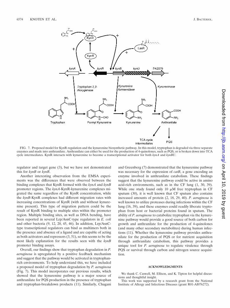
Overall, our findings show that tryptophan degradation in P.aeruginosa is upregulated by a positive feedback mechanismand suggest that the pathway would be activated in tryptophan-rich environments. To help understand this, we have includeda proposed model of tryptophan degradation by P. aeruginosa(Fig. 7). This model incorporates our previous results, whichshowed that the kynurenine pathway is a major source ofanthranilate for PQS production in the presence of tryptophanand tryptophan-breakdown products (11). Similarly, Chugani
and Greenberg (7) demonstrated that the kynurenine pathwaywas necessary for the expression of catB, a gene encoding anenzyme involved in anthranilate catabolism. These findingssuggest that the kynurenine pathway could be active in aminoacid-rich environments, such as in the CF lung (1, 30, 39).While one study found only 10 �M free tryptophan in CFsputum (30), it is well known that CF sputum also containsincreased amounts of protein (2, 18, 29, 40). P. aeruginosa iswell known to utilize proteases during infections within the CFlung (16, 39), and these enzymes could readily liberate trypto-phan from host or bacterial proteins found in sputum. Theability of P. aeruginosa to catabolize tryptophan via the kynure-nine pathway would provide a good source of both carbon forgrowth and anthranilate for the production of 4-quinolones(and many other secondary metabolites) during human infec-tions (11). Whether the kynurenine pathway provides anthra-nilate for the production of PQS or for nutrient acquisitionthrough anthranilate catabolism, this pathway provides aunique tool for P. aeruginosa to regulate virulence throughPQS or survival through carbon and nitrogen source acquisi-tion.
ACKNOWLEDGMENTS
We thank C. Caswell, M. Ellison, and K. Tipton for helpful discus-sions and thoughtful insight.
This work was supported by a research grant from the NationalInstitute of Allergy and Infectious Diseases (grant R01-AI076272).
FIG. 7. Proposed model for KynR regulation and the kynurenine biosynthetic pathway. In this model, tryptophan is degraded via three separateenzymes and made into anthranilate. Anthranilate can either be used for the production of 4-quinolones, such as PQS, or is broken down into TCAcycle intermediates. KynR interacts with kynurenine to become a transcriptional activator for both kynA and kynBU.
6574 KNOTEN ET AL. J. BACTERIOL.
on April 10, 2019 by guest
http://jb.asm.org/
Dow
nloaded from
REFERENCES
1. Balibar, C. J., and C. T. Walsh. 2006. In vitro biosynthesis of violacein fromL-tryptophan by the enzymes VioA-E from Chromobacterium violaceum.Biochemistry 45:15444–15457.
2. Barth, A. L., and T. L. Pitt. 1996. The high amino-acid content of sputumfrom cystic fibrosis patients promotes growth of auxotrophic Pseudomonasaeruginosa. J. Med. Microbiol. 45:110–119.
3. Brinkman, A. B., T. J. Ettema, W. M. de Vos, and J. van der Oost. 2003. TheLrp family of transcriptional regulators. Mol. Microbiol. 48:287–294.
4. Burns, J. L., et al. 2001. Longitudinal assessment of Pseudomonas aeruginosain young children with cystic fibrosis. J. Infect. Dis. 183:444–452.
5. Chang, A. C., and S. N. Cohen. 1978. Construction and characterization ofamplifiable multicopy DNA cloning vehicles derived from the P15A crypticminiplasmid. J. Bacteriol. 134:1141–1156.
6. Choi, K. H., A. Kumar, and H. P. Schweizer. 2006. A 10-min method forpreparation of highly electrocompetent Pseudomonas aeruginosa cells: ap-plication for DNA fragment transfer between chromosomes and plasmidtransformation. J. Microbiol. Methods 64:391–397.
7. Chugani, S., and E. P. Greenberg. 2010. LuxR homolog-independent generegulation by acyl-homoserine lactones in Pseudomonas aeruginosa. Proc.Natl. Acad. Sci. U. S. A. 107:10673–10678.
8. Collier, D. N., et al. 2002. A bacterial cell to cell signal in the lungs of cysticfibrosis patients. FEMS Microbiol. Lett. 215:41–46.
9. Cui, Y., Q. Wang, G. D. Stormo, and J. M. Calvo. 1995. A consensussequence for binding of Lrp to DNA. J. Bacteriol. 177:4872–4880.
10. Deziel, E., et al. 2004. Analysis of Pseudomonas aeruginosa 4-hydroxy-2-alkylquinolines (HAQs) reveals a role for 4-hydroxy-2-heptylquinoline incell-to-cell communication. Proc. Natl. Acad.Sci. U. S. A. 101:1339–1344.
11. Farrow, J. M., III, and E. C. Pesci. 2007. Two distinct pathways supplyanthranilate as a precursor of the Pseudomonas quinolone signal. J. Bacte-riol. 189:3425–3433.
12. Friedberg, D., M. Midkiff, and J. M. Calvo. 2001. Global versus local regu-latory roles for Lrp-related proteins: Haemophilus influenzae as a case study.J. Bacteriol. 183:4004–4011.
13. Gallagher, L. A., and C. Manoil. 2001. Pseudomonas aeruginosa PAO1 killsCaenorhabditis elegans by cyanide poisoning. J. Bacteriol. 183:6207–6214.
14. Gambello, M. J., and B. H. Iglewski. 1991. Cloning and characterization ofthe Pseudomonas aeruginosa lasR gene, a transcriptional activator of elastaseexpression. J. Bacteriol. 173:3000–3009.
15. Gilbert, K. B., T. H. Kim, R. Gupta, E. P. Greenberg, and M. Schuster. 2009.Global position analysis of the Pseudomonas aeruginosa quorum-sensingtranscription factor LasR. Mol. Microbiol. 73:1072–1085.
16. Henke, M. O., et al. 2011. Serine proteases degrade airway mucins in cysticfibrosis. Infect. Immun. 79:3438–3444.
17. Hoang, T. T., R. R. Karkhoff-Schweizer, A. J. Kutchma, and H. P. Schweizer.1998. A broad-host-range Flp-FRT recombination system for site-specificexcision of chromosomally-located DNA sequences: application for isolationof unmarked Pseudomonas aeruginosa mutants. Gene 212:77–86.
18. Hoiby, N. 1998. Pseudomonas in cystic fibrosis: past, present, and future.Cystic Fibrosis Trust, London, England.
19. Holloway, B. W., V. Krishnapillai, and A. F. Morgan. 1979. Chromosomalgenetics of Pseudomonas. Microbiol. Rev. 43:73–102.
20. Jafri, S., S. Chen, and J. M. Calvo. 2002. ilvIH operon expression in Esch-erichia coli requires Lrp binding to two distinct regions of DNA. J. Bacteriol.184:5293–5300.
21. Kersten, R. D., and P. C. Dorrestein. 2009. Secondary metabolomics: naturalproducts mass spectrometry goes global. ACS Chem. Biol. 4:599–601.
22. Kurnasov, O., et al. 2003. NAD biosynthesis: identification of the tryptophanto quinolinate pathway in bacteria. Chem. Biol. 10:1195–1204.
23. Kurnasov, O., et al. 2003. Aerobic tryptophan degradation pathway in bac-teria: novel kynurenine formamidase. FEMS Microbiol. Lett. 227:219–227.
24. Lepine, F., S. Milot, E. Deziel, J. He, and L. G. Rahme. 2004. Electrospray/mass spectrometric identification and analysis of 4-hydroxy-2-alkylquinolines(HAQs) produced by Pseudomonas aeruginosa. J. Am. Soc. Mass Spec.15:862–869.
25. Lyczak, J. B., C. L. Cannon, and G. B. Pier. 2000. Establishment of Pseu-domonas aeruginosa infection: lessons from a versatile opportunist. MicrobesInfect. 2:1051–1060.
26. Madhusudhan, K. T., N. Huang, and J. R. Sokatch. 1995. Characterizationof BkdR-DNA binding in the expression of the bkd operon of Pseudomonasputida. J. Bacteriol. 177:636–641.
27. Mahajan-Miklos, S., M. W. Tan, L. G. Rahme, and F. M. Ausubel. 1999.
Molecular mechanisms of bacterial virulence elucidated using a Pseudomo-nas aeruginosa-Caenorhabditis elegans pathogenesis model. Cell 96:47–56.
28. Matthijs, S., et al. 2004. The Pseudomonas siderophore quinolobactin issynthesized from xanthurenic acid, an intermediate of the kynurenine path-way. Mol. Microbiol. 52:371–384.
29. Ohman, D. E., and A. M. Chakrabarty. 1982. Utilization of human respira-tory secretions by mucoid Pseudomonas aeruginosa of cystic fibrosis origin.Infect. Immun. 37:662–669.
30. Palmer, K. L., L. M. Aye, and M. Whiteley. 2007. Nutritional cues controlPseudomonas aeruginosa multicellular behavior in cystic fibrosis sputum. J.Bacteriol. 189:8079–8087.
31. Palmer, K. L., L. M. Mashburn, P. K. Singh, and M. Whiteley. 2005. Cysticfibrosis sputum supports growth and cues key aspects of Pseudomonas aerugi-nosa physiology. J. Bacteriol. 187:5267–5277.
32. Preston, M. J., et al. 1997. Contribution of proteases and LasR to thevirulence of Pseudomonas aeruginosa during corneal infections. Infect. Im-mun. 65:3086–3090.
33. Qiu, D., F. H. Damron, T. Mima, H. P. Schweizer, and H. D. Yu. 2008.PBAD-based shuttle vectors for functional analysis of toxic and highly regu-lated genes in Pseudomonas and Burkholderia spp. and other bacteria. Appl.Environ. Microbiol. 74:7422–7426.
34. Rahme, L. G., et al. 1997. Use of model plant hosts to identify Pseudomonasaeruginosa virulence factors. Proc. Natl. Acad. Sci. U. S. A. 94:13245–13250.
35. Ramos, J. L. (ed.). 2004. Pseudomonas. Kluwer Academic/Plenum Publish-ers, New York, NY.
36. Schuster, M., C. P. Lostroh, T. Ogi, and E. P. Greenberg. 2003. Identifica-tion, timing, and signal specificity of Pseudomonas aeruginosa quorum-con-trolled genes: a transcriptome analysis. J. Bacteriol. 185:2066–2079.
37. Stuart, B., J. H. Lin, and P. J. Mogayzel, Jr. 2010. Early eradication ofPseudomonas aeruginosa in patients with cystic fibrosis. Paediatr. Respir.Rev. 11:177–184.
38. Sullivan, N. L., D. Tzeranis, Y. Wang, P. T. So, and D. Newman. 2011.Quantifying the dynamics of bacterial secondary metabolites by spectralmultiphoton microscopy. ACS Chem. Biol. 6:893–899.
39. Suter, S. 1994. The role of bacterial proteases in the pathogenesis of cysticfibrosis. Am. J. Respir. Crit. Care Med. 150:S118–S122.
40. Thomas, S. R., A. Ray, M. E. Hodson, and T. L. Pitt. 2000. Increased sputumamino acid concentrations and auxotrophy of Pseudomonas aeruginosa insevere cystic fibrosis lung disease. Thorax 55:795–797.
41. Vederas, J. C., E. Schleicher, M. D. Tsai, and H. G. Floss. 1978. Stereo-chemistry and mechanism of reactions catalyzed by tryptophanase Esche-richia coli. J. Biol. Chem. 253:5350–5354.
42. Vogel, H. J., and D. M. Bonner. 1956. Acetylornithinase of Escherichia coli:partial purification and some properties. J. Biol. Chem. 218:97–106.
43. Wagner, V. E., D. Bushnell, L. Passador, A. I. Brooks, and B. H. Iglewski.2003. Microarray analysis of Pseudomonas aeruginosa quorum-sensing regu-lons: effects of growth phase and environment. J. Bacteriol. 185:2080–2095.
44. Walters, M., and V. Sperandio. 2006. Quorum sensing in Escherichia coli andSalmonella. Int. J. Med. Microbiol. 296:125–131.
45. Wang, Q., and J. M. Calvo. 1993. Lrp, a global regulatory protein of Esch-erichia coli, binds co-operatively to multiple sites and activates transcriptionof ilvIH. J. Mol. Biol. 229:306–318.
46. Wang, Q., and J. M. Calvo. 1993. Lrp, a major regulatory protein in Esch-erichia coli, bends DNA and can organize the assembly of a higher-ordernucleoprotein structure. EMBO J. 12:2495–2501.
47. Warrens, A. N., M. D. Jones, and R. I. Lechler. 1997. Splicing by overlapextension by PCR using asymmetric amplification: an improved techniquefor the generation of hybrid proteins of immunological interest. Gene 186:29–35.
48. Woodcock, D. M., et al. 1989. Quantitative evaluation of Escherichia coli hoststrains for tolerance to cytosine methylation in plasmid and phage recombi-nants. Nucleic Acids Res. 17:3469–3478.
49. Xiao, G., et al. 2006. MvfR, a key Pseudomonas aeruginosa pathogenicityLTTR-class regulatory protein, has dual ligands. Mol. Microbiol. 62:1689–1699.
50. Yanofsky, C. 2007. RNA-based regulation of genes of tryptophan synthesisand degradation, in bacteria. RNA 13:1141–1154.
51. Yokoyama, K., et al. 2006. Feast/famine regulatory proteins (FFRPs): Esch-erichia coli Lrp, AsnC and related archaeal transcription factors. FEMSMicrobiol. Rev. 30:89–108.
52. Zegarra-Moran, O., et al. 2004. Double mechanism for apical tryptophandepletion in polarized human bronchial epithelium. J. Immunol. 173:542–549.
VOL. 193, 2011 KynR CONTROLS THE KYNURENINE PATHWAY 6575
on April 10, 2019 by guest
http://jb.asm.org/
Dow
nloaded from