asnc imaging guidelines asnc imaging guidelines for … · asnc imaging guidelines asnc imaging...
TRANSCRIPT
ASNC IMAGING GUIDELINES
ASNC imaging guidelines for SPECT nuclearcardiology procedures: Stress, protocols,and tracers
Milena J. Henzlova, MD,a W. Lane Duvall, MD,b Andrew J. Einstein, MD,c
Mark I. Travin, MD,d and Hein J. Verberne, MDe
a Mount Sinai Medical Center, New York, NYb Hartford Hospital, Hartford, CTc New York Presbyterian Hospital, Columbia University Medical Center, New York, NYd Montefiore Medical Center, Albert Einstein College of Medicine, Bronx, NYe Academic Medical Center, Amsterdam, The Netherlands
doi:10.1007/s12350-015-0387-x
Abbreviations
A2A Adenosine 2a
AHA American Heart Association
ALARA As low as reasonably achievable
AV Atrioventricular
BP Blood pressure
CBF Coronary blood flow
CAD Coronary artery disease
CPET Cardiopulmonary exercise testing
DSP Deconvolution of septal penetration
ECG Electrocardiogram
EF Ejection fraction
ESRD End-stage renal disease
HF Heart failure
HFrEF Heart failure (with) reduced ejection
fraction
HMR Heart-to-mediastinum ratio
ICD Implantable cardioversion defibrillator
IV Intravenous
LBBB Left bundle branch block
LEHR Low energy high resolution
LVEF Left ventricular ejection fraction
MBq Megabecquerels
mCi Millicuries
MPI Myocardial perfusion imaging
MRI Magnetic resonance imaging
mSv Millisievert
NE Norepinephrine
NET1 Norepinephrine transporter-1
NPO Nil per os (nothing by mouth)
NYHA New York Heart Association
PET Positron emission tomography
ROI Region of interest
SPECT Single-photon emission computed
tomography
TAVR Transcatheter aortic valve replacement
WR Washout rate
WPW Wolff-Parkinson White
EXERCISE STRESS TEST
Exercise testing has been used for more than 60
years for diagnostic purposes in symptomatic patients
[including patients with acute chest pain without
ischemia by electrocardiogram (ECG) and serum mark-
ers] and for prognosis and risk stratification in patients
with known coronary artery disease (CAD), such as
history of myocardial infarction or documented CAD by
coronary angiography or computed tomography angiog-
raphy and in those with high risk for the presence of
CAD, such as patients with diabetes mellitus, peripheral,
or cerebral vascular disease.Reprint requests: Milena J. Henzlova, MD, Mount Sinai Medical
Center, New York, NY; [email protected]
J Nucl Cardiol 2016;23:606–39.
1071-3581/$34.00
Copyright � 2016 American Society of Nuclear Cardiology.
606
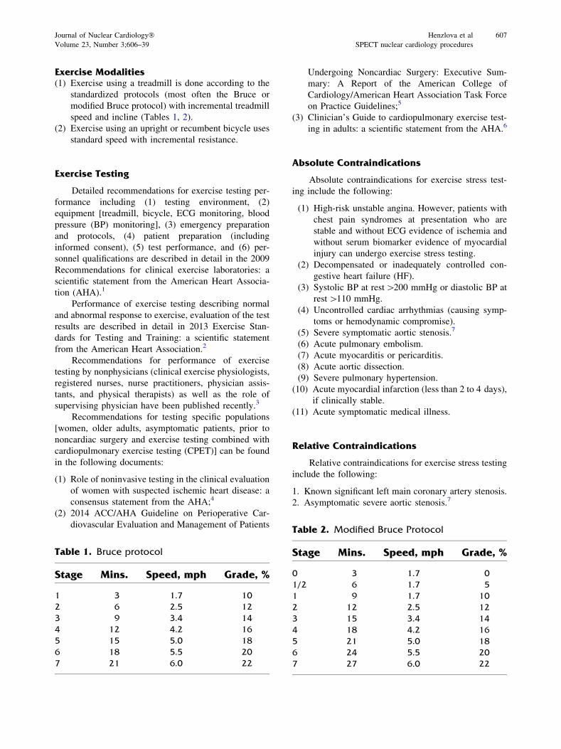
Exercise Modalities(1) Exercise using a treadmill is done according to the
standardized protocols (most often the Bruce or
modified Bruce protocol) with incremental treadmill
speed and incline (Tables 1, 2).
(2) Exercise using an upright or recumbent bicycle uses
standard speed with incremental resistance.
Exercise Testing
Detailed recommendations for exercise testing per-
formance including (1) testing environment, (2)
equipment [treadmill, bicycle, ECG monitoring, blood
pressure (BP) monitoring], (3) emergency preparation
and protocols, (4) patient preparation (including
informed consent), (5) test performance, and (6) per-
sonnel qualifications are described in detail in the 2009
Recommendations for clinical exercise laboratories: a
scientific statement from the American Heart Associa-
tion (AHA).1
Performance of exercise testing describing normal
and abnormal response to exercise, evaluation of the test
results are described in detail in 2013 Exercise Stan-
dards for Testing and Training: a scientific statement
from the American Heart Association.2
Recommendations for performance of exercise
testing by nonphysicians (clinical exercise physiologists,
registered nurses, nurse practitioners, physician assis-
tants, and physical therapists) as well as the role of
supervising physician have been published recently.3
Recommendations for testing specific populations
[women, older adults, asymptomatic patients, prior to
noncardiac surgery and exercise testing combined with
cardiopulmonary exercise testing (CPET)] can be found
in the following documents:
(1) Role of noninvasive testing in the clinical evaluation
of women with suspected ischemic heart disease: a
consensus statement from the AHA;4
(2) 2014 ACC/AHA Guideline on Perioperative Car-
diovascular Evaluation and Management of Patients
Undergoing Noncardiac Surgery: Executive Sum-
mary: A Report of the American College of
Cardiology/American Heart Association Task Force
on Practice Guidelines;5
(3) Clinician’s Guide to cardiopulmonary exercise test-
ing in adults: a scientific statement from the AHA.6
Absolute Contraindications
Absolute contraindications for exercise stress test-
ing include the following:
(1) High-risk unstable angina. However, patients with
chest pain syndromes at presentation who are
stable and without ECG evidence of ischemia and
without serum biomarker evidence of myocardial
injury can undergo exercise stress testing.
(2) Decompensated or inadequately controlled con-
gestive heart failure (HF).
(3) Systolic BP at rest[200 mmHg or diastolic BP at
rest[110 mmHg.
(4) Uncontrolled cardiac arrhythmias (causing symp-
toms or hemodynamic compromise).
(5) Severe symptomatic aortic stenosis.7
(6) Acute pulmonary embolism.
(7) Acute myocarditis or pericarditis.
(8) Acute aortic dissection.
(9) Severe pulmonary hypertension.
(10) Acute myocardial infarction (less than 2 to 4 days),
if clinically stable.
(11) Acute symptomatic medical illness.
Relative Contraindications
Relative contraindications for exercise stress testing
include the following:
1. Known significant left main coronary artery stenosis.
2. Asymptomatic severe aortic stenosis.7
Table 1. Bruce protocol
Stage Mins. Speed, mph Grade, %
1 3 1.7 10
2 6 2.5 12
3 9 3.4 14
4 12 4.2 16
5 15 5.0 18
6 18 5.5 20
7 21 6.0 22
Table 2. Modified Bruce Protocol
Stage Mins. Speed, mph Grade, %
0 3 1.7 0
1/2 6 1.7 5
1 9 1.7 10
2 12 2.5 12
3 15 3.4 14
4 18 4.2 16
5 21 5.0 18
6 24 5.5 20
7 27 6.0 22
Journal of Nuclear Cardiology� Henzlova et al 607
Volume 23, Number 3;606–39 SPECT nuclear cardiology procedures

3. Hypertrophic obstructive cardiomyopathy or other
forms of severe left ventricular outflow tract
obstruction.
4. Significant tachyarrhythmias or bradyarrhythmias.
5. High-degree atrioventricular (AV) block.
6. Electrolyte abnormalities.
7. Mental or physical impairment leading to inability to
exercise adequately.
8. If combined with imaging, patients with complete
left bundle branch block (LBBB), permanent pace-
makers, and ventricular pre-excitation [Wolff-
Parkinson-White (WPW) syndrome] should prefer-
entially undergo pharmacologic vasodilator stress
test (not a dobutamine stress test).
Limitations
Exercise stress testing has a lower diagnostic value
in patients who cannot achieve an adequate heart rate
and BP response due to a noncardiac physical limitation,
such as pulmonary, peripheral vascular or musculoskele-
tal abnormalities, or due to lack of motivation. These
patients should be considered for pharmacologic stress
testing with myocardial perfusion imaging. Also, for a
meaningful test evaluation, exercise should last at least 4
to 6 minutes.
Procedure(1) Patient preparation: nothing should be eaten at least
3 hours before the test. Patients scheduled for later
in the morning or afternoon may have a light
breakfast (e.g., cereal, fruit). Caffeine should be
avoided for at least 12 hours similar to vasodilatory
stress testing because exercise stress tests, at times,
need to be converted to a pharmacologic stress test.
If possible, insulin-dependent diabetics should be
scheduled for the morning hours.
(2) BP medication(s) with antianginal properties (b-blocker, calcium channel blocker, and nitrates) will
lower a stress test’s diagnostic utility.8 Generally,
discontinuation of these medicines is left to the
discretion of the referring physician. Regularly
taken medication should be recorded prior to testing.
(3) An intravenous (IV) cannula (larger size than 24-
gauge is preferred) should be inserted for radio-
pharmaceutical injection.
(4) The electrocardiogram should be monitored contin-
uously during the exercise test and for at least 4
minutes into the recovery phase. In addition, the
resting heart rate should return to close to baseline
and exercise-induced ST-segment changes and
symptoms should resolve. A 12-lead electrocardio-
gram should be (automatically) obtained at every
stage of exercise, at peak exercise, and at the
termination of recovery phase. In addition, in case
of abnormalities (e.g., arrhythmias, etc.) a 12-lead
electrocardiogram should be obtained.
(5) The heart rate and BP should be recorded at least
every 3 minutes during exercise, at peak exercise,
and for at least 4 minutes into the recovery phase.
(6) The end point of all exercise tests should be
symptoms (moderate to severe chest pain, excessive
shortness of breath, fatigue). Achievement of 85%
of maximum, age-adjusted, predicted heart rate is
not an indication for termination of the test. Note: In
patients with known CAD (and particularly if tested
taking regular medication), the prognostic value of
the test is preserved without reaching 85% of
maximum predicted heart rate.
(7) The radiopharmaceutical should be injected as
close to peak exercise as possible. Patients should
be encouraged to exercise for at least 1 minute
after the radiotracer injection. If needed, treadmill
speed and/or inclination can be decreased after
tracer injection.
(8) Patients referred for a diagnostic stress test may be
converted to a pharmacologic stress test or a
combination of both if they cannot exercise ade-
quately for a meaningful period of time.
Indications for Early Termination ofExercise
Indications for early termination of exercise include
the following:
(1) Moderate to severe angina pectoris.
(2) Marked dyspnea.
(3) Fatigue.
(4) Ataxia, dizziness, or near-syncope.
(5) Signs of poor perfusion (cyanosis and pallor).
(6) Patient’s request to terminate the test.
(7) Excessive ST-segment depression ([2 mm from
baseline).
(8) ST elevation ([1 mm) in leads without diagnostic
Q-waves (except for leads V1 or aVR).
(9) Sustained supraventricular or ventricular
tachycardia.
(10) Development of LBBB or intraventricular conduc-
tion delay that cannot be distinguished from
ventricular tachycardia.
(11) Drop in systolic BP of greater than 10 mmHg from
baseline, despite an increase in workload, when
accompanied by other evidence of ischemia.
(12) Hypertensive response (systolic BP [230 mmHg
and/or diastolic pressure[115 mmHg).
608 Henzlova et al Journal of Nuclear Cardiology�SPECT nuclear cardiology procedures May/June 2016
(13) Inability to monitor the electrocardiogram or
systolic BP.
(14) In patients with implantable cardioverter defibril-
lators, when the heart rate attained is within
20 beats per minute of the lowest heart rate at
which therapy (antitachycardia pacing or shock) is
programmed to be delivered.
Achievement of 85% of maximum, age-adjusted,predicted heart rate is not an indication for earlytermination of the stress test.
For review of serious complications of myocar-dial stress testing and their management, refer to thearticle by Dilsizian and colleagues.9
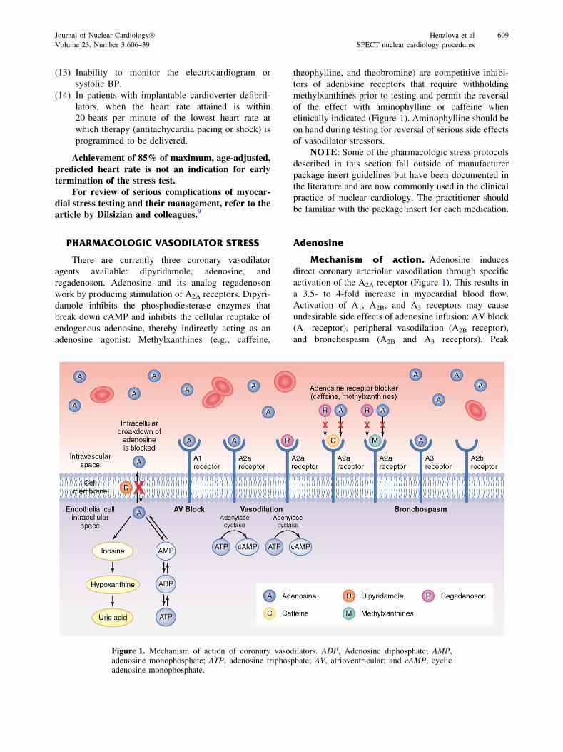
PHARMACOLOGIC VASODILATOR STRESS
There are currently three coronary vasodilator
agents available: dipyridamole, adenosine, and
regadenoson. Adenosine and its analog regadenoson
work by producing stimulation of A2A receptors. Dipyri-
damole inhibits the phosphodiesterase enzymes that
break down cAMP and inhibits the cellular reuptake of
endogenous adenosine, thereby indirectly acting as an
adenosine agonist. Methylxanthines (e.g., caffeine,
theophylline, and theobromine) are competitive inhibi-
tors of adenosine receptors that require withholding
methylxanthines prior to testing and permit the reversal
of the effect with aminophylline or caffeine when
clinically indicated (Figure 1). Aminophylline should be
on hand during testing for reversal of serious side effects
of vasodilator stressors.
NOTE: Some of the pharmacologic stress protocols
described in this section fall outside of manufacturer
package insert guidelines but have been documented in
the literature and are now commonly used in the clinical
practice of nuclear cardiology. The practitioner should
be familiar with the package insert for each medication.
Adenosine
Mechanism of action. Adenosine induces
direct coronary arteriolar vasodilation through specific
activation of the A2A receptor (Figure 1). This results in
a 3.5- to 4-fold increase in myocardial blood flow.
Activation of A1, A2B, and A3 receptors may cause
undesirable side effects of adenosine infusion: AV block
(A1 receptor), peripheral vasodilation (A2B receptor),
and bronchospasm (A2B and A3 receptors). Peak
Figure 1. Mechanism of action of coronary vasodilators. ADP, Adenosine diphosphate; AMP,adenosine monophosphate; ATP, adenosine triphosphate; AV, atrioventricular; and cAMP, cyclicadenosine monophosphate.
Journal of Nuclear Cardiology� Henzlova et al 609
Volume 23, Number 3;606–39 SPECT nuclear cardiology procedures
vasodilation after adenosine administration occurs
within 1 to 2 minutes after the start of the infusion.
The half-life of adenosine is approximately 10 seconds.
It is either phosphorylated to adenosine monophosphate
by adenosine kinase or degraded to inosine by adenosine
deaminase.
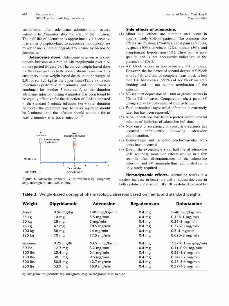
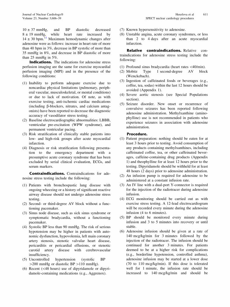
Adenosine dose. Adenosine is given as a con-
tinuous infusion at a rate of 140 mcg/kg/min over a 6-
minute period (Figure 2). The correct weight-based dose
for the obese and morbidly obese patients is unclear. It is
customary to use weight-based doses up to the weight of
250 lbs (or 125 kg) as the upper limit (Table 3). Tracer
injection is performed at 3 minutes, and the infusion is
continued for another 3 minutes. A shorter duration
adenosine infusion, lasting 4 minutes, has been found to
be equally effective for the detection of CAD compared
to the standard 6-minute infusion. For shorter duration
protocols, the minimum time to tracer injection should
be 2 minutes, and the infusion should continue for at
least 2 minutes after tracer injection.10
Side effects of adenosine.(1) Minor side effects are common and occur in
approximately 80% of patients. The common side
effects are flushing (35-40%), chest pain (25-30%),
dyspnea (20%), dizziness (7%), nausea (5%), and
symptomatic hypotension (5%). Chest pain is non-
specific and is not necessarily indicative of the
presence of CAD.
(2) AV block occurs in approximately 8% of cases.
However, the incidence of second-degree AV block
is only 4%, and that of complete heart block is less
than 1%. Most cases ([95%) of AV block are self-
limiting and do not require termination of the
infusion.
(3) ST-segment depression of 1 mm or greater occurs in
5% to 7% of cases. Compared to chest pain, ST
changes may be indicative of true ischemia.
(4) Fatal or nonfatal myocardial infarction is extremely
rare, but has been reported.11
(5) Atrial fibrillation has been reported within several
minutes of initiation of adenosine infusion.
(6) New onset or recurrence of convulsive seizures has
occurred infrequently following adenosine
administration.
(7) Hemorrhagic and ischemic cerebrovascular acci-
dents have occurred.
(8) Due to the exceedingly short half-life of adenosine
(\10 seconds), most side effects resolve in a few
seconds after discontinuation of the adenosine
infusion, and IV aminophylline administration is
only rarely required.
Hemodynamic effects. Adenosine results in a
modest increase in heart rate and a modest decrease in
both systolic and diastolic BPs. BP systolic decreased by
Figure 2. Adenosine protocol. IV, Intravenous; kg, kilogram;mcg, microgram; and min, minute.
Table 3. Weight-based dosing of pharmacologic stressors based on metric and standard weights
Weight Dipyridamole Adenosine Regadenoson Dobutamine
Metric 0.56 mg/kg 140 mcg/kg/min 0.4 mg 5–40 mcg/kg/min
25 kg 14 mg 3.5 mg/min 0.4 mg 0.125–1 mg/min
50 kg 28 mg 7 mg/min 0.4 mg 0.25–2 mg/min
75 kg 42 mg 10.5 mg/min 0.4 mg 0.375–3 mg/min
100 kg 56 mg 14 mg/min 0.4 mg 0.5–4 mg/min
125 kg 70 mg 17.5 mg/min 0.4 mg 0.625–5 mg/min
Standard 0.25 mg/lb 63.5 mcg/lb/min 0.4 mg 2.3–18.1 mcg/kg/min
50 lbs 12.7 mg 3.2 mg/min 0.4 mg 0.11–0.91 mg/min
100 lbs 25.4 mg 6.4 mg/min 0.4 mg 0.23–1.8 mg/min
150 lbs 38.1 mg 9.5 mg/min 0.4 mg 0.34–2.7 mg/min
200 lbs 50.5 mg 12.7 mg/min 0.4 mg 0.45–3.6 mg/min
250 lbs 63.5 mg 15.9 mg/min 0.4 mg 0.57–4.5 mg/min
kg, kilogram; lbs, pounds; mg, milligram; mcg, micrograms; min, minute
610 Henzlova et al Journal of Nuclear Cardiology�SPECT nuclear cardiology procedures May/June 2016
10 ± 37 mmHg, and BP diastolic decreased
8 ± 19 mmHg, while heart rate increased by
14 ± 30 bpm.11 Maximum hemodynamic changes after
adenosine were as follows: increase in heart rate of more
than 40 bpm in 3%, decrease in BP systolic of more than
35 mmHg in 8%, and decrease in BP diastolic of more
than 25 mmHg in 5%.
Indications. The indications for adenosine stress
perfusion imaging are the same for exercise myocardial
perfusion imaging (MPI) and in the presence of the
following conditions:
(1) Inability to perform adequate exercise due to
noncardiac physical limitations (pulmonary, periph-
eral vascular, musculoskeletal, or mental conditions)
or due to lack of motivation. Of note, as with
exercise testing, anti-ischemic cardiac medications
(including b-blockers, nitrates, and calcium antag-
onists) have been reported to decrease the diagnostic
accuracy of vasodilator stress testing.
(2) Baseline electrocardiographic abnormalities: LBBB,
ventricular pre-excitation (WPW syndrome), and
permanent ventricular pacing.
(3) Risk stratification of clinically stable patients into
low- and high-risk groups after acute myocardial
infarction.
(4) Diagnosis or risk stratification following presenta-
tion to the emergency department with a
presumptive acute coronary syndrome that has been
excluded by serial clinical evaluation, ECGs, and
serum markers.
Contraindications. Contraindications for ade-
nosine stress testing include the following:
(1) Patients with bronchospastic lung disease with
ongoing wheezing or a history of significant reactive
airway disease should not undergo adenosine stress
testing.
(2) Second- or third-degree AV block without a func-
tioning pacemaker.
(3) Sinus node disease, such as sick sinus syndrome or
symptomatic bradycardia, without a functioning
pacemaker.
(4) Systolic BP less than 90 mmHg. The risk of serious
hypotension may be higher in patients with auto-
nomic dysfunction, hypovolemia, left main coronary
artery stenosis, stenotic valvular heart disease,
pericarditis or pericardial effusions, or stenotic
carotid artery disease with cerebrovascular
insufficiency.
(5) Uncontrolled hypertension (systolic BP
[200 mmHg or diastolic BP[110 mmHg).
(6) Recent (\48 hours) use of dipyridamole or dipyri-
damole-containing medications (e.g., Aggrenox).
(7) Known hypersensitivity to adenosine.
(8) Unstable angina, acute coronary syndromes, or less
than 2 to 4 days after an acute myocardial
infarction.
Relative contraindications. Relative con-
traindications for adenosine stress testing include the
following:
(1) Profound sinus bradycardia (heart rates\40/min).
(2) Mobitz Type 1 second-degree AV block
(Wenckebach).
(3) Ingestion of caffeinated foods or beverages (e.g.,
coffee, tea, sodas) within the last 12 hours should be
avoided (Appendix 1).
(4) Severe aortic stenosis (see Special Populations
section).
(5) Seizure disorder. New onset or recurrence of
convulsive seizures has been reported following
adenosine administration. Methylxanthine (amino-
phylline) use is not recommended in patients who
experience seizures in association with adenosine
administration.
Procedure.(1) Patient preparation: nothing should be eaten for at
least 3 hours prior to testing. Avoid consumption of
any products containing methylxanthines, including
caffeinated coffee, tea, or other caffeinated bever-
ages, caffeine-containing drug products (Appendix
1) and theophylline for at least 12 hours prior to the
testing. Dipyridamole should be withheld for at least
48 hours (2 days) prior to adenosine administration.
(2) An infusion pump is required for adenosine to be
administered at a constant infusion rate.
(3) An IV line with a dual-port Y-connector is required
for the injection of the radiotracer during adenosine
infusion.
(4) ECG monitoring should be carried out as with
exercise stress testing. A 12-lead electrocardiogram
will be recorded every minute during the adenosine
infusion (4 to 6 minutes).
(5) BP should be monitored every minute during
infusion and 3 to 5 minutes into recovery or until
stable.
(6) Adenosine infusion should be given at a rate of
140 mcg/kg/min for 3 minutes followed by the
injection of the radiotracer. The infusion should be
continued for another 3 minutes. For patients
deemed to be at a higher risk for complications
(e.g., borderline hypotension, controlled asthma),
adenosine infusion may be started at a lower dose
(70 to 110 mcg/kg/min). If this dose is tolerated
well for 1 minute, the infusion rate should be
increased to 140 mcg/kg/min and should be
Journal of Nuclear Cardiology� Henzlova et al 611
Volume 23, Number 3;606–39 SPECT nuclear cardiology procedures
continued for 4 minutes. The radiotracer should be
injected 1 minute after starting the 140-mcg/kg/min
dose. If the shortened protocol is used (4-minute
adenosine infusion), tracer is injected after 2 min-
utes and is continued for 2 minutes after tracer
injection. Adenosine infusion has to continue during
tracer injection. Thus, if two IVs are not employed,
tracer injection needs to be slow using the dual-port
Y-connector. If the adenosine infusion is inter-
rupted, it needs to be restarted immediately.
Combination of low-level exercise withadenosine infusion. Patients who are ambulatory
may undergo low-level exercise (e.g., treadmill
1.7 mph, 0% grade) during the adenosine infusion. This
results in a significant reduction in the side effects of
adenosine (e.g., flushing, dizziness, nausea, and head-
ache) and attenuates the adenosine-induced drop in
BP.12 Image quality is improved by decreasing high
hepatic and gut radiotracer uptake, which is common
with pharmacologic stress perfusion imaging. Low-level
exercise is not recommended in patients with LBBB,
WPW, and ventricular pacing due to heart rate-related
imaging artifacts.
Indications for early termination of ade-nosine infusion. The adenosine infusion should be
stopped early under any of the following circumstances:
(1) Severe hypotension (systolic BP\80 mmHg).
(2) Development of symptomatic, persistent second-
degree or complete AV block.
(3) Other significant cardiac arrhythmia.
(4) Wheezing.
(5) Severe chest pain associated with ST depression of
2 mm or greater.
(6) Signs of poor perfusion (pallor, cyanosis, cold skin).
(7) Technical problems with the monitoring equipment.
(8) Patient’s request to stop.
(9) Note: For signs or symptoms not significant enough
to terminate the test, one can consider shortening the
time of infusion from 6 to 4 minutes. The adenosine
infusion should be terminated early if there are
pronounced hemodynamic changes or wheezing or
other symptoms, which are not enough to stop the
test.
Reversal of complications and side effectsof Adenosine. Most side effects are self-limiting due
to the short half-life of adenosine (\10 seconds).
Indications for reversal of adenosine using IV amino-
phylline (50 to 250 mg intravenously at least 1 minute
after the tracer injection) include the following:
1. Severe hypotension (systolic BP\80 mmHg).
2. Development of symptomatic, persistent second-
degree or complete heart block.
3. Other significant cardiac arrhythmia.
4. Wheezing.
5. Severe chest pain associated with ST depression of 2
mm or greater.
6. Signs of poor perfusion (pallor, cyanosis, cold skin).
Cost/time/pharmacy. Medicare Part B drug
average sales price is calculated by the manufacturer
every calendar quarter and submitted to CMS (https://
www.cms.gov/Medicare/Medicare-Fee-for-Service-Part-
B-Drugs/McrPartBDrugAvgSalesPrice/index.html). The
average price for a single study at 140-mcg/kg/min
adenosine over a 6-minute period during the 7 quarters
since 2013 was $65 for a 75-kg patient (Table 4).
Regadenoson
Mechanism of action. Regadenoson is an A2A
adenosine receptor agonist (Figure 1). Regadenoson is a
Table 4. Medicare Part B drug average sales price during the 7 quarters since 1/2013 (https://www.cms.gov/Medicare/Medicare-Fee-for-Service-Part-B-Drugs/McrPartBDrugAvgSalesPrice/index.html)
HCPCS code Medication Dosage Average cost
J1245 Dipyridamole 0.56 mg/kg� $6.30
Adenosine 140 mcg/kg/min* $64.55
J0150
J2785
Regadenoson 0.4 mg $213.26
J1250 Dobutamine 250 mg IV bag $6.59
IV, Intravenous; kg, kilogram; mcg, microgram; mg, milligram; min, minute� 75-kg patient*6-minute infusion for 75-kg patient
612 Henzlova et al Journal of Nuclear Cardiology�SPECT nuclear cardiology procedures May/June 2016
high-affinity agonist for the A2A adenosine receptor,
with at least 10-fold lower affinity for the A1 adenosine
receptor, and weak, if any, affinity for the A2B and A3
adenosine receptors. Activation of the A2A adenosine
receptor by regadenoson produces coronary vasodilation
and increases coronary blood flow (CBF) by the same
mechanism by which adenosine and dipyridamole pro-
duce coronary vasodilation. The maximal plasma
concentration of regadenoson is achieved within 1 to
4 minutes after injection and parallels the onset of the
pharmacodynamic response. The half-life of this initial
phase is approximately 2 to 4 minutes. An intermediate
phase follows, with a half-life on average of 30 minutes
coinciding with loss of the pharmacodynamic effect.
The last phase consists of a decline in plasma concen-
tration with a half-life of approximately 2 hours.
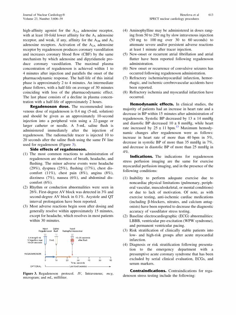
Regadenoson dose. The recommended intra-
venous dose of regadenoson is 0.4 mg (5-mL solution)
and should be given as an approximately 10-second
injection into a peripheral vein using a 22-gauge or
larger catheter or needle. A 5-mL saline flush is
administered immediately after the injection of
regadenoson. The radionuclide tracer is injected 10 to
20 seconds after the saline flush using the same IV line
used for regadenoson (Figure 3).
Side effects of regadenoson.(1) The most common reactions to administration of
regadenoson are shortness of breath, headache, and
flushing. The minor adverse events were headache
(29%), dyspnea (25%), flushing (17%), chest dis-
comfort (11%), chest pain (8%), angina (8%),
dizziness (7%), nausea (6%), and abdominal dis-
comfort (6%).
(2) Rhythm or conduction abnormalities were seen in
26%. First-degree AV block was detected in 3% and
second-degree AV block in 0.1%. Asystole and QT
interval prolongation have been reported.
(3) Most adverse reactions begin soon after dosing and
generally resolve within approximately 15 minutes,
except for headache, which resolves in most patients
within 30 minutes.
(4) Aminophylline may be administered in doses rang-
ing from 50 to 250 mg by slow intravenous injection
(50 mg to 100 mg over 30 to 60 seconds) to
attenuate severe and/or persistent adverse reactions
at least 1 minute after tracer injection.
(5) New-onset or recurrent atrial fibrillation and atrial
flutter have been reported following regadenoson
administration.
(6) New onset or recurrence of convulsive seizures has
occurred following regadenoson administration.
(7) Refractory ischemia/myocardial infarction, hemor-
rhagic, and ischemic cerebrovascular accidents have
been reported.
(8) Refractory ischemia and myocardial infarction have
occurred.
Hemodynamic effects. In clinical studies, the
majority of patients had an increase in heart rate and a
decrease in BP within 15 minutes after administration of
regadenoson. Systolic BP decreased by 13 ± 14 mmHg
and diastolic BP decreased 10 ± 8 mmHg, while heart
rate increased by 25 ± 11 bpm.13 Maximum hemody-
namic changes after regadenoson were as follows:
increase in heart rate of more than 40 bpm in 5%,
decrease in systolic BP of more than 35 mmHg in 7%,
and decrease in diastolic BP of more than 25 mmHg in
4%.
Indications. The indications for regadenoson
stress perfusion imaging are the same for exercise
myocardial perfusion imaging, and in the presence of the
following conditions:
(1) Inability to perform adequate exercise due to
noncardiac physical limitations (pulmonary, periph-
eral vascular, musculoskeletal, or mental conditions)
or due to lack of motivation. Of note, as with
exercise testing, anti-ischemic cardiac medications
(including b-blockers, nitrates, and calcium antag-
onists) have been reported to decrease the diagnostic
accuracy of vasodilator stress testing.
(2) Baseline electrocardiographic (ECG) abnormalities:
LBBB, ventricular pre-excitation (WPW syndrome),
and permanent ventricular pacing.
(3) Risk stratification of clinically stable patients into
low- and high-risk groups after acute myocardial
infarction.
(4) Diagnosis or risk stratification following presenta-
tion to the emergency department with a
presumptive acute coronary syndrome that has been
excluded by serial clinical evaluation, ECGs, and
serum markers.
Contraindications. Contraindications for rega-
denoson stress testing include the following:Figure 3. Regadenoson protocol. IV, Intravenous; mcg,microgram; and mL, milliliter.
Journal of Nuclear Cardiology� Henzlova et al 613
Volume 23, Number 3;606–39 SPECT nuclear cardiology procedures
(1) Patients with bronchospastic lung disease with
ongoing wheezing or a history of significant reactive
airway disease should not undergo regadenoson stress
testing.
(2) Second- or third-degree AV block or sinus node
dysfunction without a functioning pacemaker.
(3) Sinus node disease, such as sick sinus syndrome or
symptomatic bradycardia, without a functioning
pacemaker.
(4) Systolic BP less than 90 mmHg. The risk of serious
hypotension may be higher in patients with auto-
nomic dysfunction, hypovolemia, left main coronary
artery stenosis, stenotic valvular heart disease,
pericarditis or pericardial effusions, or stenotic
carotid artery disease with cerebrovascular
insufficiency.
(5) Uncontrolled hypertension (systolic BP
[200 mmHg or diastolic BP[110 mmHg).
(6) Recent (\48 hours) use of dipyridamole or dipyri-
damole-containing medications (e.g., Aggrenox).
(7) Known hypersensitivity to adenosine or
regadenoson.
(8) Unstable angina, acute coronary syndrome, or less
than 2 to 4 days after an acute myocardial
infarction.
Relative contraindications. Relative con-
traindications for regadenoson stress testing include
the following:
(1) Profound sinus bradycardia (heart rate\40/min).
(2) Mobitz Type 1 second-degree AV block
(Wenckebach).
(3) Severe Aortic Stenosis (see ‘‘Special Populations’’
section).
(4) Ingestion of caffeinated foods or beverages (e.g.,
coffee, tea, sodas) within the last 12 hours should be
avoided (Appendix 1).
(5) Seizure disorder. Regadenoson may lower seizure
threshold, and aminophylline should not be used in
cases of seizures associated with regadenoson.
These seizures may be of new onset, or may be
recurrences. In addition, some seizures are
prolonged and may require urgent anticonvulsive
management.14,15
Procedure.(1) Patient preparation: nothing should be eaten for at
least 3 hours prior to testing. Avoid consumption of
any products containing methylxanthines, including
caffeinated coffee, tea, or other caffeinated bever-
ages, caffeine-containing drug products (Appendix
A), and theophylline for at least 12 hours prior to
the testing. Dipyridamole should be withheld for at
least 2 days prior to regadenoson administration.
(2) ECG monitoring should be carried out as with
exercise stress testing. A 12-lead electrocardiogram
will be recorded every minute during the infusion.
(3) BP should be monitored every minute during
infusion and 3 to 5 minutes into recovery or until
stable.
(4) Regadenoson (5 mL, containing 0.4 mg of regade-
noson) should be given as a rapid (approximately
10 seconds) injection into a peripheral vein using a
22-gauge or larger catheter or needle. Administer a
5-mL saline flush immediately after the injection of
regadenoson. Administer the radionuclide MPI
agent 10 to 20 seconds after the saline flush. The
radionuclide may be injected directly into the same
catheter as regadenoson.
Combination of exercise with regadeno-son administration. Patients who are ambulatory
may undergo low-level exercise (e.g., treadmill 1.7 mph,
0% grade) for 1.5 minutes followed by regadenoson
administration, tracer injection, and an additional 2 min-
utes of exercise.16,17 Combining low-level exercise with
regadenoson resulted in improved image quality, and was
well tolerated without an increase in adverse events. Low-
level exercise supplementation is not recommended in
patients with LBBB,WPW, and ventricular pacing due to
heart rate-related imaging artifacts.
Ambulatory patients with uncertain functional
capacity who do not reach their target heart rate may
receive regadenoson to supplement submaximal exercise
stress with preserved image quality.18-21 Regadenoson
administration has been studied at peak exercise holding
the maximally attained stage, at reduced exercise, during
walk recovery, and at rest. While two of the studies
found no differences in the safety and side-effect profile
based on the time of regadenoson administration, one
study suggested more exaggerated BP responses when
administered at peak exercise.
Reversal of complications and side effectsof regadenoson. Indications for reversal of regade-
noson (50- to 250-mg aminophylline intravenously at
least 1 minute after the tracer injection) include the
following:Figure 4. Dipyridamole protocol. IV, Intravenous; kg, kilo-gram; and mg, milligram.
614 Henzlova et al Journal of Nuclear Cardiology�SPECT nuclear cardiology procedures May/June 2016
(1) Severe hypotension (systolic BP\80 mmHg).
(2) Development of symptomatic, persistent second-
degree or complete heart block.
(3) Other significant cardiac arrhythmia.
(4) Wheezing.
(5) Severe chest pain associated with ST depression of
2 mm or greater.
(6) Signs of poor perfusion (pallor, cyanosis, cold skin).
Cost/time/pharmacy. Medicare Part B drug
average sales price is calculated by the manufacturer
every calendar quarter and submitted to CMS (https://
www.cms.gov/Medicare/Medicare-Fee-for-Service-Part-
B-Drugs/McrPartBDrugAvgSalesPrice/index.html). The
average price for 0.4 mg of regadenoson during the 7
quarters since 2013 was $213.26 (Table 4).
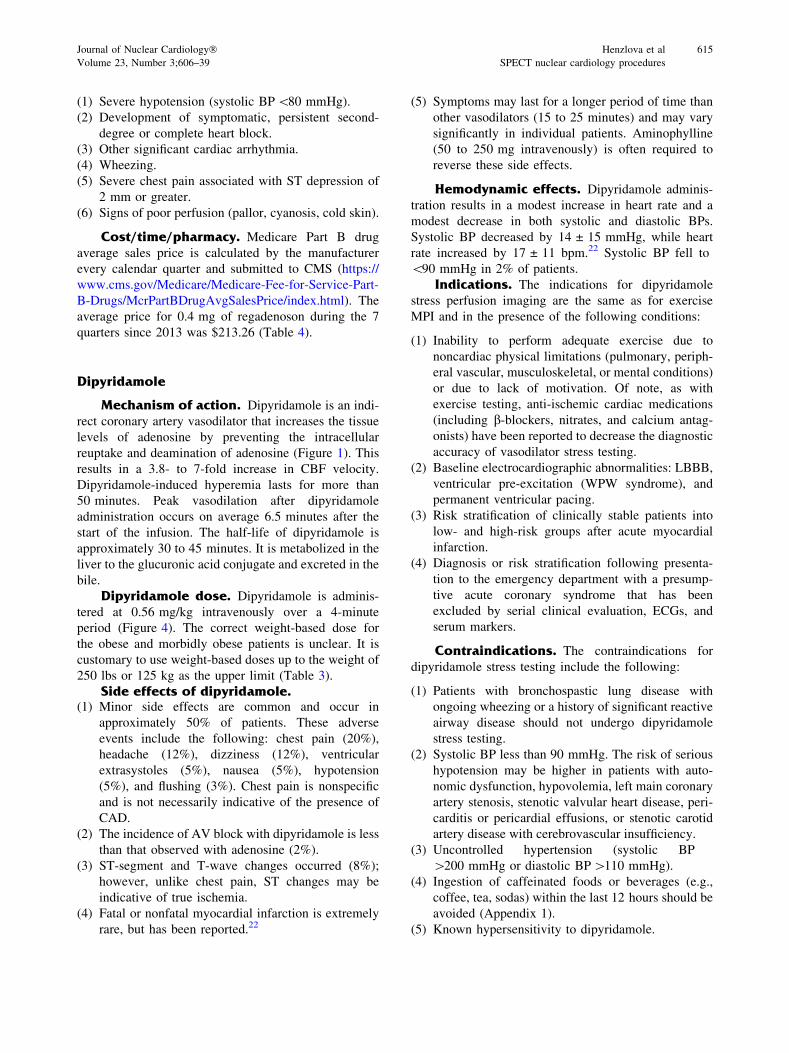
Dipyridamole
Mechanism of action. Dipyridamole is an indi-
rect coronary artery vasodilator that increases the tissue
levels of adenosine by preventing the intracellular
reuptake and deamination of adenosine (Figure 1). This
results in a 3.8- to 7-fold increase in CBF velocity.
Dipyridamole-induced hyperemia lasts for more than
50 minutes. Peak vasodilation after dipyridamole
administration occurs on average 6.5 minutes after the
start of the infusion. The half-life of dipyridamole is
approximately 30 to 45 minutes. It is metabolized in the
liver to the glucuronic acid conjugate and excreted in the
bile.
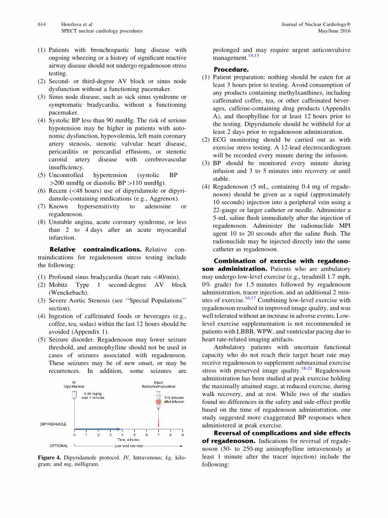
Dipyridamole dose. Dipyridamole is adminis-
tered at 0.56 mg/kg intravenously over a 4-minute
period (Figure 4). The correct weight-based dose for
the obese and morbidly obese patients is unclear. It is
customary to use weight-based doses up to the weight of
250 lbs or 125 kg as the upper limit (Table 3).
Side effects of dipyridamole.(1) Minor side effects are common and occur in
approximately 50% of patients. These adverse
events include the following: chest pain (20%),
headache (12%), dizziness (12%), ventricular
extrasystoles (5%), nausea (5%), hypotension
(5%), and flushing (3%). Chest pain is nonspecific
and is not necessarily indicative of the presence of
CAD.
(2) The incidence of AV block with dipyridamole is less
than that observed with adenosine (2%).
(3) ST-segment and T-wave changes occurred (8%);
however, unlike chest pain, ST changes may be
indicative of true ischemia.
(4) Fatal or nonfatal myocardial infarction is extremely
rare, but has been reported.22
(5) Symptoms may last for a longer period of time than
other vasodilators (15 to 25 minutes) and may vary
significantly in individual patients. Aminophylline
(50 to 250 mg intravenously) is often required to
reverse these side effects.
Hemodynamic effects. Dipyridamole adminis-
tration results in a modest increase in heart rate and a
modest decrease in both systolic and diastolic BPs.
Systolic BP decreased by 14 ± 15 mmHg, while heart
rate increased by 17 ± 11 bpm.22 Systolic BP fell to
\90 mmHg in 2% of patients.
Indications. The indications for dipyridamole
stress perfusion imaging are the same as for exercise
MPI and in the presence of the following conditions:
(1) Inability to perform adequate exercise due to
noncardiac physical limitations (pulmonary, periph-
eral vascular, musculoskeletal, or mental conditions)
or due to lack of motivation. Of note, as with
exercise testing, anti-ischemic cardiac medications
(including b-blockers, nitrates, and calcium antag-
onists) have been reported to decrease the diagnostic
accuracy of vasodilator stress testing.
(2) Baseline electrocardiographic abnormalities: LBBB,
ventricular pre-excitation (WPW syndrome), and
permanent ventricular pacing.
(3) Risk stratification of clinically stable patients into
low- and high-risk groups after acute myocardial
infarction.
(4) Diagnosis or risk stratification following presenta-
tion to the emergency department with a presump-
tive acute coronary syndrome that has been
excluded by serial clinical evaluation, ECGs, and
serum markers.
Contraindications. The contraindications for
dipyridamole stress testing include the following:
(1) Patients with bronchospastic lung disease with
ongoing wheezing or a history of significant reactive
airway disease should not undergo dipyridamole
stress testing.
(2) Systolic BP less than 90 mmHg. The risk of serious
hypotension may be higher in patients with auto-
nomic dysfunction, hypovolemia, left main coronary
artery stenosis, stenotic valvular heart disease, peri-
carditis or pericardial effusions, or stenotic carotid
artery disease with cerebrovascular insufficiency.
(3) Uncontrolled hypertension (systolic BP
[200 mmHg or diastolic BP[110 mmHg).
(4) Ingestion of caffeinated foods or beverages (e.g.,
coffee, tea, sodas) within the last 12 hours should be
avoided (Appendix 1).
(5) Known hypersensitivity to dipyridamole.
Journal of Nuclear Cardiology� Henzlova et al 615
Volume 23, Number 3;606–39 SPECT nuclear cardiology procedures
(6) Unstable angina, acute coronary syndrome, or less
than 2 to 4 days after an acute myocardial
infarction.
Note: In patients taking oral dipyridamole, IV
dipyridamole may be administered safely and
efficaciously.
Relative contraindications. Relative con-
traindications for dipyridamole stress testing include
the following:
(1) Profound sinus bradycardia (heart rates\40/min).
(2) Second- or third-degree AV block without a func-
tioning pacemaker.
(3) Severe aortic stenosis (see Special Populations
section).
(4) Seizure disorder. Methylxanthine (aminophylline)
use is not recommended in patients who experience
seizures in association with dipyridamole stress
testing.
Procedure.(1) Patient preparation: nothing should be eaten for at
least 3 hours prior to testing. Avoid consumption of
any products containing methylxanthines, including
caffeinated coffee, tea, or other caffeinated bever-
ages, caffeine-containing drug products (Appendix
1), and theophylline for at least 12 hours prior to the
testing.
(2) ECG monitoring should be carried out as with
exercise stress testing. A 12-lead electrocardiogram
will be recorded every minute during the infusion.
(3) BP and ECG should be monitored every minute
during infusion and 3 to 5 minutes into recovery or
until stable.
(4) The drug is infused intravenously over 4 minutes.
Although an infusion pump is preferable, dipyri-
damole can also be administered by hand injection
or drip. The radiotracer is injected 3 to 5 minutes
after the completion of dipyridamole infusion.
Combination of low-level exercise withdipyridamole infusion. Patients who are ambula-
tory may undergo low-level exercise (e.g., treadmill
1.7 mph, 0% grade) for 4 to 6 minutes soon after the
completion of dipyridamole infusion. Radiotracer is
injected during this low-level exercise, and the exercise
continues for an additional 2 minutes to allow for tracer
uptake in the myocardium. This significantly reduces the
side effects and improves image quality.23-26 Low-level
exercise supplementation is not recommended for
patients with LBBB, WPW, and ventricular pacing due
to heart rate-related imaging artifacts.
Indications for early termination ofdipyridamole infusion. The dipyridamole infusion
should be stopped early under any of the following
circumstances:
(1) Severe hypotension (systolic BP\80 mmHg).
(2) Development of symptomatic, persistent second-
degree or complete heart block.
(3) Other significant cardiac arrhythmia.
(4) Wheezing.
(5) Severe chest pain associated with ST depression of
2 mm or greater.
(6) Signs of poor perfusion (pallor, cyanosis, cold skin).
(7) Technical problems with the monitoring equipment.
(8) Patient’s request to stop.
Reversal of complications and side effectsof dipyridamole. Indications for reversal of dipyri-
damole (50- to 250-mg aminophylline intravenously at
least 1 minute after the tracer injection) include the
following:
(1) Severe hypotension (systolic BP\80 mmHg).
(2) Development of symptomatic, persistent second-
degree or complete heart block.
(3) Other significant cardiac arrhythmia.
(4) Wheezing.
(5) Severe chest pain associated with ST depression of
2 mm or greater.
(6) Signs of poor perfusion (pallor, cyanosis, cold skin).
(7) Can be considered in the presence of less-severe
side effects or ischemic ECG changes if at least
1 minute has elapsed since radiotracer injection.
Cost/time/pharmacy. The average price based
on Medicare Part B drug average sales price is calcu-
lated by the manufacturer every calendar quarter and
submitted to CMS (https://www.cms.gov/Medicare/Me
dicare-Fee-for-Service-Part-B-Drugs/McrPartBDrugAv
gSalesPrice/index.html) for a single study at 0.56-mg/kgFigure 5. Dobutamine protocol. IV, intravenous; kg, kilo-gram; mcg, microgram; and min, minute.
616 Henzlova et al Journal of Nuclear Cardiology�SPECT nuclear cardiology procedures May/June 2016
dipyridamole for a 75-kg patient during the 7 quarters
since 2013 was $6.30 (Table 4).
PHARMACOLOGIC SYNTHETICCATECHOLAMINE STRESS
There is one synthetic catecholamine stress agent
available: dobutamine. This agent works by stimulating
the b receptor, resulting in an increase in heart rate, BP,
and myocardial contractility similar to exercise.
Dobutamine
Mechanism of action. Dobutamine infusion
results in direct b1 and b2 stimulation with a dose-
related increase in heart rate, BP, and myocardial
contractility. Dobutamine increases regional myocardial
blood flow based on physiologic principles of coronary
flow reserve. A similar dose-related increase in subepi-
cardial and subendocardial blood flow occurs within
vascular beds supplied by normal coronary arteries.
However, blood flow increases minimally within vas-
cular beds supplied by significantly stenosed arteries,
with most of the increase occurring within the subepi-
cardium rather than the subendocardium. At a dose of
20 mcg/kg/min, however, dobutamine-induced coronary
flow heterogeneity is similar to exercise but less than
that induced by adenosine or dipyridamole. The plasma
half-life of dobutamine is 2 minutes with the onset of
action within 1 to 2 minutes; however, up to 10 minutes
may be required to obtain the peak effect (Figure 5).
Dobutamine dose. Dobutamine is infused
incrementally starting at a dose of 5 or 10 mcg/kg/
min, which is increased at 3-minute intervals to 20, 30,
and 40 mcg/kg/min. Radiotracer is injected at peak heart
rate with dobutamine infusion continuing for 1 minute
following tracer injection. As with exercise stress,
achieving greater than 85% of the predicted heart rate
is desirable.
Side effects of dobutamine.1. The common side effects are palpitation (29%), chest
pain (31%), headache (14%), flushing (14%), dysp-
nea (14%), and significant supraventricular or
ventricular arrhythmias (8% to 10%).
2. Ischemic ST-segment depression occurs in approxi-
mately one-third of patients undergoing dobutamine
infusion.
3. Severe side effects may require IV administration of
a short-acting b-blocker (esmolol, 0.5 mg/kg over
1 minute). IV metoprolol (5 mg) can also be used.
Hemodynamic effects. The hemodynamic
response to dobutamine infusion is dose dependent and
varies based on the maximal infusion rate obtained. In
studies titrating to a maximal dose of 40 mcg/kg/min,
heart rate increased 45 ± 18 bpm, while systolic BP
increased 30 ± 21 mmHg in one study, and
12 ± 29 mmHg in another.27,28
Indications. Indications for dobutamine stress
testing include the following:
(1) Dobutamine is a secondary pharmacologic stressor
that is recommended only in patients who cannot
undergo exercise stress and who also have con-
traindications to pharmacologic vasodilator stressors
(mainly bronchospastic airway disease).
(2) Dobutamine perfusion imaging has not been studied
as extensively as vasodilator stress perfusion imag-
ing in the evaluation and prognostication of patients
with CAD.
Contraindications. Contraindications for dobu-
tamine stress testing include the following:
(1) Unstable angina, acute coronary syndrome, or less
than 2 to 4 days after an acute myocardial infarction.
(2) Hemodynamically significant left ventricular out-
flow tract obstruction.
(3) Atrial tachyarrhythmias with uncontrolled ventricu-
lar response.
(4) Prior history of ventricular tachycardia.
(5) Uncontrolled hypertension (systolic BP
[200 mmHg or diastolic BP[110 mmHg).
(6) Patients with aortic dissection.
(7) Known hypersensitivity to dobutamine.
Relative contraindications.(1) Patients who are on b-blockers where the heart rate
and inotropic responses to dobutamine will be
attenuated.
(2) Severe aortic stenosis (see ‘‘Special Populations’’
section).
(3) Patients with with symptomatic or large aortic
aneurysm.
(4) Left bundle branch block.
(5) Paced ventricular rhythm.
Procedure.(1) Patient preparation: nothing should be eaten for at
least 3 hours.
(2) An infusion pump is necessary for dobutamine
administration.
(3) An IV line with a dual-port Y-connector is required
for injecting radioisotope during dobutamine
infusion.
(4) ECG monitoring and BP monitoring should
be performed as with other pharmacologic
stressors.
(5) Dobutamine infusion should start at a dose of 5 to
10 mcg/kg/min. The dobutamine dose should then
Journal of Nuclear Cardiology� Henzlova et al 617
Volume 23, Number 3;606–39 SPECT nuclear cardiology procedures
be increased at 3-minute intervals up to a maximum
of 40 mcg/kg/min. The radiotracer should be
injected at 1 minute into the highest dobutamine
dose (achieving at least 85% of maximal predicted
heart disease is desirable), and dobutamine infusion
should be continued for 1 minute after the radio-
tracer injection.
(6) Some investigators recommend the addition of
atropine (divided doses of 0.25 to 0.5 mg up to 1
to 2 mg) in patients who do not achieve target heart
rate with dobutamine alone.
Indications for early termination of dobu-tamine infusion. The dobutamine infusion should be
stopped early under any of the following circumstances:
(1) Achieving [85% of the age-predicted peak heart
rate (after maintaining for 1 minute following
radiotracer injection).
(2) Severe hypotension (systolic BP\80 mmHg).
(3) Severe hypertension (systolic BP [230 mmHg or
diastolic pressure[115 mmHg)
(4) Significant cardiac arrhythmia. Termination for
ventricular tachycardia or atrial tachyarrhythmia is
more likely with dobutamine than with other
stressors.
(5) Severe chest pain associated with ST depression of 2
mm or greater. Termination for ST-segment depres-
sion is more likely with dobutamine than with other
stressors.
(6) Signs of poor perfusion (pallor, cyanosis, cold skin).
(7) Technical problems with the monitoring equipment.
(8) Patient’s request to stop.
Reversal of complications and side effectsof dobutamine. Severe side effects, arrhythmia, or
ST changes may require IV administration of a short-
acting b-blocker (esmolol, 0.5 mg/kg over 1 minute). IV
metoprolol (5 mg) can also be used.
(1) Severe hypertension (systolic BP [220/
110 mmHg).
(2) Significant cardiac arrhythmia.
(3) Severe chest pain associated with ST depression of 2
mm or greater.
Cost/time/pharmacy. The average price based
on Medicare Part B drug average sales price is calcu-
lated by the manufacturer every calendar quarter and
submitted to CMS (https://www.cms.gov/Medicare/Med
icare-Fee-for-Service-Part-B-Drugs/McrPartBDrugAvg
SalesPrice/index.html) for a 250-mg IV bag of dobu-
tamine during the 7 quarters since 2013 was $6.59
(Table 4).
For review of serious complications of myocar-dial stress testing and their management, refer to thearticle by Dilsizian and colleagues.9
SPECIAL POPULATIONS FORPHARMACOLOGIC STRESS
Obesity
The correct dose for the obese and morbidly obese
patient is unclear. It is customary to use a weight-based
dose of IV dipyridamole, IV adenosine, and IV dobu-
tamine up to the weight of 250 lbs as the upper limit.
Left Bundle Branch Block, VentricularPacing, Ventricular Pre-excitation (WPWSyndrome)
Pharmacologic stress with coronary vasodilators
(not IV dobutamine) is the preferred stress modality with
above rest ECG patterns, both for safety (inability to
monitor for ischemia) and diagnostic reasons (e.g., the
presence of septal perfusion defects, not always related
to obstructive CAD). If rate-dependent LBBB is diag-
nosed during exercise, the test should be converted to a
pharmacologic stress test.
Reactive Airway Disease
Adenosine, regadenoson, and dipyridamole should
be administered with caution in patients with a history of
reactive airway disease or severe obstructive pulmonary
disease. Aminophylline should be available for the
treatment of vasodilator-induced bronchospasm. Dobu-
tamine is the preferred agent for pharmacologic stress in
patients with a history of significant reactive airway
disease or severe obstructive pulmonary disease.
Several studies have shown that a titrated dose
adenosine infusion preceded by an inhaled bronchodila-
tor was well tolerated in patients with mild reactive
airway disease from COPD or asthma.29-31 Because of
its selectivity for the A2A receptor and not the A2B
receptor, regadenoson shows promise to avoid bron-
choreactivity seen in adenosine and dipyridamole. Two
small pilot studies of moderate to severe COPD and
moderate asthma showed no statistically significant
difference between regadenoson and placebo in bron-
choconstrictive reactions as measured by
spirometry.32,33 While several other studies have exam-
ined the issue,34 the only randomized study enrolled 999
patients with spirometry defined COPD or asthma and
found no difference in the rate of bronchoconstriction
618 Henzlova et al Journal of Nuclear Cardiology�SPECT nuclear cardiology procedures May/June 2016
between regadenoson and placebo, but did find more
dyspnea.35
Renal Disease
Dipyridamole, adenosine, and dobutamine have no
contraindications to renal dysfunction. The package
label of regadenoson states that no serious events were
reported through 24 hours of follow-up in stage III or IV
renal impairment. In an initial pharmacologic model,
regadenoson elimination half-life appeared to be pro-
longed with decreasing renal function.36 Subsequent
published literature, including 423 patients with end-
stage renal disease (ESRD) and 745 patients with
impaired renal function but not ESRD, has found that
the drug was safe, with minimal side effects, and
hemodynamic responses similar to patients with normal
renal function.37-41
Aortic Stenosis
The use of vasodilator stress for MPI in patients
with significant aortic stenosis is limited as surgical
valve replacement is recommended for symptomatic
patients necessitating an invasive angiogram as opposed
to noninvasive testing. Occasionally, MPI was preferred
to angiography due to specific clinical considerations,
which has become a more frequent occurrence with the
advent of transcatheter aortic valve replacement
(TAVR). A number of small studies have evaluated
the diagnostic accuracy and to varying degrees the
safety and tolerability of dipyridamole (141
patients),42-44 adenosine (180 patients),45-48 and regade-
noson (50 patients).49 When assessed, there were no
significant differences in the hemodynamic responses
between patients with severe aortic stenosis and con-
trols.43,48 In a group of 50 pre-TAVR patients with a
mean EF of 39%, transient hypotension occurred in 16%
of subjects receiving regadenoson.49
There is insufficient safety data on SPECT dobu-
tamine stress in patients with severe aortic stenosis.
Pre Solid-Organ Transplantation
The role of cardiac disease evaluation prior to solid-
organ transplantation (e.g., kidney and liver) candidates
was discussed recently in an expert consensus document
by the AHA.50
The safety of regadenoson has been studied in
patients with renal impairment (see Renal Disease
section) and also in end-stage liver disease. In a study
of 168 patients, regadenoson was found to be safe in
end-stage liver disease patients with no significant
adverse events, and a lower heart-rate response but
similar BP response was seen.37 There is a case report of
three cases of sinus arrest with adenosine in liver
transplant patients with graft failure, so caution should
be taken in this particular patient population.51
Women
The role of noninvasive testing in the evaluation of
women with suspected ischemic heart disease was
recently covered in an AHA consensus statement.4
Caffeine and Coronary Vasodilators
Caffeine, a methylxanthine alkaloid derivative, is a
competitive inhibitor of the adenosine receptor due to its
similar molecular structure to adenosine. Caffeine binds
to the adenosine receptor without activating it, thereby
requires withholding methylxanthines prior to testing
with adenosine, dipyridamole, or regadenoson. The half-
life of caffeine varies widely among individuals based
on factors such as age, liver function, medication,
smoking use, and pregnancy, and is approximately
5 hours in a healthy adult.52 Caffeine is most commonly
found in coffee, tea, soda, energy drinks, and chocolate
(Appendix 1). While the caffeine content depends on the
particular product and method of preparation, the
caffeine contents of some common products are shown
in the table in Appendix 1. Decaffeinated coffee still
contains up to 13.9 mg of caffeine. A number of studies
have shown that even when patients report abstinence
from caffeine for 12 to 24 hours, a substantial portion of
patients have detectable serum caffeine levels.
The effect of caffeine on the coronary hyperemia
induced by adenosine, dipyridamole, and regadenoson
may be explained by the difference in their mechanism
of action. While dipyridamole is an indirect agonist on
the A2A receptor by increasing the concentration of
endogenous adenosine, adenosine is a direct stimulator,
and regadenoson has a higher A2A receptor affinity. A
number of studies have shown limited increase in
myocardial blood induced by dipyridamole after caf-
feine use as well as attenuated myocardial perfusion
defects.53-58 Two studies assessing the coronary hyper-
emia induced by adenosine found no significant
attenuation of the effect by caffeine.56,59 It is unclear
whether the degree of attenuation of the magnitude of
coronary hyperemia caused by caffeine would result in a
clinically significant change to myocardial perfusion
results.60 A number of studies have investigated the
effects of caffeine consumption on the MPI results using
adenosine stress finding some effect on imaging,61 but
often no significant effect.62,63 The data on regadenoson
are somewhat mixed with an animal study reporting that
caffeine did not affect the maximum increase in
Journal of Nuclear Cardiology� Henzlova et al 619
Volume 23, Number 3;606–39 SPECT nuclear cardiology procedures

Table
5.CurrentSPECTmyocardialperfusionim
agingprotocols:recommendedradiopharm
aceuticalactivitiesandtheircorresp
onding
radiationeffectivedose
s
Firstinjection
Seco
ndinjection
Total
Totaldose
ifGiven
at
Activity
(mCi)
Activity
(MBq)
Dose
(mSv)
Given
at
Activity
(mCi)
Activity
(MBq)
Dose
(mSv)
Dose
(mSv)
Stress
only
(mSv)
Tc-9
9m
protocols
Tc-9
9m
one-d
aystress-first/
stress-o
nly
Stress
8–1
2296–4
44
2.0–3
.0(Rest)
24–3
6888–1
332
7.0–1
0.5
9.0–1
3.5
2.0–3
.0
Tc-9
9m
one-d
ayrest/stress
Rest
8–1
2296–4
44
2.3–3
.5Stress
24–3
6888–1
332
6.1–9
.18.4–1
2.6
n/a
Tc-9
9m
two-d
aystress/rest
Stress
8–1
2296–4
44
2.0–3
.0(Rest)
8–1
2888–1
332
2.3–3
.54.3–6
.52.0–3
.0
Tc-9
9m
two-d
aystress/rest—
largepatient
Stress
18–3
0666–1
110
4.5–7
.6(Rest)
18–3
0666–1
110
5.2–8
.79.8–1
6.3
4.5–7
.6
Tc-9
9m
two-d
ayrest/stress
Rest
8–1
2296–4
44
2.3–3
.5Stress
8–1
2296–4
44
2.0–3
.04.3–6
.5n/a
Tc-9
9m
two-d
ayrest/stress
large
patient
Rest
18–3
0666–1
110
5.2–8
.7Stress
18–3
0666–1
110
4.5–7
.69.8–1
6.3
n/a
Tl-201protocols
Tl-201stress/redistributionrest
Stress
2.5–3
.592.5–1
29.5
10.9–1
5.3
n/a
n/a
n/a
n/a
10.9–1
5.3
10.9–1
5.3
Tl-201stress/redistributionrest/
reinjection
Stress
2.5–3
.592.5–1
29.5
10.9–1
5.3
Rest
1–2
37–7
44.4–8
.815.3–2
4.1
n/a
Tl-201rest/redistribution
Rest
2.5–3
.592.5–1
29.5
10.9–1
5.3
n/a
n/a
n/a
n/a
10.9–1
5.3
n/a
Dual-isotopeTl-201rest/Tc-9
9m
stress
Rest
2.5–3
.592.5–1
29.5
10.9–1
5.3
Stress
8–1
2296–4
44
2.0–3
.013.0–1
8.3
n/a
Dual-isotopeTl-201rest/Tc-9
9m
stress—largepatient
Rest
3.0–3
.5111–1
29.5
13.1–1
5.3
Stress
18–3
0666–1
110
4.5–7
.617.7–2
2.9
n/a
I-123protocol
MIBG
Rest
10
370
4.6
n/a
n/a
n/a
n/a
4.6
n/a
Newertechnologyreduced-d
ose
protocols
Tc-9
9m
one-d
aystress-first/
stress-o
nly
Stress
4–6
148–2
22
1.0–1
.5(Rest)
12–1
8444–6
66
3.5–5
.24.5–6
.71.0–1
.5
Tc-9
9m
one-d
ayrest/stress
Rest
4–6
148–2
22
1.2–1
.7Stress
12–1
8444–6
66
3.0–4
.54.2–6
.3n/a
Tc-9
9m
two-d
aystress/rest
Stress
4–6
148–2
22
1.0–1
.5(Rest)
4–6
148–2
22
1.2–1
.72.2–3
.31.0–1
.5
Tc-9
9m
two-d
aystress/rest—
largepatient
Stress
9–1
5333–5
55
2.3–3
.8(Rest)
9–1
5333–5
55
2.6–4
.44.9–8
.12.3–3
.8
Tc-9
9m
two-d
ayrest/stress
Rest
4–6
148–2
22
1.2–1
.7Stress
4–6
148–2
22
1.0–1
.52.2–3
.3n/a
Tc-9
9m
two-d
ayrest/stress—
largepatient
Rest
9–1
5333–5
55
2.6–4
.4Stress
9–1
5333–5
55
2.3–3
.84.9–8
.1n/a
620 Henzlova et al Journal of Nuclear Cardiology�SPECT nuclear cardiology procedures May/June 2016
coronary blood flow, but that the duration of hyperemia
was shorter.64 Regadenoson-induced hyperemic myocar-
dial blood flow measured by PET was not affected by
caffeine ingestion,65 but reversible defects on SPECT
myocardial perfusion imaging were smaller with caf-
feine consumption.66
Patients should abstain from caffeinated products
for at least 12 hours, which allows for selection of any
of the three vasodilator stressors.
RADIOTRACERS AND PROTOCOLS
There are a variety of different protocols currently
used for SPECT myocardial perfusion imaging. Table 5
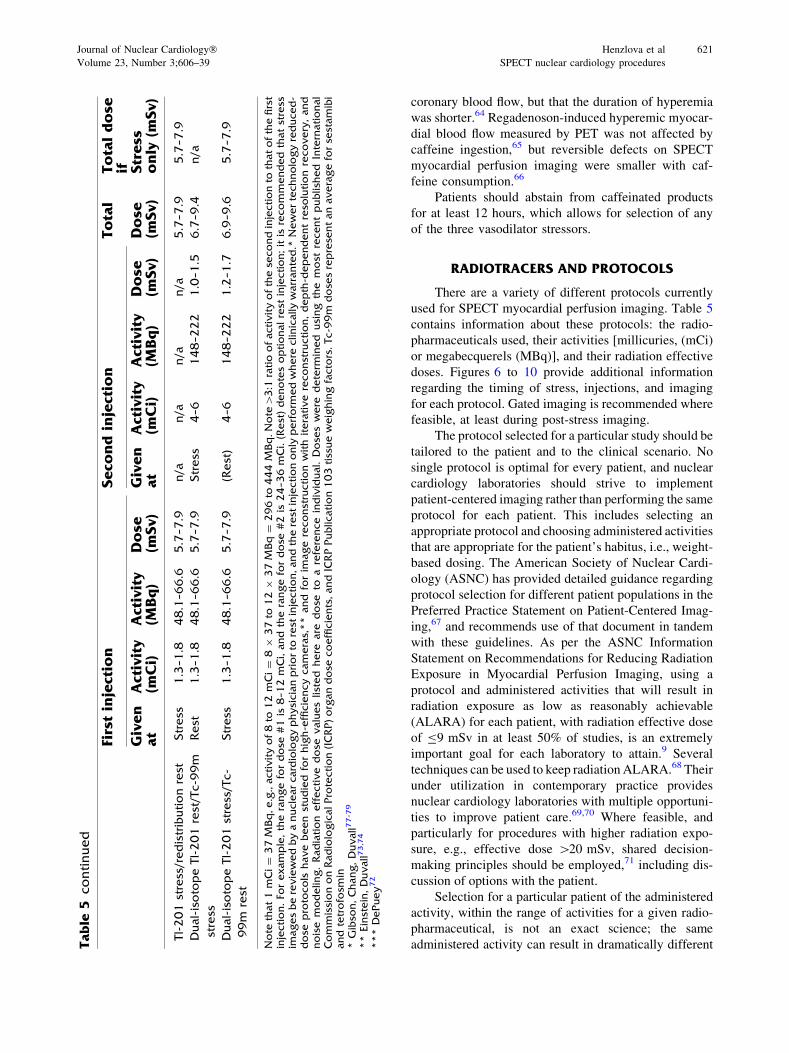
contains information about these protocols: the radio-
pharmaceuticals used, their activities [millicuries, (mCi)
or megabecquerels (MBq)], and their radiation effective
doses. Figures 6 to 10 provide additional information
regarding the timing of stress, injections, and imaging
for each protocol. Gated imaging is recommended where
feasible, at least during post-stress imaging.
The protocol selected for a particular study should be
tailored to the patient and to the clinical scenario. No
single protocol is optimal for every patient, and nuclear
cardiology laboratories should strive to implement
patient-centered imaging rather than performing the same
protocol for each patient. This includes selecting an
appropriate protocol and choosing administered activities
that are appropriate for the patient’s habitus, i.e., weight-
based dosing. The American Society of Nuclear Cardi-
ology (ASNC) has provided detailed guidance regarding
protocol selection for different patient populations in the
Preferred Practice Statement on Patient-Centered Imag-
ing,67 and recommends use of that document in tandem
with these guidelines. As per the ASNC Information
Statement on Recommendations for Reducing Radiation
Exposure in Myocardial Perfusion Imaging, using a
protocol and administered activities that will result in
radiation exposure as low as reasonably achievable
(ALARA) for each patient, with radiation effective dose
of B9 mSv in at least 50% of studies, is an extremely
important goal for each laboratory to attain.9 Several
techniques can be used to keep radiation ALARA.68 Their
under utilization in contemporary practice provides
nuclear cardiology laboratories with multiple opportuni-
ties to improve patient care.69,70 Where feasible, and
particularly for procedures with higher radiation expo-
sure, e.g., effective dose [20 mSv, shared decision-
making principles should be employed,71 including dis-
cussion of options with the patient.
Selection for a particular patient of the administered
activity, within the range of activities for a given radio-
pharmaceutical, is not an exact science; the same
administered activity can result in dramatically differentTable
5continued
Firstinjection
Seco
ndinjection
Total
Totaldose
ifGiven
at
Activity
(mCi)
Activity
(MBq)
Dose
(mSv)
Given
at
Activity
(mCi)
Activity
(MBq)
Dose
(mSv)
Dose
(mSv)
Stress
only
(mSv)
Tl-201stress/redistributionrest
Stress
1.3–1
.848.1–6
6.6
5.7–7
.9n/a
n/a
n/a
n/a
5.7–7
.95.7–7
.9
Dual-isotopeTl-201rest/Tc-9
9m
stress
Rest
1.3–1
.848.1–6
6.6
5.7–7
.9Stress
4–6
148–2
22
1.0–1
.56.7–9
.4n/a
Dual-isotopeTl-201stress/Tc-
99m
rest
Stress
1.3–1
.848.1–6
6.6
5.7–7
.9(Rest)
4–6
148–2
22
1.2–1
.76.9–9
.65.7–7
.9
Note
that1mCi=
37M
Bq,e.g.,activityof8to
12mCi=
89
37to
129
37M
Bq=
296to
444M
Bq.Note
[3:1
ratioofactivityofthese
condinjectionto
thatofthefirst
injection.Fo
rexample,therangefordose
#1is
8–1
2mCi,andtherangefordose
#2is
24–3
6mCi.(Rest)denotesoptionalrest
injection;itis
recommendedthatstress
imagesbereviewedbyanuclearcardiologyphysicianpriorto
rest
injection,andtherest
injectiononly
perform
edwhere
clinically
warranted.*
Newertechnologyreduced-
dose
protocols
havebeenstudied
forhigh-efficiencycameras,**
and
forim
agereconstructionwithiterativereconstruction,depth-d
ependentreso
lutionrecovery,and
noise
modelin
g.Radiation
effective
dose
valueslisted
here
are
dose
toareference
individual.Dose
swere
determ
ined
using
the
most
recentpublish
ed
International
CommissiononRadiologicalProtection(ICRP)organdose
coefficients,andICRPPublication103tissueweighingfactors.Tc-9
9m
dose
sreprese
ntanaverageforse
stamibi
andtetrofosm
in*Gibso
n,Chang,Duvall7
7-7
9
**Einstein,Duvall7
3,74
***DePuey72
Journal of Nuclear Cardiology� Henzlova et al 621
Volume 23, Number 3;606–39 SPECT nuclear cardiology procedures
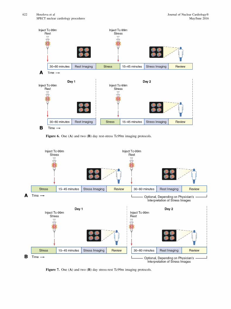
Figure 6. One (A) and two (B) day rest-stress Tc99m imaging protocols.
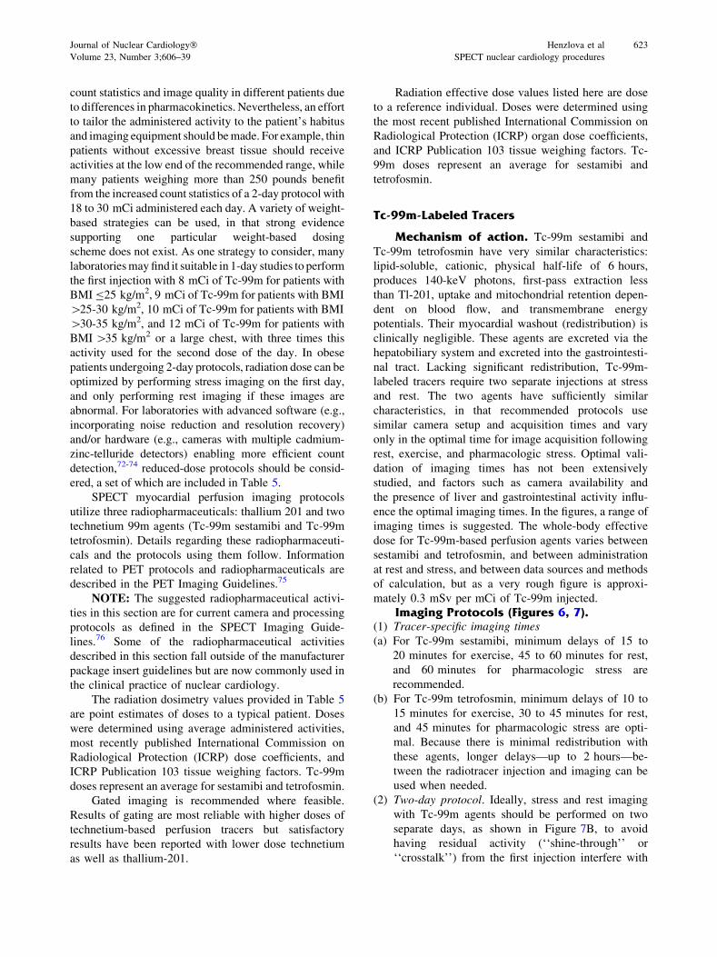
Figure 7. One (A) and two (B) day stress-rest Tc99m imaging protocols.
622 Henzlova et al Journal of Nuclear Cardiology�SPECT nuclear cardiology procedures May/June 2016
count statistics and image quality in different patients due
to differences in pharmacokinetics. Nevertheless, an effort
to tailor the administered activity to the patient’s habitus
and imaging equipment should bemade. For example, thin
patients without excessive breast tissue should receive
activities at the low end of the recommended range, while
many patients weighing more than 250 pounds benefit
from the increased count statistics of a 2-day protocol with
18 to 30 mCi administered each day. A variety of weight-
based strategies can be used, in that strong evidence
supporting one particular weight-based dosing
scheme does not exist. As one strategy to consider, many
laboratoriesmayfind it suitable in 1-day studies to perform
the first injection with 8 mCi of Tc-99m for patients with
BMIB25 kg/m2, 9 mCi of Tc-99m for patients with BMI
[25-30 kg/m2, 10 mCi of Tc-99m for patients with BMI
[30-35 kg/m2, and 12 mCi of Tc-99m for patients with
BMI[35 kg/m2 or a large chest, with three times this
activity used for the second dose of the day. In obese
patients undergoing 2-day protocols, radiation dose can be
optimized by performing stress imaging on the first day,
and only performing rest imaging if these images are
abnormal. For laboratories with advanced software (e.g.,
incorporating noise reduction and resolution recovery)
and/or hardware (e.g., cameras with multiple cadmium-
zinc-telluride detectors) enabling more efficient count
detection,72-74 reduced-dose protocols should be consid-
ered, a set of which are included in Table 5.
SPECT myocardial perfusion imaging protocols
utilize three radiopharmaceuticals: thallium 201 and two
technetium 99m agents (Tc-99m sestamibi and Tc-99m
tetrofosmin). Details regarding these radiopharmaceuti-
cals and the protocols using them follow. Information
related to PET protocols and radiopharmaceuticals are
described in the PET Imaging Guidelines.75
NOTE: The suggested radiopharmaceutical activi-
ties in this section are for current camera and processing
protocols as defined in the SPECT Imaging Guide-
lines.76 Some of the radiopharmaceutical activities
described in this section fall outside of the manufacturer
package insert guidelines but are now commonly used in
the clinical practice of nuclear cardiology.
The radiation dosimetry values provided in Table 5
are point estimates of doses to a typical patient. Doses
were determined using average administered activities,
most recently published International Commission on
Radiological Protection (ICRP) dose coefficients, and
ICRP Publication 103 tissue weighing factors. Tc-99m
doses represent an average for sestamibi and tetrofosmin.
Gated imaging is recommended where feasible.
Results of gating are most reliable with higher doses of
technetium-based perfusion tracers but satisfactory
results have been reported with lower dose technetium
as well as thallium-201.
Radiation effective dose values listed here are dose
to a reference individual. Doses were determined using
the most recent published International Commission on
Radiological Protection (ICRP) organ dose coefficients,
and ICRP Publication 103 tissue weighing factors. Tc-
99m doses represent an average for sestamibi and
tetrofosmin.
Tc-99m-Labeled Tracers
Mechanism of action. Tc-99m sestamibi and
Tc-99m tetrofosmin have very similar characteristics:
lipid-soluble, cationic, physical half-life of 6 hours,
produces 140-keV photons, first-pass extraction less
than Tl-201, uptake and mitochondrial retention depen-
dent on blood flow, and transmembrane energy
potentials. Their myocardial washout (redistribution) is
clinically negligible. These agents are excreted via the
hepatobiliary system and excreted into the gastrointesti-
nal tract. Lacking significant redistribution, Tc-99m-
labeled tracers require two separate injections at stress
and rest. The two agents have sufficiently similar
characteristics, in that recommended protocols use
similar camera setup and acquisition times and vary
only in the optimal time for image acquisition following
rest, exercise, and pharmacologic stress. Optimal vali-
dation of imaging times has not been extensively
studied, and factors such as camera availability and
the presence of liver and gastrointestinal activity influ-
ence the optimal imaging times. In the figures, a range of
imaging times is suggested. The whole-body effective
dose for Tc-99m-based perfusion agents varies between
sestamibi and tetrofosmin, and between administration
at rest and stress, and between data sources and methods
of calculation, but as a very rough figure is approxi-
mately 0.3 mSv per mCi of Tc-99m injected.
Imaging Protocols (Figures 6, 7).(1) Tracer-specific imaging times
(a) For Tc-99m sestamibi, minimum delays of 15 to
20 minutes for exercise, 45 to 60 minutes for rest,
and 60 minutes for pharmacologic stress are
recommended.
(b) For Tc-99m tetrofosmin, minimum delays of 10 to
15 minutes for exercise, 30 to 45 minutes for rest,
and 45 minutes for pharmacologic stress are opti-
mal. Because there is minimal redistribution with
these agents, longer delays—up to 2 hours—be-
tween the radiotracer injection and imaging can be
used when needed.
(2) Two-day protocol. Ideally, stress and rest imaging
with Tc-99m agents should be performed on two
separate days, as shown in Figure 7B, to avoid
having residual activity (‘‘shine-through’’ or
‘‘crosstalk’’) from the first injection interfere with
Journal of Nuclear Cardiology� Henzlova et al 623
Volume 23, Number 3;606–39 SPECT nuclear cardiology procedures
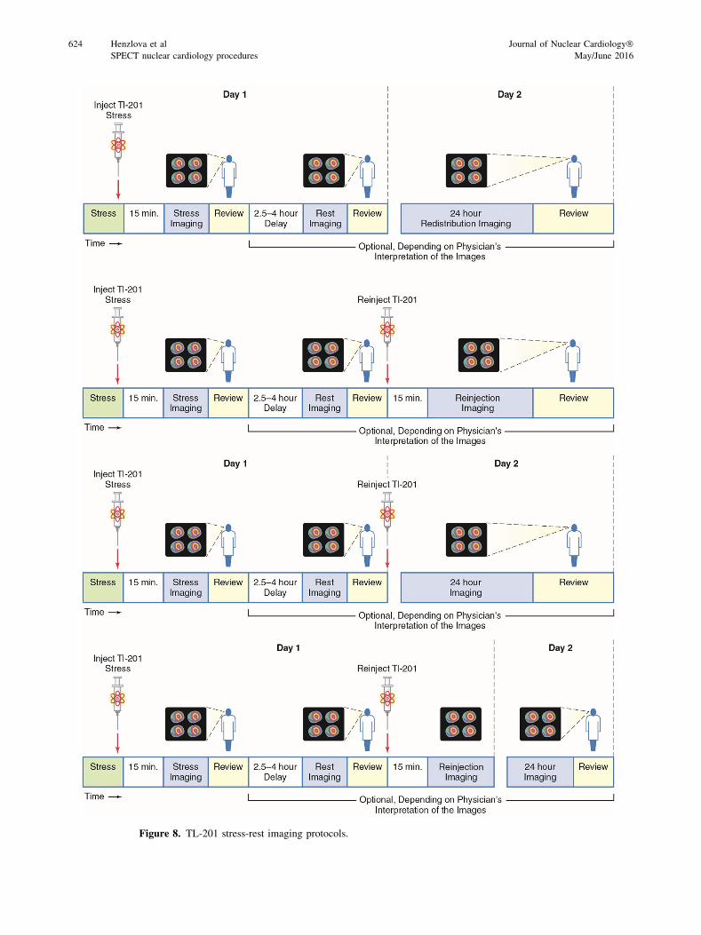
Figure 8. TL-201 stress-rest imaging protocols.
624 Henzlova et al Journal of Nuclear Cardiology�SPECT nuclear cardiology procedures May/June 2016
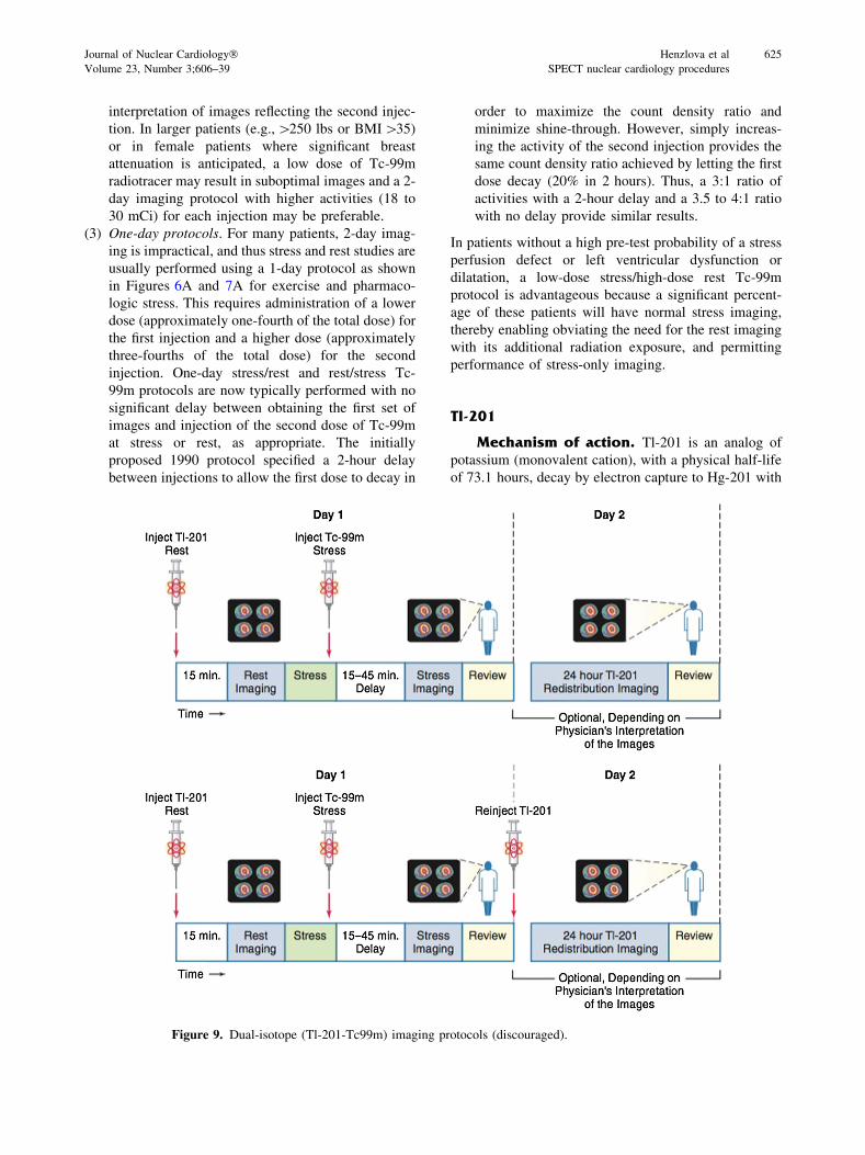
interpretation of images reflecting the second injec-
tion. In larger patients (e.g.,[250 lbs or BMI[35)
or in female patients where significant breast
attenuation is anticipated, a low dose of Tc-99m
radiotracer may result in suboptimal images and a 2-
day imaging protocol with higher activities (18 to
30 mCi) for each injection may be preferable.
(3) One-day protocols. For many patients, 2-day imag-
ing is impractical, and thus stress and rest studies are
usually performed using a 1-day protocol as shown
in Figures 6A and 7A for exercise and pharmaco-
logic stress. This requires administration of a lower
dose (approximately one-fourth of the total dose) for
the first injection and a higher dose (approximately
three-fourths of the total dose) for the second
injection. One-day stress/rest and rest/stress Tc-
99m protocols are now typically performed with no
significant delay between obtaining the first set of
images and injection of the second dose of Tc-99m
at stress or rest, as appropriate. The initially
proposed 1990 protocol specified a 2-hour delay
between injections to allow the first dose to decay in
order to maximize the count density ratio and
minimize shine-through. However, simply increas-
ing the activity of the second injection provides the
same count density ratio achieved by letting the first
dose decay (20% in 2 hours). Thus, a 3:1 ratio of
activities with a 2-hour delay and a 3.5 to 4:1 ratio
with no delay provide similar results.
In patients without a high pre-test probability of a stress
perfusion defect or left ventricular dysfunction or
dilatation, a low-dose stress/high-dose rest Tc-99m
protocol is advantageous because a significant percent-
age of these patients will have normal stress imaging,
thereby enabling obviating the need for the rest imaging
with its additional radiation exposure, and permitting
performance of stress-only imaging.
Tl-201
Mechanism of action. Tl-201 is an analog of
potassium (monovalent cation), with a physical half-life
of 73.1 hours, decay by electron capture to Hg-201 with
Figure 9. Dual-isotope (Tl-201-Tc99m) imaging protocols (discouraged).
Journal of Nuclear Cardiology� Henzlova et al 625
Volume 23, Number 3;606–39 SPECT nuclear cardiology procedures
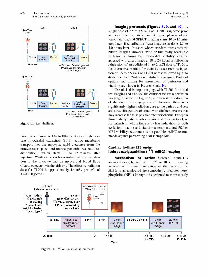
principal emission of 68- to 80-keV X-rays, high first-
pass myocardial extraction (85%), active membrane
transport into the myocyte, rapid clearance from the
intravascular space, and monoexponential washout (re-
distribution), which starts 10 to 15 minutes after
injection. Washout depends on initial tracer concentra-
tion in the myocyte and on myocardial blood flow.
Clearance occurs via the kidneys. The effective radiation
dose for Tl-201 is approximately 4.4 mSv per mCi of
Tl-201 injected.
Imaging protocols (Figures 8, 9, and 10). A
single dose of 2.5 to 3.5 mCi of Tl-201 is injected prior
to peak exercise stress or at peak pharmacologic
vasodilatation, and SPECT imaging starts 10 to 15 min-
utes later. Redistribution (rest) imaging is done 2.5 to
4.0 hours later. In cases where standard stress-redistri-
bution imaging shows a fixed or minimally reversible
perfusion abnormality, myocardial viability can be
assessed with a rest image at 18 to 24 hours or following
reinjection of an additional 1- to 2-mCi dose of Tl-201.
An alternative method for viability assessment is injec-
tion of 2.5 to 3.5 mCi of Tl-201 at rest followed by 3- to
4-hour or 18- to 24-hour redistribution imaging. Protocol
options and timing for assessment of perfusion and
viability are shown in Figures 8 and 10.
Use of dual-isotope imaging, with Tl-201 for initial
rest imaging and aTc-99 labeled tracer for stress perfusion
imaging, as shown in Figure 9, allows a shorter duration
of the entire imaging protocol. However, there is a
significantly higher radiation dose to the patient, and rest
and stress images are obtained with different tracers that
may increase the false positive rate for ischemia. Except in
those elderly patients who require a shorter protocol, or
for patients in whom there is a clear indication for both
perfusion imaging and viability assessment, and PET or
MRI viability assessment is not possible, ASNC recom-
mends against performing dual-isotope MPI.
Cardiac Iodine-123 meta-iodobenzylguanidine (123I-mIBG) Imaging
Mechanism of action. Cardiac iodine-123
meta-iodobenzylguanidine (123I-mIBG) imaging
assesses sympathetic innervation of the myocardium.
MIBG is an analog of the sympathetic mediator nore-
pinephrine (NE), although it is designed to more closely
Figure 11. 123I-mIBG imaging protocols.
Figure 10. Rest thallium.
626 Henzlova et al Journal of Nuclear Cardiology�SPECT nuclear cardiology procedures May/June 2016
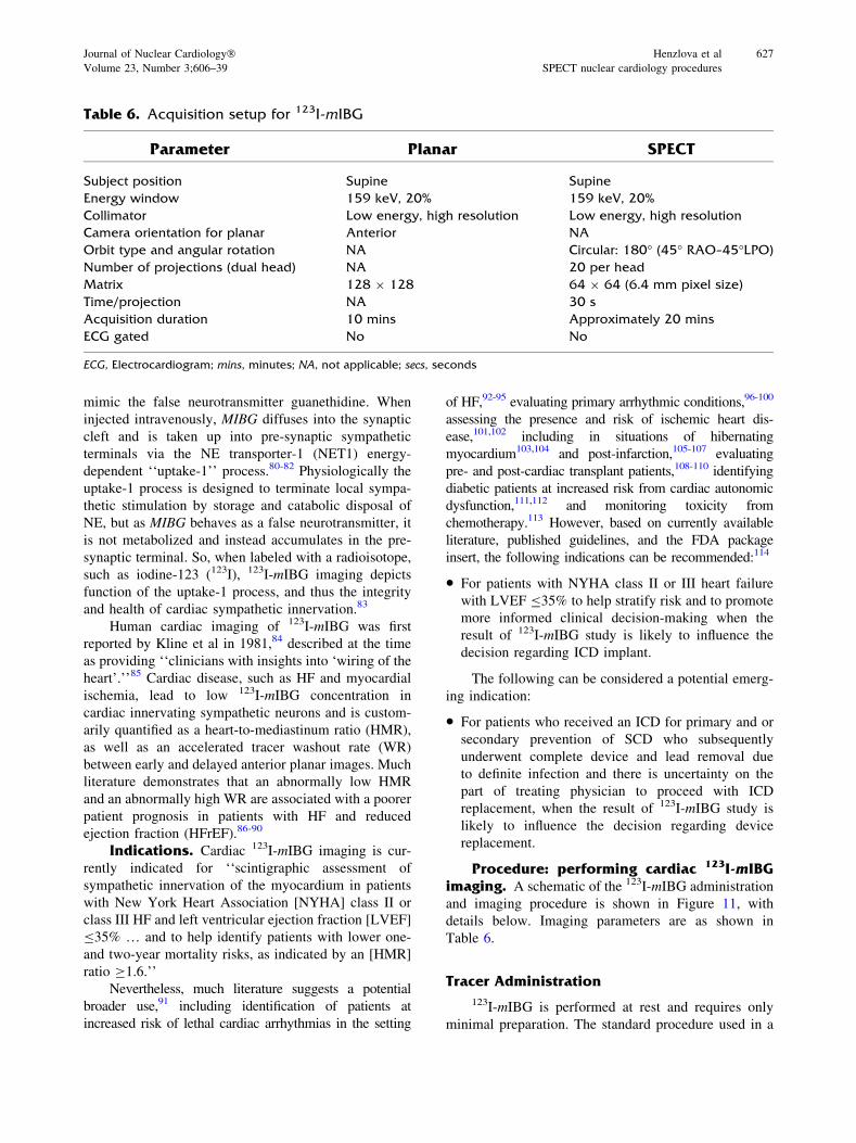
mimic the false neurotransmitter guanethidine. When
injected intravenously, MIBG diffuses into the synaptic
cleft and is taken up into pre-synaptic sympathetic
terminals via the NE transporter-1 (NET1) energy-
dependent ‘‘uptake-1’’ process.80-82 Physiologically the
uptake-1 process is designed to terminate local sympa-
thetic stimulation by storage and catabolic disposal of
NE, but as MIBG behaves as a false neurotransmitter, it
is not metabolized and instead accumulates in the pre-
synaptic terminal. So, when labeled with a radioisotope,
such as iodine-123 (123I), 123I-mIBG imaging depicts
function of the uptake-1 process, and thus the integrity
and health of cardiac sympathetic innervation.83
Human cardiac imaging of 123I-mIBG was first
reported by Kline et al in 1981,84 described at the time
as providing ‘‘clinicians with insights into ‘wiring of the
heart’.’’85 Cardiac disease, such as HF and myocardial
ischemia, lead to low 123I-mIBG concentration in
cardiac innervating sympathetic neurons and is custom-
arily quantified as a heart-to-mediastinum ratio (HMR),
as well as an accelerated tracer washout rate (WR)
between early and delayed anterior planar images. Much
literature demonstrates that an abnormally low HMR
and an abnormally high WR are associated with a poorer
patient prognosis in patients with HF and reduced
ejection fraction (HFrEF).86-90
Indications. Cardiac 123I-mIBG imaging is cur-
rently indicated for ‘‘scintigraphic assessment of
sympathetic innervation of the myocardium in patients
with New York Heart Association [NYHA] class II or
class III HF and left ventricular ejection fraction [LVEF]
B35% … and to help identify patients with lower one-
and two-year mortality risks, as indicated by an [HMR]
ratio C1.6.’’
Nevertheless, much literature suggests a potential
broader use,91 including identification of patients at
increased risk of lethal cardiac arrhythmias in the setting
of HF,92-95 evaluating primary arrhythmic conditions,96-100
assessing the presence and risk of ischemic heart dis-
ease,101,102 including in situations of hibernating
myocardium103,104 and post-infarction,105-107 evaluating
pre- and post-cardiac transplant patients,108-110 identifying
diabetic patients at increased risk from cardiac autonomic
dysfunction,111,112 and monitoring toxicity from
chemotherapy.113 However, based on currently available
literature, published guidelines, and the FDA package
insert, the following indications can be recommended:114
• For patients with NYHA class II or III heart failure
with LVEF B35% to help stratify risk and to promote
more informed clinical decision-making when the
result of 123I-mIBG study is likely to influence the
decision regarding ICD implant.
The following can be considered a potential emerg-
ing indication:
• For patients who received an ICD for primary and or
secondary prevention of SCD who subsequently
underwent complete device and lead removal due
to definite infection and there is uncertainty on the
part of treating physician to proceed with ICD
replacement, when the result of 123I-mIBG study is
likely to influence the decision regarding device
replacement.
Procedure: performing cardiac 123I-mIBGimaging. A schematic of the 123I-mIBG administration
and imaging procedure is shown in Figure 11, with
details below. Imaging parameters are as shown in
Table 6.
Tracer Administration
123I-mIBG is performed at rest and requires only
minimal preparation. The standard procedure used in a
Table 6. Acquisition setup for 123I-mIBG
Parameter Planar SPECT
Subject position Supine Supine
Energy window 159 keV, 20% 159 keV, 20%
Collimator Low energy, high resolution Low energy, high resolution
Camera orientation for planar Anterior NA
Orbit type and angular rotation NA Circular: 180� (45� RAO–45�LPO)
Number of projections (dual head) NA 20 per head
Matrix 128 9 128 64 9 64 (6.4 mm pixel size)
Time/projection NA 30 s
Acquisition duration 10 mins Approximately 20 mins
ECG gated No No
ECG, Electrocardiogram; mins, minutes; NA, not applicable; secs, seconds
Journal of Nuclear Cardiology� Henzlova et al 627
Volume 23, Number 3;606–39 SPECT nuclear cardiology procedures
recent multicenter study was to keep the patient NPO
(except for water) after midnight on the day of imag-
ing.115 It is accepted that standard HF medications such
as ß-blockers, angiotensin-converting enzyme inhibitors
(ACE-I), and/or angiotensin receptor blockers (ARBs)
need not be held.116-119 However, it is recommended to
temporarily discontinue medications and substances
known to interfere directly with the mechanisms of
NE uptake and granule storage, such as opioids, cocaine,
tramadol, tricyclic antidepressants sypathomimetrics,
some antihypertensive and cardiovascular agents, and
antipsychotics (see detail in Table 7).118 In addition,
foods containing vanillin and catecholamine-like com-
pounds (e.g., chocolate and blue cheese) should be
stopped.120,121 A subanalysis of data from the ADMIRE-
HF study showed that neuropsychiatric medications with
high potency can result in a falsely low HMR and thus
an overestimation of cardiac risk.122
There are differing views regarding the need for
pre-test administration of iodine to prevent thyroid
uptake of I-123. Historically, such blockade had been
undertaken to shield the thyroid from exposure to
unbound radionuclide iodine impurities, but with mod-
ern production methods the amount of these is minimal,
and many feel that pre-treatment is unnecessary. Nev-
ertheless, a recent study showed that even with an 123I-
mIBG preparation of 98% radiochemical purity,
unblocked patients have a 50% higher 4-hour thyroid
accumulation of unbound 123I compared with pre-treated
patients, with an estimated 70-mGy exposure from a 10-
mCi administered dose.123 Thus, it is recommended that
pre-treatment should be individually determined, to be
considered in perhaps younger patients, but less worth-
while in elderly patients with multiple comorbidities and
potential risk of iodine allergy. If desired, thyroid
blockade can be achieved with oral administration of
solutions of potassium iodide or Lugol’s (130-mg iodide
for adults, body weight adjusted for children), or for
patients allergic to iodine, potassium perchlorate
(500 mg for adults, weight adjusted for children), orally
administered at least 30 minutes prior to MIBG
injection.118
Older studies used a dose of 3 to 5 mCi [111 to 185
megabecquerels (MBq)] administered over 1 minute.
However, as it is often difficult to obtain satisfactory
SPECT images using these doses, especially in patients
with severe cardiac dysfunction, investigators have
recently been using up to 10 mCi (370 MBq).80,115 A
10-mCi dose results in radiation exposure of approxi-
mately 5 mSv, with highest exposure to the bladder,
liver, spleen, gall bladder, heart, and adrenals; the
absorbed dose may be higher in patients with severe
renal impairment.118
It has been recommended that patients lie quietly in a
supine position for about 5 to 15 minutes before admin-
istration. As initial images are acquired a few minutes
later, the patient should be lying under the camera or in
close proximity. The tracer is then administered slowly
over 1 to 2 minutes, followed by a saline flush.
Adverse Reactions
Adverse reactions to 123I-mIBG are uncommon.
Among the side effects reported when 123I-mIBG is
administered too quickly are palpitations, shortness of
breath, heat sensations, transient hypertension, and
abdominal cramps. A rare anaphylactic reaction is also
possible.118
Imaging Techniques
While acquisition and processing of planar and
SPECT 123I-mIBG images are most often performed
with techniques typical for perfusion imaging, there are
special issues that need to be considered that will likely
result in changes in methodology over time. The current
key parameters for 123I-mIBG image interpretation—
HMR and washout—require accurate and robust quan-
titative analysis of counts in the heart and adjacent
background regions, and thus could vary in relation to
issues, such as the acquisition field of view, the type of
collimator used, and how the multiple energy emissions
of 123I are taken into account during acquisition and
processing. In addition, data in the literature are based
on images obtained without using correction for atten-
uation, Compton scatter or depth-dependent loss of
spatial resolution, with use of such techniques likely to
produce different values. Finally, increased use of solid-
state cameras will likely produce different quantitative123I-mIBG results from what has been reported for
standard Anger cameras.
Anterior planar 123I-mIBG images are acquired for
10 minutes with the patient supine approximately
15 minutes (early) and beginning approximately
3 hours, 50 minutes (late) after tracer administration,
and stored in a 128 9 128 or 256 9 256 matrix.115,118
The imaging field of view should include as much of the
heart and upper chest as possible, with avoidance of
positioning the heart too close to the edge of the field or
too close to the center. Positioning should be consistent
for the early and late planar images.
With a large field of view, anterior planar image is
important for obtaining the desired HMR. The technique
for obtaining suitably equivalent images from small-
field-of-view SPECT cameras is unclear. In a report
from a pilot group of 67 subjects, HMRs calculated from
628 Henzlova et al Journal of Nuclear Cardiology�SPECT nuclear cardiology procedures May/June 2016
SPECT images using mean counts between heart and
mediastinum volumes of interest drawn on transaxial
images were equivalent to those obtained by planar
techniques for differentiating subjects with normal
versus abnormal MIBG uptake.124 More investigations
are required to validate this further, including for images
obtained with solid-state cameras.
Although the utility of SPECT imaging assessment
of regional defects is currently uncertain, based on
theoretic consideration106,125,126 and some literature data
particularly for primary arrhythmias,96-100 tomographic
acquisitions are commonly performed immediately fol-
lowing both the early and late planar images. They are
obtained with a minimum of 60 projections at 30 sec-
onds/stop over a 180� arc (45� right anterior oblique to
45�left posterior oblique) and stored in a 64 9 64
matrix.
Much literature is based on 123I-mIBG image
acquisition using low-energy high-resolution (LEHR)
collimators, with a symmetrically centered energy win-
dow of 20% around the main 159-keV isotope
photopeak. However, HMR values vary depending on
the collimator used, different for low-energy versus
medium-energy collimators, but also varying among
different low-energy collimator brands.127-129 In part
these differences relate to 123I also emitting multiple
low-abundance high-energy ([400 keV) photons, espe-
cially a 529-keV emission. While on an intrinsic 123I
spectrum, the higher energy emissions are small in
relation to the principle 159-keV peak, a low-energy
collimator blocks many of the 159-keV emissions while
allowing septal penetration of the higher energy photons
that degrade image quality and affect accuracy of
quantitative values, such as HMR.130,131 Although most
clinical literature is based on use of a LEHR, in fact the
reported HMRs are severely underestimated in relation
to the true value.132 Several methods have been explored
attempting to overcome this problem when an LEHR is
used, including mathematical deconvolution of septal
penetration (DSP),130 image acquisition using 123I-dual
window acquisition (IDW),133 decreasing the 159-kEV
energy window to 15%.132 Although it has been sug-
gested that a medium-energy collimator may be
preferred,129 use of a calibration phantom to derive a
conversion coefficient for each camera-collimation sys-
tem may be a better approach.134,135
There have been recent reports on the 123I-mIBG
imaging with solid-state cameras. While data from these
are encouraging, given the aforementioned discussion,
one must consider the image that derived quantitative
parameters from these camera(s) may differ from those
reported in the literature upon which clinical use is
recommended.136-138 Solid-state cameras may provide
the image resolution and detail necessary for higher
quality SPECT images that can allow assessment of
regional sympathetic activity that might be superior to
HMR in predicting adverse cardiac events, particularly
arrhythmias.139
Derivation of the HMR and Washout(Figures 12, 13)
As the HMR is currently the key result of a 123I-
mIBG study, it is important it be derived in a standard
way. While there have been attempts to automate the
process,140 at present the standard is to manually draw
the cardiac and mediastinal regions of interest (ROI).
While various techniques have been reported for
deriving heart counts,141-143 the currently accepted
method118 is an irregular ROI defining the epicardial
border of the heart, as this technique permits the most
consistency and results in the best estimate of cardiac
uptake in counts per pixel. If the myocardium is not
visualized well, the ROI should be based on the presumed
location of the heart. One should try to avoid adjacent
lung and liver, but counts from these organs sometimes
need to be included if that is the only way to get sufficient
myocardial counts. Mediastinal counts are derived from a
square ROI (7 9 7 pixels) in the upper mediastinum
below the lung apices andmidway between the lungs, and
within these constraints positioning the ROI in the area of
lowest counts. Mediastinal counts per pixel are deter-
mined. The HMR is then calculated by dividing the mean
counts/pixel in the total myocardium ROI by the mean
counts/pixel in the mediastinal ROI. The technique
appears to be fairly robust with a recent study showing
only minimal, inconsequential changes in HMR when
there are changes in the visually defined heart ROI.144
Although there are various methods of washout rate
determination reported, recent European guidelines
indicate the following.118
WRBKGcorrected ¼fHe �Meg � fðHl �MlÞ � 1:21�g
ðHe �MeÞ� 100:
*123I decay corrected for 3 hour and 45 minutes, e is
the early images, l the late images, BKG the back-
ground, H the heart counts per pixel, M the mediastinal
counts per pixel, and WR is the washout rate.
There is some controversy regarding whether back-
ground subtraction should be performed. Obtaining the
WR without background subtraction has been shown to
result in lower variability, but also lower value.145
Journal of Nuclear Cardiology� Henzlova et al 629
Volume 23, Number 3;606–39 SPECT nuclear cardiology procedures
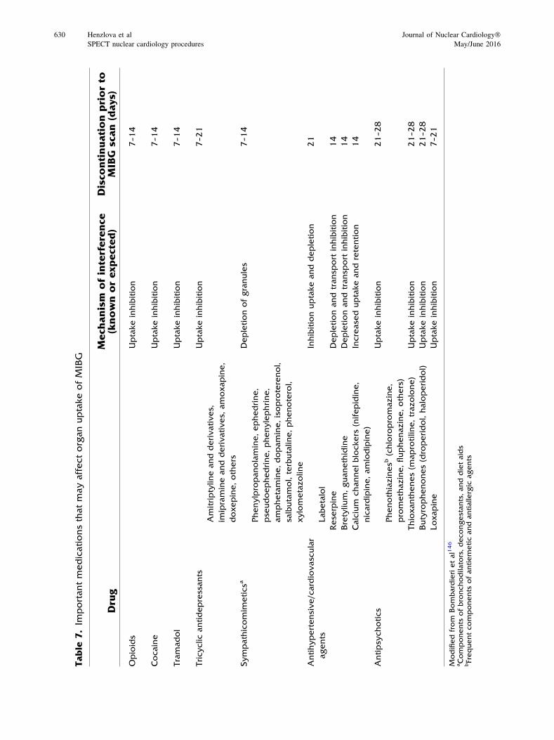
Table
7.Im
portantmedicationsthatmayaffectorganuptakeofMIBG
Dru
gMech
anism
ofinterference
(knownorexpected)
Disco
ntinuationpriorto
MIBG
scan(days)
Opioids
Uptakeinhibition
7–1
4
Cocaine
Uptakeinhibition
7–1
4
Tramadol
Uptakeinhibition
7–1
4
Tricyclicantidepressants
Amitriptylineandderivatives,
imipramineandderivatives,
amoxapine,
doxepine,others
Uptakeinhibition
7–2
1
Sympathicomim
eticsa
Phenylpropanolamine,ephedrine,
pse
udoephedrine,phenylephrine,
amphetamine,dopamine,isoproterenol,
salbutamol,terbutaline,phenoterol,
xylometazoline
Depletionofgranules
7–1
4
Antihypertensive/cardiovascular
agents
Labetalol
Inhibitionuptakeanddepletion
21
Rese
rpine
Depletionandtransp
ort
inhibition
14
Bretylium,guanethidine
Depletionandtransp
ort
inhibition
14
Calcium
channelblockers
(nifepidine,
nicardipine,amlodipine)
Increase
duptakeandretention
14
Antipsy
chotics
Phenothiazinesb
(chloropromazine,
promethazine,fluphenazine,others)
Uptakeinhibition
21–2
8
Thioxanthenes(m
aprotiline,trazolone)
Uptakeinhibition
21–2
8
Butyrophenones(droperidol,haloperidol)
Uptakeinhibition
21–2
8
Loxapine
Uptakeinhibition
7–2
1
Modifiedfrom
Bombardierietal146
aComponents
ofbronchodilators,decongestants,anddietaids
bFrequentcomponents
ofantiemeticandantialle
rgic
agents
630 Henzlova et al Journal of Nuclear Cardiology�SPECT nuclear cardiology procedures May/June 2016
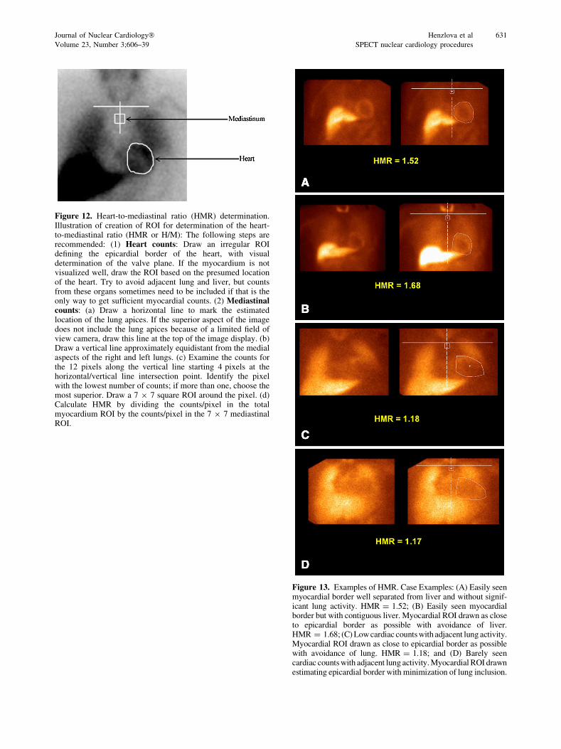
Figure 13. Examples of HMR. Case Examples: (A) Easily seenmyocardial border well separated from liver and without signif-icant lung activity. HMR = 1.52; (B) Easily seen myocardialborder but with contiguous liver. Myocardial ROI drawn as closeto epicardial border as possible with avoidance of liver.HMR = 1.68; (C)Lowcardiac countswith adjacent lungactivity.Myocardial ROI drawn as close to epicardial border as possiblewith avoidance of lung. HMR = 1.18; and (D) Barely seencardiac countswith adjacent lung activity.Myocardial ROI drawnestimating epicardial border with minimization of lung inclusion.
Figure 12. Heart-to-mediastinal ratio (HMR) determination.Illustration of creation of ROI for determination of the heart-to-mediastinal ratio (HMR or H/M): The following steps arerecommended: (1) Heart counts: Draw an irregular ROIdefining the epicardial border of the heart, with visualdetermination of the valve plane. If the myocardium is notvisualized well, draw the ROI based on the presumed locationof the heart. Try to avoid adjacent lung and liver, but countsfrom these organs sometimes need to be included if that is theonly way to get sufficient myocardial counts. (2) Mediastinalcounts: (a) Draw a horizontal line to mark the estimatedlocation of the lung apices. If the superior aspect of the imagedoes not include the lung apices because of a limited field ofview camera, draw this line at the top of the image display. (b)Draw a vertical line approximately equidistant from the medialaspects of the right and left lungs. (c) Examine the counts forthe 12 pixels along the vertical line starting 4 pixels at thehorizontal/vertical line intersection point. Identify the pixelwith the lowest number of counts; if more than one, choose themost superior. Draw a 7 9 7 square ROI around the pixel. (d)Calculate HMR by dividing the counts/pixel in the totalmyocardium ROI by the counts/pixel in the 7 9 7 mediastinalROI.
Journal of Nuclear Cardiology� Henzlova et al 631
Volume 23, Number 3;606–39 SPECT nuclear cardiology procedures
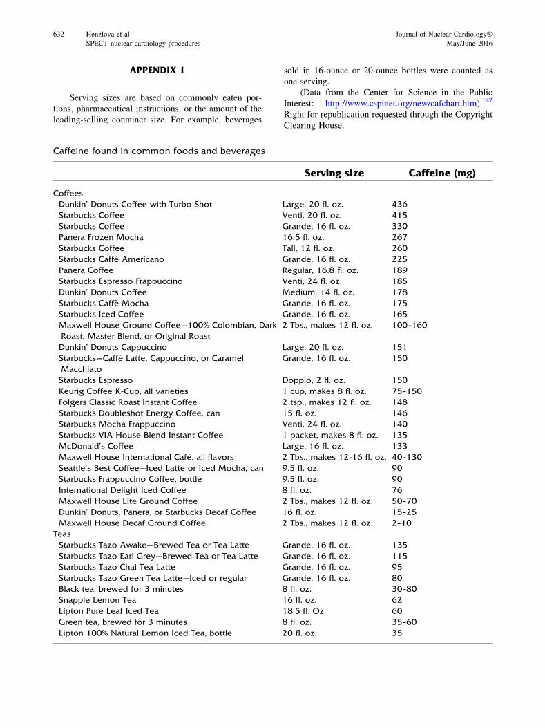
APPENDIX 1
Serving sizes are based on commonly eaten por-
tions, pharmaceutical instructions, or the amount of the
leading-selling container size. For example, beverages
sold in 16-ounce or 20-ounce bottles were counted as
one serving.
(Data from the Center for Science in the Public
Interest: http://www.cspinet.org/new/cafchart.htm).147
Right for republication requested through the Copyright
Clearing House.
Caffeine found in common foods and beverages
Serving size Caffeine (mg)
Coffees
Dunkin’ Donuts Coffee with Turbo Shot Large, 20 fl. oz. 436
Starbucks Coffee Venti, 20 fl. oz. 415
Starbucks Coffee Grande, 16 fl. oz. 330
Panera Frozen Mocha 16.5 fl. oz. 267
Starbucks Coffee Tall, 12 fl. oz. 260
Starbucks Caffe Americano Grande, 16 fl. oz. 225
Panera Coffee Regular, 16.8 fl. oz. 189
Starbucks Espresso Frappuccino Venti, 24 fl. oz. 185
Dunkin’ Donuts Coffee Medium, 14 fl. oz. 178
Starbucks Caffe Mocha Grande, 16 fl. oz. 175
Starbucks Iced Coffee Grande, 16 fl. oz. 165
Maxwell House Ground Coffee—100% Colombian, Dark
Roast, Master Blend, or Original Roast
2 Tbs., makes 12 fl. oz. 100–160
Dunkin’ Donuts Cappuccino Large, 20 fl. oz. 151
Starbucks—Caffe Latte, Cappuccino, or Caramel
Macchiato
Grande, 16 fl. oz. 150
Starbucks Espresso Doppio, 2 fl. oz. 150
Keurig Coffee K-Cup, all varieties 1 cup, makes 8 fl. oz. 75–150
Folgers Classic Roast Instant Coffee 2 tsp., makes 12 fl. oz. 148
Starbucks Doubleshot Energy Coffee, can 15 fl. oz. 146
Starbucks Mocha Frappuccino Venti, 24 fl. oz. 140
Starbucks VIA House Blend Instant Coffee 1 packet, makes 8 fl. oz. 135
McDonald’s Coffee Large, 16 fl. oz. 133
Maxwell House International Cafe, all flavors 2 Tbs., makes 12-16 fl. oz. 40–130
Seattle’s Best Coffee—Iced Latte or Iced Mocha, can 9.5 fl. oz. 90
Starbucks Frappuccino Coffee, bottle 9.5 fl. oz. 90
International Delight Iced Coffee 8 fl. oz. 76
Maxwell House Lite Ground Coffee 2 Tbs., makes 12 fl. oz. 50–70
Dunkin’ Donuts, Panera, or Starbucks Decaf Coffee 16 fl. oz. 15–25
Maxwell House Decaf Ground Coffee 2 Tbs., makes 12 fl. oz. 2–10
Teas
Starbucks Tazo Awake—Brewed Tea or Tea Latte Grande, 16 fl. oz. 135
Starbucks Tazo Earl Grey—Brewed Tea or Tea Latte Grande, 16 fl. oz. 115
Starbucks Tazo Chai Tea Latte Grande, 16 fl. oz. 95
Starbucks Tazo Green Tea Latte—Iced or regular Grande, 16 fl. oz. 80
Black tea, brewed for 3 minutes 8 fl. oz. 30–80
Snapple Lemon Tea 16 fl. oz. 62
Lipton Pure Leaf Iced Tea 18.5 fl. Oz. 60
Green tea, brewed for 3 minutes 8 fl. oz. 35–60
Lipton 100% Natural Lemon Iced Tea, bottle 20 fl. oz. 35
632 Henzlova et al Journal of Nuclear Cardiology�SPECT nuclear cardiology procedures May/June 2016
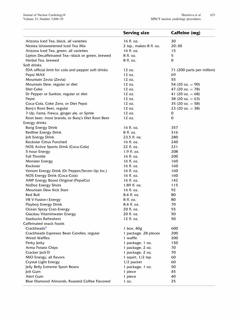
Serving size Caffeine (mg)
Arizona Iced Tea, black, all varieties 16 fl. oz. 30
Nestea Unsweetened Iced Tea Mix 2 tsp., makes 8 fl. oz. 20–30
Arizona Iced Tea, green, all varieties 16 fl. oz. 15
Lipton Decaffeinated Tea—black or green, brewed 8 fl. oz. 5
Herbal Tea, brewed 8 fl. oz. 0
Soft drinks
FDA official limit for cola and pepper soft drinks 12 oz. 71 (200 parts per million)
Pepsi MAX 12 oz. 69
Mountain Zevia (Zevia) 12 oz. 55
Mountain Dew, regular or diet 12 oz. 54 (20 oz. = 90)
Diet Coke 12 oz. 47 (20 oz. = 78)
Dr Pepper or Sunkist, regular or diet 12 oz. 41 (20 oz. = 68)
Pepsi 12 oz. 38 (20 oz. = 63)
Coca-Cola, Coke Zero, or Diet Pepsi 12 oz. 35 (20 oz. = 58)
Barq’s Root Beer, regular 12 oz. 23 (20 oz. = 38)
7-Up, Fanta, Fresca, ginger ale, or Sprite 12 oz. 0
Root beer, most brands, or Barq’s Diet Root Beer 12 oz. 0
Energy drinks
Bang Energy Drink 16 fl. oz. 357
Redline Energy Drink 8 fl. oz. 316
Jolt Energy Drink 23.5 fl. oz. 280
Rockstar Citrus Punched 16 fl. oz. 240
NOS Active Sports Drink (Coca-Cola) 22 fl. oz. 221
5-hour Energy 1.9 fl. oz. 208
Full Throttle 16 fl. oz. 200
Monster Energy 16 fl. oz. 160
Rockstar 16 fl. oz. 160
Venom Energy Drink (Dr Pepper/Seven Up Inc.) 16 fl. oz. 160
NOS Energy Drink (Coca-Cola) 16 fl. oz. 160
AMP Energy Boost Original (PepsiCo) 16 fl. oz. 142
NoDoz Energy Shots 1.89 fl. oz. 115
Mountain Dew Kick Start 16 fl. oz. 92
Red Bull 8.4 fl. oz. 80
V8 V-Fusion?Energy 8 fl. oz. 80
Playboy Energy Drink 8.4 fl. oz. 70
Ocean Spray Cran-Energy 20 fl. oz. 55
Glaceau Vitaminwater Energy 20 fl. oz. 50
Starbucks Refreshers 12 fl. oz. 50
Caffeinated snack foods
Crackheads2 1 box, 40g 600
Crackheads Espresso Bean Candies, regular 1 package, 28 pieces 200
Wired Waffles 1 waffle 200
Perky Jerky 1 package, 1 oz. 150
Arma Potato Chips 1 package, 2 oz. 70
Cracker Jack’D 1 package, 2 oz. 70
MiO Energy, all flavors 1 squirt, 1/2 tsp. 60
Crystal Light Energy 1/2 packet 60
Jelly Belly Extreme Sport Beans 1 package, 1 oz. 50
Jolt Gum 1 piece 45
Alert Gum 1 piece 40
Blue Diamond Almonds, Roasted Coffee Flavored 1 oz. 25
Journal of Nuclear Cardiology� Henzlova et al 633
Volume 23, Number 3;606–39 SPECT nuclear cardiology procedures
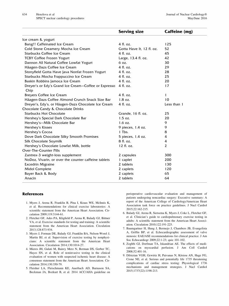
References
1. Myers J, Arena R, Franklin B, Pina I, Kraus WE, McInnis K,
et al. Recommendations for clinical exercise laboratories: A
scientific statement from the American Heart Association. Cir-
culation 2009;119:3144-61.
2. Fletcher GF, Ades PA, Kligfield P, Arena R, Balady GJ, Bittner
VA, et al. Exercise standards for testing and training: A scientific
statement from the American Heart Association. Circulation
2013;128:873-934.
3. Myers J, Forman DE, Balady GJ, Franklin BA, Nelson-Worel J,
Martin BJ, et al. Supervision of exercise testing by nonphysi-
cians: A scientific statement from the American Heart
Association. Circulation 2014;130:1014-27.
4. Mieres JH, Gulati M, Bairey Merz N, Berman DS, Gerber TC,
Hayes SN, et al. Role of noninvasive testing in the clinical
evaluation of women with suspected ischemic heart disease: A
consensus statement from the American Heart Association. Cir-
culation 2014;130:350-79.
5. Fleisher LA, Fleischmann KE, Auerbach AD, Barnason SA,
Beckman JA, Bozkurt B, et al. 2014 ACC/AHA guideline on
perioperative cardiovascular evaluation and management of
patients undergoing noncardiac surgery: Executive summary: A
report of the American College of Cardiology/American Heart
Association task force on practice guidelines. J Nucl Cardiol
2015;22:162-215.
6. Balady GJ, Arena R, Sietsema K, Myers J, Coke L, Fletcher GF,
et al. Clinician’s guide to cardiopulmonary exercise testing in
adults: A scientific statement from the American Heart Associ-
ation. Circulation 2010;122:191-225.
7. Baumgartner H, Hung J, Bermejo J, Chambers JB, Evangelista
A, Griffin BP, et al. Echocardiographic assessment of valve
stenosis: EAE/ASE recommendations for clinical practice. J Am
Soc Echocardiogr 2009;22:1-23; quiz 101-102.
8. Zoghbi GJ, Dorfman TA, Iskandrian AE. The effects of medi-
cations on myocardial perfusion. J Am Coll Cardiol
2008;52:401-16.
9. Dilsizian VGH, Gewirtz H, Paivanas N, Kitsiou AN, Hage FG,
Crone NE, et al. Serious and potentially life 1735 threatening
complications of cardiac stress testing: Physiological 1736
mechanisms and management strategies. J Nucl Cardiol
2015;1737(22):1198-213.
Serving size Caffeine (mg)
Ice cream & yogurt
Bang!! Caffeinated Ice Cream 4 fl. oz. 125
Cold Stone Creamery Mocha Ice Cream Gotta Have It, 12 fl. oz. 52
Starbucks Coffee Ice Cream 4 fl. oz. 45
TCBY Coffee Frozen Yogurt Large, 13.4 fl. oz. 42
Dannon All Natural Coffee Lowfat Yogurt 6 oz. 30
Haagen-Dazs Coffee Ice Cream 4 fl. oz. 29
Stonyfield Gotta Have Java Nonfat Frozen Yogurt 4 fl. oz. 28
Starbucks Mocha Frappuccino Ice Cream 4 fl. oz. 25
Baskin Robbins Jamoca Ice Cream 4 fl. oz. 20
Dreyer’s or Edy’s Grand Ice Cream—Coffee or Espresso
Chip
4 fl. oz. 17
Breyers Coffee Ice Cream 4 fl. oz. 1
Haagen-Dazs Coffee Almond Crunch Snack Size Bar 1.8 oz. 10
Dreyer’s, Edy’s, or Haagen-Dazs Chocolate Ice Cream 4 fl. oz. Less than 1
Chocolate Candy & Chocolate Drinks
Starbucks Hot Chocolate Grande, 16 fl. oz. 25
Hershey’s Special Dark Chocolate Bar 1.5 oz. 20
Hershey’s—Milk Chocolate Bar 1.6 oz. 9
Hershey’s Kisses 9 pieces, 1.4 oz. 9
Hershey’s Cocoa 1 Tbs. 8
Dove Dark Chocolate Silky Smooth Promises 5 pieces, 1.4 oz. 4
Silk Chocolate Soymilk 8 fl. oz. 4
Hershey’s Chocolate Lowfat Milk, bottle 12 fl. oz. 2
Over-The-Counter Pills
Zantrex-3 weight-loss supplement 2 capsules 300
NoDoz, Vivarin, or over the counter caffeine tablets 1 caplet 200
Excedrin Migraine 2 tablets 130
Midol Complete 2 caplets 120
Bayer Back & Body 2 caplets 65
Anacin 2 tablets 64
634 Henzlova et al Journal of Nuclear Cardiology�SPECT nuclear cardiology procedures May/June 2016
10. Bokhari S, Ficaro EP, McCallister BD Jr. Adenosine stress
protocols for myocardial perfusion imaging. J Nucl Cardiol
2007;14:415-6.
11. Cerqueira MD, Verani MS, Schwaiger M, Heo J, Iskandrian AS.
Safety profile of adenosine stress perfusion imaging: Results
from the adenoscan multicenter trial registry. J Am Coll Cardiol
1994;23:384-9.
12. Thomas GS, Miyamoto MI. Should simultaneous exercise
become the standard for adenosine myocardial perfusion imag-
ing? Am J Cardiol 2004;94:3D-10D; discussion 10D-11D.
13. Iskandrian AE, Bateman TM, Belardinelli L, Blackburn B,
Cerqueira MD, Hendel RC, et al. Adenosine versus regadenoson
comparative evaluation in myocardial perfusion imaging: Results
of the ADVANCE phase 3 multicenter international trial. J Nucl
Cardiol 2007;14:645-58.
14. Agarwal V, DePuey EG. Regadenoson and seizures: A real
clinical concern. J Nucl Cardiol 2014;21:869-70.
15. Page RL 2nd, Spurck P, Bainbridge JL, Michalek J, Quaife RA.
Seizures associated with regadenoson: A case series. J Nucl
Cardiol 2012;19:389-91.
16. Cabrera R, Husain Z, Palani G, Karthikeyan AS, Choudhry Z,
Dhanalakota S, et al. Comparison of hemodynamic and stress
testing variables in patients undergoing regadenoson stress
myocardial perfusion imaging to regadenoson with adjunctive
low-level exercise myocardial perfusion imaging. J Nucl Cardiol
2013;20:336-43; quiz 344-335.
17. Thomas GS, Thompson RC, Miyamoto MI, Ip TK, Rice DL,
Milikien D, et al. The RegEx trial: A randomized, double-blind,
placebo- and active-controlled pilot study combining regadeno-
son, a selective a(2a) adenosine agonist, with low-level exercise,
in patients undergoing myocardial perfusion imaging. J Nucl
Cardiol 2009;16:63-72.
18. AlJaroudi WA, Alraies MC, Cerquiera MD, Jaber WA. Safety
and tolerability of regadenoson in 514 SPECT mpi patients with
and without coronary artery disease and submaximal exercise
heart rate response. Eur J Nucl Med Mol Imaging 2013;40:341-8.
19. ParkerMW,Morales DC, SlimHB, Karthikeyan AS, Choudhry Z,
Dhanalakota S, et al. A strategy of symptom-limited exercise with
regadenoson-as-needed for stress myocardial perfusion imaging:
A randomized controlled trial. J Nucl Cardiol 2013;20:185-96.
20. Ross MI, Wu E, Wilkins JT, Gupta D, Shen S, Aulwes D, et al.
Safety and feasibility of adjunctive regadenoson injection at peak
exercise during exercise myocardial perfusion imaging: The Both
Exercise and Regadenoson Stress Test (BERST) trial. J Nucl
Cardiol 2013;20:197-204.
21. Thompson RC, Patil H, Thompson EC, Thomas GS, Al-Amoodi
M, Kennedy KF, et al. Regadenoson pharmacologic stress for
myocardial perfusion imaging: A three-way comparison between
regadenoson administered at peak exercise, during walk recovery,
or no-exercise. J Nucl Cardiol 2013;20:214-21; quiz 222-216.
22. Lette J, Tatum JL, Fraser S, Miller DD,Waters DD, Heller G, et al.
Safety of dipyridamole testing in 73,806 patients: The multicenter
dipyridamole safety study. J Nucl Cardiol 1995;2:3-17.
23. Candell-Riera J, Santana-Boado C, Castell-Conesa J, Aguade-
Bruix S, Olona M, Palet J, et al. Simultaneous dipyri-
damole/maximal subjective exercise with 99mtc-mibi SPECT:
Improved diagnostic yield in coronary artery disease. J Am Coll
Cardiol 1997;29:531-6.
24. Casale PN, Guiney TE, Strauss HW, Boucher CA. Simultaneous
low level treadmill exercise and intravenous dipyridamole stress
thallium imaging. Am J Cardiol 1988;62:799-802.
25. Stern S, Greenberg ID, Corne RA. Quantification of walking
exercise required for improvement of dipyridamole thallium-201
image quality. J Nucl Med 1992;33:2061-6.
26. Vitola JV, Brambatti JC, Caligaris F, Lesse CR, Nogueira PR,
Joaquim AI, et al. Exercise supplementation to dipyridamole
prevents hypotension, improves electrocardiogram sensitivity,
and increases heart-to-liver activity ratio on tc-99m sestamibi
imaging. J Nucl Cardiol 2001;8:652-9.
27. Hays JT, Mahmarian JJ, Cochran AJ, Verani MS. Dobutamine
thallium-201 tomography for evaluating patients with suspected
coronary artery disease unable to undergo exercise or vasodilator
pharmacologic stress testing. J AmColl Cardiol 1993;21:1583-90.
28. Marwick T, Willemart B, D’Hondt AM, Baudhuin T, Wijns W,
Detry JM, et al. Selection of the optimal nonexercise stress for
the evaluation of ischemic regional myocardial dysfunction and
malperfusion. Comparison of dobutamine and adenosine using
echocardiography and 99mtc-mibi single photon emission com-
puted tomography. Circulation 1993;87:345-54.
29. Johnston DL, Scanlon PD, Hodge DO, Glynn RB, Hung JC,
Gibbons RJ. Pulmonary function monitoring during adenosine
myocardial perfusion scintigraphy in patients with chronic
obstructive pulmonary disease. Mayo Clin Proc 1999;74:339-46.
30. Reyes E, Loong CY, Wechalekar K, Latus K, Anagnostopoulos
C, Underwood SR. Side effect profile and tolerability of adeno-
sine myocardial perfusion scintigraphy in patients with mild
asthma or chronic obstructive pulmonary disease. J Nucl Cardiol
2007;14:827-34.
31. Sundram F, Notghi A, Smith NB. Pharmacological stress
myocardial perfusion scintigraphy: Use of a modified adenosine
protocol in patients with asthma. Nucl Med Commun
2009;30:217-25.
32. Leaker BR, O’Connor B, Hansel TT, Barnes PJ, Meng L, Mathur
VS, et al. Safety of regadenoson, an adenosine A2A receptor
agonist for myocardial perfusion imaging, in mild asthma and
moderate asthma patients: A randomized, double-blind, placebo-
controlled trial. J Nucl Cardiol 2008;15:329-36.
33. Thomas GS, Tammelin BR, Schiffman GL, Marquez R, Rice DL,
Milikien D, et al. Safety of regadenoson, a selective adenosine
A2A agonist, in patients with chronic obstructive pulmonary
disease: A randomized, double-blind, placebo-controlled trial
(RegCOPD trial). J Nucl Cardiol 2008;15:319-28.
34. Golzar Y, Doukky R. Regadenoson use in patients with chronic
obstructive pulmonary disease: The state of current knowledge.
Int J Chron Obstruct Pulmon Dis 2014;9:129-37.
35. Prenner BM, Bukofzer S, Behm S, Feaheny K, McNutt BE. A
randomized, double-blind, placebo-controlled study assessing the
safety and tolerability of regadenoson in subjects with asthma or
chronic obstructive pulmonary disease. J Nucl Cardiol
2012;19:681-92.
36. Gordi T, Blackburn B, Lieu H. Regadenoson pharmacokinetics
and tolerability in subjects with impaired renal function. J Clin
Pharmacol 2007;47:825-33.
37. Aljaroudi W, Iqbal F, Koneru J, Bhambhvani P, Heo J, Iskan-
drian AE. Safety of regadenoson in patients with end-stage liver
disease. J Nucl Cardiol 2011;18:90-5.
38. Ananthasubramaniam K, Weiss R, McNutt B, Klauke B, Feaheny
K, Bukofzer S. A randomized, double-blind, placebo-controlled
study of the safety and tolerance of regadenoson in subjects with
stage 3 or 4 chronic kidney disease. J Nucl Cardiol 2012;19:319-
29.
39. Doukky R, Rangel MO, Wassouf M, Dick R, Alqaid A. Morales
Demori R. The safety and tolerability of regadenoson in patients
with end-stage renal disease: The first prospective evaluation. J
Nucl Cardiol 2013;20:205-13.
40. Laighold S, Druz R. Initial clinical experience with a selective
A2A receptor agonist, regadenoson, in a patient with end-stage
renal disease on hemodialysis. J Nucl Cardiol 2009;16:478-80.
Journal of Nuclear Cardiology� Henzlova et al 635
Volume 23, Number 3;606–39 SPECT nuclear cardiology procedures
41. Palani G, Husain Z, Salinas RC, Karthikeyan V, Karthikeyan AS,
Ananthasubramaniam K. Safety of regadenoson as a pharmaco-
logic stress agent for myocardial perfusion imaging in chronic
kidney disease patients not on hemodialysis. J Nucl Cardiol
2011;18:605-11.
42. Avakian SD, Grinberg M, Meneguetti JC, Ramires JA, Mansur
AP. SPECT dipyridamole scintigraphy for detecting coronary
artery disease in patients with isolated severe aortic stenosis. Int J
Cardiol 2001;81:21-7.
43. Demirkol MO, Yaymaci B, Debes H, Basaran Y, Turan F.
Dipyridamole myocardial perfusion tomography in patients with
severe aortic stenosis. Cardiology 2002;97:37-42.
44. Kupari M, Virtanen KS, Turto H, Viitasalo M, Manttari M,
Lindroos M, et al. Exclusion of coronary artery disease by
exercise thallium-201 tomography in patients with aortic valve
stenosis. Am J Cardiol 1992;70:635-40.
45. Burgstahler C, Kunze M, Gawaz MP, Rasche V, Wohrle J,
Hombach V, et al. Adenosine stress first pass perfusion for the
detection of coronary artery disease in patients with aortic
stenosis: A feasibility study. Int J Cardiovasc Imaging
2008;24:195-200.
46. Patsilinakos SP, Kranidis AI, Antonelis IP, Filippatos G, Hous-
sianakou IK, Zamanis NI, et al. Detection of coronary artery
disease in patients with severe aortic stenosis with noninvasive
methods. Angiology 1999;50:309-17.
47. Patsilinakos SP, Spanodimos S, Rontoyanni F, Kranidis A,
Antonelis IP, Sotirellos K, et al. Adenosine stress myocardial
perfusion tomographic imaging in patients with significant aortic
stenosis. J Nucl Cardiol 2004;11:20-5.
48. Samuels B, Kiat H, Friedman JD, Berman DS. Adenosine
pharmacologic stress myocardial perfusion tomographic imaging
in patients with significant aortic stenosis. Diagnostic efficacy
and comparison of clinical, hemodynamic and electrocardio-
graphic variables with 100 age-matched control subjects. J Am
Coll Cardiol 1995;25:99-106.
49. Cremer PC, Khalaf S, Lou J, Rodriguez L, Cerqueira MD, Jaber
WA. Stress positron emission tomography is safe and can guide
coronary revascularization in high-risk patients being considered
for transcatheter aortic valve replacement. J Nucl Cardiol
2014;21:1001-10.
50. Lentine KL, Costa SP, Weir MR, Robb JF, Fleisher LA, Kasiske
BL, et al. Cardiac disease evaluation and management among
kidney and liver transplantation candidates: A scientific state-
ment from the American Heart Association and the American
College of Cardiology Foundation. J Am Coll Cardiol
2012;60:434-80.
51. Giedd KN, Bokhari S, Daniele TP, Johnson LL. Sinus arrest
during adenosine stress testing in liver transplant recipients with
graft failure: Three case reports and a review of the literature. J
Nucl Cardiol 2005;12:696-702.
52. Kovacs D, Pivonka R, Khosla PG, Khosla S. Effect of caffeine on
myocardial perfusion imaging using single photon emission
computed tomography during adenosine pharmacologic stress.
Am J Ther 2008;15:431-4.
53. Bottcher M, Czernin J, Sun KT, Phelps ME, Schelbert HR. Effect
of caffeine on myocardial blood flow at rest and during phar-
macological vasodilation. J Nucl Med 1995;36:2016-21.
54. Kubo S, Tadamura E, Toyoda H, Mamede M, Yamamuro M,
Magata Y, et al. Effect of caffeine intake on myocardial hyper-
emic flow induced by adenosine triphosphate and dipyridamole. J
Nucl Med 2004;45:730-8.
55. Lapeyre AC 3rd, Goraya TY, Johnston DL, Gibbons RJ. The
impact of caffeine on vasodilator stress perfusion studies. J Nucl
Cardiol 2004;11:506-11.
56. Salcedo J, Kern MJ. Effects of caffeine and theophylline on
coronary hyperemia induced by adenosine or dipyridamole.
Catheter Cardiovasc Interv 2009;74:598-605.
57. Smits P, Aengevaeren WR, Corstens FH, Thien T. Caffeine
reduces dipyridamole-induced myocardial ischemia. J Nucl Med
1989;30:1723-6.
58. Smits P, Corstens FH, Aengevaeren WR, Wackers FJ, Thien T.
False-negative dipyridamole-thallium-201 myocardial imaging
after caffeine infusion. J Nucl Med 1991;32:1538-41.
59. Aqel RA, Zoghbi GJ, Trimm JR, Baldwin SA, Iskandrian AE.
Effect of caffeine administered intravenously on intracoronary-
administered adenosine-induced coronary hemodynamics in
patientswith coronary artery disease.AmJCardiol 2004;93:343-6.
60. Hage FG, Iskandrian AE. The effect of caffeine on adenosine
myocardial perfusion imaging: Time to reassess? J Nucl Cardiol
2012;19:415-9.
61. Reyes E, Loong CY, Harbinson M, Donovan J, Anagnostopoulos
C, Underwood SR. High-dose adenosine overcomes the attenu-
ation of myocardial perfusion reserve caused by caffeine. J Am
Coll Cardiol 2008;52:2008-16.
62. Lee JC, Fraser JF, Barnett AG, Johnson LP, Wilson MG,
McHenry CM, et al. Effect of caffeine on adenosine-induced
reversible perfusion defects assessed by automated analysis. J
Nucl Cardiol 2012;19:474-81.
63. Zoghbi GJ, Htay T, Aqel R, Blackmon L, Heo J, Iskandrian AE.
Effect of caffeine on ischemia detection by adenosine single-
photon emission computed tomography perfusion imaging. J Am
Coll Cardiol 2006;47:2296-302.
64. Zhao G, Messina E, Xu X, Ochoa M, Sun HL, Leung K, et al.
Caffeine attenuates the duration of coronary vasodilation and
changes in hemodynamics induced by regadenoson (cvt-3146), a
novel adenosine A2A receptor agonist. J Cardiovasc Pharmacol
2007;49:369-75.
65. Gaemperli O, Schepis T, Koepfli P, Siegrist PT, Fleischman S,
Nguyen P, et al. Interaction of caffeine with regadenoson-
induced hyperemic myocardial blood flow as measured by
positron emission tomography: A randomized, double-blind,
placebo-controlled crossover trial. J Am Coll Cardiol 2008;51:
328-9.
66. Tejani FH, Thompson RC, Kristy R, Bukofzer S. Effect of caf-
feine on SPECT myocardial perfusion imaging during
regadenoson pharmacologic stress: A prospective, randomized,
multicenter study. Int J Cardiovasc Imaging 2014;30:979-89.
67. Depuey EG, Mahmarian JJ, Miller TD, Einstein AJ, Hansen CL,
Holly TA, et al. Patient-centered imaging. J Nucl Cardiol
2012;19:185-215.
68. Fazel R, Gerber TC, Balter S, Brenner DJ, Carr JJ, Cerqueira
MD, et al. Approaches to enhancing radiation safety in cardio-
vascular imaging: A scientific statement from the American
Heart Association. Circulation 2014;130:1730-48.
69. Einstein AJ, Pascual TN, Mercuri M, Karthikeyan G, Vitola JV,
Mahmarian JJ, et al. Current worldwide nuclear cardiology
practices and radiation exposure: Results from the 65 country
IAEA nuclear cardiology protocols cross-sectional study
(INCAPS). Eur Heart J 2015;36:1689-96.
70. Einstein AJ, Tilkemeier P, Fazel R, Rakotoarivelo H, Shaw LJ,
American Society of Nuclear Cardiology. Radiation safety in
nuclear cardiology-current knowledge and practice: Results from
the 2011 American Society of Nuclear Cardiology Member
Survey. JAMA Intern Med 2013;173:1021-3.
71. Einstein AJ, Berman DS, Min JK, Hendel RC, Gerber TC, Carr
JJ, et al. Patient-centered imaging: Shared decision making for
cardiac imaging procedures with exposure to ionizing radiation. J
Am Coll Cardiol 2014;63:1480-9.
636 Henzlova et al Journal of Nuclear Cardiology�SPECT nuclear cardiology procedures May/June 2016
72. DePuey EG, Bommireddipalli S, Clark J, Leykekhman A,
Thompson LB, Friedman M. A comparison of the image quality
of full-time myocardial perfusion SPECT vs wide beam recon-
struction half-time and half-dose SPECT. J Nucl Cardiol
2011;18:273-80.
73. Duvall WL, Croft LB, Ginsberg ES, Einstein AJ, Guma KA,
George T, et al. Reduced isotope dose and imaging time with a
high-efficiency CZT SPECT camera. J Nucl Cardiol
2011;18:847-57.
74. Einstein AJ, Blankstein R, Andrews H, Fish M, Padgett R, Hayes
SW, et al. Comparison of image quality, myocardial perfusion,
and left ventricular function between standard imaging and sin-
gle-injection ultra-low-dose imaging using a high-efficiency
SPECT camera: The millisievert study. J Nucl Med
2014;55:1430-7.
75. Machac J, Bacharach SL, Bateman TM, Bax JJ, Beanlands R,
Bengel F, et al. Positron emission tomography myocardial per-
fusion and glucose metabolism imaging. J Nucl Cardiol
2006;13:e121-51.
76. Hansen CL, Goldstein RA, Akinboboye OO, Berman DS, Bot-
vinick EH, Churchwell KB, et al. Myocardial perfusion and
function: Single photon emission computed tomography. J Nucl
Cardiol 2007;14:e39-60.
77. Chang SM, Nabi F, Xu J, Raza U, Mahmarian JJ. Normal stress-
only versus standard stress/rest myocardial perfusion imaging:
Similar patient mortality with reduced radiation exposure. J Am
Coll Cardiol 2010;55:221-30.
78. Duvall WL, Wijetunga MN, Klein TM, Razzouk L, Godbold J,
Croft LB, et al. The prognosis of a normal stress-only tc-99m
myocardial perfusion imaging study. J Nucl Cardiol 2010;17:370-7.
79. Gibson PB, Demus D, Noto R, Hudson W, Johnson LL. Low
event rate for stress-only perfusion imaging in patients evaluated
for chest pain. J Am Coll Cardiol 2002;39:999-1004.
80. Flotats A, Carrio I. Cardiac neurotransmission SPECT imaging. J
Nucl Cardiol 2004;11:587-602.
81. Haider N, Baliga RR, Chandrashekhar Y, Narula J. Adrenergic
excess, hNET1 down-regulation, and compromised mIBG uptake
in heart failure poverty in the presence of plenty. JACC Car-
diovasc Imaging 2010;3:71-5.
82. Hattori N, Schwaiger M. Metaiodobenzylguanidine scintigraphy
of the heart: What have we learnt clinically? Eur J Nucl Med
2000;27:1-6.
83. Sisson JC, Wieland DM. Radiolabeled meta-iodobenzylguani-
dine: Pharmacology and clinical studies. Am J Physiol Imaging
1986;1:96-103.
84. Kline RC, Swanson DP, Wieland DM, Thrall JH, Gross MD, Pitt
B, et al. Myocardial imaging in man with I-123 meta-iodoben-
zylguanidine. J Nucl Med 1981;22:129-32.
85. Raffel DM, Wieland DM. Development of mibg as a cardiac inner-
vation imaging agent. JACC Cardiovasc Imaging 2010;3:111-6.
86. Jacobson AF, Senior R, Cerqueira MD, Wong ND, Thomas GS,
Lopez VA, et al. Myocardial iodine-123 meta-iodobenzylguani-
dine imaging and cardiac events in heart failure. Results of the
prospective admire-hf (adreview myocardial imaging for risk
evaluation in heart failure) study. J Am Coll Cardiol
2010;55:2212-21.
87. Merlet P, Valette H, Dubois-Rande JL, Moyse D, Duboc D, Dove
P, et al. Prognostic value of cardiac metaiodobenzylguanidine
imaging in patients with heart failure. J Nucl Med 1992;33:471-7.
88. Nakata T, Nakajima K, Yamashina S, Yamada T, Momose M,
Kasama S, et al. A pooled analysis of multicenter cohort studies
of (123)I-mIBG imaging of sympathetic innervation for assess-
ment of long-term prognosis in heart failure. JACC Cardiovasc
Imaging 2013;6:772-84.
89. Ogita H, Shimonagata T, Fukunami M, Kumagai K, Yamada T,
Asano Y, et al. Prognostic significance of cardiac (123)I
metaiodobenzylguanidine imaging for mortality and morbidity in
patients with chronic heart failure: A prospective study. Heart
2001;86:656-60.
90. Verberne HJ, Brewster LM, Somsen GA, van Eck-Smit BL.
Prognostic value of myocardial 123I-metaiodobenzylguanidine
(mIBG) parameters in patients with heart failure: A systematic
review. Eur Heart J 2008;29:1147-59.
91. Travin MI. Cardiac autonomic imaging with SPECT tracers. J
Nucl Cardiol 2013;20:128-43; quiz 146.
92. AlBadarin FJ, Wimmer AP, Kennedy KF, Jacobson AF, Bateman
TM. The utility of admire-hf risk score in predicting serious
arrhythmic events in heart failure patients: Incremental prog-
nostic benefit of cardiac 123I-mIBG scintigraphy. J Nucl Cardiol
2014;21:756-62; quiz 753-755, 763-755.
93. Arora R, Ferrick KJ, Nakata T, Kaplan RC, Rozengarten M, Latif
F, et al. I-123 mIBG imaging and heart rate variability analysis to
predict the need for an implantable cardioverter defibrillator. J
Nucl Cardiol 2003;10:121-31.
94. Klein T, Dilsizian V, Cao Q, Chen W, Dickfeld TM. The
potential role of iodine-123 metaiodobenzylguanidine imaging
for identifying sustained ventricular tachycardia in patients with
cardiomyopathy. Curr Cardiol Rep 2013;15:359.
95. Nagahara D, Nakata T, Hashimoto A, Wakabayashi T, Kyuma
M, Noda R, et al. Predicting the need for an implantable car-
dioverter defibrillator using cardiac metaiodobenzylguanidine
activity together with plasma natriuretic peptide concentration or
left ventricular function. J Nucl Med 2008;49:225-33.
96. Miranda CH, Figueiredo AB, Maciel BC, Marin-Neto JA,
Simoes MV. Sustained ventricular tachycardia is associated with
regional myocardial sympathetic denervation assessed with 123i-
metaiodobenzylguanidine in chronic chagas cardiomyopathy. J
Nucl Med 2011;52:504-10.
97. Mitrani RD, Klein LS, Miles WM, Hackett FK, Burt RW,
Wellman HN, et al. Regional cardiac sympathetic denervation in
patients with ventricular tachycardia in the absence of coronary
artery disease. J Am Coll Cardiol 1993;22:1344-53.
98. Paul M, Schafers M, Kies P, Acil T, Schafers K, Breithardt G,
et al. Impact of sympathetic innervation on recurrent life-
threatening arrhythmias in the follow-up of patients with idio-
pathic ventricular fibrillation. Eur J Nucl Med Mol Imaging
2006;33:866-70.
99. Paul M, Wichter T, Kies P, Gerss J, Wollmann C, Rahbar K,
et al. Cardiac sympathetic dysfunction in genotyped patients with
arrhythmogenic right ventricular cardiomyopathy and risk of
recurrent ventricular tachyarrhythmias. J Nucl Med
2011;52:1559-65.
100. Wichter T, Matheja P, Eckardt L, Kies P, Schafers K, Schulze-
Bahr E, et al. Cardiac autonomic dysfunction in brugada syn-
drome. Circulation 2002;105:702-6.
101. Tomoda H, Yoshioka K, Shiina Y, Tagawa R, Ide M, Suzuki Y.
Regional sympathetic denervation detected by iodine 123
metaiodobenzylguanidine in non-q-wave myocardial infarction
and unstable angina. Am Heart J 1994;128:452-8.
102. Watanabe K, Takahashi T, Miyajima S, Hirokawa Y, Tanabe N,
Kato K, et al. Myocardial sympathetic denervation, fatty acid
metabolism, and left ventricular wall motion in vasospastic
angina. J Nucl Med 2002;43:1476-81.
103. Fallavollita JA, Canty JM Jr. Dysinnervated but viable myo-
cardium in ischemic heart disease. J Nucl Cardiol 2010;17:1107-
15.
104. Fallavollita JA, Heavey BM, Luisi AJ Jr, Michalek SM, Baldwa
S, Mashtare TL Jr, et al. Regional myocardial sympathetic
Journal of Nuclear Cardiology� Henzlova et al 637
Volume 23, Number 3;606–39 SPECT nuclear cardiology procedures
denervation predicts the risk of sudden cardiac arrest in ischemic
cardiomyopathy. J Am Coll Cardiol 2014;63:141-9.
105. McGhie AI, Corbett JR, Akers MS, Kulkarni P, Sills MN, Kre-
mers M, et al. Regional cardiac adrenergic function using I-123
meta-iodobenzylguanidine tomographic imaging after acute
myocardial infarction. Am J Cardiol 1991;67:236-42.
106. Simoes MV, Barthel P, Matsunari I, Nekolla SG, Schomig A,
Schwaiger M, et al. Presence of sympathetically denervated but
viable myocardium and its electrophysiologic correlates after
early revascularised, acute myocardial infarction. Eur Heart J
2004;25:551-7.
107. Stanton MS, Tuli MM, Radtke NL, Heger JJ, Miles WM, Mock
BH, et al. Regional sympathetic denervation after myocardial
infarction in humans detected noninvasively using I-123-
metaiodobenzylguanidine. J Am Coll Cardiol 1989;14:1519-26.
108. Bengel FM, Ueberfuhr P, Schiepel N, Nekolla SG, Reichart B,
Schwaiger M. Effect of sympathetic reinnervation on cardiac
performance after heart transplantation. N Engl J Med
2001;345:731-8.
109. Flotats A, Carrio I. Value of radionuclide studies in cardiac
transplantation. Ann Nucl Med 2006;20:13-21.
110. Gerson MC, McGuire N, Wagoner LE. Sympathetic nervous
system function as measured by I-123 metaiodobenzylguanidine
predicts transplant-free survival in heart failure patients with
idiopathic dilated cardiomyopathy. J Card Fail 2003;9:384-91.
111. Hattori N, Tamaki N, Hayashi T, Masuda I, Kudoh T, Tateno M,
et al. Regional abnormality of iodine-123-mIBG in diabetic
hearts. J Nucl Med 1996;37:1985-90.
112. Nagamachi S, Fujita S, Nishii R, Futami S, Tamura S, Mizuta M,
et al. Prognostic value of cardiac i-123 metaiodobenzylguanidine
imaging in patients with non-insulin-dependent diabetes mellitus.
J Nucl Cardiol 2006;13:34-42.
113. Carrio I, Estorch M, Berna L, Lopez-Pousa J, Tabernero J, Torres
G. Indium-111-antimyosin and iodine-123-mIBG studies in early
assessment of doxorubicin cardiotoxicity. J Nucl Med
1995;36:2044-9.
114. Sciammarella M, Gerson M, Buxton AE, Bartley SC, Doukky R,
Merlino DA, et al. Model coverage policy: Myocardial sympa-
thetic innervation imaging: Iodine-123meta-iodobenzylguanidine
(123I-mIBG). J Nucl Cardiol 2015;22:804-11.
115. Jacobson AF, Lombard J, Banerjee G, Camici PG. 123I-mIBG
scintigraphy to predict risk for adverse cardiac outcomes in heart
failure patients: Design of two prospective multicenter interna-
tional trials. J Nucl Cardiol 2009;16:113-21.
116. Agostini D, Carrio I, Verberne HJ. How to use myocardial 123I-
mIBG scintigraphy in chronic heart failure. Eur J Nucl Med Mol
Imaging 2009;36:555-9.
117. Carrio I, Cowie MR, Yamazaki J, Udelson J, Camici PG. Cardiac
sympathetic imaging with mIBG in heart failure. JACC Car-
diovasc Imaging 2010;3:92-100.
118. Flotats A, Carrio I, Agostini D, Le Guludec D, Marcassa C,
Schafers M, et al. Proposal for standardization of 123i-
metaiodobenzylguanidine (mIBG) cardiac sympathetic imaging
by the eanm cardiovascular committee and the european council
of nuclear Cardiology Eur J Nucl Med Mol Imaging
2010;37:1802-12.
119. Yamashina S, Yamazaki J. Neuronal imaging using SPECT. Eur
J Nucl Med Mol Imaging 2007;34(Suppl 1):S62-73.
120. Solanki KK, Bomanji J, Moyes J, Mather SJ, Trainer PJ, Britton
KE. A pharmacological guide to medicines which interfere with
the biodistribution of radiolabelled meta-iodobenzylguanidine
(mibg). Nucl Med Commun 1992;13:513-21.
121. Wafelman AR, Hoefnagel CA, Maes RA, Beijnen JH. Radioio-
dinated metaiodobenzylguanidine: A review of its biodistribution
and pharmacokinetics, drug interactions, cytotoxicity and
dosimetry. Eur J Nucl Med 1994;21:545-59.
122. Jacobson A, Travin M. Impact of neuropsychiatric medications
on cardiac uptake of 123I- mIBG in heart failure subjects. J Nucl
Med 2013;54:1708.
123. Friedman NC, Hassan A, Grady E, Matsuoka DT, Jacobson AF.
Efficacy of thyroid blockade on thyroid radioiodine uptake in123I-mIBG imaging. J Nucl Med 2014;55:211-5.
124. Chen J, Folks RD, Verdes L, Manatunga DN, Jacobson AF,
Garcia EV. Quantitative i-123 mIBG SPECT in differentiating
abnormal and normal mibg myocardial uptake. J Nucl Cardiol
2012;19:92-9.
125. Inoue H, Zipes DP. Results of sympathetic denervation in the
canine heart: Supersensitivity that may be arrhythmogenic. Cir-
culation 1987;75:877-87.
126. Minardo JD, Tuli MM, Mock BH, Weiner RE, Pride HP, Well-
man HN, et al. Scintigraphic and electrophysiological evidence
of canine myocardial sympathetic denervation and reinnervation
produced by myocardial infarction or phenol application. Cir-
culation 1988;78:1008-19.
127. Dobbeleir AA, Hambye AS, Franken PR. Influence of high-en-
ergy photons on the spectrum of iodine-123 with low- and
medium-energy collimators: Consequences for imaging with
123i-labelled compounds in clinical practice. Eur J Nucl Med
1999;26:655-8.
128. Inoue Y, Suzuki A, Shirouzu I, Machida T, Yoshizawa Y, Akita
F, et al. Effect of collimator choice on quantitative assessment of
cardiac iodine 123 mIBG uptake. J Nucl Cardiol 2003;10:623-32.
129. Verberne HJ, Feenstra C, de Jong WM, Somsen GA, van Eck-
Smit BL, Busemann Sokole E. Influence of collimator choice and
simulated clinical conditions on 123I-mIBG heart/mediastinum
ratios: A phantom study. Eur J Nucl Med 2005;32:1100-7.
130. Chen J, Garcia EV, Galt JR, Folks RD, Carrio I. Optimized
acquisition and processing protocols for i-123 cardiac SPECT
imaging. J Nucl Cardiol 2006;13:251-60.
131. Chen J, Garcia EV, Galt JR, Folks RD, Carrio I. Improved
quantification in 123i cardiac SPECT imaging with deconvolu-
tion of septal penetration. Nucl Med Commun 2006;27:551-8.
132. Inoue Y, Abe Y, Itoh Y, Asano Y, Kikuchi K, Sakamoto Y, et al.
Acquisition protocols and correction methods for estimation of
the heart-to-mediastinum ratio in 123I-metaiodobenzylguanidine
cardiac sympathetic imaging. J Nucl Med 2013;54:707-707.
133. Matsuo S, Nakajima K, Okuda K, Kawano M, Ishikawa T,
Hosoya T, et al. Standardization of the heart-to-mediastinum
ratio of 123i-labelled-metaiodobenzylguanidine uptake using the
dual energy window method: Feasibility of correction with dif-
ferent camera-collimator combinations. Eur J Nucl Med
2009;36:560-6.
134. Nakajima K, Okuda K, Matsuo S, Yoshita M, Taki J, Yamada M,
et al. Standardization of metaiodobenzylguanidine heart to
mediastinum ratio using a calibration phantom: Effects of cor-
rection on normal databases and a multicentre study. Eur J Nucl
Med 2012;39:113-9.
135. Nakajima K, Okuda K, Yoshimura M, Matsuo S, Wakabayashi H,
Imanishi Y, et al. Multicenter cross-calibration of i-123
metaiodobenzylguanidine heart-to-mediastinum ratios to over-
come camera-collimator variations. J Nucl Cardiol 2014;21:970-8.
136. Bellevre D, Manrique A, Legallois D, Bross S, Baavour R, Roth
N, et al. First determination of the heart-to-mediastinum ratio in
i-123-mIBG cardiac adrenergic CZT imaging in patients with
heart failure. A d-SPECT versus a-SPECT prospective study. Eur
J Nucl Med Mol Imaging 2015;42:1912-9.
137. Rouzet F, De Paola Chequer R, Milliner M, BenAzzouna R,
Mikail N, Askienazy S, et al. Comparison of mIBG heart-to-
638 Henzlova et al Journal of Nuclear Cardiology�SPECT nuclear cardiology procedures May/June 2016
mediastinum ratio determined on conventional and cardiac CZT
camera. J Nucl Med 2014;55(supplement 1):133.
138. Strydhorst J, Wells RG, Ruddy T. Phantom validation of 123I-
mIBG imaging with a dedicated solid state SPECT camera. J
Nucl Med 2014;55(supplement 1):1689.
139. Gimelli A, Liga R, Giorgetti A, Genovesi D, Marzullo P.
Assessment of myocardial adrenergic innervation with a solid-
state dedicated cardiac cadmium-zinc-telluride camera: First
clinical experience. Eur Heart J Cardiovasc Imaging 2014;15:
575-85.
140. Okuda K, Nakajima K, Hosoya T, Ishikawa T, Konishi T, Mat-
subara K, et al. Semi-automated algorithm for calculating heart-
to-mediastinum ratio in cardiac iodine-123 mIBG imaging. J
Nucl Cardiol 2011;18:82-9.
141. Agostini D, Belin A, Amar MH, Darlas Y, Hamon M, Grollier G,
et al. Improvement of cardiac neuronal function after carvedilol
treatment in dilated cardiomyopathy: A 123I-mIBG scintigraphic
study. J Nucl Med 2000;41:845-51.
142. Gerson MC, Craft LL, McGuire N, Suresh DP, Abraham WT,
Wagoner LE. Carvedilol improves left ventricular function in
heart failure patients with idiopathic dilated cardiomyopathy and
a wide range of sympathetic nervous system function as
measured by iodine 123 metaiodobenzylguanidine. J Nucl Car-
diol 2002;9:608-15.
143. Yamada T, Shimonagata T, Fukunami M, Kumagai K, Ogita H,
Hirata A, et al. Comparison of the prognostic value of cardiac
iodine-123 metaiodobenzylguanidine imaging and heart rate
variability in patients with chronic heart failure: A prospective
study. J Am Coll Cardiol 2003;41:231-8.
144. Jacobson AF, Matsuoka DT. Influence of myocardial region of
interest definition on quantitative analysis of planar 123I-mIBG
images. Eur J Nucl Med Mol Imaging 2013;40:558-64.
145. Veltman CE, Boogers MJ, Meinardi JE, Al Younis I, Dibbets-
Schneider P, Van der Wall EE, et al. Reproducibility of planar
(123)i-meta-iodobenzylguanidine (mIBG) myocardial scintigra-
phy in patients with heart failure. Eur J Nucl Med Mol Imaging
2012;39:1599-608.
146. Bombardieri E, Giammarile F, Aktolun C, Baum RP, Bischof
Delaloye A, Maffioli L, et al. 131i/123I-metaiodobenzylguanidine
(mIBG) scintigraphy: Procedure guidelines for tumour imaging.
Eur J Nucl Med Mol Imaging 2003;30:132-9.
147. Center for Science in the Public Interest. Caffeine content of food
& drugs. 2014.Table containing the caffeine content of food and
drugs. http://www.cspinet.org/new/cafchart.htm.
Journal of Nuclear Cardiology� Henzlova et al 639
Volume 23, Number 3;606–39 SPECT nuclear cardiology procedures