kinesiology taping techniques for the upper...
TRANSCRIPT

12/9/14
1
Kinesiology Taping Techniques for the Upper
Extremity Kristin Valdes, OTD,OT, CHT
Marybeth Laplant, PT, CHT Mary Loughlin, OTR/L,CHT,CKTP
Evidence?
n Despite its popularity and widespread clinical use, there is relatively little evidence to support the effectiveness of elastic therapeutic tape/Kinesiotape, let alone for specific neck and upper extremity conditions
n A scoping review of the use of elastic therapeutic tape for neck or upper extremity conditions
n Journal of Hand Therapy n 2014 n Taylor et al.
Reviewed
n Of the 14 studies that were included in this scoping review, seven articles were randomized controlled trials (RCTs), four were single-group repeated measure studies, two were quasi-experimental studies, & one was a case report
n Looked at the impact on taping on
n Pain n Motion n Strength n Patient Preference
Pain
n Six out of eight studies that examined changes in pain for carpal tunnel syndrome, biceps tendonitis, medial epicondylitis, rotator cuff pain, neck pain and shoulder pain found statistically significant differences in favor of elastic therapeutic tape
n But…. n No long term follow-up
measurements obtained n elastic therapeutic tape
may be able to provide an immediate short-term reduction in pain

12/9/14
2
Impact on range of motion
n Four of these studies involved changes in shoulder range of motion and two studies examined cervical range of motion. Of these, three studies found statistical significance for increased range of motion.
Impact on strength
n Three studies (one with a healthy athlete population, and two with athletes with upper limb conditions) examined changes in strength. Two measured grip strength, and one lower trapezius muscle strength following immediate application of elastic therapeutic tape. All found no statistically significant difference between groups.
Patient preference
n Only one study examined patients' preference of elastic therapeutic tape compared to an alternative intervention (in this case, bandaging for lymphedema management). They found statistically significant results to support the argument that elastic therapeutic tape was preferred by participants (p < 0.05). Reasons included longer wearing times, less difficult usage, increased comfort and convenience.
Benefits of Taping
n Another benefit that was found as part of this scoping review was that elastic therapeutic tape is a treatment modality that has few side-effects. No adverse events were reported in any of these studies as a result of the use of elastic therapeutic tape.
n The findings of this scoping review suggest that elastic therapeutic tape may play a role in reducing short-term neck and upper extremity pain and that it may be a more convenient and comfortable alternative to existing conservative treatments.

12/9/14
3
Kaneko, S & Takaski, H JOSPT 2011
n Forearm Pain, Diagnosed as Intersection Syndrome, Managed by Taping: A Case Series
514 | july 2011 | volume 41 | number 7 | journal of orthopaedic & sports physical therapy
[ CASE REPORT ]
1Staff Occupational Therapist, Shinoro Orthopedic, Hokkaido, Japan; PhD candidate, Sapporo Medical University, The Graduate School of Health Sciences, Department of Occupational Therapy, Hokkaido, Japan. 3PhD candidate, The University of Queensland, School of Health and Rehabilitation Science, Division of Physiotherapy, Queensland, Australia. The patients reported in this study were seen and treated at the Shinoro Orthopedic. All patients provided informed consent to be included in this case series and their anonymity was guaranteed. The opinions or assertions contained herein are the private views of the authors. The authors affirm that we have no financial affiliation (including research funding) or involvement with any commercial organization that has a direct financial interest in any matter included in this manuscript. Address correspondence to Hiroshi Takasaki, Division of Physiotherapy, School of Health and Rehabilitation Science, The University of Queensland, Brisbane, Queensland 4072, Australia. E-mail: [email protected]
Intersection syndrome, an overuse injury affecting the forearm, has been reported in sporting activity involving the upper limb, such as rowing, canoeing, racket sports, weight lifting, and skiing.16 People who have intersection syndrome report pain, crepitus,
and/or swelling in the dorsal forearm, 4 to 8 cm proximal to Lister’s tubercle,4 where the muscle bellies of the abductor pollicis longus (APL) and extensor pollicis brevis (EPB) cross the underlying extensor
carpi radialis longus (ECRL) and exten-sor carpi radialis brevis (ECRB).5 The pathophysiological basis for intersection
! STUDY DESIGN: Case series.
! BACKGROUND: Intersection syndrome is an overuse injury of the forearm. Taping has been de-scribed for the management of soft tissue injuries, yet there has been no report for the management of intersection syndrome using this method. The purpose of this case series was, therefore, to describe the efficacy of taping for the management of intersection syndrome.
! CASE DESCRIPTION: Five patients with inter-section syndrome were managed by taping, in an effort to reduce crepitus induced by thumb move-ments. Nonstretch sports tape was applied, with an ulnarly directed tension force across the dorsal aspect of the forearm. Taping was performed daily for 3 weeks. Follow-up took place at 1, 2, 3, and 4 weeks, and at 1 year from the initial consultation.
! OUTCOMES: All patients demonstrated com-
plete elimination of crepitus with the application of tape. Crepitus induced by wrist movements, ten-derness over the dorsal forearm, and swelling were no longer present at 3-week follow-up. Disability identified by the disability/symptom subscale of the Disabilities of the Arm, Shoulder and Hand questionnaire decreased at 3-week follow-up, and this reduction was maintained at 4-week and 1-year follow-ups.
! DISCUSSION: Taping improved symptoms and function in this small case series. One possible explanation for this improvement may be the alteration of soft tissue alignment.
! LEVEL OF EVIDENCE: Therapy, level 4. J Orthop Sports Phys Ther 2011;41(7):514-519, Epub 6 April 2011. doi:10.2519/jospt.2011.3569
! KEY WORDS: overuse syndrome , tape, thumb, wrist
SHOUTA KANEKO, OT, MSc1 • HIROSHI TAKASAKI, PT, MSc2
Forearm Pain, Diagnosed as Intersection Syndrome,
Managed by Taping: A Case Series
syndrome is uncertain, but 2 potential mechanisms are considered. The first may be friction between the tendons of
the APL and EPB, and those of the ECRL and ECRB; the second may be stenosis, due to entrapment within the second dorsal compartment that houses the ECRL and ECRB.4,10,20 Aso et al1 argued in support of the former mechanism, due to the presence of pain on palpation and crepitus over the intersection of the APL and EPB, and the ECRL and ECRB, rather than the distal area of the second dorsal compartment, and due to thumb movements that accompany crepitus. A key feature of intersection syndrome on magnetic resonance imaging (MRI) is peritendinous edema around the first and second extensor compartment ten-dons, which extends proximally from the intersection between the APL and EPB, and the ECRL and ECRB.4,14
Current management of intersection syndrome comprises a combination of rest, nonsteroidal anti-inflammatory drugs, and splinting.10,16 One report in-dicated that 60% of patients responded to this form of management within 2 to 3 weeks.4 However, splinting the wrist in 15° to 20° of extension restricts wrist and thumb movements, possibly leading to difficulty with daily living and work activities.10 Steroid injection is recom-mended for those failing to respond to
������.DQHNR�LQGG������ ��������������������30
Jour
nal o
f Orth
opae
dic
& S
ports
Phy
sical
The
rapy
®
Dow
nloa
ded
from
ww
w.jo
spt.o
rg a
t on
Dec
embe
r 2, 2
014.
For
per
sona
l use
onl
y. N
o ot
her u
ses w
ithou
t per
miss
ion.
C
opyr
ight
© 2
011
Jour
nal o
f Orth
opae
dic
& S
ports
Phy
sical
The
rapy
®. A
ll rig
hts r
eser
ved.
Taping Technique
n The taping direction for each patient was determined by assessing crepitus during thumb movements, while manual force was applied across the soft tissue of the dorsal aspect of the forearm.
Taping Technique
n Tape was then applied in an attempt to replicate and maintain the manually applied force across the muscle-tendon unit. The distal end of the tape was applied first to the muscle bellies of the APL and EPB. Tension was exerted with the free end of the tape as it was applied across the dorsal forearm, perpendicular to its long axis
n A 2nd layer of tape was used to reinforce the 1st layer
Application and Wearing of the Tape
n The tape was removed at night, and each patient was instructed to maintain the taping regimen for 3 weeks and allowed to continue work.
The patients were also advised to perform their normal daily activities. Following the 3-week intervention, all patients were advised to use the symptomatic limb during activities of daily living and to work without tape. They were instructed to reapply the tape if they had any return of symptoms

12/9/14
4
Outcomes
All patients reported a complete elimination of crepitus with the application of tape. Crepitus induced by wrist movements, tenderness over the dorsal forearm, and swelling were no longer present at 3-week follow-up. Disability identified by the disability/symptom subscale of the Disabilities of the Arm, Shoulder and Hand questionnaire decreased at 3-week follow-up, and this reduction was maintained at 4-week and 1-year follow-ups.
Scapular Taping
n Lin et al showed taping caused increased serratus anterior activity and decreased upper trapezius activity while there was no effect on lower trapezius
The Effects of Scapular Taping on Electromyographic Muscle Activityand Proprioception Feedback in Healthy Shoulders
Jiu-jenq Lin,1,2∗ Cheng-Ju Hung,1 Pey-Lin Yang1
1School and Graduate Institute of Physical Therapy, College of Medicine, National Taiwan University, 3 F, 17, Xu-Zhou Road, Taipei 100, Taiwan,2Department of Physical Medicine and Rehabilitation; Physical Therapy Center, National Taiwan University Hospital, Taipei 100, Taiwan
Received 2 August 2009; accepted 12 February 2010Published online 6 July 2010 in Wiley Online Library (wileyonlinelibrary.com). DOI 10.1002/jor.21146
ABSTRACT: We investigated the effects of scapular tape on the electromyographic (EMG) activity of the upper trapezius (UT), lowertrapezius (LT), serratus anterior (SA), anterior deltoid (AD), and shoulder proprioception in 12 healthy shoulders. Participants wereblindfolded and required to complete a target end/mid range position with the hand. They performed six trials under two experimentalconditions; no tape and therapeutic tape. EMG activity was measured by surface electrodes, and proprioception was measured by theFASTRAK electromagnetic motion tracking system. Two-way repeated measures ANOVA showed that UT and AD activities decreased2.65% (p = 0.001), and SA muscular activities increased 1.9% (p = 0.015) in the taping condition. The proprioceptive feedback magnitudewas significantly lower in the taping condition than in the no taping condition (11.9◦, p < 0.005). Additionally, correlation coefficients werehigher than 0.5 between muscle activity and proprioceptive feedback with the taping condition; UT and magnitude in the mid range task(R = 0.516); LT and magnitude in the end range task (R = −0.524); and SA and magnitude in the mid range task (R = −0.576). The resultssuggest that scapular tape affects the muscle activity of UT, AD, and SA, and that the effects are related to proprioception feedback.These results implicate that the mechanisms by which scapular taping induces effects can be explained by neuromuscular control andproprioceptive feedback factors. © 2010 Orthopaedic Research Society. Published by Wiley Periodicals, Inc. J Orthop Res 29: 53–57, 2011
Keywords: shoulder proprioception; scapular taping; electromyography; upper trapezius; lower trapezius (LT); serratus anterior (SA);anterior deltoid (AD)
Shoulder pain and related glenohumeral joint move-ment dysfunction are common conditions.1,2 Thesedysfunctions are aggravated by frequent use of the armat or above the shoulder level. Consequently, shoulderimpingement, rotator cuff disease, glenohumeral jointinstability, or adhesive capsulitis may develop.2,3 Scapu-lar or humeral movement alterations are thought to berelated to these conditions. A number of factors havebeen proposed to affect scapular or humeral movement,including soft tissue tightness, muscle activity, and bonyalignment.4,5 Treatments for these complaints aim toaddress these aspects to correct scapular or humeralmovements.4,6
Application of tape is widely used in rehabilitationand prevention of these shoulder complaints.7–10 Therationale for taping is that it affords protection and sup-port for a joint during functional movement.7 Althoughit is unclear if tape protects the glenohumeral jointposition,8,9 immediate symptoms improve with scapulartaping and relief of symptoms is greater during func-tional movement than in static positions.8,10
Evidence to support that control of the scapula canbe altered by taping is limited.8,11 Selkowitz et al.8found that compared to the no taping condition, scapu-
∗ Associate Professor (School and Graduate Institute of Phys-ical Therapy) and Adjunct Physical Therapist (Department ofRehabilitation and Physical Medicine and Physical TherapyCenter).Correspondence to: Jiu-jenq Lin (T: 886-2-33668126; F: 886-2-33668161; E-mail: [email protected])© 2010 Orthopaedic Research Society. Published by Wiley Periodicals, Inc.
lar taping decreased upper trapezius (UT) activity andincreased lower trapezius (LT) activity in people withsuspected shoulder impingement during a functionaloverhead-reaching task. In contrast, using a scapulartaping technique on healthy individuals, Alexander etal.11 found a decreased amplitude of the LT H-reflex,indicating an inhibitory influence of taping. On theother hand, Cools et al.7 found no significant differencesbetween scapular taping and no taping for the upper,middle, and LT, and the serratus anterior (SA). Tapemay adjust muscle activity via proprioceptive feedback,a sensory modality allowing a person to identify theposition of a limb in space and perceive limb motion.12
Subjects with stiff shoulder have reduced proprioceptivefeedback during arm elevation,13 but others reportedthat scapular taping did not affect joint repositioningduring active shoulder movements.9 Just how elec-tromyography (EMG) and proprioception are changedby taping and whether the altered EMG amplitude isassociated with proprioception should be determined.
The purpose of our study was to determine if EMGactivity in the scapular muscles is influenced by theapplication of tape over the muscle belly of the UT mus-cle by removing the confounding effects of pain andby including an asymptomatic group. Based on clin-ical assumptions and previous investigations,7,8,14–17
we assumed that with taping, UT activity woulddecrease and the LT/SA would increase. Additionally,we hypothesized that taping would improve propriocep-tion feedback. Although previous studies8,15,16 partiallysupported these hypotheses in symptomatic subjects,
JOURNAL OF ORTHOPAEDIC RESEARCH JANUARY 2011 53
Tape Application
n Used an “I” shaped Kinesiotape
n Subjects were asked to fully retract and depress their scapula and maintain the posture.
n At the same time, we applied the Kinesiotape from the inferior margin of the medial 1/3 of the clavicle to T12 with full stretching of the tape
Shoulder taping to improve ROM
n The results suggest that KT can increase shoulder ROM. Stretching was not found to have an effect on shoulder ROM, regardless of whether it was used alone or in combination with KT. 24 ❚ MARCH 2013 INTERNATIONAL JOURNAL OF ATHLETIC THERAPY & TRAINING
© 2013 Human Kinetics - IJATT 18(2), pp. 24-28
Kinesio tape (KT) is a treatment method theorized to improve joint range of motion (ROM) in the neck and lumbar spine,1-4 but the effect of KT on the shoulder has
not been investigated. Shoulder injuries that are common in ath-letics include instabil-ity, impingement, and rotator cuff tendinopa-thy.5 The complexity of shoulder girdle function contributes to the vari-ety of injury types, any of which may be associ-ated with restriction of glenohumeral ROM.5-9 Shoulder ROM is highly influenced by scapular
dyskinesis/instability, muscle tightness, tendon thickening, and capsular restrictions resulting from scar tissue formation.5,9-12 KT is currently used in clinical practice in
The Effects of Kinesio Tape and Stretching on Shoulder ROM
RESEARCH REPORT
Ai Ujino, MS, ATC, LAT; Lindsey E. Eberman, PhD, ATC, LAT; Leamor Kahanov, EdD, LAT,
State University
conjunction with joint mobilization, ROM exercises, and active/passive stretches, but no evidence of a beneficial effect on shoulder ROM is available.1-4,13
KT is believed to have therapeutic effects that promote edema reduction, pain control, increased ROM, and blood and lymphatic flow within underlying tissue.2,14-22 Because of its elasticity, KT is theorized to increase interstitial space by lifting the skin over the targeted treatment area, which is the mecha-nism believed to decrease pain, increase blood and lymphatic circulation, and increase joint mobility.14,15,23,24 Multiple therapeutic interventions are often administered con-comitantly,13 which provided the rationale for assessment of KT with stretching for improvement of shoulder ROM.25-29
Procedures and FindingsWe used a quasi-experimental post-test design with three groups: (a) KT only (KT),
Kinesio tape alone increased glenohumeral total arc of motion in healthy individuals.
Stretching and the combination of Kinesio tape and stretching did not change range of motion in healthy populations.
Stretching and the combination of Kinesio tape and stretching may have a greater impact in unhealthy, motion restricted patients.
Key PointsKey Points
Context: Kinesio tape (KT) is theorized to increase joint range of motion (ROM) by a mechanism that dif-fers from that of stretching exercises. Objective: To investigate the combined effects of KT and stretching on shoulder ROM. Participants: Healthy volunteers (n = 71) ranging in age from 18–40 years, with no his-tory of shoulder injury (29 males and 42 females). Interventions: Participants were randomly assigned to three treatment groups (KT only, Stretch, and KT/Stretch). Main Outcome Measures: Posttreatment ROM measurements were obtained with a digital inclinometer on day 1 and day 4. Results: Analysis of variance identified a significant difference among groups for the magnitude of change in shoulder ROM, F(2,68) = 3.268, p = 0.044, which was greatest for the KT group (mean change = 9.20 ± 17.91). Conclusion: The results suggest that KT can increase shoulder ROM. Stretching was not found to have an effect on shoulder ROM, regardless of whether it was used alone or in combination with KT.

12/9/14
5
Tape needed
One “I” strip of tape cut ½ lengthwise until 1 ½ inches from one end One “I” shaped piece
Technique
n One “I”strip of tape covered the skin surface from the anterior portion of the glenoid rim to the inferior border of the surface from the medial portion of the spine of scapula to the anterior portion of the glenoid rim with 50% stretch
Y strip
n The upper portion of the “Y” strip was pulled diagonally in a superior and anterior direction. Prior to application of the lower portion, the shoulder was positioned in ER
Anterior View
n Analysis of variance identified a significant difference among groups for the magnitude of change in shoulder ROM, F(2, 68) = 3.268, p = 0.044, and the
n KT group demonstrated the greatest increase in ROM between day 1 and day 4

12/9/14
6
To Improve ROM with Adhesive Capsulitis Innovative Journal of Medical and Health Science 3: 2 March – April (2013) 45 - 51.
Contents lists available at www.innovativejournal.in
INNOVATIVE JOURNAL OF MEDICAL AND HEALTH SCIENCE
Journal homepage:http://www.innovativejournal.in/index.php/ijmhs
45
EFFICACY OF KINESOTAPING AS AN ADJUNCT TO POSITIONAL STREATCHING OF CORACOHUMERAL LIGAMENTS IN PATIENTS WITH PRIMARY ADHESIVE
CAPSULITIS.
Pradeepshankar1, Renukadevi .M 2, Nirnay Gowda2, �Ǥ������4, Harish Pai5.
1J.S.S. College of Physiotherapy, Department of Physiotherapy, J.S.S. Hospital, mysore. Karnataka , India
ARTICLE INFO ABSTRACT
Corresponding Author: Pradeepshankar
Associate Professor, J.S.S. College of Physiotherapy, Department of Physiotherapy, J.S.S. Hospital, mysore. Karnataka , India [email protected] Keywords: Kineso Tape, Positional Stretching , Coracohumeral Ligament
Adhesive capsulitis is a condition of uncertain aetiology characterized by a progressive loss of both active and passive range of motion of shoulder. Approximately 2-3 % of adults aged between 40-65 years develop adhesive capsulitis with greater occurrence in women than man. The Etiological factors contributing for primary adhesive capsulitis is iopathic in nature. However the pathological changes in primary adhesive capsulitis has been proposed by many researchers, According to Desai the primary area of pathology in adhesive capsulitis is Coracohumeral ligament (CHL) and rotator interval. Various physiotherapy intervention has being applied to adhesive capsulitis of shoulder not considering importance of primary pathology of coracohumeral ligament and scapula dyskinesis effect on adhesive capsulitis. So the study is proposed to find the efficacy of kineso tape adjunct to positional streatching of CHL against only CHL positional stretching in adhesive capsulitis. Methodology; Subjects fulfilling the inclusion criteria were randomly divided into two group’s Group A and Group B, twenty subjects in each group. Each subject underwent assessment to predict the base line values of parameters like VAS, and ROM and DASH SCORE. Level of inferior angle of scapula with respect to T7 level noted in neutral standing position. Group A underwent treatment with positional stretching for CHL alone while Group B subjects were treated with kinesio tape in addition to positional stretching of CHL. The subjects received 3 sessions of treatment per week for 4 weeks. Result shown highly significant improvement in all parameter pre and post of each group. Between group CHL positional streatching with Kineso tape shown significant improvement in all parameter than only CHL positional streatching. Conclusion; kinesotape with CHL positional stretching is effective in overcome pain and disability in patient suffering from adhesive capsulitis.
©2013, IJMHS, All Right Reserved
INTRODUCTION Adhesive capsulitis is a condition of uncertain aetiology characterized by a progressive loss of both active and passive range of motion of shoulder1. Approximately 2-3 % of adults aged between 40-65 years develop adhesive capsulitis with greater occurrence in women and usually non dominant arm is involved2. The primary adhesive capsulitis is Etiologic factors contributing is idiopathic in nature. However the pathological changes in primary adhesive capsulitis has been proposed by many researchers as follows According to DePalma stated that pathological changes in adhesive capsulitis occur primarily in normally flexible fibrous capsule, which becomes progressively non elastic and shrunken. Initially the capsule becomes contracted, with loss of inferior capsular fold. Later stages there will be increased capsular fibrosis and resulting in loss of elasticity of tissues. J. S.Neviaser3
stated that the absence of synovial fluid in the glenohumeral joint , tightly contracted and thickened joint capsule, cellular changes of chronic inflammation with fibrotic and perivascular infiltration in the synovial layer of capsule with reparative inflammatory process are the pathological process of adhesive capsulitis. According to Desai the primary area of pathology in adhesive capsulitis is Coracohumeral ligament (CHL) and rotator interval9
Coracohumeral ligament plays a major role in counteracting the downward pull of gravitational force on arm and in maintaining glenohumeral relation. They proposed that (mengiardis, Omari et al) in adhesive capsulitis there will be fibroblastic proliferation and thickening of coracohumeral ligament and the capsule at the rotator cuff interval there will be complete obliteration of the fat triangle under the coracoid process are the most
n Kinesotape with CHL positional stretching is effective in overcome pain and disability in patient suffering from adhesive capsulitis.
Initially the duration of stretch is applied for 5 min and progressed to 15 min by the end of second week
n For deltoid muscle; a kinesio Y strip is applied with paper-off tension from insertion to origin. The first tail of y strip is applied to the anterior Deltoid with the arm in horizontal abduction and external rotation, along the outer border of the anterior deltoid to acromioclavicular joint. The second tail is applied to posterior deltoid with the arm in horizontal adduction and internal rotation, along the outer border of posterior deltoid to the acromioclavicular joint. Last 2 inches been applied without tension. For assisting external rotation by releasing tension of internal rotation; kinesio Y strip is applied to the base of posteriolateral border of humerus
n Very light to light tension (15-25%) is applied to the tails of the kinesio Y strip. The superior tail is applied inferior to clavicle and end of the sternoclavicular joint and the inferior tail is applied following the lower fibers of the pectoralis major to the costochondral joint

12/9/14
7
n Lastly, we performed the taping of the teres minor muscle. The I-type strip was placed on the lower facet of the greater tuberosity of the humerus with no tension. Then, the patient abducted the shoulder in horizontal flexion with internal rotation. We placed the rest of the strip along the axillary border of the scapula with light (15–25%) tension
Kinesiology Taping vs. Athletic Taping
ATHLETIC TAPING n Stabilization n Restrictive/supportive n Restricts fluid exchange n Worn a few hrs n Examples: McConnell
tape, Leukotape, Endura Sports Tape
KINESIOLOGY TAPING
n Stabilization n Allows mobility n Increased circulation n Withstands fluids n Can be worn about 3
days n Examples: Kinesiotape,
Rock Tape, Spider Tech
Kinesiology Taping
-Elastic properties -Weight, thickness, elasticity of tape similar to skin -One way longitudinal stretch -Allows free movement and for normal tissue
expansion
Kinesiology Taping benefits
n Provides support n Reduces muscle fatigue n Stimulates muscles to strengthen when weak n Afferent Sensory Stimulation( pain relief) n Increases vascularity/improves lymphatic flow
(clearing inflammation) n Promotes movement across kinetic and fascial
chains

12/9/14
8
With inflammation, tissue restrictions or other limitations, there becomes increased resistance and decreased space between the skin, fascia and muscle.
Contraindications/Precautions
n DVT n Open Wounds n Infection n Fragile/sensitive skin n Heart Failure n CHF with edema n Respiratory Conditions
n Diabetes n Kidney Disease n Ulnar side of elbow
(caution due to ulnar nerve)
n Currently under treatment for Cancer
n Pregnancy n Skin irritation

12/9/14
9
Things to remember
n Tell pts never blow dry the tape n Tell pts do not use hot packs or heat treatments
with the tape on n No oils or lotions on skin when applying n May need to shave hair off the affected area to
have good contact n Check skin before applying
General Principles
n Anchors/ends are applied with no tension n Tape is made to stretch in the longitudinal direction only n Tape can be left on 3-5 days n Skin needs to rest at least 24 hrs after tape application, before
reapplication. n Remove tape immediately and gently if irritation/sensitivity. (can
do 24 hr. test patch with no tension) n Teach pts how to remove the tape. n Must wait 30 min before going in a pool or doing a sweaty
workout in order for tape to adhere well.
Stretch and Recoil Principles
n Stretch the tape away from the anchor and the tail will recoil back to the anchor
n To encourage shortening of a muscle (to facilitate) apply tape ORIGIN to INSERTION
n To encourage elongation of a muscle (to inhibit) tape INSERTION TO ORIGIN.
n In cases of edema applications, lymphatic flow will be directed towards the anchor
Anchor Tail End
No tension Portion of tape where tension is applied
No tension
Typically 1-2 inches
Typically 1-2 inches
In case of higher tension(>50%), use rule of thirds
In cases of higher tension(>50%), use rule of thirds

12/9/14
10
More General Principles
n Majority of techniques 15-25 percent tension. (Most skin irritation due to too much tension.)
n Rule of thirds for higher tension taping(>50% tension)
n For all basic application techniques, the muscle/tissue to be treated should be put in a stretched position in combination with the stretch capabilities of the tape. This will create convolutions as the skin is lifted.
Basic Tape Cuts
n I strip: Tension is focused within the therapeutic zone directly over the target tissue
n Y strip: tension is dispersed through and between two tails over target tissue
n X cut: Tension is focused directly over target tissue and dispersed through tails
n Fan cut: Tension is dispersed to over target tissue through multiple tails
Shapes of Tape Cuts Buttonhole cuts
n Can be used so that the tape can be anchored over the digits

12/9/14
11
Tape Application
n Prior to taping, make sure skin is clean n Round edges of tape n Typically the joint is moved through a full active
or passive motion to provide a stretch on the tissue while tape is applied. In some cases, alternate positioning may be recommended.
n Lightly rubbing the tape activates the adhesive
Removal of Tape
n Remove in direction of hair growth n May gently roll it off the skin using 1 hand while
other hand supports the skin n May be removed while showering or in a bath n Can use baby oil, canola oil, moisturizer, hand
lotion to assist with tape removal
Types of applications
n General muscle applications n Advanced Corrective techniques -mechanical correction -fascia correction -space correction -ligament/tendon correction -functional correction -lymphatic correction
How much tension? n Paper off tension=10-15%
MUSCLE APPLICATIONS-to inhibit 15-25%, insertion to origin -to facilitate 15-35%, origin to insertion
ADVANCED TECHNIQUES n Space correction-10-35% n Fascial correction-10-25% for superficial fascia (advanced oscillating
technique) n Lymphatic correction-0-20% n Tendon correction 50-75% n Functional correction (“spring assist” or “limit”)50-75% n Mechanical correction (“postural hold”) 50-75% n Ligament correction 75-100%

12/9/14
12
Case study-CTS
n 50 year old woman complains of paresthesias in the thumb, index, and middle fingers
n Her CTS symptoms have been exacerbated with a full day of paddling/boating
n What type of taping technique would you recommend?
CTS
n Measure from palm of hand to medial epicondyle. Apply tape distal to proximal, with 15-25% tension, while wrist and fingers are extended.
n Measure 2/3 around wrist. Apply15-25% tension over carpal tunnel while wrist extended.
Case study-lateral epicondylitis
n 41 year old male complains of localized pain at the lateral epicondyle, and trigger points and myofascial restrictions in the proximal dorsal forearm musculature after recent performing of repetitive throwing drills with his son’s baseball team.
n What kind of taping would you recommend?

12/9/14
13
Lateral Epicondylitis
n Measure distal to 2nd and 3rd metacarpal insertion region to lateral epicondyle. Cut “Y” leaving anchor uncut.
n Apply distal to proximal with 15-25% tension while wrist is flexed and elbow extended.
Case study-Dequervain’s
n 31 year old dental hygienist progressive onset of pain in first dorsal compartment
n Positive finklesteins test n What kind of taping technique would you
recommend?
Dequervains
n Measure from IP joint of thumb to mid-shaft portion of ulna. Cut tape in half for a 1” piece. Small slit in end (“Y”)for distal anchor. Use 15-25% tension apply distal to proximal. If possible, apply with thumb in flexed position.

12/9/14
14
Case study-wrist sprain
n 16 year old lacrosse player diagnosed with wrist sprain sustained when hyper-flexing wrist during play
n Has had a course of rest and rehab n Has not returned to quite 100% and wants
nonrestrictive support during return to play
Wrist Sprain
n Measure volar palm to dorsal mid forearm. Cut buttonholes for RF/MF. No tension on distal and proximal anchor/ends in tent position. 25-50% center tape tension. Move pt into flexion to secure middle of tape.
n Measure 2/3 around wrist. Apply dorsal piece 50% tension (mechanical correction) in neutral. Apply ends with wrist extended.
Case study-mallet finger
n 60 year old male sustained a soft tissue mallet injury while playing in weekend softball game
n Pt did a course of 8 weeks mallet splinting n Pt has orders to begin AROM and wean splint n Pt had some compliance issues during splinting phase , but
demonstrates full extension with effort when mallet splint removed.
n Healing is considered “fragile” n Pt wants to return to weekend sports activities and is very active
during the week n Afraid of lag developing if pt moves too fast/aggressively
Mallet Finger
n Need Two small I strips about ½ in in width (one about 4” long, one about 1” long).
n Apply with finger in ext. n Apply no tension anchor
volar distal phal, 50% tension tendon correction over distal and middle phal, paper off tension to no tension rest of finger, no tension on end.
n Apply a second tendon correction50% tension with small piece. No tension on ends that wrap around finger.

12/9/14
15
Case study- CMC OA
n 55 year old female professional flute player diagnosed with CMC OA.
n Has found custom and prefab splinting options to work well for most activities except for flute playing.
n What type of taping would you recommend?
Moulton, et al, 2001
n Results: 30degrees of MP flexion effectively unloaded the most volar surface of the trapeziometacarpal joint regardless of the presence or severity of arthritic disease present.
n Author’s conclusions: In the presence of hyperextension, early intervention to stabilize the MP joint may slow the natural progression of OA at the trapeziometacarpal jt.
York and Park, 2008
n For CMC OA, the splint or tape should induce palmar abduction, slight flexion and medial rotation which increase natural stability and increase fitting of the joint surfaces.
n The goals: increase stability, reduce pain, decrease inflammation, improve function, reduce mechanical stress that may be causing the instability.
CMC OA
n Measure distance from dorsal wrist at scaphoid, the going volarly through web and around to volar portion of wrist at scaphoid. Cut 3-1” wide strips at this length.
n Anchor 1st strip at dorsal aspect dist radius. 50% stretch on tape as it is wound around volar aspect of thumb through 1st web. End on volar dist.radius. No tension on anchor or end. Repeat with 2nd and 3rd strips starting more distally to encompass the MP joint.
n Pt positions thumb CMC in 35 deg palmar abd, slight flexion, MP in slight flexion.

12/9/14
16
Case study- scar
n 45 year old female secretary s/p right scaphoid cyst removal with bone graft
n Pt has tried traditional scar management techniques including scar massage and scar pad use
n Pt displeased that scar is pitting and is also restricted with medial/lateral glide
n What type of taping would you use?
Scar
n Position pt in maximum elongation position. Stretch the middle of an I strip 25-50% and apply on top of scar, no tension on ends.
n For side pieces. Apply anchor with no tension on side you want tissue to move towards, 25-50% tension on tail.
Case study- edema
n 25 year old male construction worker hit in dorsal hand by a board
n X-rays negative for fracture n Significant dorsal hand swelling noted n What type of taping would you recommend?
12/9/14
17
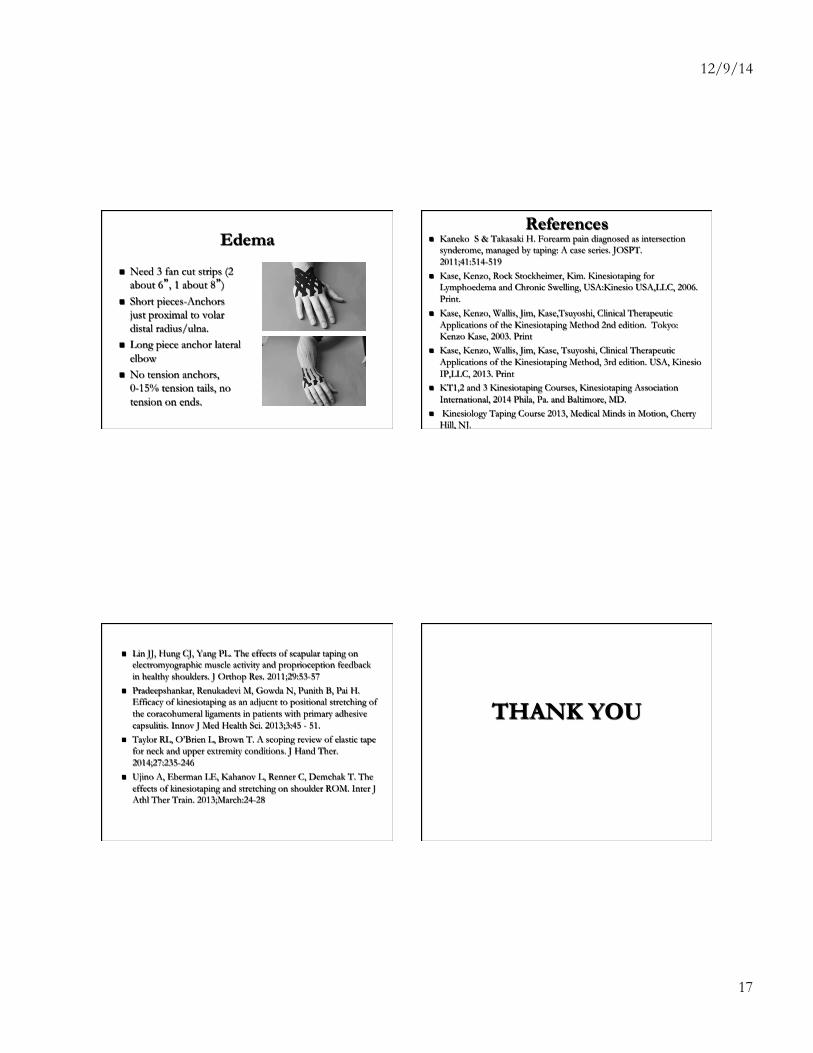
Edema
n Need 3 fan cut strips (2 about 6”, 1 about 8”)
n Short pieces-Anchors just proximal to volar distal radius/ulna.
n Long piece anchor lateral elbow
n No tension anchors, 0-15% tension tails, no tension on ends.
References n Kaneko S & Takasaki H. Forearm pain diagnosed as intersection
synderome, managed by taping: A case series. JOSPT. 2011;41:514-519
n Kase, Kenzo, Rock Stockheimer, Kim. Kinesiotaping for Lymphoedema and Chronic Swelling, USA:Kinesio USA,LLC, 2006. Print.
n Kase, Kenzo, Wallis, Jim, Kase,Tsuyoshi, Clinical Therapeutic Applications of the Kinesiotaping Method 2nd edition. Tokyo: Kenzo Kase, 2003. Print
n Kase, Kenzo, Wallis, Jim, Kase, Tsuyoshi, Clinical Therapeutic Applications of the Kinesiotaping Method, 3rd edition. USA, Kinesio IP,LLC, 2013. Print
n KT1,2 and 3 Kinesiotaping Courses, Kinesiotaping Association International, 2014 Phila, Pa. and Baltimore, MD.
n Kinesiology Taping Course 2013, Medical Minds in Motion, Cherry Hill, NJ.
n Lin JJ, Hung CJ, Yang PL. The effects of scapular taping on electromyographic muscle activity and proprioception feedback in healthy shoulders. J Orthop Res. 2011;29:53-57
n Pradeepshankar, Renukadevi M, Gowda N, Punith B, Pai H. Efficacy of kinesiotaping as an adjucnt to positional stretching of the coracohumeral ligaments in patients with primary adhesive capsulitis. Innov J Med Health Sci. 2013;3:45 - 51.
n Taylor RL, O’Brien L, Brown T. A scoping review of elastic tape for neck and upper extremity conditions. J Hand Ther. 2014;27:235-246
n Ujino A, Eberman LE, Kahanov L, Renner C, Demchak T. The effects of kinesiotaping and stretching on shoulder ROM. Inter J Athl Ther Train. 2013;March:24-28
THANK YOU