kent academic repository et al. 2016 neuroscience_tdcs... · angius, luca and pageaux, benjamin and...
TRANSCRIPT

Kent Academic RepositoryFull text document (pdf)
Copyright & reuse
Content in the Kent Academic Repository is made available for research purposes. Unless otherwise stated all
content is protected by copyright and in the absence of an open licence (eg Creative Commons), permissions
for further reuse of content should be sought from the publisher, author or other copyright holder.
Versions of research
The version in the Kent Academic Repository may differ from the final published version.
Users are advised to check http://kar.kent.ac.uk for the status of the paper. Users should always cite the
published version of record.
Enquiries
For any further enquiries regarding the licence status of this document, please contact:
If you believe this document infringes copyright then please contact the KAR admin team with the take-down
information provided at http://kar.kent.ac.uk/contact.html
Citation for published version
Angius, Luca and Pageaux, Benjamin and Hopker, James G. and Marcora, Samuele Maria andMauger, Alexis R. (2016) Transcranial Direct Current Stimulation Improves Isometric Timeto Exhaustion of the Knee Extensors. Neuroscience, 339 . pp. 363-375. ISSN 0306-4522.
DOI
https://doi.org/10.1016/j.neuroscience.2016.10.028
Link to record in KAR
http://kar.kent.ac.uk/59658/
Document Version
Publisher pdf

TRANSCRANIAL DIRECT CURRENT STIMULATION IMPROVESISOMETRIC TIME TO EXHAUSTION OF THE KNEE EXTENSORS
L. ANGIUS, a B. PAGEAUX, b J. HOPKER, a
S. M. MARCORA a AND A. R. MAUGER a*aEndurance Research Group, School of Sport and
Exercise Sciences, University of Kent, Chatham Maritime, UK
b Laboratoire INSERM U1093, Universite de Bourgogne,
Dijon, France
Abstract—Transcranial direct current stimulation (tDCS)
can increase cortical excitability of a targeted brain area,
which may affect endurance exercise performance. How-
ever, optimal electrode placement for tDCS remains unclear.
We tested the effect of two different tDCS electrode mon-
tages for improving exercise performance. Nine subjects
underwent a control (CON), placebo (SHAM) and two differ-
ent tDCS montage sessions in a randomized design. In one
tDCS session, the anodal electrode was placed over the left
motor cortex and the cathodal on contralateral forehead
(HEAD), while for the other montage the anodal electrode
was placed over the left motor cortex and cathodal electrode
above the shoulder (SHOULDER). tDCS was delivered for
10 min at 2.0 mA, after which participants performed an iso-
metric time to exhaustion (TTE) test of the right knee exten-
sors. Peripheral and central neuromuscular parameters
were assessed at baseline, after tDCS application and after
TTE. Heart rate (HR), ratings of perceived exertion (RPE),
and leg muscle exercise-induced muscle pain (PAIN) were
monitored during the TTE. TTE was longer and RPE lower
in the SHOULDER condition (P< 0.05). Central and periph-
eral parameters, and HR and PAIN did not present any differ-
ences between conditions after tDCS stimulation (P> 0.05).
In all conditions maximal voluntary contraction (MVC) sig-
nificantly decreased after the TTE (P< 0.05) while motor-
evoked potential area (MEP) increased after TTE
(P< 0.05). These findings demonstrate that SHOULDER
montage is more effective than HEAD montage to improve
endurance performance, likely through avoiding the nega-
tive effects of the cathode on excitability. � 2016 The
Author(s). Published by Elsevier Ltd on behalf of IBRO. This
is an open access article under the CC BY-NC-ND license
(http://creativecommons.org/licenses/by-nc-nd/4.0/).
Key words: tDCS, exercise, performance, brain stimulation.
INTRODUCTION
Muscle fatigue, defined as an exercise-induced reduction
in the maximal force/power production of a muscle group
(Gandevia, 2001), is known to be associated with
changes at or distal to the neuromuscular junction (i.e.
peripheral fatigue) and/or failure to recruit the active mus-
cle group (i.e. central fatigue) (Gandevia, 2001). How-
ever, because the process of fatigue also involves
multiple structures of the cortico-spinal tract, it is also
important to recognize that a failure to generate output
from the motor cortex (M1) can also result in reduced
muscle force – this is termed supraspinal fatigue
(Gandevia et al., 1996; Taylor et al., 1996; Taylor and
Gandevia, 2008). Supraspinal fatigue can occur during
exercise involving both isometric and dynamic contrac-
tions (Taylor et al., 1996; Gandevia, 2001; Søgaard
et al., 2006) which has been observed to develop from
exercise onset and continues until exhaustion along with
peripheral parameters (Taylor et al., 1996; Gandevia,
2001; Søgaard et al., 2006). Non-invasive techniques
such as the transcranial magnetic stimulation (TMS) have
been extensively used to stimulate the M1 during muscle
contraction in order to investigate the impact of suprasp-
inal fatigue during exercise (Taylor et al., 1996;
Gandevia, 2001; Søgaard et al., 2006). However, despite
a significant number of studies and the development/
refinement of non-invasive methods, the physiological
mechanisms of supraspinal fatigue are still not well estab-
lished (Gandevia, 2001; Taylor and Gandevia, 2008).
However, there is evidence to suggest that the descend-
ing output from the M1 is not adequate during fatiguing
exercise (Taylor et al., 1996; Gandevia, 2001; Liu et al.,
2002). In the study of Søgaard and colleagues (2006)
the superimposed twitch (Tw) evoked by TMS over M1
increased until exhaustion, indicating a suboptimal output
from the M1. Furthermore, in the study of Liu et al. (2002)
functional magnetic resonance imaging (fMRI) revealed a
significant reduction in brain activation during the last 60 s
of a sustained (125 s) handgrip maximal voluntary con-
traction (MVC). If a suboptimal output from the M1 con-
tributes to supraspinal fatigue, then any intervention
which moderates this reduction could plausibly improve
exercise performance. Anodal transcranial direct current
stimulation (tDCS) of the M1 has reliably been shown to
increase cortical excitability (Nitsche and Paulus, 2001),
http://dx.doi.org/10.1016/j.neuroscience.2016.10.0280306-4522/� 2016 The Author(s). Published by Elsevier Ltd on behalf of IBRO.This is an open access article under the CC BY-NC-ND license (http://creativecommons.org/licenses/by-nc-nd/4.0/).
*Corresponding author. Address: School of Sport and ExerciseSciences, University of Kent, Chatham Maritime, Kent ME4 4AG,UK. Fax: +44-0-1634-888890.
E-mail address: [email protected] (A. R. Mauger).Abbreviations: BLa�, blood lactate; CSP, cortical silent period; EMG,electromyography; Hb diff, hemoglobin difference; HHb,deoxyhemoglobin; HR, heart rate; MEP, motor-evoked potential;MVC, maximal voluntary contraction; NIRS, near-infraredspectroscopy; O2Hb, oxyhemoglobin; PAIN, exercise-induced musclepain; RMS, root mean square; RPE, rating of perceived exertion; SMA,supplementary motor area; tDCS, transcranial direct currentstimulation; tHb, total hemoglobin; TTE, time to exhaustion; Tw,twitch; VAL, voluntary activation level; VL, vastus lateralis.
Neuroscience 339 (2016) 363–375
363

and so this procedure may have the potential to attenuate
the development of supraspinal fatigue. Briefly, tDCS
involves the application of a weak electrical current
through the brain between two electrodes, which conse-
quently excite (i.e. anodal stimulation) or inhibit (i.e.
cathodal stimulation) the targeted brain area by altering
resting membrane potential (Nitsche and Paulus, 2001;
Nitsche et al., 2005). Recently, a series of experiments
investigating the effect of tDCS prior to exercise have
been conducted. In several studies, endurance perfor-
mance appeared to improve following tDCS stimulation
(Cogiamanian et al., 2007; Williams et al., 2013; Okano
et al., 2015), however other studies reported no effect
(Kan et al., 2013; Lampropoulou and Nowicky, 2013;
Muthalib et al., 2013; Angius et al., 2015). The variation
in changes to exercise performance arising from tDCS
are potentially a consequence of different experimental
and methodological set up. Aside from the absence of a
placebo control in many of the above studies, a notable
methodological difference is the use of a cephalic or
extracephalic electrode montage. A cephalic electrode
montage involves placing the anodal electrode over the
M1 (or main target area) and the cathodal electrode (i.e.
reference) placed over the contralateral prefrontal area
(Williams et al., 2013; Angius et al., 2015; Okano et al.,
2015). An extracephalic set up places the cathodal elec-
trode on the opposite shoulder (Cogiamanian et al.,
2007; Kan et al., 2013; Lampropoulou and Nowicky,
2013; Muthalib et al., 2013), rather than the contralateral
area of the head. This is because the tDCS anode
increases excitability over the area that it is placed,
whereas the cathode decreases excitability. Therefore,
in the studies which used a cephalic montage (Angius
et al., 2015), the unwanted effects of decreased excitabil-
ity in the brain area under the cathode may have negated
the positive effects of the anodal stimulation. Using an
extracephalic montage may avoid this problem and
explain why exercise performance differences tend to be
more apparent in the studies that use this approach
(Cogiamanian et al., 2007; Kan et al., 2013). In addition
to differences in electrode location, there are notable dis-
crepancies between exercise-based tDCS studies with
regard to the relative participant fatigue state in which
tDCS is applied. Indeed, tDCS has been administered in
a pre-fatigued state (Abdelmoula et al., 2016;
Cogiamanian et al., 2007), during exercise (Williams
et al., 2013) or at rest (Angius et al., 2015). These differ-
ences are relevant because a rapid and adaptive change
in the corticospinal response has been shown to depend
on the exercise intensity and exercise duration
(Gandevia, 2001), which will dictate the level of central
and/or peripheral fatigue in the individual. Therefore, brain
response and exercise performance might change
according to the ‘‘state” of the corticospinal tract prior to
tDCS administration, thus making the effect of tDCS and
study comparisons unclear.
The literature supporting the use of tDCS to moderate
exercise performance is limited, with methodological
differences contributing to apparent discrepancy in their
findings. There is also a dearth of literature detailing
changes in neuromuscular parameters following tDCS
and exercise. Specifically, we hypothesized that an
extracephalic montage might be more beneficial to
improve exercise capacity compared to cephalic
montage. Therefore, the purpose of the present study
was to examine the effect of a tDCS M1 cephalic and
extracephalic electrode montage on lower limb isometric
exercise. Using TMS and peripheral stimulation to
quantify changes in neuromuscular parameters, the
study aimed to clarify the optimal electrode montage to
improve endurance performance and detail any
neuromuscular changes that paralleled this.
EXPERIMENTAL PROCEDURES
Ethical approval
For the present investigation, each participant was
informed about the procedures of the study but not of
the aims and hypothesis. Written informed consent was
given by all participants. Study ethics were approved by
the School’s Research Ethics Advisory Group (approval
number Prop82_2012_13), which conformed to the
standards set by the Declaration of Helsinki.
Participants
Nine recreationally active males (mean ± SD; age = 23
± 2 year, height = 179 ± 7 cm, weight = 76 ± 9 kg)
participated in the present study. None of the
participants had any history of cardiorespiratory,
metabolic or mental disorder/disease or was taking any
medication at the time of the study. Each participant
gave their written informed consent and was informed
about the procedures of the study but not of the aims
and hypothesis. All experimental protocols and
procedures were approved by the local ethics
committee. All tests were conducted in a temperature-
controlled room (20 �C, relative humidity 50%), within 2–
5 days of each other and at the same time of the day
for each participant. Each participant was informed
about the procedures of the study but not of the aims
and hypothesis. Throughout the experiment, all
participants were asked to keep their normal eating
behaviors and refrain from vigorous exercise (24 h
prior), drinking alcohol (48 h), caffeine (8 h prior) and
analgesics (6 h prior) prior to any test occasion.
Experimental protocol
Each participant visited the laboratory on five different
occasions. During the first visit, participants were
familiarized with the laboratory and all the experimental
procedures. In the four subsequent visits, using a
single-blind, randomized and counter-balanced design,
all participants underwent a control (CON), placebo
(SHAM) and cephalic (HEAD) and extra-cephalic
(SHOULDER) testing session.
Endurance task (time to exhaustion test; TTE)
To assess endurance performance, participants
performed a submaximal isometric TTE task of the right
364 L. Angius et al. / Neuroscience 339 (2016) 363–375

knee extensor muscles at 20% of their MVC, which was
performed during each visit. During the TTE each
participant received visual feedback on a computer
monitor showing the target force. The task terminated
when their force went below the required target value
for more than 3 s. None of the participants were aware
of the time elapsed during the test and results of all the
sessions were provided only after the completion of all
visits. Participants’ perception of effort was measured
using the 15-point rating of perceived exertion (RPE)
scale (Borg, 1998) every 20 s of the TTE task. Leg muscle
pain was assessed every 20 s by using a 10-point numer-
ical scale (Cook et al., 1997). Heart rate (HR) was moni-
tored continuously and averaged for every 20 s elapsed.
Blood lactate concentration (B[La�]) was obtained by col-
lecting a 10 ll samples of capillary blood immediately
after the TTE task. Each sample was then analyzed after
the completion of all experimental procedures to deter-
mine the lactate concentration (Biosen; EFK Diagnostics,
London, UK).
Neuromuscular tests
After a brief, standardized warm-up with submaximal
isometric contractions, all participants performed a 5-s
MVC with superimposed doublet stimulation, followed
(4-s interval) by a resting potentiated doublet. The MVC
produced during this test was used to calculate the
participants’ 20% MVC used in the subsequent TTE
task of that visit. Ten seconds after the MVC
participants performed a series of four submaximal
contractions at 50% of the MVC (3 s duration) with
superimposed TMS and one with superimposed femoral
stimulation. Each contraction was interspaced by 3 s.
Neuromuscular assessment tests were performed prior
to tDCS, post tDCS and immediately after the TTE task
(see Fig. 1).
TMS procedure
TMS was used to assess the level of cortical excitability of
the M1. The stimulation site was determined by a TMS
stimulator (Magstim 2002, The Magstim Company Ltd,
Whitland, UK) with the concave double coil (110 mm
diameter) placed over the contralateral M1 of the
exercising leg. The stimulation site was determined
when the largest motor-evoked potential (MEP)
response of the vastus lateralis (VL) was obtained
together with a small MEP response (<10%) of the
antagonist muscle (biceps femoris, BF). Once the site
was determined, this was marked on the participant’s
scalp with a marker pen. After determining the
stimulation site, stimulus intensity was set according to
the highest MEP response elicited during a 3-s
submaximal contraction at 50% MVC. To determine this,
stimulation intensity commenced at 45% and was
subsequently ramped up in increments of 5% until a
plateau in MEP response was observed. Procedures for
stimulation location and intensity were performed for
each participant at the beginning of each visit. The
intensity of stimulation across participants and visits was
63 ± 8% of the maximum stimulator output.
Femoral nerve stimulation
Transcutaneous electrically-evoked femoral nerve
stimulation was delivered by using a high-voltage
constant-current stimulator (model DS7 modified,
Digitimer, Hertfordshire, UK). The femoral nerve was
stimulated using a cathode surface electrode
(Swaromed, Nessler Medizintechnik, Innsbruck, Austria)
positioned over the femoral triangle while the anode
electrode (Phoenix Healthcare Products Ltd.,
Nottingham, UK) was placed in the gluteal fold. The
stimulation intensity (mean current 288 ± 64 mA) was
increased by 20 mA until the action potential (M-wave)
demonstrated no further increase (Mmax) at rest and
during submaximal 50% MVC contractions. The final
intensity stimulation was then set at 130% Mmax. Both
Mmax and TMS intensities were determined at the
beginning of each experimental session and were kept
constant throughout that visit.
Mechanical recordings
All the experimental procedures were performed on an
isokinetic dynamometer (Cybex NORM isokinetic
dynamometer, CMSi, Computer 267 Sports Medicine
Inc., Stoughton, USA). All tests were performed with the
right leg at a knee joint angle of 90� of flexion
Fig. 1. Overall view of the experimental protocol. Maximal muscular wave (Mwave); motor-evoked potential (MEP); maximal voluntary contraction
(MVC); transcranial direct current stimulation (tDCS); time to exhaustion (TTE).
L. Angius et al. / Neuroscience 339 (2016) 363–375 365

(0�= knee fully extended) and a hip angle of 90�. The
set-up for each participant was recorded in the
familiarization session and kept constant in all
subsequent visits. Mechanical signals were digitized on-
line at a sampling frequency of 1 kHz using a computer,
and stored for analysis with commercially available
software (Acqknowledge 4.2 for MP Systems, Biopac
Systems Inc., Goleta, USA).
Electromyographic recordings
Electromyography (EMG) of the VL was recorded with
two surface electrodes (Swaromed, Nessler
Medizintechnik, Innsbruck, Austria) while the reference
electrode was placed over the patella of the right knee.
The skin was shaved and cleaned using alcohol swabs.
Myoelectrical signals were amplified with a bandwidth
frequency ranging from 10 Hz to 500 Hz (gain = 500),
digitized on-line at a sampling frequency of 2 kHz using
a computer, and stored for analysis with commercially
available software (Acqknowledge 4.2 for MP Systems,
Biopac Systems Inc., Goleta, USA).
Near-infrared spectroscopy (NIRS) procedures
Brain oxygenation was monitored via near infrared
spectroscopy using a portable device (Artinis, Zetten,
The Netherlands). Two probes were placed on the left
and right prefrontal cortex region of the forehead (Fp1
and Fp2, according to the international EEG 10–20
system) using a transmitter-receptor distance of 4 cm.
NIRS data were recorded for four minutes at rest and
were used as baseline. Subsequently, NIRS data were
collected both during tDCS and the TTE task with a
sampling frequency of 10 Hz.
tDCS procedure
tDCS was delivered by a direct current stimulator (TCT
Research Limited, Hong Kong) using a pair of rubber
electrodes in a 4 � 3-cm water-soaked synthetic
sponge. Two different montages were used for the
present investigation: (1) anodal placed over the left M1
with the cathodal placed above dorsolateral right
prefrontal cortex (HEAD); (2) anodal placed over the left
M1 with the cathodal was placed over the shoulder
(SHOULDER). For the SHAM session, electrodes were
placed in the same position for HEAD while in the
control no electrodes were placed on the participant.
During HEAD and SHOULDER conditions the current
was applied with an intensity of 2.0 mA for 10 min,
whereas during the SHAM condition stimulation lasted
30 s and subsequently ramped down to no stimulation.
Data analysis
Peak force during the MVC of knee extensor muscles was
considered as the peak torque attained during the MVC,
while voluntary activation level (VAL) during the MVC
was estimated according to the following formula:
VAL ¼ 100 � ð1� superimposed doublet amplitude=
potentiated doublet amplitudeÞ
The root mean square (RMS) of the EMG signal was
automatically calculated with the software and the peak-
to-peak amplitude of the resting M-waves were
calculated and averaged for the stimulations. The
following parameters were also analyzed: peak torque
doublet, peak Tw. EMG amplitude during the MVC was
quantified as the RMS for a 0.5-s interval at peak torque
(250-ms interval either side of the peak torque).
Maximal RMS values obtained during the MVC
(RMSMVC) were normalized by the resting M-wave RMS
(RMSM) to obtain the RMSMVC/RMSM ratio in order to
take into account peripheral influences, including
neuromuscular propagation and changes in impedance
during the EMG recordings. The MEP area (MEParea),
was calculated and averaged for the four stimulations,
and then normalized for the M-wave obtained during the
50% MVC contraction. Cortical silent period (CSP)
duration of the MEP was determined by the same
experimenter from the onset of the MEP to the return of
continuous EMG signal (Saisanen et al., 2008). Because
of continuous measures, VL RMS was plotted as 0%,
25%, 50%, 75% and 100% of each TTE. 0% corre-
sponded to the first 5 s of the TTE while for 25%, 50%,
75% and 100%, the signal was analyzed and averaged
for the last 5 s for each percentage. NIRS data were aver-
aged for the last 60 s during baseline measurement, while
during tDCS administration NIRS data were averaged for
the last 60 s every two min (i.e. min 2, 4, 6, 8 and 10). Dur-
ing exercise, data were averaged for 5 s respectively at
the 0%, 25%, 50%, 75% and 100% of each TTE. The
Beer–Lambert Law was used to calculate changes in
tissue oxygenation. Relative concentration changes
were measured from resting baseline for oxyhe-
moglobin (DO2Hb), deoxyhemoglobin (DHHb), total
hemoglobin (DtHb = O2Hb+ HHb) and hemoglobin dif-
ference (DHb diff = O2Hb� HHb). DtHb was calculated
to give an index of change in regional blood volume. Indi-
vidual values of RPE, exercise-induced muscle pain
(PAIN) and HR obtained during the TTE were plotted
against the absolute TTE time for each condition, then
the curve for each variable was mathematically fitted by
a linear equation to obtain the slope.
Statistical analysis
All data are presented as mean ± SD. Assumptions of
statistical tests such as normal distribution and
sphericity of data were checked before running each
individual statistical analysis. The effect of tDCS
montage on TTE time and B[La�] were assessed by
using a one-way ANOVA with repeated measures. The
same statistical analysis was performed to compare the
slope of RPE, PAIN and HR obtained during the TTE.
Fully repeated measures 4 � 3 way ANOVAs were used
to test the effect of condition (HEAD, SHOUDLER,
SHAM and CONTROL) and time (baseline, post-tDCS
and post TTE) on MVC, VAL, Doublet, VL RMS during
TTE, MEParea/Mwave, and CSP. Three way 4 � 2 � 5
ANOVAs were used to test the effect of condition
(HEAD, SHOUDLER, SHAM and CONTROL), prefrontal
cortex side (left vs. right side) and time on DO2Hb,
DHHb, DHb diff, DtHb and TSI during tDCS stimulation.
366 L. Angius et al. / Neuroscience 339 (2016) 363–375

Three-way 4 � 2 � 6 ANOVAs were used to test the
effect of condition (HEAD, SHOUDLER, SHAM and
CONTROL), prefrontal cortex side (left vs. right side)
and time on DO2Hb, DHHb, DHb diff, DtHb and TSI
obtained during the TTE. Bonferroni post hoc tests was
used when appropriate. The a level was set at
P< 0.05. Statistics were calculated using SPSS version
20.
RESULTS
TTE was significantly longer (F(3,24) = 7.84, P 6 0.001,
gp2 = 0.49) in the SHOULDER condition compared to
the HEAD, SHAM and CON conditions (219 ± 136 s,
191 ± 124 s, 173 ± 114 s and 187 ± 121 s,
respectively). This was accompanied by a significantly
lower RPE slope in the SHOULDER condition (F(3,24)
= 5.29, P 6 0.006, gp2 = 0.88) (see Fig. 2). No
significant differences between conditions were
observed for B[La�] (F(3,24) = 0.06, P= 0.99,
gp2 = 0.00) (4.81 ± 206, 4.70 ± 210, 4.67 ± 241, 4.97
± 2.07 mmol l�1 respectively for SHOULDER, HEAD,
SHAM and CON) or HR slope (F(3,24) = 0.031,
P= 0.90, gp2 = 0.03) (5.36 ± 2.49, 5.60 ± 3.62, 5.53
± 3.11 and 4.85 ± 3.54 respectively for SHOULDER,
HEAD, SHAM and CON) or PAIN slope (F(3,24)
= 0.50, P= 0.68, gp2 = 0.05) (1.02 ± 0.69, 1.00
± 0.53, 0.95 ± 0.57 and 0.91 ± 0.60 respectively for
SHOULDER, HEAD, SHAM and CON).
Neuromuscular parameters
MVC torque decreased significantly at exhaustion (F
(2,16) = 24.85, P 6 0.001, gp2 = 0.75) but did not differ
between conditions (F(3,24) = 0.68, P= 0.56,
gp2 = 0.07). RMS of VL increased over time (F(3,24)
= 2.40, P 6 0.001, gp2 = 0.87) but did not differ between
conditions (F(3,24) = 0.68, P= 0.38, gp2 = 0.07) (see
Fig. 3).
Peripheral fatigue
Doublet amplitude decreased significantly only at
exhaustion (F(2,16) = 36.92, P 6 0.001, gp2 = 0.82) but
did not differ between conditions (F(3,24) = 0.70,
P= 0.55, gp2 = 0.08). Tw decreased only at exhaustion
(F(2,16) = 36.92, P 6 0.001, gp2 = 0.82) but did not
differ between conditions (F(3,24) = 0.70, P= 0.55,
gp2 = 0.08). Mamp at 50% MVC was significantly higher
only at exhaustion (F(3,24) = 11.09, P 6 0.001,
gp2 = 0.58) but did not differ between conditions (F
(3,24), P= 0.28, gp2 = 0.14). Marea at 50% MVC was
significantly different only at exhaustion (F(3,24)
= 10.21, P 6 0.001, gp2 = 0.56) but did not differ
between conditions (F(3,24), P= 0.95, gp2 = 0.21) (see
Fig. 3).
Central fatigue
VAL decreased significantly only at exhaustion
(F(2,16) = 15.27, P 6 0.001, gp2 = 0.99) but did not
differ between conditions (F(3,24) = 1.19, P= 0.33,
gp2 = 0.13). RMSMVC/RMSMwave of the VL did not
change over time (F(2,16) = 1.23, P= 0.85, gp2 = 0.13)
and did not differ between conditions (F(3,24) = 0.499,
P= 0.68, gp2 = 0.05).
Cortical excitability
MEParea increased only at exhaustion (F(2,16) = 5.18,
P 6 0.018, gp2 = 0.39) but did not differ between
conditions (F(3,24) = 0.10, P= 0.96, gp2 = 0.01).
MEParea/Marea ratio increased only at exhaustion
(F(2,16) = 6.21, P 6 0.01, gp2 = 0.43) but did not
differ between conditions (F(2,16) = 6.21, P= 0.91,
gp2 = 0.01). CSP increased only at exhaustion
(F(2,16) = 5.48, P 6 0.015, gp2 = 0.40) but did not
differ between conditions (F(3,24) = 0.49, P= 0.37,
gp2 = 0.98) (see Fig. 3).
NIRS parameters during tDCS stimulation
DO2Hb did not change over time (F(4,32) = 0.98,
P= 0.42, gp2 = 0.27) and no differences between
conditions (F(3,24) = 0.30, P= 0.99, gp2 = 0.55) or side
(F(1,8) = 3.87, P= 0.85, gp2 = 0.41) were found. DHHb
did not change over time (F(4,32) = 0.92, P= 0.23,
gp2 = 0.25) and no differences between conditions
(F(3,24) = 0.75, P= 0.39, gp2 = 0.18) or side (F(1,8) =
0.62, P= 0.45, gp2 = 0.10) were found. DtHb did not
Fig. 2. Physiological and perceptual response of all tests performed. Panel A shows time to exhaustion (TTE) performance. Panel B shows the
slope values of ratings of perceived exertion (RPE). *P 6 0.05 significant from HEAD, CONTROL and SHAM. Data are presented as mean ± SD
(n= 9).
L. Angius et al. / Neuroscience 339 (2016) 363–375 367

change over time (F(4,32) = 1.36, P= 0.77, gp2 = 0.37)
and no differences between conditions (F(3,24) = 0.29,
P= 0.10, gp2 = 0.09) or side (F(1,8) = 1.30, P= 0.28,
gp2 = 0.17) were found. DHbdiff did not change over
time (F(4,32) = 2.58, P= 0.15, gp2 = 0.65) and no
differences between conditions (F(3,24) = 0.87,
P= 0.32, gp2 = 0.21) or side (F(1,8) = 0.02, P= 0.87,
gp2 = 0.53) were found. Tissue saturation index did not
change over time (F(4,28) = 0.10, P= 0.63, gp2 = 0.06)
and no differences between conditions (F(3,21) = 0.83,
P= 0.65, gp2 = 0.20) or side (F(1,7) = 0.10, P= 0.755,
gp2 = 0.05) were found (see Tables 1 and 2).
Fig. 3. Overall response neuromuscular parameters during the various phases of the experiment. Panel A shows maximal voluntary contraction
(MVC); Panel B shows voluntary activation level (VAL); Panel C shows peak torque of the doublet; Panel D shows MEParea/Mwave ratio; Panel E
shows cortical silent period (CSP); Panel F shows root mean square of vastus lateralis (VL RMS) during time to exhaustion (TTE). *P 6 0.05
significant from baseline and post tDCS; #P< 0.05, significant main effect of time. Data are presented as mean ± SD (n= 9).
368 L. Angius et al. / Neuroscience 339 (2016) 363–375
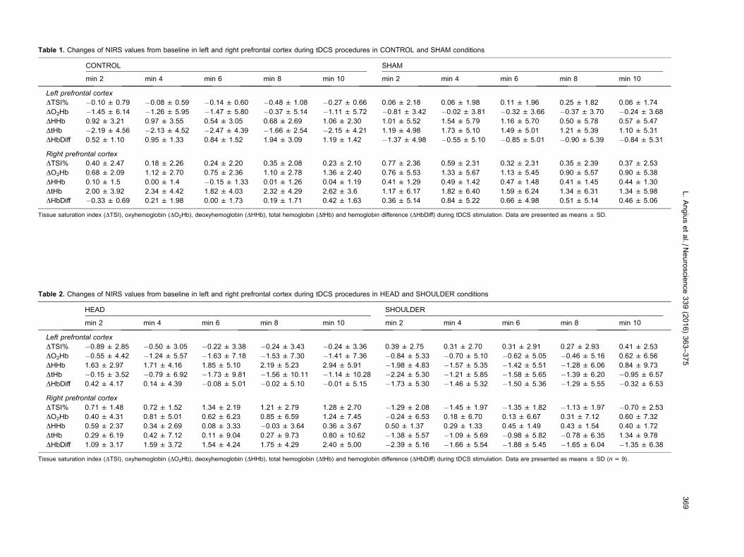
Table 1. Changes of NIRS values from baseline in left and right prefrontal cortex during tDCS procedures in CONTROL and SHAM conditions
CONTROL SHAM
min 2 min 4 min 6 min 8 min 10 min 2 min 4 min 6 min 8 min 10
Left prefrontal cortex
DTSI% �0.10 ± 0.79 �0.08 ± 0.59 �0.14 ± 0.60 �0.48 ± 1.08 �0.27 ± 0.66 0.06 ± 2.18 0.06 ± 1.98 0.11 ± 1.96 0.25 ± 1.82 0.06 ± 1.74
DO2Hb �1.45 ± 6.14 �1.26 ± 5.95 �1.47 ± 5.80 �0.37 ± 5.14 �1.11 ± 5.72 �0.81 ± 3.42 �0.02 ± 3.81 �0.32 ± 3.66 �0.37 ± 3.70 �0.24 ± 3.68
DHHb 0.92 ± 3.21 0.97 ± 3.55 0.54 ± 3.05 0.68 ± 2.69 1.06 ± 2.30 1.01 ± 5.52 1.54 ± 5.79 1.16 ± 5.70 0.50 ± 5.78 0.57 ± 5.47
DtHb �2.19 ± 4.56 �2.13 ± 4.52 �2.47 ± 4.39 �1.66 ± 2.54 �2.15 ± 4.21 1.19 ± 4.98 1.73 ± 5.10 1.49 ± 5.01 1.21 ± 5.39 1.10 ± 5.31
DHbDiff 0.52 ± 1.10 0.95 ± 1.33 0.84 ± 1.52 1.94 ± 3.09 1.19 ± 1.42 �1.37 ± 4.98 �0.55 ± 5.10 �0.85 ± 5.01 �0.90 ± 5.39 �0.84 ± 5.31
Right prefrontal cortex
DTSI% 0.40 ± 2.47 0.18 ± 2.26 0.24 ± 2.20 0.35 ± 2.08 0.23 ± 2.10 0.77 ± 2.36 0.59 ± 2.31 0.32 ± 2.31 0.35 ± 2.39 0.37 ± 2.53
DO2Hb 0.68 ± 2.09 1.12 ± 2.70 0.75 ± 2.36 1.10 ± 2.78 1.36 ± 2.40 0.76 ± 5.53 1.33 ± 5.67 1.13 ± 5.45 0.90 ± 5.57 0.90 ± 5.38
DHHb 0.10 ± 1.5 0.00 ± 1.4 �0.15 ± 1.33 0.01 ± 1.26 0.04 ± 1.19 0.41 ± 1.29 0.49 ± 1.42 0.47 ± 1.48 0.41 ± 1.45 0.44 ± 1.30
DtHb 2.00 ± 3.92 2.34 ± 4.42 1.82 ± 4.03 2.32 ± 4.29 2.62 ± 3.6 1.17 ± 6.17 1.82 ± 6.40 1.59 ± 6.24 1.34 ± 6.31 1.34 ± 5.98
DHbDiff �0.33 ± 0.69 0.21 ± 1.98 0.00 ± 1.73 0.19 ± 1.71 0.42 ± 1.63 0.36 ± 5.14 0.84 ± 5.22 0.66 ± 4.98 0.51 ± 5.14 0.46 ± 5.06
Tissue saturation index (DTSI), oxyhemoglobin (DO2Hb), deoxyhemoglobin (DHHb), total hemoglobin (DtHb) and hemoglobin difference (DHbDiff) during tDCS stimulation. Data are presented as means ± SD.
Table 2. Changes of NIRS values from baseline in left and right prefrontal cortex during tDCS procedures in HEAD and SHOULDER conditions
HEAD SHOULDER
min 2 min 4 min 6 min 8 min 10 min 2 min 4 min 6 min 8 min 10
Left prefrontal cortex
DTSI% �0.89 ± 2.85 �0.50 ± 3.05 �0.22 ± 3.38 �0.24 ± 3.43 �0.24 ± 3.36 0.39 ± 2.75 0.31 ± 2.70 0.31 ± 2.91 0.27 ± 2.93 0.41 ± 2.53
DO2Hb �0.55 ± 4.42 �1.24 ± 5.57 �1.63 ± 7.18 �1.53 ± 7.30 �1.41 ± 7.36 �0.84 ± 5.33 �0.70 ± 5.10 �0.62 ± 5.05 �0.46 ± 5.16 0.62 ± 6.56
DHHb 1.63 ± 2.97 1.71 ± 4.16 1.85 ± 5.10 2.19 ± 5.23 2.94 ± 5.91 �1.98 ± 4.83 �1.57 ± 5.35 �1.42 ± 5.51 �1.28 ± 6.06 0.84 ± 9.73
DtHb �0.15 ± 3.52 �0.79 ± 6.92 �1.73 ± 9.81 �1.56 ± 10.11 �1.14 ± 10.28 �2.24 ± 5.30 �1.21 ± 5.85 �1.58 ± 5.65 �1.39 ± 6.20 �0.95 ± 6.57
DHbDiff 0.42 ± 4.17 0.14 ± 4.39 �0.08 ± 5.01 �0.02 ± 5.10 �0.01 ± 5.15 �1.73 ± 5.30 �1.46 ± 5.32 �1.50 ± 5.36 �1.29 ± 5.55 �0.32 ± 6.53
Right prefrontal cortex
DTSI% 0.71 ± 1.48 0.72 ± 1.52 1.34 ± 2.19 1.21 ± 2.79 1.28 ± 2.70 �1.29 ± 2.08 �1.45 ± 1.97 �1.35 ± 1.82 �1.13 ± 1.97 �0.70 ± 2.53
DO2Hb 0.40 ± 4.31 0.81 ± 5.01 0.62 ± 6.23 0.85 ± 6.59 1.24 ± 7.45 �0.24 ± 6.53 0.18 ± 6.70 0.13 ± 6.67 0.31 ± 7.12 0.60 ± 7.32
DHHb 0.59 ± 2.37 0.34 ± 2.69 0.08 ± 3.33 �0.03 ± 3.64 0.36 ± 3.67 0.50 ± 1.37 0.29 ± 1.33 0.45 ± 1.49 0.43 ± 1.54 0.40 ± 1.72
DtHb 0.29 ± 6.19 0.42 ± 7.12 0.11 ± 9.04 0.27 ± 9.73 0.80 ± 10.62 �1.38 ± 5.57 �1.09 ± 5.69 �0.98 ± 5.82 �0.78 ± 6.35 1.34 ± 9.78
DHbDiff 1.09 ± 3.17 1.59 ± 3.72 1.54 ± 4.24 1.75 ± 4.29 2.40 ± 5.00 �2.39 ± 5.16 �1.66 ± 5.54 �1.88 ± 5.45 �1.65 ± 6.04 �1.35 ± 6.38
Tissue saturation index (DTSI), oxyhemoglobin (DO2Hb), deoxyhemoglobin (DHHb), total hemoglobin (DtHb) and hemoglobin difference (DHbDiff) during tDCS stimulation. Data are presented as means ± SD (n= 9).
L.Angiusetal./N
euroscience339(2016)363–375
369

NIRS parameters during TTE
DO2Hb increased over time (F(5,40) = 30.58, P 6 0.001,
gp2 = 1.00) but no differences were observed between
conditions (F(3,24) = 1.96, P= 0.24, gp2 = 0.44) or side
(F(1,8) = 0.04, P= 0.84, gp2 = 0.05) were found. DHHb
increased over time (F(5,40) = 38.11, P> 0.001,
gp2 = 1.00) and no differences between conditions (F
(3,24) = 0.74, P= 0.43, gp2 = 0.18) or side (F(1,8)
= 2.88, P= 0.12, gp2 = 0.32) were found. DtHb
increased over time (F(5,40) = 21.13, P 6 0.001,
gp2 = 1.00) and no differences between conditions (F
(3,24) = 0.57, P= 0.55, gp2 = 0.15) or side (F(1,8)
= 1.14, P= 0.31, gp2 = 0.15) were found. DHbDiff
decreased over time (F(5,40) = 38.11, P 6 0.001,
gp2 = 0.10) and no differences between conditions (F
(3,24) = 0.74, P= 0.43, gp2 = 0.18) or side (F(1,8)
= 2.88, P= 0.12, gp2 = 0.32) were found. Tissue
saturation decreased over time (F(5,40) = 21.13,
P= 0.003, gp2 = 0.10) and no differences between
conditions (F(3,24) = 0.57, P= 0.55, gp2 = 0.15) or side
(F(1,8) = 1.14, P= 0.31, gp2 = 0.15) were found. (see
Tables 3–6).
DISCUSSION
This is the first study showing an improvement in
isometric endurance performance of the lower limbs
following tDCS stimulation. Our findings suggest that in
order to improve lower limb endurance performance, an
extracephalic electrode montage is more effective than
cephalic montage.
Effect of tDCS on isometric endurance performance
and perceptual parameters
This study showed for the first time that only anodal tDCS
stimulation with an extracephalic montage improves
endurance performance of the knee extensors.
Following tDCS, an improvement in isometric endurance
performance has been previously demonstrated in
elbow flexor muscles (Cogiamanian et al., 2007;
Williams et al., 2013) and these authors associated the
improvement in performance with an augmented cortical
excitability of the motor, premotor and somatosensory
area with a potentially enhanced descending drive to the
motoneuronal pool. However, it is important to note that
two other studies showed no improvement in isometric
performance following tDCS (Kan et al., 2013; Muthalib
et al., 2013) which might be a consequence of different
experimental designs.
In the current experiment, isometric endurance
performance was longer in the SHOULDER condition,
where the anode was placed over the M1 and the
cathode placed on the shoulder (thus avoiding any
decreased excitability of right prefrontal cortex induced
by the cathode). A potential explanation for this
improvement in isometric endurance performance is
perception of effort during the TTE task, which
increased more slowly over time in the SHOULDER
condition. It has previously been proposed that during
sustained exercise, the increase in perception of effort
over time reflects, at least in part, the increase in
activity of premotor and/or motor areas of the brain (i.e.
central motor command) necessary to compensate the
decline in force-generating capacity of the
neuromuscular system (de Morree et al., 2012, 2014;
Marcora et al., 2008). This proposal is based on evidence
supporting the hypothesis that the sensory signals for per-
ception of effort are corollary discharges from premotor
and/or motor areas of the brain (Marcora, 2009;
Takarada et al., 2014; Zenon et al., 2015).
In our experiment, two different reasons might explain
the reduction in RPE in the SHOULDER condition. The
first is that anodal stimulation of the M1 facilitated the
descending drive to the muscle, thus reducing activity of
premotor areas and participants perceiving less effort for
the same force produced. In support of this, previous
experimental findings have demonstrated that
manipulation of the activity of the M1 and
supplementary motor area (SMA) influenced perception
of effort. In accordance with this, the study of Takarada
and colleagues (2014) demonstrated that suppression of
the activity of the M1 by repetitive TMS (rTMS) increases
perception of effort, thus making participants perceive the
Table 3. Changes of NIRS values from baseline in left and right prefrontal cortex during the isometric time to exhaustion in the CONTROL condition
CONTROL
0% 25% 50% 75% 100% EXH
Left prefrontal cortex
DTSI% �0.20 ± 0.90* �0.81 ± 0.93* �1.58 ± 1.42* �1.81 ± 1.26* �2.29 ± 1.63* �2.48 ± 1.75*
DO2Hb 7.20 ± 5.59* 9.69 ± 6.20* 11.50 ± 7.92* 13.66 ± 7.22* 15.24 ± 8.06* 15.74 ± 9.68*
DHHb 1.44 ± 1.60* 1.19 ± 1.52* 0.35 ± 1.49* 0.57 ± 1.71* 1.16 ± 1.84* 1.57 ± 2.19*
DtHb 8.65 ± 5.37* 10.88 ± 6.30* 11.84 ± 7.99* 14.23 ± 6.98* 16.40 ± 8.15* 17.31 ± 10.15*
DHbDiff 5.76 ± 6.24* 8.50 ± 6.47* 11.15 ± 8.14* 13.09 ± 7.83* 14.08 ± 8.38* 14.16 ± 9.75*
Right prefrontal cortex
DTSI% 0.13 ± 2.50* �0.62 ± 2.63* �0.83 ± 2.30* �1.40 ± 2.80* �2.80 ± 4.41* �3.56 ± 4.57*
DO2Hb 5.12 ± 5.72* 8.61 ± 7.17* 10.22 ± 9.83* 16.09 ± 10.41* 16.44 ± 8.80* 16.33 ± 9.00*
DHHb 0.66 ± 2.99* 0.37 ± 2.98* �0.60 ± 3.54* 0.13 ± 3.00* �0.08 ± 2.79* �0.26 ± 2.91*
DtHb 5.78 ± 7.58* 8.99 ± 8.97* 9.62 ± 12.05* 16.22 ± 11.89* 16.36 ± 9.83* 16.07 ± 9.94*
DHbDiff 4.45 ± 5.09* 8.24 ± 6.34* 10.81 ± 8.55* 15.97 ± 9.67* 16.53 ± 8.60* 16.59 ± 8.95*
Tissue saturation index (DTSI), oxyhemoglobin (DO2Hb), deoxyhemoglobin (DHHb), total hemoglobin (DtHb) and hemoglobin difference (DHbDiff) during tDCS stimulation.* P< 0.05, significant main effect of time. Data are presented as means ± SD (n= 9).
370 L. Angius et al. / Neuroscience 339 (2016) 363–375

voluntary contraction as harder. Furthermore, another
study performed by Zenon et al. (2015) demonstrated that
disrupting neural activity in SMA and M1 led to a signifi-
cant alteration of perception of effort. In the HEAD condi-
tion, the positive effect of anodal stimulation over M1 on
perception of effort and isometric endurance performance
Table 4. Changes of NIRS values from baseline in left and right prefrontal cortex during the isometric time to exhaustion in the SHAM condition
SHAM
0% 25% 50% 75% 100% EXH
Left prefrontal cortex
DTSI% �3.26 ± 2.25* �3.37 ± 2.77* �3.73 ± 2.87* �5.36 ± 2.41* �5.13 ± 3.81* �5.50 ± 3.49*
DO2Hb 6.75 ± 7.31* 7.29 ± 6.54* 8.80 ± 7.09* 12.38 ± 6.52* 13.87 ± 7.39* 14.04 ± 7.90*
DHHb 2.24 ± 1.94* 1.40 ± 1.88* 1.17 ± 2.69* 0.87 ± 2.61* 1.12 ± 3.29* 1.28 ± 3.12*
DtHb 7.16 ± 5.64* 6.92 ± 5.14* 8.60 ± 6.45* 11.79 ± 7.46* 14.26 ± 9.28* 14.59 ± 9.50*
DHbDiff 2.69 ± 6.03* 4.12 ± 4.88* 6.27 ± 6.35* 10.04 ± 5.78* 12.03 ± 5.95* 12.03 ± 6.71*
Right prefrontal cortex
DTSI% �1.74 ± 2.20* �1.61 ± 2.21* �1.92 ± 2.59* �3.69 ± 3.54* �4.23 ± 4.91* �4.56 ± 6.70*
DO2Hb 3.51 ± 5.32* 3.70 ± 5.41* 4.96 ± 5.95* 10.20 ± 7.60* 12.13 ± 6.05* 11.89 ± 6.53*
DHHb 1.01 ± 1.84* 0.16 ± 1.40* �0.25 ± 1.65* �0.20 ± 2.21* �0.62 ± 2.46* �0.59 ± 2.41*
DtHb 3.96 ± 4.26* 3.23 ± 4.55* 4.52 ± 5.35* 9.63 ± 8.56* 11.54 ± 6.68* 11.33 ± 7.07*
DHbDiff 4.82 ± 8.43* 5.81 ± 6.86* 7.91 ± 8.25* 12.92 ± 6.87* 15.67 ± 6.86* 15.39 ± 7.07*
Tissue saturation index (DTSI), oxyhemoglobin (DO2Hb), deoxyhemoglobin (DHHb), total hemoglobin (DtHb) and hemoglobin difference (DHbDiff) during tDCS stimulation.* P< 0.05, significant main effect of time. Data are presented as means ± SD (n= 9).
Table 5. Changes of NIRS values from baseline in left and right prefrontal cortex during the isometric time to exhaustion in the HEAD condition
HEAD
0% 25% 50% 75% 100% EXH
Left prefrontal cortex
DTSI% �1.39 ± 1.97* �1.68 ± 1.89* �2.14 ± 2.16* �2.58 ± 2.86* �2.52 ± 2.73* �3.10 ± 3.05*
DO2Hb 1.96 ± 5.32* 3.75 ± 5.41* 7.19 ± 4.69* 7.37 ± 5.83* 10.56 ± 6.02* 10.54 ± 6.32*
DHHb 1.33 ± 3.06* 0.73 ± 2.51* 0.07 ± 2.59* �0.02 ± 2.63* 0.19 ± 2.16* �0.10 ± 3.94*
DtHb 5.52 ± 10.34* 6.70 ± 9.43* 9.48 ± 9.47* 10.69 ± 10.13* 12.96 ± 9.08* 12.67 ± 10.43*
DHbDiff 2.85 ± 7.71* 5.24 ± 6.71* 9.34 ± 5.80* 10.72 ± 6.07* 12.59 ± 6.39* 12.86 ± 6.63*
Right prefrontal cortex
DTSI% �0.45 ± 3.48* �0.39 ± 3.10* �0.61 ± 3.41* �1.14 ± 3.44* �0.72 ± 4.38* �1.11 ± 4*.
DO2Hb 1.89 ± 8.15* 3.34 ± 7.48* 7.36 ± 6.36* 10.30 ± 6.23* 12.61 ± 5.06* 12.99 ± 9.22*
DHHb 0.64 ± 1.50* 0.04 ± 1.76* �0.55 ± 2.23* �0.74 ± 2.96* �0.66 ± 2.56* �1.49 ± 4.84*
DtHb 4.20 ± 8.10* 5.05 ± 7.60* 8.48 ± 6.10* 11.23 ± 6.36* 13.62 ± 6.59* 13.17 ± 12.58*
DHbDiff �2.64 ± 8.18* �0.59 ± 7.46* 4.02 ± 6.51* 7.14 ± 6.49* 9.38 ± 7.31* 10.59 ± 7.33*
Tissue saturation index (DTSI), oxyhemoglobin (DO2Hb), deoxyhemoglobin (DHHb), total hemoglobin (DtHb) and hemoglobin difference (DHbDiff) during tDCS stimulation.* P< 0.05, significant main effect of time. Data are presented as means ± SD (n= 9).
Table 6. Changes of NIRS values from baseline in left and right prefrontal cortex during the isometric time to exhaustion in the SHOULDER condition
SHOULDER
0% 25% 50% 75% 100% EXH
Left prefrontal cortex
DTSI% 0.17 ± 2.34* 0.06 ± 2.39* 0.12 ± 2.46* �0.42 ± 2.83* �0.45 ± 2.98* �0.16 ± 2.97*
DO2Hb �0.29 ± 4.91* 2.00 ± 4.89* 5.78 ± 6.62* 4.84 ± 6.64* 6.30 ± 5.37* 6.48 ± 7.41*
DHHb 2.43 ± 1.74* 2.17 ± 1.96* 1.53 ± 2.78* 1.04 ± 3.31* 1.08 ± 2.93* 1.49 ± 3.48*
DtHb 8.81 ± 9.02* 10.83 ± 9.75* 13.98 ± 12.35* 12.55 ± 10.53* 14.04 ± 10.05* 14.64 ± 9.47*
DHbDiff 6.08 ± 4.86* 8.62 ± 4.89* 13.04 ± 6.36* 12.59 ± 6.77* 14.01 ± 5.84* 13.78 ± 6.16*
Right prefrontal cortex
DTSI% �2.17 ± 8.46* �2.05 ± 9.57* �2.29 ± 11.29* �2.92 ± 12.05* �4.00 ± 12.59* �4.54 ± 14.02*
DO2Hb 0.33 ± 6.34* 2.63 ± 5.78* 6.45 ± 6.26* 8.66 ± 8.62* 11.51 ± 9.19* 11.23 ± 11.89*
DHHb 0.94 ± 4.59* 0.68 ± 3.81* �0.11 ± 4.33* �0.03 ± 4.45* �0.01 ± 4.25* 0.31 ± 4.59*
DtHb 4.32 ± 8.69* 6.36 ± 7.28* 9.41 ± 9.61* 11.80 ± 8.02* 14.61 ± 8.82* 14.66 ± 9.11*
DHbDiff 4.79 ± 7.71* 7.34 ± 8.22* 11.97 ± 10.54* 14.20 ± 9.14* 16.99 ± 10.01* 16.39 ± 7.67*
Tissue saturation index (DTSI), oxyhemoglobin (DO2Hb), deoxyhemoglobin (DHHb), total hemoglobin (DtHb) and hemoglobin difference (DHbDiff) during tDCS stimulation.* P< 0.05, significant main effect of time. Data are presented as means ± SD (n= 9).
L. Angius et al. / Neuroscience 339 (2016) 363–375 371

could have been counteracted by a negative effect of
cathodal stimulation over the right dorsolateral prefrontal
cortex. This brain area is involved in mood and emotion
regulation (Ochsner et al., 2002), and it may be part of
a system-regulating exercise performance (Robertson
and Marino, 2016). Therefore, it is plausible that its catho-
dal stimulation may negatively affect endurance perfor-
mance as recently proposed by Angius and colleagues
(2015) where cycling TTE was not affected following
tDCS stimulation with the same HEAD montage used in
this study. While a facilitated descending drive to the mus-
cle is perhaps the most likely explanation for the observed
effect on RPE and TTE in the SHOULDER condition, this
hypothesis should be approached with some caution
given that there was no apparent effect on cortical
excitability following tDCS observed in this study (see
Fig. 3). However, this is likely due to specific neuromuscu-
lar assessment protocols used in the current study, or that
the muscles in the leg were the target for stimulation, as
tDCS of the M1 is well-established to increase M1
excitability (Nitsche and Paulus, 2000; Nitsche et al.,
2005; Jeffery et al., 2007; Madhavan and Stinear,
2010). This discussion is expanded below in the section
‘Effects of tDCS on neuromuscular parameters’. It should
also be noted that the benefits of the anodal tDCS stimu-
lation could have been extended to other areas of the
brain (i.e. spatial effect), including the cortical brain areas
such as the SMA, premotor cortex and somatosensory
areas, or sub-cortical brain areas such as red nucleus
and reticular formation (Lang et al., 2005). The data from
the current study cannot confirm whether this may have
occurred or the potential functional significance of such
an effect, but the potential spatial effect of tDCS on other
brain areas should not be discounted in explaining the
observed ergogenic effect in this study.
Effect of prolonged exhaustive isometric exercise on
neuromuscular function
In line with previous experiments (Pageaux et al., 2013),
prolonged isometric submaximal contraction of knee
extensor induced a significant increase in muscle fatigue
as demonstrated by the reduced MVC immediately after
exhaustion. Our data demonstrate that the increase in
muscle fatigue was caused by both peripheral and central
mechanisms as supported by the decrement of Doublet,
Tw and VAL. However, it should be noted that contrary
to previous studies (Pageaux et al., 2013), the ratio
RMSMVC/RMSMwave EMG did change after exhaustion.
This ratio has been previously used in different studies
to detect any change of central parameters after exhaus-
tion (Pageaux et al., 2013, 2015). However, conflicting
results have meant that this metric has been criticized
(Farina, 2006). Our data further confirm that the quantifi-
cation and assessment of central fatigue should instead
be performed using the Tw interpolation technique
(Gandevia et al., 2013). In the current study, MEParea
and the MEParea/Marea ratio increased at exhaustion when
compared to baseline, thus demonstrating an increase in
cortical excitability at exhaustion. Similar findings were
shown in previous experiments involving both isometric
and dynamic muscle contractions (Jubeau et al., 2014;
Temesi et al., 2014; Pageaux et al., 2015). However,
these findings contrast with the study of Gruet and
colleagues (2014) where MEP did not change at exhaus-
tion after an intermittent exhaustive isometric task of the
knee extensors at 50% MVC when compared to baseline.
These findings suggest that MEP response at exhaustion
may differ according to the regime of the muscle contrac-
tion, thus showing a task specificity. Similarly to previous
studies (Taylor et al., 1996; Gruet et al., 2014; Pageaux
et al., 2015), CSP duration significantly increased immedi-
ately after exercise. Lengthening of the CSP has been
associated with the increase of intracortical inhibition of
cortical and sub-cortical areas (Taylor et al., 1996;
Gandevia, 2001), impairment of the motoneuron respon-
siveness (McNeil et al., 2011) and stimulation of
mecano–metabo-sensitive muscle afferents (Hilty et al.,
2011). However, in the current study, as CSP was not dif-
ferent between conditions it is unlikely that tDCS elicited
an effect on these measures.
Effects of tDCS on neuromuscular parameters
To the best of our knowledge, this is the first study to
investigate the effect of tDCS on VAL or during maximal
contraction of knee extensors. tDCS administration
appeared to elicit no effect on the neuromuscular
response and consequently we did not find any change
in either central or peripheral parameters. The effect of
tDCS on maximal force production has mainly focused
on upper limb muscles (i.e. elbow flexors) without any
improvement in MVC (Cogiamanian et al., 2007; Kan
et al., 2013; Lampropoulou and Nowicky, 2013), although
none of these studies involved the super imposed stimu-
lation technique during MVC to assess VAL. However, it
is likely that these parameters would not be affected by
acute administration of tDCS as they are already maxi-
mal, so any further increase in VAL or MVC might be
not achievable. Indeed, as proposed by Khan et al.
(2013) and Hummel et al. (2006), tDCS does not further
enhance motor function when there is little or no potential
improvement. MEP parameters obtained by TMS have
been extensively used as index of cortical excitability of
the M1 following tDCS stimulation. An increase in cortical
excitability supported by an increase in MEP response
lasting up to 60 min (depending on the type and duration
of stimulation) (Nitsche and Paulus, 2001) has been reli-
ably shown following anodal tDCS stimulation both at rest
and during submaximal contractions (Nitsche and Paulus,
2000; Nitsche et al., 2005; Jeffery et al., 2007; Madhavan
and Stinear, 2010). Contrary to what was initially
expected, in our experiment cortical parameters did not
change following tDCS. It is likely that this inconsistency
was caused by the different assessment protocol used
or the muscles investigated. Experimental evidence
regarding the excitability of the lower limb area of the
M1 in the healthy individual is very limited with only a
few studies demonstrating a modest effect of tDCS
(Jeffery et al., 2007; Madhavan and Stinear, 2010;
Tatemoto et al., 2013). Jeffery and colleagues (2007)
specified that stimulation of the leg area of the M1 might
be less inclined to tDCS intervention compared to the
hand area of the M1 because it has a deeper location to
372 L. Angius et al. / Neuroscience 339 (2016) 363–375

the scalp. However, the fact that endurance performance
was improved in the current study suggests that tDCS did
elicit an effect on the M1. An additional cause might be the
intensity chosen for the submaximal contractions in the
neuromuscular tests. Isometric contractions at 50% of
MVC have been previously used to provide a more stable
and consistent response of CSP (Saisanen et al., 2008;
Pageaux et al., 2015). However, it has been shown that
the largest MEP response occurs with a contraction at
50% MVC with no further increases observed beyond this
(Goodall et al., 2009; Sidhu et al., 2009). Therefore, it
might be possible that any changes to MEP response
as a result of tDCS were masked as a result of the 50%
MVC. As changes to MEP response have been already
reliably shown following tDCS, we chose to use a 50%
MVC so that any potential changes to CSP could be more
accurately quantified. In the current study, CSP did not
differ between each condition. Few previous studies have
investigated the effect of tDCS stimulation on CSP, with
contrasting outcomes (Horvath et al., 2014). To date, only
the study of Tremblay et al. (2013) showed a decrease in
CSP following anodal tDCS stimulation, which the authors
attributed to a reduction of GABAB-related inhibition on
the M1. In the study of Tremblay et al. (2013), cortical
response was assessed during 20% MVC of first dorsal
interosseus following a 20-min anodal tDCS stimulation.
Therefore, it may be that the differing results may be
caused by the duration of tDCS stimulation or the muscle
investigated.
The HEAD montage used in this experiment is the
same used in numerous experiments to relieve pain
(Boggio et al., 2008; Lefaucheur et al., 2008; Kan et al.,
2013; Angius et al., 2015). However, in accordance with
previous findings related to pain and exercise perfor-
mance (Kan et al., 2013; Angius et al., 2015), this mon-
tage was not able to reduce exercise-induced pain. Kan
et al. (2013) found no change in performance of a single
joint isometric contraction, while Angius et al. (2015)
found no change in high-intensity cycling TTE. It should
be noted that the nature of the pain stimulus induced to
monitor the well-established analgesic effect of tDCS
(Boggio et al., 2008; Lefaucheur et al., 2008) is very differ-
ent to the nature of exercise-induced pain and this may
explain the different findings. Indeed, while tDCS has
been shown to reduce pain during a cold pressor test,
no change in pain was found during exercise (Angius
et al., 2015). Furthermore, many other factors during
exercise (including distraction and attention) might reduce
the benefits of tDCS (Angius et al., 2015).
In addition to the above factors, the cathodal electrode
placed over the contralateral prefrontal area in the HEAD
montage likely changed the direction of electrical flow
through the brain. Several experiments using computer-
based models have demonstrated that the propagation
of the electrical field in the brain is mainly affected by the
type and position of the electrodes over the scalp
(Wagner et al., 2007; Miranda et al., 2013; Bai et al.,
2014). Accordingly, any possible benefits following anodal
stimulation of the M1may have been negated by the dorso
lateral prefrontal cortex (DLPFC) cathodal stimulation.
Therefore, in support of previous findings, it is unlikely that
the observed changes in performance observed in the cur-
rent study were not related to analgesia, but rather a mod-
eration of the participant’s perception of effort.
Effect of tDCS and exercise on NIRS parameters
When activated, brain tissues require more oxygen and
glucose availability, which are supported by an increase
in cerebral blood flow. Changes in cortical excitability
during and following tDCS stimulation with subsequent
increase in metabolism and regional blood flow are well
documented (Lang et al., 2005; Paquette et al., 2011).
In our experiment, we used the NIRS technique over left
and right prefrontal cortex to non-invasively monitor oxy-
gen consumption both during tDCS stimulation and exer-
cise. Contrary to previous findings, our data did not
indicate any change in oxygen consumption during tDCS
and no differences were found between the left and right
prefrontal cortex when the cathodal electrode was placed
over the right prefrontal cortex. By using fNIRS technique,
Merzagora and colleagues (2010) documented an
increase and decrease in oxygen consumption respec-
tively during anodal and cathodal stimulation and is there-
fore in contrast to our data. Further study is therefore
needed to confirm this effect (or lack of). For the NIRS
response during exercise, our data are in agreement with
previous findings (Rupp and Perrey, 2009; Muthalib et al.,
2013), with no differences found between conditions.
Analogous findings were reported by Muthalib et al.
(2013) where anodal tDCS did not affect prefrontal oxy-
genation during isometric elbow flexor exercise. The lack
of change in NIRS parameters between conditions is
likely caused by the effect of exercise-induced cerebral
response overcoming any differences following tDCS
stimulation. The distance between the tDCS stimulation
site (M1) and the site monitored by NIRS is also a likely
reason for the lack of observed effect of NIRS parameters
monitored in this study. However, difficulties in obtaining
NIRS data from the M1 area (compounded by the require-
ment of the anodal tDCS electrode placement above the
M1) necessitated the placement of the NIRS probe over
the prefrontal area. While this study suggests that NIRS
placement over the prefrontal area when tDCS is used
to stimulate the M1 is perhaps not warranted, this tech-
nique may still have some utility when tDCS is used to
stimulate the prefrontal cortex.
CONCLUSION
This is the first study comparing the effect of different
tDCS electrode montages on neuromuscular,
physiological and perceptual parameters of exercise
performance of the knee extensor muscles. In summary,
this study demonstrated that an extracephalic shoulder
montage is more effective than a cephalic head
montage in improving isometric endurance performance
of the lower limb. This performance improvement was
paralleled by a reduced perception of effort. This study
provides important methodological and physiological
guidance in developing appropriate techniques for the
application of tDCS on exercise in the lower limbs.
L. Angius et al. / Neuroscience 339 (2016) 363–375 373

FUNDING
This research did not receive any specific grant from
funding agencies in the public, commercial, or not-for-
profit sectors.
Acknowledgment—The authors want to thank Mr. Eric Clark for
his support in the acquisition of the data.
REFERENCES
Abdelmoula A, Baudry S, Duchateau J (2016) Anodal transcranial
direct current stimulation enhances time to task failure of a
submaximal contraction of elbow flexors without changing
corticospinal excitability. Neuroscience 13:94–103.
Angius L, Hopker JG, Marcora SM, Mauger AR (2015) The effect of
transcranial direct current stimulation of the motor cortex on
exercise-induced pain. Eur J Appl Physiol 115:2311–2319.
Bai S, Dokos S, Ho K-A, Loo C (2014) A computational modelling
study of transcranial direct current stimulation montages used in
depression. NeuroImage 87:332–344.
Boggio PS, Zaghi S, Lopes M, Fregni F (2008) Modulatory effects of
anodal transcranial direct current stimulation on perception and
pain thresholds in healthy volunteers. Eur J Neurol
15:1124–1130.
Borg G (1998), Borg’s Perceived Exertion and Pain Scales. Human
Kinetics.
Cogiamanian F, Marceglia S, Ardolino G, Barbieri S, Priori A (2007)
Improved isometric force endurance after transcranial direct
current stimulation over the human motor cortical areas. Eur J
Neurosci 26:242–249.
Cook DB, O’Connor PJ, Eubanks SA, Smith JC, Lee M (1997)
Naturally occurring muscle pain during exercise: assessment and
experimental evidence. Med Sci Sports Exerc 29:999–1012.
de Morree HM, Klein C, Marcora SM (2012) Perception of effort
reflects central motor command during movement execution.
Psychophysiology 49:1242–1253.
de Morree HM, Klein C, Marcora SM (2014) Cortical substrates of the
effects of caffeine and time-on-task on perception of effort. J Appl
Physiol 117:1514–1523.
Farina D (2006) Interpretation of the surface electromyogram in
dynamic contractions. Exerc Sport Sci Rev 34:121–127.
Gandevia SC (2001) Spinal and supraspinal factors in human muscle
fatigue. Physiol Rev 81:1725–1789.
Gandevia SC, Allen GM, Butler JE, Taylor JL (1996) Supraspinal
factors in human muscle fatigue: evidence for suboptimal output
from the motor cortex. J Physiol 490(Pt 2):529–536.
Gandevia SC, McNeil CJ, Carroll TJ, Taylor JL (2013) Twitch
interpolation: superimposed twitches decline progressively
during a tetanic contraction of human adductor pollicis. J
Physiol 591:1373–1383.
Goodall S, Romer LM, Ross EZ (2009) Voluntary activation of human
knee extensors measured using transcranial magnetic
stimulation. Exp Physiol 94:995–1004.
Gruet M, Temesi J, Rupp T, Levy P, Verges S, Millet GY (2014)
Dynamics of corticospinal changes during and after high-intensity
quadriceps exercise. Exp Physiol 99:1053–1064.
Hilty L, Lutz K, Maurer K, Rodenkirch T, Spengler CM, Boutellier U,
Jancke L, Amann M (2011) Spinal opioid receptor-sensitive
muscle afferents contribute to the fatigue-induced increase in
intracortical inhibition in healthy humans. Exp Physiol
96:505–517.
Horvath JC, Carter O, Forte JD (2014), Transcranial direct current
stimulation: five important issues we aren’t discussing (but
probably should be). Front Syst Neurosci 8 Available at: http://
www.ncbi.nlm.nih.gov/pmc/articles/PMC3901383/ [Accessed
November 9, 2015].
Hummel FC, Voller B, Celnik P, Floel A, Giraux P, Gerloff C, Cohen
LG (2006) Effects of brain polarization on reaction times and pinch
force in chronic stroke. BMC Neurosci 7:73.
Jeffery DT, Norton JA, Roy FD, Gorassini MA (2007) Effects of
transcranial direct current stimulation on the excitability of the leg
motor cortex. Exp Brain Res 182:281–287.
Jubeau M, Rupp T, Perrey S, Temesi J, Wuyam B, Levy P, Verges S,
Millet GY (2014) Changes in voluntary activation assessed by
transcranial magnetic stimulation during prolonged cycling
exercise. PLoS One 9:e89157.
Kan B, Dundas JE, Nosaka K (2013) Effect of transcranial direct
current stimulation on elbow flexor maximal voluntary isometric
strength and endurance. Appl Physiol Nutr Metab 38:734–739.
Khan B, Hodics T, Hervey N, Kondraske G, Stowe AM, Alexandrakis
G (2013) Functional near-infrared spectroscopy maps cortical
plasticity underlying altered motor performance induced by
transcranial direct current stimulation. J Biomed Opt 18:116003.
Lampropoulou SI, Nowicky AV (2013) The effect of transcranial direct
current stimulation on perception of effort in an isolated isometric
elbow flexion task. Mot Control 17:412–426.
Lang N, Siebner HR, Ward NS, Lee L, Nitsche MA, Paulus W,
Rothwell JC, Lemon RN, Frackowiak RS (2005) How does
transcranial DC stimulation of the primary motor cortex alter
regional neuronal activity in the human brain? Eur J Neurosci
22:495–504.
Lefaucheur J-P, Antal A, Ahdab R, Ciampi de Andrade D, Fregni F,
Khedr EM, Nitsche M, Paulus W (2008) The use of repetitive
transcranial magnetic stimulation (rTMS) and transcranial direct
current stimulation (tDCS) to relieve pain. Brain Stimulat 1:337–344.
Liu JZ, Dai TH, Sahgal V, Brown RW, Yue GH (2002) Nonlinear
cortical modulation of muscle fatigue: a functional MRI study.
Brain Res 957:320–329.
Madhavan S, Stinear JW (2010) Focal and bi-directional modulation
of lower limb motor cortex using anodal transcranial direct current
stimulation. Brain Stimulat 3:42.
Marcora S (2009) Perception of effort during exercise is independent
of afferent feedback from skeletal muscles, heart, and lungs. J
Appl Physiol 1985(106):2060–2062.
Marcora SM, Bosio A, de Morree HM (2008) Locomotor muscle
fatigue increases cardiorespiratory responses and reduces
performance during intense cycling exercise independently from
metabolic stress. Am J Physiol Regul Integr Comp Physiol 294:
R874–R883.
McNeil CJ, Giesebrecht S, Gandevia SC, Taylor JL (2011) Behaviour
of the motoneurone pool in a fatiguing submaximal contraction. J
Physiol 589:3533–3544.
Merzagora AC, Foffani G, Panyavin I, Mordillo-Mateos L, Aguilar J,
Onaral B, Oliviero A (2010) Prefrontal hemodynamic changes
produced by anodal direct current stimulation. NeuroImage
49:2304–2310.
Miranda PC, Mekonnen A, Salvador R, Ruffini G (2013) The electric
field in the cortex during transcranial current stimulation.
NeuroImage 70:48–58.
Muthalib M, Kan B, Nosaka K, Perrey S (2013) Effects of transcranial
direct current stimulation of the motor cortex on prefrontal cortex
activation during a neuromuscular fatigue task: an fNIRS study.
Adv Exp Med Biol 789:73–79.
Nitsche MA, Paulus W (2000) Excitability changes induced in the
human motor cortex by weak transcranial direct current
stimulation. J Physiol 527(Pt 3):633–639.
Nitsche MA, Paulus W (2001) Sustained excitability elevations
induced by transcranial DC motor cortex stimulation in humans.
Neurology 57:1899–1901.
Nitsche MA, Seeber A, Frommann K, Klein CC, Rochford C, Nitsche
MS, Fricke K, Liebetanz D, Lang N, Antal A, Paulus W, Tergau F
(2005) Modulating parameters of excitability during and after
transcranial direct current stimulation of the human motor cortex.
J Physiol 568:291–303.
Ochsner KN, Bunge SA, Gross JJ, Gabrieli JDE (2002) Rethinking
feelings: an FMRI study of the cognitive regulation of emotion. J
Cogn Neurosci 14:1215–1229.
374 L. Angius et al. / Neuroscience 339 (2016) 363–375

Okano AH, Fontes EB, Montenegro RA, Farinatti P.de TV, Cyrino ES,
Li LM, Bikson M, Noakes TD (2015) Brain stimulation modulates
the autonomic nervous system, rating of perceived exertion and
performance during maximal exercise. Br J Sports Med
49:1213–1218.
Pageaux B, Marcora SM, Lepers R (2013) Prolonged mental exertion
does not alter neuromuscular function of the knee extensors. Med
Sci Sports Exerc 45:2254–2264.
Pageaux B, Angius L, Hopker JG, Lepers R, Marcora SM (2015)
Central alterations of neuromuscular function and feedback from
group III–IV muscle afferents following exhaustive high-intensity
one-leg dynamic exercise. Am J Physiol Regul Integr Comp
Physiol 308:R1008–1020.
Paquette C, Sidel M, Radinska BA, Soucy J-P, Thiel A (2011)
Bilateral transcranial direct current stimulation modulates
activation-induced regional blood flow changes during voluntary
movement. J Cereb Blood Flow Metab 31:2086–2095.
Robertson CV, Marino FE (2016) A role for the prefrontal cortex in
exercise tolerance and termination. J Appl Physiol 120:464–466.
Rupp T, Perrey S (2009) Effect of severe hypoxia on prefrontal cortex
and muscle oxygenation responses at rest and during exhaustive
exercise. Adv Exp Med Biol 645:329–334.
Saisanen L, Pirinen E, Teitti S, Kononen M, Julkunen P, Maatta S,
Karhu J (2008) Factors influencing cortical silent period: optimized
stimulus location, intensity and muscle contraction. J Neurosci
Methods 169:231–238.
Sidhu SK, Bentley DJ, Carroll TJ (2009) Cortical voluntary activation
of the human knee extensors can be reliably estimated using
transcranial magnetic stimulation. Muscle Nerve 39:186–196.
Søgaard K, Gandevia SC, Todd G, Petersen NT, Taylor JL (2006)
The effect of sustained low-intensity contractions on supraspinal
fatigue in human elbow flexor muscles. J Physiol 573:511–523.
Takarada Y, Mima T, Abe M, Nakatsuka M, Taira M (2014) Inhibition
of the primary motor cortex can alter one’s ‘‘sense of effort”:
effects of low-frequency rTMS. Neurosci Res 89:54–60.
Tatemoto T, Yamaguchi T, Otaka Y, Kondo K, Tanaka S (2013),
Anodal Transcranial Direct Current Stimulation over the Lower
Limb Motor Cortex Increases the Cortical Excitability with
Extracephalic Reference Electrodes. In: Converging Clinical and
Engineering Research on Neurorehabilitation (Pons JL, Torricelli
D, Pajaro M, eds), pp 829–834 Biosystems & Biorobotics.
Springer Berlin Heidelberg. Available at: http://link.springer.com/
chapter/10.1007/978-3-642-34546-3_135 [Accessed November
9, 2015].
Taylor JL, Gandevia SC (2008) A comparison of central aspects of
fatigue in submaximal and maximal voluntary contractions. J Appl
Physiol 104:542–550.
Taylor JL, Butler JE, Allen GM, Gandevia SC (1996) Changes in
motor cortical excitability during human muscle fatigue. J Physiol
490(Pt 2):519–528.
Temesi J, Rupp T, Martin V, Arnal PJ, Feasson L, Verges S, Millet
GY (2014) Central fatigue assessed by transcranial magnetic
stimulation in ultratrail running. Med Sci Sports Exerc
46:1166–1175.
Tremblay S, Beaule V, Lepage J-F, Theoret H (2013) Anodal
transcranial direct current stimulation modulates GABAB-related
intracortical inhibition in the M1 of healthy individuals.
NeuroReport 24:46–50.
Wagner T, Fregni F, Fecteau S, Grodzinsky A, Zahn M, Pascual-
Leone A (2007) Transcranial direct current stimulation: a
computer-based human model study. NeuroImage
35:1113–1124.
Williams PS, Hoffman RL, Clark BC (2013) Preliminary evidence that
anodal transcranial direct current stimulation enhances time to
task failure of a sustained submaximal contraction Hug F, ed.
PLoS One 8:e81418.
Zenon A, Sidibe M, Olivier E (2015) Disrupting the supplementary
motor area makes physical effort appear less effortful. J Neurosci
35:8737–8744.
(Accepted 7 October 2016)(Available online 14 October 2016)
L. Angius et al. / Neuroscience 339 (2016) 363–375 375