jurnal abnormalitas
TRANSCRIPT
8/13/2019 jurnal abnormalitas
http://slidepdf.com/reader/full/jurnal-abnormalitas 1/9
Chromosome aberrations, micronucleus and sperm head abnormalities in micetreated with Delvocid, a food preservative
Pınar Goc Rasgele * , Fisun KaymakTrakya University, Faculty of Sciences and Arts, Department of Biology, 22030 Edirne, Turkey
a r t i c l e i n f o
Article history:Received 19 August 2009Accepted 11 December 2009
Keywords:DelvocidMice bone marrowChromosome aberrationMicronucleusSpermTestosteroneFood additive
a b s t r a c t
Delvocid is used as preservative in foods. The genotoxic effects of the food preservative Delvocidwere evaluated using chromosome aberrations and micronucleus test in bone marrow cells andsperm head abnormality assays in mice. Blood samples were taken from mice and levels of total tes-tosterone in serum were also determined. Delvocid was intraperitoneally (ip) injected at 200, 400and 800 mg/kg.
Delvocid did not induce chromosome aberrations but signicantly increased the number of micro-nucleated polychromatic erythrocytes in bone marrow and sperm head abnormalities at all concen-trations and treatment periods. It also decreased MI at all concentrations for 6, 12 and 24 htreatment periods. Delvocid decreased PCE/NCE ratio at all concentrations for 48 h in female mice,for 24 and 48 h treatment periods in male mice. At the 800 mg/kg concentration, Delvocid decreasedPCE/NCE ratio for 24 and 72 h in female mice. A dose dependent increase was observed in the per-centage of sperm head abnormalities. The levels of serum testosterone decreased dose-dependently.
The obtained results indicate that Delvocid is not clastogenic, but it is aneugenic in mice bonemarrow and it is a potential germ cell mutagen in sperm cells.
2009 Elsevier Ltd. All rights reserved.
1. Introduction
In recent years, the widespread use of food additives has im-proved due to developing industry, increasing population and con-sumption of food. So it is essential to nd new food sources andpreserve them for a long time without molding. Different methodswere developed and many chemical preservatives were used forthis purpose. It was reported that certain food additives, especiallyantimicrobial agents are genotoxic in different test systems ( Njagiand Gopalan, 1982; Luca et al., 1987; Ak ın and Sümer, 1991;Rencüzogullar ı et al., 2001). But, there are still many food additiveswhose genotoxic effects are unknown.
Delvocid is a food additive of which natamycin is the active sub-stance and is used to inhibit yeast and fungi growth on cheese andsausages ( EMEA, 1998). It is produced from Streptomyces natalensis .
Delvocid did not induce reverse mutation in Salmonellatyphimurium TA1535, TA1538, TA98 and TA100 strains and in Esch-erichia coli WP2uvrA and WP2 strains, and was not mutagenic inBacillus subtilis (WHO, 2006). Cox et al. (1973) reported that nata-mycin is not clastogenic in male and female mice, Levinskas et al.(1966) suggested that fertility, gestation, lactation and viabilityindices did not differ in male and female rats receiving diets con-taining natamycin ( WHO, 2006). Natamycin did not induce allergicreactions in 111 patients ( Grupper, 1964 ) and 73 workers ( Malten,1967 ).
Natamycin was found to be a non-mutagenic in Salmonella/mammalian microsome mutation assay in S. typhimurium strains,in a mouse lymphoma mutation assay, in chromosome aberrationassay with CHO cells and it is not effective on the reproductive per-formance in rats, and it has low acute toxicity in dogs and rabbits(EMEA, 1998). In these studies, natamycin is not mutagenic. Delvo-cid is a commercial form of natamycin. There is no study availableon the genotoxic effects of natamycin. But the chemicals that areused as additives may disrupt the properties of substances. So,Delvocid may have different peculiarities from natamycin ( Hollandet al., 2002). No report is found about the genotoxic effect of Delvo-cid in mice. In thepresent study, it is aimed to investigate genotoxiceffects of Delvocid by chromosome aberration, micronucleus andsperm head abnormality assays in mice.
0278-6915/$ - see front matter 2009 Elsevier Ltd. All rights reserved.doi:10.1016/j.fct.2009.12.007
Abbreviations: CA, chromosome aberration; CHO, Chinese hamster over; MI,mitotic index; MMC, mitomycin C; MN, micronucleus; MNNCE, micronucleatednormochromatic erythrocyte; MNPCE, micronucleated polychromatic erythrocyte;NCE, normochromatic erythrocyte; PCE, polychromatic erythrocyte; RI, replicationindex; SCE, sister chromatid exchange; SE, standard error.
* Corresponding author. Tel.: +90 284 235 28 25; fax: +90 284 235 40 10.E-mail addresses: [email protected] (P.G. Rasgele), [email protected]
(F. Kaymak).
Food and Chemical Toxicology 48 (2010) 789–797
Contents lists available at ScienceDirect
Food and Chemical Toxicology
j ou rna l homepage : www.e l sev i e r. com/ loca t e / foodchemtox
R E T R
A C T E D

8/13/2019 jurnal abnormalitas
http://slidepdf.com/reader/full/jurnal-abnormalitas 2/9
2. Materials and methods
2.1. Test chemicals
Delvocid (CAS No. 7681-93-8; 50% natamycin, 50% lactose) (Fig. 1) whose effec-tive substance is Natamycin was used as a test material. Giemsa (Cat. No. 109204,CAS No. 51811-82-6) and May Grunwald (Cat. No. 101424) were obtained fromMerck. Fetal calf serum (Cat. No. N4762) and mitomycin C (Cat. No. M0503, CASNo. 50-07-7) were purchased from Sigma Aldrich. MMC was used as positive con-trol, distilled water was used as negative control.
2.2. Animals and dose
Male and female mice ( Mus musculus ) (8–12 weeks of age, with average bodyweight of 20–25 g), were purchased from Trakya University Scientic Research Cen-ter. The animals were maintained in closely inbred colony under conventional lab-oratory conditions at a room temperature of 25 ± 5 C and in 12 h dark and 12 hlight cycles. Food pellets and water were provided ad libitum. Five groups were pre-pared for the chromosome aberration assay (ve animals each), micronucleus assay(ve animals each) and sperm head abnormality assay (three animals each). Threeof these were experiment groups. One of these was the positive, and the other onewas the negative control group.
According to van Eeken and Wubs (1976) , the LD50 of Delvocid (intraperitoneal)was found to be 1600 mg/kg bw ( WHO, 2006). In the study, mice were injected with200, 400 and 800 mg/kg bw (1/8, 1/4, 1/2 LD 50, respectively) concentrations of Delvocid intraperitoneally.
2.3. Chromosome aberration assay
Delvocid was dissolved in distilled water and was injected intraperitoneally tofemale and male mice (10–12 weeks) in 6, 12 and 24 h periods.
Bone marrow chromosomes were prepared according to the method of Preston et al. (1987) . In order to arrest cells at metaphase, colchicine(0.01%) was injected intraperitoneally 3 h before cervical dislocation. Then,the bone marrow from a femur was ushed out in 1% sodium citrate, the sus-pension was centrifuged for 5 min at 1000 rpm. The cells were incubated at
37 C for 25 min with hypotonic solution (1% sodium citrate) and xed withxative (1:3 acetic acid:methanol) three times. The cells were spread on glassslides and left to dry. The slides were stained with 10% Giemsa in Sörensenbuffer for 10 min.
One hundred well-spread metaphase were examined for each concentrationand treatment period. Chromosomal aberrations were investigated at 1000 mag-nication. For MI, 3000 cells were scored from each animal. The gaps were not eval-uated as chromosomal aberrations according to Mace et al. (1978) .
2.4. Micronucleus assay
Female and male mice (8–10 weeks) were treated with the same concentrationsintraperitoneally for 24, 48 and 72 h. Bone marrow smears were done according tothe methods of Schmid (1975) and Aaron et al. (1989) with minor modications.The bone marrow cells were ushed out with fetal calf serum, and the suspensionwas centrifuged for 10 min at 2000 rpm. The pellets were spread on a slide glassand xed with methanol. The slides were stained with May Grunwald for 3 min,May Grunwald:distilled water (1:1) for 2 min, 10% Giemsa in Sörensen buffer for10 min. A total of 1000 erythrocytes were scored for each animal at a magnicationof 1000. The numbers of micronucleated PCE and micronucleated NCE werecounted. PCE/NCE ratio was calculated.
2.5. Sperm head abnormality assay
Male mice (10–12 weeks) were intraperitoneally injected Delvocid at the sameconcentrations for 6, 12 and 24 h. The smears were prepared according to the meth-ods of Wyrobek and Bruce (1975) with minor modications. The animals were sac-riced by cervical dislocation. Both of the cauda epididymuses were dissected out,cut into pieces in 5 ml of physiological saline solution, ltered and smears weremade. The smears were xed in methanol and were stained with 10% Giemsa inSörensen buffer for 10 min. One thousand sperms per animal were scored andsperm head abnormalities were determined.
Fig. 1. Chemical structure of Delvocid.
Fig. 3. (a) 1 MNNCE (800 mg/kg, 48 h); (b) 2 MNNCE (800 mg/kg, 24 h) ( 1000).
Fig. 2. (a) 1 MNPCE observed after 800 mg/kg Delvocid treatment for 24 h ( 1000). (b) 2 MNPCE observed after 800 mg/kg Delvocid treatment for 24 h ( 1000).
790 P.G. Rasgele, F. Kaymak / Food and Chemical Toxicology 48 (2010) 789–797
R E T R
A C T E D

8/13/2019 jurnal abnormalitas
http://slidepdf.com/reader/full/jurnal-abnormalitas 3/9
2.6. Testosterone measurement
Blood samples were taken from mice, levels of total testosterone were mea-sured using Immulite 2000 assay at Trakya University, Medical Faculty, CenterLaboratory.
2.7. Statistical analysis
Variance analysis of datawas doneusing STATISTICAAXA 7.1 computer program.Fisher’s exact test and X 2 test were used for CA and MI, respectively. For parametricdata, Kalmogorov Smirnov test was used, and the signicance between groups wasdetermined using the one-way analysis of the variance (ANOVA), followed by a posthoctest. IfANOVA wassignicant, Dunnett’stest wasperformed( Zar,1999 ).For non-parametricdata,theKruskal–WallistestwascarriedoutfollowedbytheMann–Whit-ney U test. Dose–response relationship was determined using Pearson correlationanalysis. P < 0.05 was considered as the level of signicance.
3. Results
3.1. Chromosome aberration assay
In this study, the observed aberrations were chromatid gap, iso-chromatid gap, chromatid break, isochromatid break, contractionand centromeric attenuation. However, Delvocid did not induce a
signicant increase of chromosome aberrations both in femaleand male mice at all concentrations and treatment periods whencompared with the negative control ( Tables 1 and 2 ). Delvocid sig-nicantly decreased the MI both in male and female mice at alltreatment periods when compared to the negative control ( Tables1 and 2).
3.2. Micronucleus assay
The results obtained were given in Tables 3 and 4 . In femalemice, Delvocid induced a signicant increase in the frequency of micronucleated PCE at all concentrations both in 24 and 48 h.Delvocid signicantly decreased the PCE/NCE ratio at 800 mg/kgconcentration for 24 and 72 h treatments and at all the concentra-
tions for 48 h treatment periods when compared with the negativecontrol.In male mice, the 400 and 800 mg/kg concentrations of Delvocid
signicantly increased the number of micronucleated PCE for 24and 48 h treatment periods. The signicant reduction for thePCE/NCE was observed at all the concentrations for 24 and 48 hwhen compared with the control. Samples of the micronucleatedPCE and NCE were given in Figs. 2 and 3.
3.3. Sperm head abnormality assay
Delvocid induced various types of abnormalities in sperm headmorphology. Head abnormalities were banana shaped, hookless,amorphous and folded. All the concentrations tested caused a sig-
nicant increase in the frequency of abnormal sperms comparedwith the control ( Table 5). The types of sperm head abnormalitieswere presented in Fig. 4.
3.4. Serum testosterone concentration
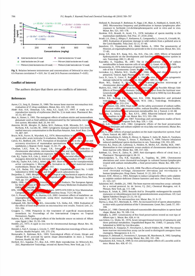
Delvocid signicantly decreased the testosterone concentra-tions in the serum at 400 and 800 mg/kg concentrations for allthe treatment periods ( Table 6). Levels of testosterone decreasedwith the increasing concentrations at these periods ( Fig. 5). Thisdecrease was concentration-dependent.
4. Discussion
Delvocid has been widely used in food products like cheese,sausage and salami ( EMEA, 1998). The widespread use of the food T a
b l e 1
F r e q u e n c y o f c h r o m o s o m e a b e r r a t i o n s a n d m i t o t i c i n d
e x i n b o n e m a r r o w c e l l s o f f e m a l e m i c e i n d u c e d b y D e l v o c i d
.
T r e a t m e n t p e r i o d s
a n d s e x
C o n c e n t r a t i o n s
T o t a l c e l l
n u m b e r
N o r m a l c e l l
n u m b e r
A b n o r m a l c e l l
n u m b e r
T o t a l a b n o r m a l i t y
( g a p )
C h r o m a t i d
g a p
I s o c h r o m a t i d
g a p
C h r o m
a t i d
b r e a k
I s o c h r o m a t i d
b r e a k
C o n t r a c t i o n
C e n t r o m e r i c
a t t e n u a t i o n
M i t o t i c
i n d e x , %
6 h
- f e m a l e
( ) C o n t r o l
1 0 0
1 0 0
0
0
0
0
0
0
0
0
3 . 1
7
( + ) C o n t r o l
1 0 0
8 5
1 5
1 2 * * *
3
0
7
0
0
5
2 . 1
0 * *
2 0 0 m g / k g
1 0 0
9 7
3
2
0
0
1
0
0
1
2 . 2
6 *
4 0 0 m g / k g
1 0 0
9 7
3
2
1
0
1
0
1
0
2 . 0 7 *
*
8 0 0 m g / k g
1 0 0
9 6
4
4
2
0
1
0
0
3
1 . 9
6 * *
1 2 h
- f e m a l e
( ) C o n t r o l
1 0 0
9 8
2
1
1
0
1
0
0
0
3 . 6
3
( + ) C o n t r o l
1 0 0
8 7
1 3
1 2 *
5
0
1 0
0
0
2
2 . 3
0 * *
2 0 0 m g / k g
1 0 0
9 8
2
1
1
0
1
0
0
0
2 . 6
6 *
4 0 0 m g / k g
1 0 0
9 5
5
2
2
0
1
0
0
1
2 . 3
7 * *
8 0 0 m g / k g
1 0 0
9 7
3
3
1
1
1
0
1
1
2 . 1
7 * * *
2 4 h
- f e m a l e
( ) C o n t r o l
1 0 0
9 9
1
1
0
0
1
0
0
0
3 . 9
3
( + ) C o n t r o l
1 0 0
8 3
1 7
9 *
1 1
0
9
0
0
0
2 . 6
7 * *
2 0 0 m g / k g
1 0 0
9 8
2
1
1
0
0
0
0
1
3 . 0 0 *
4 0 0 m g / k g
1 0 0
9 6
4
1
3
0
0
0
0
1
2 . 3
7 * * *
8 0 0 m g / k g
1 0 0
9 7
3
2
1
1
2
0
0
0
1 . 9
3 * * *
*
P 6
0 . 0 5
.
* *
P
6
0 . 0 1 .
* * *
P 6
0 . 0 0 1
.
P.G. Rasgele, F. Kaymak / Food and Chemical Toxicology 48 (2010) 789–797 791
R E T R
A C T E D

8/13/2019 jurnal abnormalitas
http://slidepdf.com/reader/full/jurnal-abnormalitas 4/9
preservatives cause serious problems of health ( Sarıkaya and Solak,2003). Therefore, it is very important to assess genotoxicity andcytotoxicity of these chemicals. Chromosome aberrations, micro-nucleus and sperm head abnormality assays are often used forevaluating genotoxic agents ( Ieradi et al., 2003 ). There is no reportabout the effects of Delvocid and natamycin on hereditary materialand sperm.
In this study, Delvocid was not clastogenic at all concentrationsand treatment periods. Also, according to WHO (2006), Delvocidwas not mutagenic in S. typhimurium , E. coli and B. subtilis . Coxet al. (1973) indicated that neither in male nor in female miceNatamycin was clastogenic ( WHO, 2006). In the studies of EMEA(1998) , it was reported that natamycin was not mutagenic and in-duced low acute toxicity in rabbits and dogs. These statements areconsistent with the results of the present study. Furthermore, somepreservatives in food products also revealed negative results ingenotoxicity tests. Boric acid did not induce CA and SCE in CHO(National Toxicology Program, 1987 ), sodium nitrite did not in-crease CA in mice, rats and rabbits ( Luca et al., 1987), sorbic acidwas not clastogenic and mutagenic in CA, SCE and Ames assays(Walker, 1990 ). Sodium and potassium metabisulphite were notteratogenic and mutagenic in mice, rats, hamsters and rabbits ( Nairand Elmore, 2003 ).
MI is used to indicate cytotoxicity of chemicals. A decreased MIreects the inhibition of cell cycle and effects the cell division neg-atively (Amorim et al., 2000 ). In the present study, Delvocid signif-icantly decreased MI compared to control and caused a toxic effect.In the present test conditions, negative correlation was found be-tween MI and concentrations and between MI and CA frequency.But the increase in the frequency of CAs was not signicant. Thisnding suggests that toxicity increased with the increasing of concentrations.
MN assay is the most widely used short-term in vivo assay foridentication of genotoxic effects ( Heddle, 1973 ). Micronuclei areconsisted of chromosome fragments or whole chromosomes whichlag behind at anaphase of mitosis and are not incorporated intodaughter nuclei. They form single or multiple micronuclei in thecytoplasm. The assay is based on the increase in frequency of micronucleated PCEs in bone marrow of the treated animals(EPA, 1996). We selected two types of erythrocytes (PCEs andNCEs) to evaluate the incidence of MN ( Vijayalaxmi and Venu,1999 ). They were easy to determine due to staining characteristicsand the extent of nuclear damage during the erythropoiesis ( Rab-bani et al., 2005 ).
In the present study, tested concentrations of Delvocid did notsignicantly induce CAs (without gaps), but the same concentra-tions of Delvocid signicantly induced MN in mice bone marrow.MN may be formed from clastogenic and aneugenic effects. But,in CA assay, a clastogenic effect was not observed. So, formationof MN suggests that Delvocid might be aneugenic. In the studies
done using the same food preservatives, different results have beenreported by many authors. Renner and Wever (1983) indicatedthat sodium metabisulphite did not induce CA, SCE and MN in micebone marrow while in the study of Rencüzogullar ı et al. (2001) , it isstated that sodium metabisulphite induced CA and SCE and de-creased MI and RI at all concentrations in human lymphocytes.According to the Kayraldız and Topakta s (2007), intraperitonealadministration of sodium metabisulphite was more effective thangavage administration. The same chemical substance can revealdifferent results. Differences in dose levels, sampling times andway of treatment may be related to these differences in response.According to results of the present study, CA frequency increasedwith the increasing concentrations. So, higher concentrations of Delvocid has a probable genotoxic effect in mice bone marrow.
According to many investigators ( Cole et al., 1979; Salamoneand Heddle, 1983 ), PCEs remain alive within the bone marrow in T a
b l e 2
F r e q u e n c y o f c h r o m o s o m e a b e r r a t i o n s a n d m i t o t i c i n d e x i n b o n e m a r r o w c e l l s o f m a l e m i c e i n d u c e d b y
D e l v o c i d
.
T r e a t m e n t p e r i o d s
a n d s e x
C o n c e n t r a t i o n s
T o t a l c e l l
n u m b e r
N o r m a l c e l l
n u m b e r
A b n o r m a l c e l l
n u m b e r
T o t a l a b n o r m a l i t y
( g a p )
C h r o m a t i d
g a p
I s o c h r o m a t i d
g a p
C h r o m
a t i d
b r e a k
I s o c h r o m a t i d
b r e a k
C o n t r a c t i o n
C e n t r o m e r i c
a t t e n u a t i o n
M i t o t i c
i n d e x %
6 h
- m a l e
( ) C o n t r o l
1 0 0
1 0 0
0
0
0
0
0
0
0
0
3 . 9
7
( + ) C o n t r o l
1 0 0
8 7
1 3
1 3 *
* *
4
0
7
0
0
6
2 . 4 0 *
* *
2 0 0 m g / k g
1 0 0
9 8
2
1
1
1
1
0
0
0
2 . 7 3 *
*
4 0 0 m g / k g
1 0 0
9 8
2
1
1
0
1
0
0
0
2 . 5 0 *
*
8 0 0 m g / k g
1 0 0
9 8
2
2
0
0
2
0
0
0
2 . 2 6 *
* *
1 2 h
- m a l e
( ) C o n t r o l
1 0 0
9 9
1
0
1
0
0
0
0
0
4 . 5
3
( + ) C o n t r o l
1 0 0
8 1
1 9
1 9 *
* *
5
0
9
1
4
5
2 . 7 3 *
* *
2 0 0 m g / k g
1 0 0
9 6
5
2
5
1
2
0
0
0
3 . 2 0 *
*
4 0 0 m g / k g
1 0 0
9 7
3
3
0
0
3
0
0
0
2 . 5 3 *
* *
8 0 0 m g / k g
1 0 0
9 4
6
5
3
0
0
0
1
4
2 . 1 3 *
* *
2 4 h
- m a l e
( ) C o n t r o l
1 0 0
9 8
2
2
0
0
1
0
1
0
4 . 3
7
( + ) C o n t r o l
1 0 0
8 0
2 0
2 1 *
* *
6
0
1 8
1
0
2
2 . 5 3 *
* *
2 0 0 m g / k g
1 0 0
9 6
4
0
5
0
0
0
0
0
3 . 0 3 *
*
4 0 0 m g / k g
1 0 0
9 7
3
3
3
1
1
0
0
2
2 . 9 6 *
*
8 0 0 m g / k g
1 0 0
8 6
1 5
7
1 1
1
1
0
2
4
1 . 8 7 *
* *
* P 6 0
. 0 5
.
* *
P
6 0
. 0 1
.
* * *
P 6
0 . 0
0 1 .
792 P.G. Rasgele, F. Kaymak / Food and Chemical Toxicology 48 (2010) 789–797
R E T R
A C T E D

8/13/2019 jurnal abnormalitas
http://slidepdf.com/reader/full/jurnal-abnormalitas 5/9
between 10 and 33 h and the number of MNPCEs is increased at 6 h
for aneugens and 10 h for clastogens ( Cole et al., 1981; Vanderker-ken et al., 1989 ), so, spindle poisons and clastogenic chemicalscould be detected in bone marrow 24 and 48 h after the treatment(Vanparys et al., 1992 ). Therefore, two treatment periods (24 and48 h) are sufcient to detect clastogens and aneugens.
In the present study, we have demonstrated that Delvocid in-creased the number of micronucleated PCEs (for 24 and 48 h; in fe-male mice at all concentrations, in male mice at 400 and 800 mg/kgconcentrations) and decreased the PCE/NCE ratio in bone marrowof mice. It has been shown by many investigators that food preser-vatives are genotoxic in a variety of test systems. Citric acid(Y ılmaz et al., 2008 ) and biphenyl ( Rencüzogullar ı et al., 2008) in-creased frequency of MN in human lymphocytes. Sodium benzoate,boric acid, citric acid, potassium citrate and sodium citrate caused
formation of MN in Allium cepa (Türkoglu, 2007). Our nding is inaccord with the results of these studies.
The PCE/NCE ratio is used to obtain information about the cell
cycle specic action of a positive chemical. A decrease in PCE/NCE ratio reects a cytotoxic effect or alterations in erythropoiesis.The PCE/NCE ratio is decreased because of the cavity formation inbone marrow when there are cytotoxic effects on the cell divisionand/or maturation of the nucleated cells ( Gollapudi et al., 1984 ). Inaddition, newly maturated NCEs remain behind the bone marrowdue to the failure of release into the peripheral blood on schedule(Von Ledebur and Schmid, 1973 ).
Generally, aneugenic substances cause MN inhibiting spindleformation. Abnormalities due to inhibition of spindle formation re-ect high toxicity of chemicals ( Haliem, 1990 ). Both MI and PCE/NCE ratio are used to monitor toxicity in CA assay and MN assay,respectively. In the present study, the tested concentrations of Delvocid did not signicantly induce CAs without gaps but the
same concentrations signicantly induced MN. The PCE/NCE ratioin bone marrow of mice decreased with the increase of cells with
Table 3
Micronucleus induction and numbers of PCEs and NCEs in Delvocid-exposed female mice bone marrow.
Treatmentperiods
Concentrations Total cell number/mice number
TotalMNPCE% ± SE
1 MNPCE ± SE 2MNPCE ± SE
3MNPCE ± SE
1MNNCE ± SE
2MNNCE ± SE
PCE/NCE ± SE
24 h-female ( ) Control 5000/5 6.20 ± 2.20 5.40 ± 2.04 0.20 ± 0.20 0 1.40 ± 0.51 0.20 ± 0.20 1.67 ± 0.20(+) Control 5000/5 56.40 ± 6.65
***
51.60 ± 4.91***
4.00 ± 1.67*
0.80 ± 0.49 9.20 ± 0.80**
0.40 ± 0.40 0.98 ± 0.12*
200 mg/kg 5000/5 21.20 ± 3.72*
21.20 ± 3.72**
1.20 ± 0.80 0 3.60 ± 1.16 0.40 ± 0.40 1.10 ± 0.14400 mg/kg 5000/5 28.80 ± 2.06
***
24.40 ± 1.72***
4.00 ± 0.89*
0.40 ± 0.40 4.80 ± 1.02*
0.40 ± 0.40 1.05 ± 0.28
800 mg/kg 5000/5 30.40 ± 0.98***
25.60 ± 1.32***
4.80 ± 0.49**
0 7.20 ± 1.02**
0.80 ± 0.49 0.88 ± 0.14*
48 h-female ( ) Control 5000/5 5.20 ± 1.02 4.80 ± 0.80 0.40 ± 0.40 0 2.40 ± 0.40 0 1.90 ± 0.19(+) Control 5000/5 51.20 ± 2.58
***
45.60 ± 1.47***
5.60 ± 1.47***
0 6.40 ± 2.04 1.20 ± 0.49*
0.90 ± 0.08**
200 mg/kg 5000/5 12.40 ± 1.72*
11.60 ± 1.72*
0.40 ± 0.40 0.40 ± 0.40 4.00 ± 1.09 0.80 ± 0.49 1.01 ± 0.29**
400 mg/kg 5000/5 16.00 ± 1.79**
14.80 ± 2.49*
0.80 ± 0.49 0.40 ± 0.40 5.20 ± 1.02*
0.40 ± 0.40 0.84 ± 0.09***
800 mg/kg 5000/5 26.80 ± 3.32***
25.60 ± 3.18***
1.20 ± 0.80 0 6.40 ± 1.72*
0.80 ± 0.80 0.99 ± 0.10**
72 h-female ( ) Control 5000/5 9.20 ± 0.80 8.80 ± 1.02 0.40 ± 0.40 0 4.00 ± 0.63 0 1.51 ± 0.18(+) Control 5000/5 33.60 ± 7.55
***
28.40 ± 6.21***
5.20 ± 1.49*
0 7.60 ± 2.13 0.80 ± 0.80 1.06 ± 0.20200 mg/kg 5000/5 12.80 ± 2.15 10.00 ± 1.78 2.80 ± 0.80 0 5.20 ± 1.62 0.80 ± 0.49 1.39 ± 0.13400 mg/kg 5000/5 10.80 ± 2.42 7.60 ± 1.60 3.20 ± 1.02 0 5.20 ± 0.80 0.80 ± 0.49 1.31 ± 0.09800 mg/kg 5000/5 23.20 ± 3.32
*
16.00 ± 1.67*
6.00 ± 1.41**
1.20 ± 0.80 5.60 ± 1.93 1.20 ± 0.49*
1.01 ± 0.10*
MNPCE: micronucleated polychromatic erythrocyte, MNNCE: micronucleated normochromatic erythrocyte, SE: standard error.*
P 6 0.05.**
P 6 0.01.***
P 6 0.001.
Table 4
Micronucleus induction and numbers of PCEs and NCEs in Delvocid-exposed male mice bone marrow.
Treatmentperiods
Concentrations Total cell number/mice number
TotalMNPCE% ± SE
1 MNPCE ± SE 2MNPCE ± SE
3MNPCE ± SE
1MNNCE ± SE
2MNNCE ± SE
PCE/NCE ± SE
24 h-male ( ) Control 5000/5 19.60 ± 0.75 17.60 ± 0.74 2.00 ± 0.63 0 8.00 ± 1.41 3.60 ± 0.74 1.48 ± 0.03(+) Control 5000/5 37.20 ± 3.83
***
32.80 ± 4.03***
3.60 ± 0.74 0.80 ± 0.49 9.20 ± 0.80 4.00 ± 4.00 0.91 ± 0.02***
200 mg/kg 5000/5 23.60 ± 3.54 21.20 ± 2.93 2.00 ± 0.63 0.40 ± 0.40 5.20 ± 1.35 0.40 ± 0.40 1.26 ± 0.09*
400 mg/kg 5000/5 34.00 ± 2.28**
29.20 ± 1.74*
4.80 ± 1.02 0 7.20 ± 0.49 4.40 ± 1.72 0.96 ± 0.05***
800 mg/kg 5000/5 30.00 ± 1.79*
24.40 ± 1.47*
5.60 ± 0.74*
0 7.60 ± 0.74 5.60 ± 0.74 0.86 ± 0.05***
48 h-male ( ) Control 5000/5 16.00 ± 2.28 14.80 ± 1.62 1.20 ± 0.80 0 5.20 ± 0.49 2.00 ± 0.89 1.70 ± 0.11(+) Control 5000/5 37.60 ± 2.71
***
34.00 ± 1.78***
3.60 ± 1.16 0 8.80 ± 1.02*
1.60 ± 0.74 0.81 ± 0.03***
200 mg/kg 5000/5 19.20 ± 2.58 16.80 ± 1.85 2.40 ± 0.98 0 9.20 ± 1.35*
0 1.00 ± 0.05***
400 mg/kg 5000/5 35.20 ± 4.32***
30.40 ± 3.18***
4.00 ± 0.63 0.80 ± 0.80 13.60 ± 1.47**
2.00 ± 1.26 0.97 ± 0.03***
800 mg/kg 5000/5 34.40 ± 1.94***
28.80 ± 1.85***
5.60 ± 0.74**
0 17.20 ± 1.35**
0.80 ± 0.80 0.81 ± 0.03***
72 h-male ( ) Control 5000/5 15.20 ± 1.36 13.60 ± 1.16 1.60 ± 0.74 0 5.60 ± 1.16 2.00 ± 0.63 1.52 ± 0.05(+) Control 5000/5 36.80 ± 2.87
***
30.80 ± 2.57***
4.80 ± 1.02*
1.20 ± 0.49*
10.40 ± 0.74*
2.00 ± 0.00 0.82 ± 0.08***
200 mg/kg 5000/5 19.20 ± 1.96 15.60 ± 1.32 3.60 ± 0.74 0 6.00 ± 0.63 1.60 ± 0.40 1.35 ± 0.10400 mg/kg 5000/5 16.80 ± 0.80 13.20 ± 1.49 3.20 ± 0.80 0.40 ± 0.40 6.40 ± 0.74 2.40 ± 0.74 1.23 ± 0.02800 mg/kg 5000/5 19.20 ± 1.96 16.40 ± 1.16 2.80 ± 1.02 0 8.40 ± 1.16 2.00 ± 0.00 1.26 ± 0.11
MNPCE: micronucleated polychromatic erythrocyte, MNNCE: micronucleated normochromatic erythrocyte, SE: standard error.*
P 6 0.05.**
P 6 0.01.***
P 6 0.001.
P.G. Rasgele, F. Kaymak / Food and Chemical Toxicology 48 (2010) 789–797 793
R E T R
A C T E D

8/13/2019 jurnal abnormalitas
http://slidepdf.com/reader/full/jurnal-abnormalitas 6/9
MN. Positive correlation was found between MI and PCE/NCE ratioin both male and female mice for 24 h treatment periods. Since itdecreased the MI and the PCE/NCE ratio in bone marrow of mice,Delvocid can be accepted as a toxic agent.
In sperm head abnormality assay, sperm morphology is used toinvestigate carcinogenic and mutagenic chemicals ( Ieradi et al.,2003 ). This assay is a sensitive and reliable method ( Giri et al.,2002 ). In this study, Delvocid signicantly increased the percent-age of abnormal sperm at all concentrations. The dose dependentincrease in the frequency of sperm head abnormalities suggeststhat Delvocid caused differentiation in male germ cells and it is agerm cell mutagen. Abdel Aziz et al. (1997) have reported thatEritrosin induced an increase in the abnormal sperm; Heindelet al. (1997) have found that boric acid affected reproduction and
fertility in rodents negatively; and in study of Oishi (2002) propylparaben affected the hormonal secretion and the male reproduc-
tion functions in rats. Our results are in agreement with thesendings.
The exact reason of the increase in the frequency of abnormalsperm is not known but there are different opinions. This increasemay be related to decrease of the fertility. Induction of abnormalsperms is assumed to be a result of naturally occurring errors dur-ing the differentiation or the consequence of an abnormal chromo-some ( Bruce et al., 1974). In addition, Y chromosomes have animportant role in determining frequency of sperm abnormalities(Krzanowska, 1976; Styrna et al., 1991 ).
According to Topham (1980) , the properties controlling spermmorphology exist on the autosomes and those agents are identiedby sperm abnormality assay and they cause minor alterations intesticular DNA (Giri et al., 2002). In the study of Chauhan et al.(2000) , it has been reported that the exogenous factors inducealterations in sperm morphology by point mutations ( Narayanaet al., 2002). Bone marrow and spermatogenic tissues are rich interms of investigation of mitotic cells, and both bone marrowand sperm abnormality assays provide reliable results ( Wanget al., 1998). According to many investigators ( Hemavathi andRahiman, 1993; Jayashree et al., 1994; Chauhan et al., 2000 ), sev-eral chemicals which induced cytotoxic effects and formation of MN in bone marrow cells caused abnormal sperms as well ( Giriet al., 2002). In the present study, Delvocid did not increase CA fre-
quency but it increased formation of MN. A signicant relation wasfound between MI which is an indicator of cytotoxicity in CA assayand percentage of abnormal sperms at all treatment periods. Thesame relation was also observed between PCE/NCE ratio and per-centage of abnormal sperm for 24 h. The increased frequency of abnormal sperm may be from toxic effects due to aneugeni. As aresult, Delvocid is considered as toxic and germ cell mutagen be-cause of its effect on hereditary material and sperm, respectively.
Desjardins (1985) and Herbert et al. (1995) have reported thattestosterone controls the development of male reproductive sys-tem and spermatogenesis ( Oishi, 2002). The epididymis is animportant organ for sperm maturation. The functions of epididy-mis and sperm may be changed by lowering hormone levels.According to Ono et al. (1999), direct sperm toxicity may exist
within the epididymis. In the sperm maturation process, sperm ac-quires structural stabilization of the head and tail ( Calvin and Bed-
Table 6
Effects of Delvocid on the serum testosterone hormone levels in male mice.
Treatment periods Concentrations Total testosterone (±SE)
6 h ( ) Control 56.00 ± 6.55(+) Control 22.33 ± 1.45
***
200 mg/kg 46.66 ± 0.88400 mg/kg 2.33 ± 1.45
***
800 mg/kg 20.00 ± 0.00***
12 h ( ) Control 43.70 ± 4.07(+) Control 20.00 ± 0.00
***
200 mg/kg 40.33 ± 0.88400 mg/kg 30.66 ± 0.88
*
800 mg/kg 25.93 ± 3.72**
24 h ( ) Control 51.00 ± 2.30(+) Control 20.00 ± 0.00
***
200 mg/kg 45.66 ± 2.40400 mg/kg 38.00 ± 1.52
*
800 mg/kg 35.00 ± 5.85**
SE: standard error.*
P 6 0.05.**
P 6 0.01.***
P 6 0.001.
Table 5
Number and mean percentage of sperm head abnormalities in control and Delvocid treated mice.
Treatmentperiods
Concentrations Number of sperms examined/number of animals
Bananashaped
Withouthook
Amorphoushead
Bent atcephalocaudal junction
Total spermabnormality
% Abnormal sperm(mean ± SE)
6 h ( ) Control 3000/3 64 36 33 33 167 5.56 ± 0.20(+) Control 3000/3 362
***
241***
119***
53 784***
26.13 ± 0.68***
200 mg/kg 3000/3 168*
137**
61***
80*
447***
14.9 ± 0.87***
400 mg/kg 3000/3 227 ** 153***
67***
99**
552***
18.4 ± 0.11***
800 mg/kg 3000/3 288***
170***
68***
177***
714***
23.8 ± 1.53***
12 h ( ) Control 3000/3 66 45 31 35 178 5.93 ± 0.18(+) Control 3000/3 413
***
250***
117***
56*
844***
28.13 ± 0.03***
200 mg/kg 3000/3 204***
101 37 89***
432***
14.4 ± 1.06***
400 mg/kg 3000/3 247***
152**
48 117***
576***
19.2 ± 0.77***
800 mg/kg 3000/3 287***
140**
92***
89***
616***
20.53 ± 1.04***
24 h ( ) Control 3000/3 61 47 37 40 187 6.23 ± 0.32(+) Control 3000/3 406
***
276***
112***
76**
882***
29.4 ± 1.05***
200 mg/kg 3000/3 189***
117**
56 70*
441***
14.7 ± 1.04***
400 mg/kg 3000/3 253***
148***
84***
138***
633***
21.1 ± 0.65***
800 mg/kg 3000/3 294***
165***
98***
98***
667***
22.23 ± 2.10***
SE: standard error.*
P 6 0.05.**
P 6 0.01.***
P 6 0.001.
794 P.G. Rasgele, F. Kaymak / Food and Chemical Toxicology 48 (2010) 789–797
R E T R
A C T E D

8/13/2019 jurnal abnormalitas
http://slidepdf.com/reader/full/jurnal-abnormalitas 7/9
ford, 1971 ), its activity (Hoskins et al., 1978 ) and fertilizing capac-ity (Yanagimachi, 1988; Miller et al., 1996 ). In the present study, asignicantly increased percentage of abnormal sperm occured inDelvocid treated mice and Delvocid decreased testosterone levels.Consequently, in male reproductive system, the development andfunctioning disorder emerged. Decreases in the levels of testoster-one have been reported after the treatment of rats with other food
preservatives ( Oishi, 2002; Jeong et al., 2005 ).
In summary, the obtained results indicate that Delvocid is notclastogenic in the CA assay but an aneugenic chemical in the MNassay. In addition, Delvocid has cytotoxic effects in mice becauseit reduced MI and PCE/NCE ratio. Delvocid may be regarded as apotential germ cell mutagen due to its effect on sperm. For this rea-son, it is necessary to be careful when using these chemicals infood as preservatives, and they should not be used in excessive
amounts in food industry.
Fig. 4. (a) Normal sperm; (b) banana shaped (200 mg/kg,12 h); (c) without hook (800 mg/kg, 12 h); (d) amorphous head sperm (400 mg/kg, 24 h); (e) bent at cephalocaudal junction (400 mg/kg, 6 h) ( 1000).
P.G. Rasgele, F. Kaymak / Food and Chemical Toxicology 48 (2010) 789–797 795
R E T R
A C T E D
8/13/2019 jurnal abnormalitas
http://slidepdf.com/reader/full/jurnal-abnormalitas 8/9
Conict of Interest
The authors declare that there are no conicts of interest.
References
Aaron, C.S., Sorg, R., Zimmer, D., 1989. The mouse bone marrow micronucleus test:evaluation of 21 drug candidates. Mutat. Res. 223, 129–140.
Abdel Aziz, A.H., Shouman, S.A., Attia, A.S., Saad, S.F., 1997. A study on thereproductive toxicity of erythrosine (Red No. 3) in male mice. Pharmacol. Res.35 (5), 457–462.
Akın, A., Sümer, S., 1991. The mutagenic effects of sodium nitrite and monosodiumglutamate used as food additives demonstrated by the Salmonella microsometest system. Microbiol. Bull. 25, 94–107.
Amorim, M.I.M., Mergler, D., Bahia, M.O., Dubeau, H.M., Miranda, D.C., Level, J.,Burbano, R.R., Lucotte, M., 2000. Cytogenetic damage related to low levels of methyl mercury contamination in the Brazilian Amazon. Ann. Acad. Bras. Cienc.72, 487–507.
Bruce, W.R., Furrer, R., Wyrobek, A.J., 1974. Abnormalities in the shape of murinesperm after acute testicular X-irradiation. Mutat. Res. 23, 381–386.
Calvin, H.I., Bedford, J.M., 1971. Formation of disulphide bonds in the nucleus andaccessory structures of mammalian spermatozoa during maturation in theepididymis. J. Reprod. Fertil. Suppl. 13, 65–75.
Chauhan, L.K.S., Pant, N., Gupta, S.K., Srivastava, S.P., 2000. Induction of chromosome aberrations, micronucleus formation and sperm abnormalities inmouse following carbofuran exposure. Mutat. Res. 465, 123–129.
Cole, R.J., Taylor, N.A., Cole, J., Arlett, C.F., 1979. Transplacental effects of chemicalmutagens detected by the micronucleus test. Nature (London) 277, 317–318.
Cole, R.J., Taylor, N.A., Cole, J., Arlett, C.F., 1981. Short terms tests for transplacentallyactive carcinogens. I. Micronucleus formation in fetal and maternal mouseerythroblasts. Mutat. Res. 80, 141–157.
Cox, G.E., Bailey, D.E., Morgareidge, K., 1973. Unpublished Report No. 1-1052Submitted to WHO by Food and Drug Research Laboratories Inc.
Desjardins, C., 1985. Morphological, physiological and biochemical aspects of malereproduction. In: Dixon, R.L. (Ed.), Reproductive Toxicology. Raven Press, NewYork, pp. 131–146.
EMEA, 1998. Committee for Veterinary Medicinal Products. The European Agencyfor the Evaluation of Medicinal Products Veterinary Medicines Evaluation Unit,EMEA/MRL/342/98-FINAL.
EPA, 1996. Health Effects Test Guidelines. OPPTS 870.5395 In Vivo Mammalian
Cytogenetic Tests: Erythrocyte Micronucleus Assay. 712-C-96-226.Giri, S., Prasad, S.B., Giri, A., Sharma, G.D., 2002. Genotoxic effects of malathion: anorganophosphorus insecticide, using three mammalian bioassays in vivo.Mutat. Res. 514, 223–231.
Gollapudi, B.B., McClintock, M.L., Linscombe, V.A., Sinha, A.K., 1984. Evaluation of the effect of food deprivation on micronucleus test results. Toxicol. Lett. 21,353–356.
Grupper, C., 1964. Pimaricin in the treatment of supercial mucocutaneousmonoliasis. In: Proceedings of the International Congress on TropicalDermatoses, Naples.
Halıem, A.S., 1990. Cytological effects of the herbicide sencor on mitosis of Alliumcepa . Egypt. J. Bot. 33, 93–104.
Heddle, J.A., 1973. A rapid in vivo test for chromosomal damage. Mutat. Res. 18,187–190.
Heindel, J., Fail, P., George, J., Grizzle, T., 1997. Reproduction toxicology of boric acid.Environ. Health Perspect. 105, 275–276.
Hemavathi, E., Rahiman, M.A., 1993. Toxicological effects of ziram, thiram anddithane M-45 assessed by sperm shape abnormalities in mice. J. Toxicol.Environ. Health 38, 393–398.
Herbert, D.C., Supakar, P.C., Roy, A.K., 1995. Male reproduction. In: Witorsch, R.L.(Ed.), Reproductive Toxicology, second ed. Raven Press, New York, pp. 3–21.
Holland, N., Duramad, P., Rothman, N., Figgs, L.W., Blair, A., Hubbard, A., Smith, M.T.,2002. Micronucleus frequency and proliferation in human lymphocytes afterexposure to herbicide 2,4-dichlorophenoxyacetic acid in vitro and in vivo.Mutat. Res. 521, 165–178.
Hoskins, D.D., Brandt, H., Acott, T.S., 1978. Initiation of sperm motility in themammalian epididymis. Fed. Proc. 37, 2534–2542.
Ieradi, L.A., Zima, J., Allegra, F., Kotlanova, E., Campanella, L., Grossi, R., Cristaldi, M.,2003. Evaluation of genotoxic damage in wild rodents from a polluted area inthe Czech Republic. Folia Zool. 52 (1), 57–66.
Jayashree, I.V., Vijayalaxmi, K.K., Abdul Rahim, A., 1994. The genotoxicity of Hinoson, an organophosphorous pesticide in the in vivo mouse. Mutat. Res. 322,77–85.
Jeong, S.H., Kim, B.Y., Kang, H.G., Ku, H.O., Cho, J.H., 2005. Effects of butylatedhydroxyanisole on the development and functions of reproductive system inrats. Toxicology 208 (1), 49–62.
Kayraldız, A., Topaktas, M., 2007. The in vivo genotoxic effects of sodiummetabisulte in bone marrow cells of rats. Russ. J. Genet. 43 (8), 905–909.
Krzanowska, H., 1976. Inheritance of sperm head abnormality types in mice and therole of the Y chromosome. Genet. Res. 28, 189–198.
Levinskas, G.J., Ribelin, W.E., Shaffer, C.B., 1966. Acute and chronic toxicity of pimaricin. Toxicol. Appl. Pharmacol. 8, 97–109.
Luca, D., Luca, V., Cotor, F., Raileanu, L., 1987. In vivo and in vitro cytogeneticdamage induced by sodium nitrite. Mutat. Res. 189 (3), 333–339.
Mace, M.L., Daskal, Y., Wray, W., 1978. Scanning electron microscopy of chromosome aberration. Mutat. Res. 52, 199–206.
Malten, K.E., 1967. Report of an Investigation Concerning Possible Allergic SideEffects of Pimaricin in Humans. Unpublished Report from the Instituut voorGeneeskunde en Maatschappij, Nijmegen. Submitted to WHO by Gist-BrocadesNV, Delft.
Miller, R.K., Kellogg, C.K., Saltzman, R.A., 1996. Reproductive and perinataltoxicology. In: Haley, T.J., Berndt, W.O. (Eds.), Toxicology. Hemisphere,Washington, pp. 212–214.
Nair, B., Elmore, A.R., 2003. Final report on the safety assessment of sodium sulte,potassium sulte, ammonium sulte, sodium bisulte, ammonium bisulte,sodium metabisulte and potassium metabisulte. Int. J. Toxicol. 2, 63–88.
Narayana, K., Souza, U.J.A., Rao, K.P.S., 2002. Ribavirin-induced sperm shapeabnormalities in Wistar rat. Mutat. Res. 513, 193–196.
National Toxicology Program, 1987. Toxicology and carcinogenesis studies of boricacid. Natl. Toxicol. Program Tech. Rep. 324, 1–126.
Njagi, G.D., Gopalan, H.N., 1982. Cytogenetic effects of the food preservativessodium benzoate and sodium sulphite on vicia faba root meristems. Mutat. Res.102 (3), 213–219.
Oishi, S., 2002. Effects of propyl paraben on the male reproductive system. FoodChem. Toxicol. 40, 1807–1813.
Ono, A., Kawashima, K., Sekita, K., Hirose, A., Ogawa, Y., Saito, M., Naito, K., Yasuhara,K., Kaneko, T., Furuya, T., Inoue, T., Kurokawa, Y., 1999. Toluene inhalationinduced epididymal sperm dysfunction in rats. Toxicology 139, 193–205.
Preston, R.J., Dean, J.B., Galloway, S., Holden, H., Mcfee, A.F., Shelby, M.D., 1987.Mammalian in vivo cytogenetic assays-analysis of chromosome aberrations inbone marrow cells. Mutat. Res. 189, 157–165.
Rabbani, S.I., Devi, K., Zahra, N., 2005. Anti-clastogenic effects of citral. Iran. J.Pharmacol. Therapeuticsijpt 4, 28–31.
Rencüzogulları, E., Üla, H.B., Kayraldız, A., Topaktas, M., 2001. Chromosomeaberrations and sister chromatid exchanges in cultured human lymphocytestreated with sodium metabisulte, a food preservative. Mutat. Res. 490, 107–112.
Rencüzogulları, E. , Parlak, S., _Ila, H.B., 2008. The effects of food protector biphenyl onsister chromatid exchange, chromosome aberrations and micronucleus inhuman lymphocytes. Drug Chem. Toxicol. 31 (2), 263–274.
Renner, H.W., Wever, J., 1983. Attempts to induce cytogenetic effects with sulphitein sulphite oxidase-decient Chinese hamsters and mice. Food Chem. Toxicol.21, 123–127.
Salamone, M.F., Heddle, J.A., 1983. The bone marrow micronucleus assay: rationalefor a revised protocol. In: de Serres, F.J. (Ed.), Chemical Mutagens, vol. 8.Plenum, New York, pp. 111–149.
Sarıkaya, R., Solak, K., 2003. Benzoik Asit’in Drosophila melanogaster ’de somatikmutasyon ve rekombinasyon testi ile genotoksisitesinin aras tırılması. GÜ, GaziEgitim Fakültesi Dergisi 23 (3), 19–32.
Schmid, W., 1975. The micronucleus test. Mutat. Res. 31, 9–15.Styrna, J., Imai, H.T., Moriwaki, K., 1991. An increased level of sperm abnormalities
in mice with a partial deletion of the Y chromosome. Genet. Res. Camb. 57, 195–199.
Topham, J.C., 1980. Chemically induced transmissible abnormalities in sperm headshape. Mutat. Res. 70, 109–114.
Türkoglu, S., 2007. Genotoxicity of ve food preservatives tested on root tips of Allium cepa L.. Mutat. Res. 626, 4–14.
van Eeken, C.J., Wubs, W., 1976. Acute intraperitoneal toxicity of natamycin andthree potential metabolites. Unpublished Report No. 15465, 11 January 1976,Submitted to WHO by Gist-Brocades.
Vanderkerken, K., Vanparys, P., Verschaeve, L., Kirsch-Volders, M., 1989. The mousebone marrow micronucleus assay can be used to distinguish aneugens fromclastogens. Mutagenesis 4, 6–11.
Vanparys, P., Deknudt, G., Vermeiren, F., Sysmans, M., Marsboom, R., 1992. Samplingtimes in micronucleus testing. Mutat. Res. 282, 191–196.
Vijayalaxmi, K.K., Venu, R., 1999. In-vivo anticlastogenic effects of L-ascorbic acid inmice. Mutat. Res. 438, 47–51.
y = 3E-05x 2 - 0.0677x + 56.854R2 = 0.9882
y = 3E-05x 2 - 0.0415x + 51.526R 2 = 0.9787
y = 2E-05x 2 - 0.0369x + 44.614R2 = 0.9502
0
10
20
30
40
50
60
0 200 400 600 800 1000Concentrations (mg/kg)
T o
t a l t e s
t o s
t e r o n e
( n g
/ m l )
total tes tos terone 6 saat total tes tosterone 12 saat
total tes tos ter one 24 s aat Polinom ( total tes tos ter one 6 s aat)
Polinom (total testosterone 24 saat) Polinom (total testosterone 12 saat)
Fig. 5. Relation between testosterone and concentrations of Delvocid in mice (for6 h Pearson correlation P = 0.01; for 12 and 24 h Pearson correlation P = 0.05).
796 P.G. Rasgele, F. Kaymak / Food and Chemical Toxicology 48 (2010) 789–797
R E T R
A C T E D

8/13/2019 jurnal abnormalitas
http://slidepdf.com/reader/full/jurnal-abnormalitas 9/9
Von Ledebur, M., Schmid, W., 1973. The micronucleus test, methodological aspects.Mutat. Res. 19, 109–117.
Walker, R., 1990. Toxicology of sorbic acid and sorbates. Food Addit. Contam. 7,671–676.
Wang, X.L., Zhen, W.J., Wu, H.Y., Feng, J., Tu, Z.H., 1998. Mutagenicity tests onepristeride in vitro and in vivo. Acta Pharmacol. Sin. 19 (6), 569–572.
WHO, 2006. WHO Food Additives Series: 48 Safety Evaluation of Certain FoodAdditives and Contaminants. <http://www.inchemorg/documents/ jecfaljecmono/v48je06.htm> .
Wyrobek, A.J., Bruce, W.R., 1975. Chemical induction of sperm abnormalities inmice. Proc. Natl. Acad. Sci. USA 72, 4425–4429.
Yanagimachi, R., 1988. Mammalian fertilization. In: Knobil, E., Neal, J., et al. (Eds.),The Physiology of Reproduction. Raven Press, New York, pp. 135–185.
Yılmaz, S., Ünal, F., Yüzbasıoglu, D., Aksoy, H., 2008. Clastogenic effects of foodadditive citric acid in human peripheral lymphocytes. Cytotechnology 56, 2.
Zar, J.H., 1999. Biostatistical Analysis, fourth ed. Prentice Hall International, NJ.
P.G. Rasgele, F. Kaymak / Food and Chemical Toxicology 48 (2010) 789–797 797
R E T R
A C T E D