journal of medical and bioengineering vol. 3, no. 3 ... · universiti kuala lumpur institute of...
TRANSCRIPT

Evaluation of Antioxidant Activity of Zingiber
Officinale (Ginger) on Formalin-Induced
Testicular Toxicity in Rats
T. I. Rasyidah1, S. Suhana
2, H. Nur-Hidayah
3, M. A. Kaswandi
4, and R. M. Noah
5
Universiti Kuala Lumpur Institute of Medical Science Technology,
A1-1, Jln TKS 1, Taman Kajang Sentral, 43000 Kajang, Selangor
Email: [email protected], {
1tehrasyidah,
3nurhidayah,
4kaswandi,
5drrahim}@mestech.unikl.edu.my
Abstract—This study was carried out to investigate the
possible antioxidant activity of Zingiber officinale (ginger)
ethanolic extract on formalin-induced testicular toxicity in
rats. Twenty male Wistar rats were randomly divided into
four groups: (1): control; (2): rats exposed with 10%
formalin; (3): rats exposed with 10% formalin and treated
with ethanolic ginger extract; (4): rats treated with ethanolic
ginger extract. Exposure of 10 % formalin was performed
through inhalation while ethanolic ginger extract was
administered orally. Determination of malondialdehyde
(MDA) and the activities of superoxide dismutase (SOD)
and catalase (CAT) were assessed upon harvested testicles.
As a result, 10% formalin exposure significantly increased
the concentration of MDA as compared to control.
Meanwhile, all groups showed significant increase in SOD
level as compared to control. There is no significant
difference of CAT activities in all experimental groups as
compared to control. However, rats exposed with formalin
and treated with ethanolic ginger extract significantly
increased the CAT activity as compared to the group of
formalin exposure only. In conclusion, 10% formalin
triggered oxidative stress in testicles with the evidence of the
significant increase of MDA concentration. Moreover,
ginger exhibit antioxidant properties which proven by the
increase of SOD and CAT activities.
Index Terms—Zingiber officinale, formalin, testicles,
oxidative stress, antioxidant
I. INTRODUCTION
Zingiber officinale (ginger) belongs to family
Zingiberaceae [1]. It is an important ingredient
traditionally used in Chinese, Ayurvedic and Tibb-Unani
herbal medicines to treat several diseases such as asthma,
stroke, and diabetes [2]. Over the centuries, the usage of
Zingiber officinale has been progressed and increased in
pharmaceutical demands. Recent study supported that
Zingiber officinale has the protective nutraceutical effect
against oxidative stress and also reproductive toxicity [3].
Among main active phytochemicals in Zingiber officinale
such as gingerols, gingerdiol, shogaols, zingerone, and
zingibrene are claimed to have antioxidant activity [4].
Some study showed that Zingiber officinale treatment
provided antioxidant effects by raising tissue
Manuscript received July 5, 2013; revised October 25, 2013.
concentrations of superoxide dismutase (SOD), catalase
(CAT) and glutathione peroxidase (GPx) [5]. These
antioxidants are important protection against oxidative
stress due to their ability to detoxify free radicals, such as
reactive oxygen species (ROS).
The imbalance of ROS production and detoxification
lead to oxidative stress in tissue. Oxidative stress is
directly proportionate to lipid peroxidation, DNA damage,
protein damage and induction of apoptosis which will
result in cell death [6]. Formaldehyde (FA) is identified
as one of the causative agent of oxidative stress.
Formaldehyde (CH2O) is a colourless, flammable,
reactive gas and readily polymerized at room temperature
with a pungent odour [7]. It is commercially available as
a solution called formalin and according to Occupational
Safety and Health Administration (OSHA) it is formed
from various proportions of formaldehyde, water, and
alcohol [8]. Formaldehyde has been routinely utilized in
medical work setting such as hospitals and laboratories.
Formaldehyde is an excellent tissue fixative and
commonly used for the preservation of tissues [9].
Therefore, exposure to FA occurs significantly among
pathologist, hospital housekeeping staff, and laboratory
workers [10]. Some epidemiological studies of industrial
workers, embalmers and pathology anatomists have
indicated association of FA exposure with elevated
cancer risks at various sites, including the brain, nasal
cavities, lung [11], pancreas [12], lymphohematopoietic
system [13], [14] and prostate [15]. Other than cancer
effects, recent study shows that long-term exposure of
formaldehyde can cause reproductive damage on male
rats by producing oxidative stress [16].
This paper describes the role of Zingiber officinale as
protective nutraceutical agent towards testicular toxicity
effect of formalin exposure by measuring the lipid
peroxidation activity, malondialdehyde (MDA) and the
levels of two antioxidant enzymes, superoxide dismutase
(SOD) and catalase (CAT) in rat testicles.
II. METHODOLOGY
A. Preparation of Plant Extract
One kilogram of fresh Zingiber officinale rhizomes were
procured from the local market in Kajang, Selangor and
Journal of Medical and Bioengineering Vol. 3, No. 3, September 2014
149©2014 Engineering and Technology Publishingdoi: 10.12720/jomb.3.3.149-153
were cleaned with tap water. The cleaned rhizomes were
peeled and cut into slices and let dried under the sunlight
for few days until constant weight was achieved.
Approximately 250g dried rhizomes were grounded and
extracted with ethanol through double boiling at 60˚C for
12 hours. The ethanol was removed by using rotary
evaporator at 40 °C. The extraction end-product is a pure
Zingiber officinale appears in dark orange colour. The
extract was kept at 4 ˚C until further uses. The extract
must first be solubilized with corn oil and 15 % of
dimethyl sulfoxide (DMSO) prior to oral administration
of the rats at 100mg/kg body weight dosage.
B. Experimental Animals
Twenty healthy male Wistar rats (weighing 150g -
200g), were housed in 38x23x10 cm transparent
polycarbonate wire-topped cages (2 rats per cage). They
were acclimatized at 12 hours light and 12 hours dark
cycle and fed with standard diet and tap water for one
week prior experiment procedure commence. Rats were
randomly divided into four groups: (1): control (received
treatment vehicle, corn oil and 15 % of dimethyl
sulfoxide); (2): rats exposed with 10% formalin (4
hrs/day, 5 days/wk, 8 wks); (3): rats exposed with 10%
formalin (4 hrs/day, 5 days/wk, 8 wks) and treated with
100 mg/kg body weight of ethanolic Zingiber officinale
extract (14 days); (4): rats treated with 100 mg/kg body
weight of ethanolic Zingiber officinale extract (14 days).
The duration of 8 weeks of formalin exposure was
performed through whole-body inhalation while ethanolic
Zingiber officinale extract was given orally.
C. The Exposure Procedure
The formalin exposure took place in a transparent
polycarbonate inhalation chamber with the dimensions
50x35x30 cm to generate a constant airstream from a
daily fixed amount of commercial aqueous solution of
formalin. The formalin (37% formaldehyde solution) was
given by means of a pipette into a flat dish which was
located on the top center of the chamber. The weight and
physical observation of rats was recorded in daily basis.
D. Biochemical Assays
After 24 hours post experiment, the rats were
sacrificed and the testicles were harvested, weighted,
divided equally and immediately frozen in liquid nitrogen
to stop the biochemical reaction in organ. The
biochemical assays were done on these tissue samples to
measure the activity of malondialdehyde (MDA) [17],
superoxide dismutase (SOD) [18], catalase (CAT) [19]
and protein estimation [20].
E. Statistical Analysis
All statistical analysis was carried out by using
Statistical Package for the Social Sciences (SPSS)
statistical software version 13.0. For biochemical analysis,
two-way ANOVA was used to compare means among
four groups. Post- Hoc Dunnett was used to make the
comparison between means. All the data were expressed
in mean ± standard error of the mean (SEM) and p value
less than 0.05 considered as significant.
III. RESULT
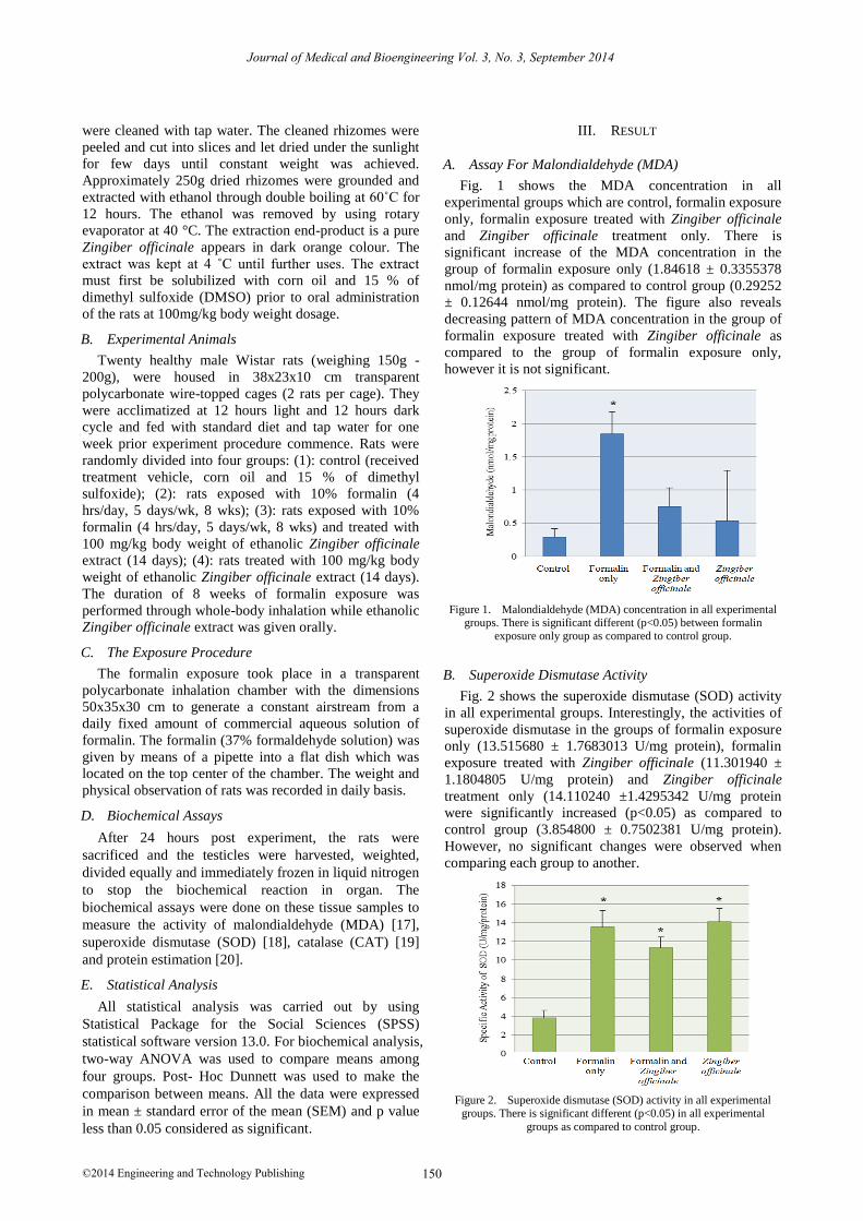
A. Assay For Malondialdehyde (MDA)
Fig. 1 shows the MDA concentration in all
experimental groups which are control, formalin exposure
only, formalin exposure treated with Zingiber officinale
and Zingiber officinale treatment only. There is
significant increase of the MDA concentration in the
group of formalin exposure only (1.84618 ± 0.3355378
nmol/mg protein) as compared to control group (0.29252
± 0.12644 nmol/mg protein). The figure also reveals
decreasing pattern of MDA concentration in the group of
formalin exposure treated with Zingiber officinale as
compared to the group of formalin exposure only,
however it is not significant.
Figure 1. Malondialdehyde (MDA) concentration in all experimental groups. There is significant different (p<0.05) between formalin
exposure only group as compared to control group.
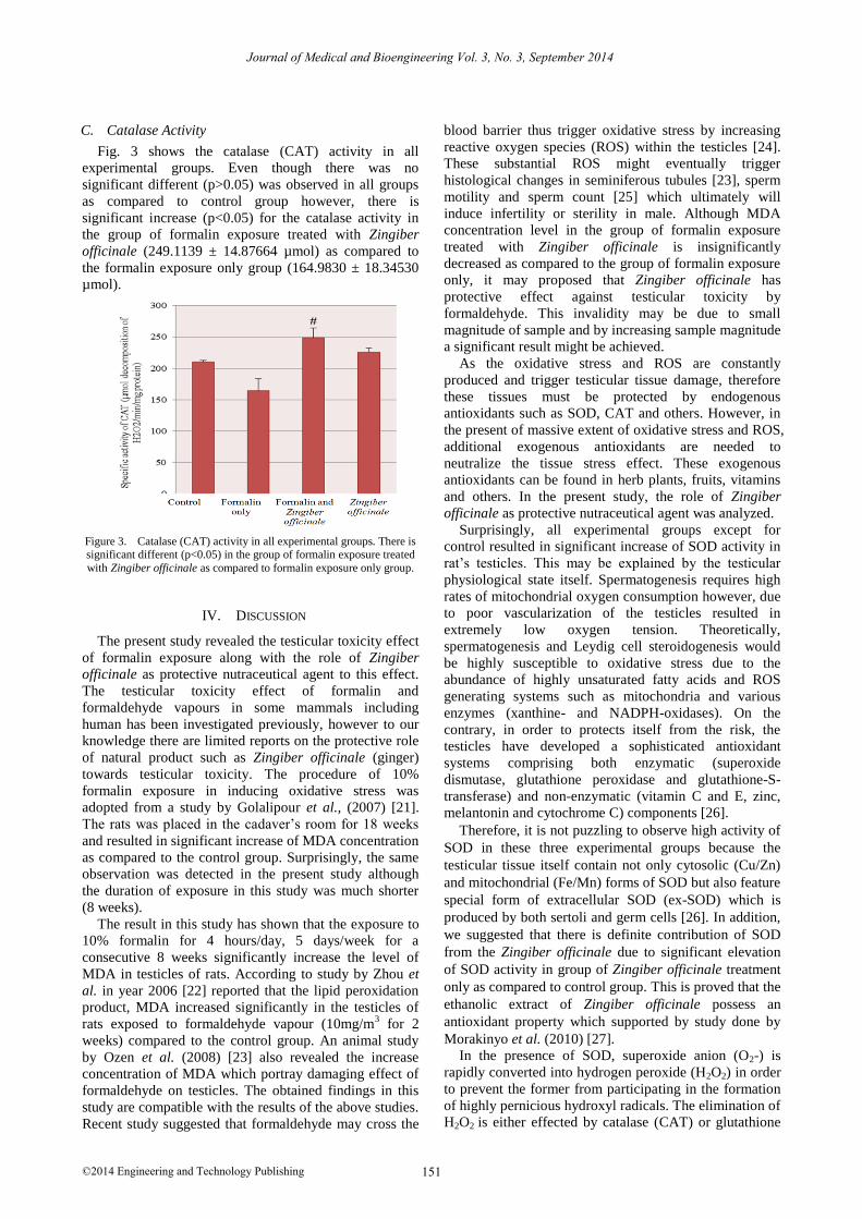
B. Superoxide Dismutase Activity
Fig. 2 shows the superoxide dismutase (SOD) activity
in all experimental groups. Interestingly, the activities of
superoxide dismutase in the groups of formalin exposure
only (13.515680 ± 1.7683013 U/mg protein), formalin
exposure treated with Zingiber officinale (11.301940 ±
1.1804805 U/mg protein) and Zingiber officinale
treatment only (14.110240 ±1.4295342 U/mg protein
were significantly increased (p<0.05) as compared to
control group (3.854800 ± 0.7502381 U/mg protein).
However, no significant changes were observed when
comparing each group to another.
Figure 2. Superoxide dismutase (SOD) activity in all experimental groups. There is significant different (p<0.05) in all experimental
groups as compared to control group.
Journal of Medical and Bioengineering Vol. 3, No. 3, September 2014
150©2014 Engineering and Technology Publishing
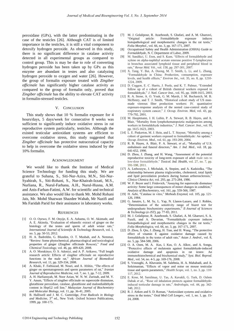
C. Catalase Activity
Fig. 3 shows the catalase (CAT) activity in all
experimental groups. Even though there was no
significant different (p>0.05) was observed in all groups
as compared to control group however, there is
significant increase (p<0.05) for the catalase activity in
the group of formalin exposure treated with Zingiber
officinale (249.1139 ± 14.87664 µmol) as compared to
the formalin exposure only group (164.9830 ± 18.34530
µmol).
Figure 3. Catalase (CAT) activity in all experimental groups. There is significant different (p<0.05) in the group of formalin exposure treated
with Zingiber officinale as compared to formalin exposure only group.
IV. DISCUSSION
The present study revealed the testicular toxicity effect
of formalin exposure along with the role of Zingiber
officinale as protective nutraceutical agent to this effect.
The testicular toxicity effect of formalin and
formaldehyde vapours in some mammals including
human has been investigated previously, however to our
knowledge there are limited reports on the protective role
of natural product such as Zingiber officinale (ginger)
towards testicular toxicity. The procedure of 10%
formalin exposure in inducing oxidative stress was
adopted from a study by Golalipour et al., (2007) [21].
The rats was placed in the cadaver’s room for 18 weeks
and resulted in significant increase of MDA concentration
as compared to the control group. Surprisingly, the same
observation was detected in the present study although
the duration of exposure in this study was much shorter
(8 weeks).
The result in this study has shown that the exposure to
10% formalin for 4 hours/day, 5 days/week for a
consecutive 8 weeks significantly increase the level of
MDA in testicles of rats. According to study by Zhou et
al. in year 2006 [22] reported that the lipid peroxidation
product, MDA increased significantly in the testicles of
rats exposed to formaldehyde vapour (10mg/m3 for 2
weeks) compared to the control group. An animal study
by Ozen et al. (2008) [23] also revealed the increase
concentration of MDA which portray damaging effect of
formaldehyde on testicles. The obtained findings in this
study are compatible with the results of the above studies.
Recent study suggested that formaldehyde may cross the
blood barrier thus trigger oxidative stress by increasing
reactive oxygen species (ROS) within the testicles [24].
These substantial ROS might eventually trigger
histological changes in seminiferous tubules [23], sperm
motility and sperm count [25] which ultimately will
induce infertility or sterility in male. Although MDA
concentration level in the group of formalin exposure
treated with Zingiber officinale is insignificantly
decreased as compared to the group of formalin exposure
only, it may proposed that Zingiber officinale has
protective effect against testicular toxicity by
formaldehyde. This invalidity may be due to small
magnitude of sample and by increasing sample magnitude
a significant result might be achieved.
As the oxidative stress and ROS are constantly
produced and trigger testicular tissue damage, therefore
these tissues must be protected by endogenous
antioxidants such as SOD, CAT and others. However, in
the present of massive extent of oxidative stress and ROS,
additional exogenous antioxidants are needed to
neutralize the tissue stress effect. These exogenous
antioxidants can be found in herb plants, fruits, vitamins
and others. In the present study, the role of Zingiber
officinale as protective nutraceutical agent was analyzed.
Surprisingly, all experimental groups except for
control resulted in significant increase of SOD activity in
rat’s testicles. This may be explained by the testicular
physiological state itself. Spermatogenesis requires high
rates of mitochondrial oxygen consumption however, due
to poor vascularization of the testicles resulted in
extremely low oxygen tension. Theoretically,
spermatogenesis and Leydig cell steroidogenesis would
be highly susceptible to oxidative stress due to the
abundance of highly unsaturated fatty acids and ROS
generating systems such as mitochondria and various
enzymes (xanthine- and NADPH-oxidases). On the
contrary, in order to protects itself from the risk, the
testicles have developed a sophisticated antioxidant
systems comprising both enzymatic (superoxide
dismutase, glutathione peroxidase and glutathione-S-
transferase) and non-enzymatic (vitamin C and E, zinc,
melantonin and cytochrome C) components [26].
Therefore, it is not puzzling to observe high activity of
SOD in these three experimental groups because the
testicular tissue itself contain not only cytosolic (Cu/Zn)
and mitochondrial (Fe/Mn) forms of SOD but also feature
special form of extracellular SOD (ex-SOD) which is
produced by both sertoli and germ cells [26]. In addition,
we suggested that there is definite contribution of SOD
from the Zingiber officinale due to significant elevation
of SOD activity in group of Zingiber officinale treatment
only as compared to control group. This is proved that the
ethanolic extract of Zingiber officinale possess an
antioxidant property which supported by study done by
Morakinyo et al. (2010) [27].
In the presence of SOD, superoxide anion (O2-) is
rapidly converted into hydrogen peroxide (H2O2) in order
to prevent the former from participating in the formation
of highly pernicious hydroxyl radicals. The elimination of
H2O2 is either effected by catalase (CAT) or glutathione
Journal of Medical and Bioengineering Vol. 3, No. 3, September 2014
151©2014 Engineering and Technology Publishing
peroxidase (GPx), with the latter predominating in the
case of the testicles [26]. Although CAT is of limited
importance in the testicles, it is still a vital component to
detoxify hydrogen peroxide. As observed in this study,
there is no significant difference in catalase activity
detected in all experimental groups as compared to
control group. This is may be due to role of converting
hydrogen peroxide has been taken up by GPx as this
enzyme are abundant in testes and also catalyzing
hydrogen peroxide to oxygen and water [26]. However,
the group of formalin exposure treated with Zingiber
officinale has significantly higher catalase activity as
compared to the group of formalin only, proved that
Zingiber officinale has the ability to elevate CAT activity
in formalin-stressed testicles.
V. CONCLUSION
This study shows that 10 % formalin exposure for 4
hours/days, 5 days/week for consecutive 8 weeks was
sufficient to significantly induce oxidative stress in rat
reproductive system particularly, testicles. Although the
existed testicular antioxidant systems are efficient to
overcome oxidative stress, this study suggested that
Zingiber officinale has protective nutraceutical capacity
to help in overcome the oxidative stress induced by the
10% formalin.
ACKNOWLEDGMENT
We would like to thank the Institute of Medical
Science Technology for funding this study. We are
grateful to Suhana, S., Siti-Nur-Azira, M.N., Siti-Nur-
Syahirah, S., Siti-Rohani, S., Nur-Khairah-Izzati, M.S.,
Nurliana, R., Nurul-Farhana, A.H., Nurul-Husna, A.M.
and Anis-Farhan-Fatimi, A.W. for scientific and technical
assistance. We also would like to thank Mr. Mohd Rohani
Jais, Mr. Mohd Shazwan Shazdee Wahab, Mr Nazifi and
Ms Faridah Parid for their assistance in laboratory works.
REFERENCES
[1] O. O. Oyewo, F. M. Onyije, E. A. Ashamu, O. W. Akintude, and A. E. Akinola, “Evaluation of ethanolic extract of ginger on the
histology of the testes and sperm of adult wistar rats,”
International Journal of Scientific & Technology Research, vol. 1, no. 5, pp. 50-53, 2012.
[2] H. A. Badreldin, G. Blunden, O. T. Musbah, and A. Nemmar,
“Review: Some phytochemical, pharmacological and toxicological properties of ginger (Zingiber officinale Roscoe),” Food and
Chemical Toxicology, vol. 46, pp. 409-420, 2008. [3] A. O. Morakinyo, O. S. Adeniyi, and A. P. Arikawe, “Full length
research article: Effects of zingiber officinale on reproductive
functions in the male rat,” African Journal of Biomedical Research, vol. 11, pp. 329-334, 2008.
[4] A. Khaki, F. Fathiazad, M. Nouri, and A. Afshin, “The effects of ginger on spermatogenesis and sperm parameters of rat,” Iranian
Journal of Reproductive Medicine, vol. 7, no. 1, pp. 7-12, 2009.
[5] A. H. Harliansyah, M. Noor Azian, W. N. W. Zurinah, and M. Y. Y. Anum, “Effects of zingiber officinale on superoxide dismutase,
glutathione peroxidase, catalase, glutathione and malondialdehyde content in HepG2 cell line,” Malaysian Journal of Biochemistry
and Molecular Biology, vol. 11, pp. 36-41, 2005.
[7] M. J. Golalipour, R. Azarhoush, S. Ghafari, and A. M. Gharravi, “Original article: Formaldehyde exposure induces
histopathological and morphometric changes in the rat testis,”
Folia Morphol., vol. 66, no. 3, pp. 167-171, 2007. [8] Occupational Safety and Health Administration (OSHA) Guide to
Formaldehyde, N. C Department of Labor, 2009. [9] M. Sandikci, U. Eren, and S. Kum, “Effects of formaldehyde and
xylene on alpha-naphthyl acetate esterase positive T-lymphocytes
in bronchus associated lymphoid tissue and peripheral blood in rats,” Revue Méd. Vét., vol. 158, pp. 297-301, 2007.
[10] X. Tang, Y. Bai, A. Duong, M. T. Smith, L. Li, and L. Zhang,
“Formaldehyde in China: Production, consumption, exposure levels, and health effects,” Environ Int., vol. 35, no. 8, pp. 1210-
1224, 2009. [11] D. Coggon, E. C. Harris, J. Poole, and K. T. Palmer, “Extended
follow up of a cohort of British chemical workers exposed to
formaldehyde,” J. Natl. Cancer Inst., vol. 95, pp. 1608-1615, 2003.
[12] R. A. Stone, A. O. Youk, G. M. Marsh, J. M. Buchanich, M. B.
McHenry, and T. J. Smith, “Historical cohort study of US man-
made vitreous fiber production workers: IV. quantitative exposure-response analysis of the nested case-control study of
respiratory system cancer,” J. Occup. Environ. Med, vol. 43, pp.
779-792, 2001. [13] M. Hauptmann, J. H. Lubin, P. A. Stewart, R. B. Hayes, and A.
Blair, “Mortality from lymphohematopoietic malignancies among
workers in formaldehyde industries,” J. Natl. Cancer. Inst., vol. 95, pp. 1615-1623, 2003.
[14] L. E. Pinkerton, M. J. Hein, and L. T. Stayner, “Mortality among a
cohort of garment workers exposed to formaldehyde: An update,” Occup. Environ. Med., vol. 61, pp. 193-200, 2004.
[15] R. B. Hayes, A. Blair, P. A. Stewart, et al., “Mortality of U.S.
embalmers and funeral directors,” Am. J. Ind. Med., vol. 18, pp. 641-652, 1990.
[16] D. Zhou, J. Zhang, and H. Wang, “Assessment of the potential reproductive toxicity of long-term exposure of adult male rats to
low-dose formaldehyde,” Toxicol. Ind. Health, vol. 27, no. 7, pp.
591-598, 2011. [17] A. Ledwozyw, J. Michalak, A. Stepien, and A. Kadziolka, “The
relationship between plasma triglycerides, cholesterol, total lipids and lipid peroxidation products during human artherosclerosis,”
Clinica Chimica Act, vol. 293, pp. 275-284, 1986.
[18] W. F. Beyer and I. Fridovich, “Assaying for superoxide dismutase activity: Some large consequences of minor changes in condition,”
Analysis of Biochemistry, vol. 161, pp. 559-566, 1987. [19] H. Aebi, “Catalase in vitro,” Methods Enzymol, vol. 105, pp. 121-
126, 1984.
[20] G. Janairo, L. M. Sy, L. Yap, N. Llanos-Lazaro, and J. Robles, “Determination of the sensitivity range of biuret test for
undergraduate biochemistry experiments,” E-Journal of Sciences & Technology (e-JST), pp. 77-83, 2011.
[21] M. J. Golalipour, R. Azarhoush, S. Ghafari, A. M. Gharravi, S. A.
Fazeli, and A. Davarian, “Formaldehyde exposure induces histopathological and morphometric changes in the rat testis,”
Folia Morphologica, vol. 66, no. 3, pp. 167-171, 2007. [22] D. Zhou, S. Qiu, J. Zhang, H. Tian, and H. Wang, “The protective
effect of vitamin E against oxidative damage caused by
formaldehyde in the testes of adult rats,” Asian J. Androl., vol. 8, no. 5, pp. 584-588, 2006.
[23] O. A. Ozen, M. A. Kus, I. Kus, O. A. Alkoc, and A. Songu,
“Protective effects of melatonin against formaldehyde-induced oxidative damage and apoptosis in rat testes: An
immunohistochemical and biochemical study,” Syst. Biol. Reprod.
Med., vol. 54, no. 4-5, pp. 169-176, 2008. [24] S. Vosoughi, A. Khavanin, M. Salehnia, H. A. Mahabadi, and A.
Soleimanian, “Effects of vapor and noise on mouse testicular tissue and sperm parameters,” Health Scope, vol. 1, no. 3, pp. 110-
117, 2012.
[25] E. Kose, M. Sarsilmaz, U. Tas, A. Kavakli, G. Turk, D. Ozlem Dabak et al., “Rose oil inhalation protects against formaldehyde-
induced testicular damage in rats,” Andrologia, vol. 44, pp. 342-348, 2012.
[26] R. J. Aitken and S. D. Roman, “Antioxidant systems and oxidative
stress in the testes,” Oxid Med Cell Longev., vol. 1, no. 1, pp. 15-24, 2008.
Journal of Medical and Bioengineering Vol. 3, No. 3, September 2014
152©2014 Engineering and Technology Publishing
[6] B. Halliwell and J. M. C. Gutteridge, Free Radicals in Biology and Medicine, 3rd ed., New York: Oxford Science Publications,
1999, pp. 100-175.
[27] A. O. Morakinyo, P. U. Achema, and O. A. Adegoke, “Effect of zingiber officinale (ginger) on sodium arsenite induced
reproductive toxicity in male rats,” Afr. J. Biomed. Res., vol. 13,
pp. 39-45, 2010.
Teh Rasyidah Ismail was born in Kuala Lumpur, Malaysia on February 2, 1978. She received her
BSc. in Biomedical Science and MSc. in
Pathology from University Putra Malaysia (UPM), Serdang Malaysia in 2001 and 2004 respectively.
She is a lecturer at the Institute of Medical Science Technology (MESTECH), Universiti
Kuala Lumpur (UNIKL), Kajang Malaysia. Her
research interests include pathogenesis, hematopathology and natural products.
Journal of Medical and Bioengineering Vol. 3, No. 3, September 2014
153©2014 Engineering and Technology Publishing