· pdf filearticle journal of biomedical ... non-small-cell lung cancer therapy is a challenge...
TRANSCRIPT

Copyright © 2016 American Scientific PublishersAll rights reservedPrinted in the United States of America
ArticleJournal of
Biomedical NanotechnologyVol. 12, 1–10, 2016www.aspbs.com/jbn
Curcumin-ER Prolonged Subcutaneous Delivery for theTreatment of Non-Small Cell Lung Cancer
Amalendu P. Ranjan1, Anindita Mukerjee1, Andrew Gdowski1, Lawrence Helson2, Annie Bouchard3,Muhammed Majeed4, and Jamboor K. Vishwanatha1�∗1Department of Molecular and Medical Genetics and Institute for Cancer Research, Graduate School of Biomedical Sciences,University of North Texas Health Science Center, Fort Worth, TX, 76107, USA2SignPath Pharmaceuticals Inc., Quakertown, PA, USA3IPS Therapeutique, Sherbrook, Quebec Canada4Sabinsa Corporation, East Windsor, NJ, USA
Non-small-cell lung cancer therapy is a challenge due to poor prognosis and low survival rate. There is an acute need foradvanced therapies having higher drug efficacy, low immunogenicity and fewer side effects which will markedly improvepatient compliance and quality of life of cancer patients. The purpose of this study was to develop a novel hybrid curcuminnanoformulation (Curcumin-ER) and evaluate the therapeutic efficacy of this formulation on a non-small cell lung cancerxenograft model. Use of curcumin, a natural anticancer agent, is majorly limited due to its poor aqueous solubility andhence it’s low systemic bioavailability. In this paper, we carried out the nanoformulation of Curcumin-ER, optimized theformulation process and determined the anticancer effects of Curcumin-ER against human A549 non-small cell lungcancer using in vitro and in vivo studies. Xenograft tumors in nude mice were treated with 20 mg/kg subcutaneousinjection of Curcumin-ER and liposomal curcumin (Lipocurc) twice a week for seven weeks. Results showed that tumorgrowth was suppressed by 52.1% by Curcumin-ER treatment and only 32.2% by Lipocurc compared to controls. Tumorsections were isolated from murine xenografts and histology and immunohistochemistry was performed. A decrease inexpression of NF�B-p65 subunit and proliferation marker, Ki-67 was observed in treated tumors. In addition, a potentanti-angiogenic effect, characterized by reduced expression of annexin A2 protein, was observed in treated tumors. Theseresults establish the effectiveness of Curcumin-ER in regressing human non-small cell lung cancer growth in the xenograftmodel using subcutaneous route of administration. The therapeutic efficacy of Curcumin-ER highlights the potential ofthis hybrid nanoformulation in treating patients with non-small cell lung cancer.
KEYWORDS: Curcumin, Curcumin-ER, Lipid-Polymer Nanoparticle, Non-Small Cell Lung Cancer, A-549.
INTRODUCTIONLung cancer has one of the highest incidences of canceramong all the carcinomas worldwide. Non-small-cell lungcancer is often diagnosed at an advanced stage, with 70%of them resulting in metastases.1 The first choice treat-ment in non-small cell lung cancer (NSCLC) is chemother-apy with gemcitabine, taxanes or vinorelbine, togetherwith a platinum drug like cisplatin or carboplatin.2�3
Chemotherapy with drugs like cisplatin is associated withadverse side effects, such as anemia, neurotoxicity and
∗Author to whom correspondence should be addressed.Email: [email protected]: 15 March 2015Revised/Accepted: 8 July 2015
nephrotoxicity.4 Poor prognosis and less than 15% of5-year survival rate5 highlights the urgent need for newdrugs or delivery systems against non-small-cell lungcancer.Advancements in the field that are in use currently have
included treatments to include less cytotoxic therapiesspecifically directed towards identifiable targets associatedwith tumor proliferation and progression. However, thesetreatment options are also plagued with immunogenic con-cerns and low bioavailability issues. For over a decade,polymeric nanoparticles have become highly sought asdrug delivery systems due to their high structural integrity,stability during storage, ease of preparation, function-alization, and controlled release capability. In addition,incorporation of active targeting moieties makes them
J. Biomed. Nanotechnol. 2016, Vol. 12, No. xx 1550-7033/2016/12/001/010 doi:10.1166/jbn.2016.2207 1

Curcumin-ER Prolonged Subcutaneous Delivery for the Treatment of Non-Small Cell Lung Cancer Ranjan et al.
vastly attractive as targeted therapeutics.6 Another classof nanoparticles, liposomes, has long been thought to bethe more ideal drug delivery system compared to poly-meric nanoparticles as they have been shown to possessimproved biocompatibility. This improved biocompatibil-ity is because liposomes are often derived from bio-logical membranes, but also can be prepared from bothnatural and synthesized phospholipids.7 The limitationwith liposome delivery is that they are easily cleared bythe reticular endothelial system (RES) leading to poorbioavailability.8 Further, liposomes suffer from lack ofstructural integrity which may result in erratic release of itscontents and low storage life.8 To address the limitationsof polymeric nanoparticles and liposomes, a new gener-ation delivery system termed lipid–polymer nanoparticleshas been developed.9 Traditionally, they have a polymercore encapsulating the therapeutic substances and a lipidlayer enveloping the polymer core, which confers biocom-patibility to the nanoparticle. In addition, the lipid layerfunctions to minimize leakage of the encapsulated con-tent during the lipid–polymer nanoparticle preparation andhelps slow down the polymer degradation rate.10
Curcumin is a natural anti-cancer agent extracted fromthe rhizome of turmeric (Curcuma longa Linn.) and hasbeen much investigated for the past few decades.11–13 It hasvery low intrinsic toxicity along with a wide range ofpharmacological activities including anti-cancer properties.Other research also reported that curcumin had anti-cancereffects via interference with the activity of the transcriptionfactor NF-�B.14 Several phase I and phase II clinical tri-als with oral administration indicate that curcumin is quitesafe and exhibits preventive and therapeutic efficacy.15–17
However, the main limitations of curcumin are its lowaqueous solubility, inactivation in the gut wall, and hencereduced bioavailability.18
In the present study, we formulated a lipid-polymer hybrid nanoformulation encapsulating curcumin(Curcumin-ER) as a possible therapy against NSCLC. Wecharacterized the hybrid nanoformulation for entrapmentefficiency, drug loading, particle size, zeta potential, sur-face morphology and its in vitro release profile. Next,we determined the pharmacokinetics of Curcumin-ER inrats and carried out xenograft studies in subcutaneousA-549 tumor bearing mice during which we demonstratedimproved cell uptake and anti-proliferative effects ofCurcumin-ER. Finally, we conducted immunoblotting andimmunohistochemical analysis in isolated tumors to deter-mine the therapeutic efficacy of Curcumin-ER as a thera-peutic for NSCLC.
MATERIALS AND METHODSMaterialsPoly(D,L-lactide-co-glycolide) 50:50; i.v. 0.77 dL/g(∼0.5% w/v in chloroform at 30 �C); m.w. 124 kDawas purchased from Lakeshore Biomaterials (Birmingham,
AL). Curcumin, 99.2% pure, was synthesized underGMP conditions by Sabinsa (NJ, USA) and obtainedthrough SignPath Pharma Inc (PA, USA). The GMP gradeLipocurc was obtained from Polymun GmbH (Vienna,Austria). The Lipocurc was made with a 9:1 ratioof DMPC (1,2-dimyristoyl-sn-glycero-3-phosphocholine)and DMPG (1,2-dimyristoyl-sn-glycero-3-phospho-rac-[1-glycerol]) with a curcumin content of 6.4 mg/ml. Ace-tonitrile, ethanol, nile red, sucrose, were purchased fromSigma Aldrich (St. Louis, MO). The human lung can-cer cell line–A 549 was obtained from ATCC (Manassas,VA). RPMI 1640 and FBS were obtained from Gibco,Invitrogen (Carlsbad, CA). Gold anti-fade mounting agentwith 4′-6-diamidino-2-phenylindole (DAPI) was purchasedfrom Invitrogen (Carlsbad, CA). Double-distilled deion-ized water was used for all the experiments.
Preparation and Optimizations of theCurcumin-ERCurcumin was dispersed in an organic phase containingPLGA in acetonitrile. The lipids, DMPC (1,2-dimyristoyl-sn-glycero-3-phosphocholine) and DMPG (1,2-dimyris-toyl-sn-glycero-3-phospho-rac-[1-glycerol]) were used at apre-determined ratio (7:3). There are previous reports ofliposomes composed of the same lipids (i.e., DMPC andDMPG), which lacked general toxicity and decreased nys-tatin toxicity.19 Hence we chose to use the same combina-tion of lipids but optimized the ratio in a previous studyto obtain the optimal formulation. DMPG and DMPCwere dissolved in 4% ethanol water. Both lipids were thenmixed and heated to 50 �C. The organic phase mixture wassubsequently added drop wise with constant stirring. Themixture was sonicated in sonication bath (FS 30, FisherScientific, PA, USA) for 5 min. The final mixture wasstirred for 3 hours on magnetic stirrers to facilitate evap-oration of the organic solvent from the formulation. Oncethe organic solvent was evaporated, the mixture was cen-trifuged and washed thrice at 4000 rpm for 15 minutesusing Amicon filters with a 10 kDa cutoff. The nanoparti-cles so obtained were resuspended in a 10% (w/v) sucrosethat acts as a cryoprotectant. Further, the nanoparticle-cryoprotectant mixture is flash frozen in liquid nitrogenfor 5 minutes and then lyophilized for 48 hrs in a glasscontainer.For optimization of the nanoformulation, design of
experiments (DOE) applying a 3-factor, 3-level Box-Behnken Design was used to establish the functional rela-tionships between operating variables (factors) and theirresponses. The factors (operating variables) chosen werepolymer concentration (X1�, amount of lipids (X2� andmolar ratio of lipids (X3�. The responses (dependent vari-ables) studied were average Particle Size (Y1�, entrap-ment efficiency (EE) (Y2� and drug loading (DL) (Y3�.The factors with their ranges and corresponding depen-dent responses are described in Supplement Table I. Thenonlinear mathematical model generated by this design
2 J. Biomed. Nanotechnol. 12, 1–10, 2016
Ranjan et al. Curcumin-ER Prolonged Subcutaneous Delivery for the Treatment of Non-Small Cell Lung Cancer
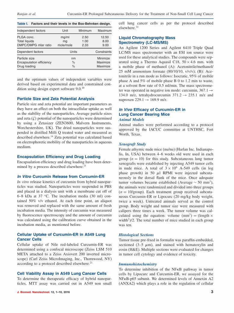
Table I. Factors and their levels in the Box-Behnken design.
Independent factors Unit Minimum Maximum
PLGA conc. mg/ml 2.50 12�50Total liquids mg 1.00 8�00DMPC/DMPG mlar ratio mole/mole 2.33 9�00
Dependent factors Units Constraints
Particle size nm MinimizeEncapsulation efficiency % MaximizeDrug loading % Maximize
and the optimum values of independent variables werederived based on experimental data and constrained con-dition using design expert software 9.0.20
Particle Size and Zeta Potential AnalysisParticle size and zeta potential are important parameters asthey have an effect on both the intracellular uptake as wellas the stability of the nanoparticles. Average particle sizesand zeta (�� potential of the nanoparticles were determinedby using a Zetasizer (ZEN3600, Malvern Instruments,Worchestershire, UK). The dried nanoparticles were sus-pended in distilled Milli Q treated water and measured asdescribed elsewhere.14 Zeta potential was calculated basedon electrophoretic mobility of the nanoparticles in aqueousmedium.
Encapsulation Efficiency and Drug LoadingEncapsulation efficiency and drug loading have been deter-mined by a process described elsewhere.21
In Vitro Curcumin Release from Curcumin-ERIn vitro release kinetics of curcumin from hybrid nanopar-ticles was studied. Nanoparticles were suspended in PBSand placed in a dialysis unit with a membrane cut off of6–8 kDa at 37 �C. The incubation media (30 ml) con-tained 50% v/v ethanol. At each time point, an aliquotwas removed and replaced with the same amount of freshincubation media. The intensity of curcumin was measuredby fluorescence spectroscopy and the amount of curcuminwas calculated using the calibration curve obtained in theincubation media, as mentioned before.
Cellular Uptake of Curcumin-ER in A549 LungCancer CellsCellular uptake of Nile red-labeled Curcumin-ER wasdetermined using a confocal microscope (Zeiss LSM 510META attached to a Zeiss Axiovert 200 inverted micro-scope) (Carl Zeiss MicroImaging, Inc., Thornwood, NY)according to a protocol described elsewhere.21
Cell Viability Assay in A549 Lung Cancer CellsTo determine the therapeutic efficacy of hybrid nanopar-ticles, MTT assay was carried out in A549 non small
cell lung cancer cells as per the protocol describedelsewhere.14
Liquid Chromatography MassSpectrometry (LC-MS/MS)An Agilent 1200 Series and Agilent 6410 Triple QuadLC/MS mass spectrometer with an ESI ion source wereused for these analytical studies. The compounds were sep-arated using a Thermo Aquasil C18, 50× 4.6 mm. witha mobile phase of methanol (A): Acetonitrile/methanol/25 mM ammonium formate (80/10/10, v/v/v); (B): Ace-tonitrile in a run mode as follows: Isocratic, 95% of mobilephase A and 5% of mobile phase B 0 to 1.2 min to waste,at a solvent flow rate of 0.5 ml/min. The mass spectrome-ter was operated in negative ion mode: curcumin, 367.1→134.0 m/z, tetrahydrocurcumin 371.2 → 235.1 m/z andnaproxen 229.1→ 169.9 m/z.
In Vivo Efficacy of Curcumin-ER inLung Cancer Bearing MiceAnimal ModelsAnimal studies were performed according to a protocolapproved by the IACUC committee at UNTHSC, FortWorth, Texas.
Xenograft StudyFemale athymic nude mice (nu/nu) (Harlan Inc. Indianapo-lis, In, USA) between 4–6 weeks old were used in eachgroup (n = 10) for this study. Subcutaneous lung tumorxenografts were established by injecting A549 tumor cellsin nude mice. A total of 3× 10⁶ A-549 cells (in logphase growth) in 50 �l RPMI were injected subcuta-neously in the dorsal flank of the mice. Once adequatetumor volumes became established (Average ∼ 50 mm3),the animals were randomized and divided into three groups(n = 10/group). Each treatment group received subcuta-neous Curcumin-ER or Lipocurc (20 mg/kg body weight,twice a week). Untreated animals served as the controlgroup. Body weight and tumor size were measured withcalipers three times a week. The tumor volume was cal-culated using the equation: volume (mm3� = (length×width2�/2. The total number of mice studied in each groupwas ten.
Histological SectionsTumor tissue pre-fixed in formalin was paraffin-embedded,sectioned (3–5 �m), and stained with hematoxylin andeosin (H&E). Multiple sections were evaluated for changesin tumor cell cytology and evidence of toxicity.
ImmunohistochemistryTo determine inhibition of the NF�B pathway in tumorcells by Lipocurc and Curcumin-ER, we assayed for theNF�B-p65 subunit. We determined levels of Annexin A2(ANXA2) which plays a role in the regulation of cellular
J. Biomed. Nanotechnol. 12, 1–10, 2016 3

Curcumin-ER Prolonged Subcutaneous Delivery for the Treatment of Non-Small Cell Lung Cancer Ranjan et al.
growth and signal transduction pathways, and SRC, a non-receptor tyrosine kinase protein in xenograft tissue withand without treatment. We also determined levels of Ki67which has a role in cell proliferation. Tumors collectedfrom the mice at the end of the study (7 weeks) weresectioned and examined. Immuno-histochemical studieswere carried out using formalin-fixed, paraffin-embeddedsections (5 �m), heat induced antigen retrieval and 1:50 to1:200 concentrations of monoclonal antibody. Antibodiesused for immunohistochemistry included antibody againstannexin A2 (BD Bioscience, San Jose, CA, USA), NF�B-p65 (e Bioscience, San Diego, CA USA), ki67 (e Bio-science, San Diego, CA USA), Secondary antibody (eithermouse or rabbit; Invitrogen, Grand island, NY, USA) wastagged to a fluorophore (Alexa 488: green; Alexa 594:red) and viewed under a confocal microscope (Zeiss LSM510 META) attached to a Zeiss Axiovert 200 inverted
Polymer (PLGA)-
Drug (Curcumin) -
BiomimicLipids(DMPC & DMPG) –
Particlesize &PDI
91.6±3.1nm0.17±0.01
Encapsulationefficiency (%)
93.7±1.9
Zeta Potential –16.3±1.0 mVDrug loading
(%) 1.27±0.4
A
B
C
Figure 1. Curcumin-ER formulation and optimization: (A) Schematic representation of Curcumin-ER; (B) Three dimensionalresponse surface plot showing the effect of molar ratio of lipids and PLGA concentration on particle size, encapsulation effi-ciency and drug loading keeping the total amount of lipids (mg) constant; (C) Physicochemical characterization of Curcumin-ERprepared under the predicted optimum conditions.
Table II. Predictd potimum formulation conditions selectedfor further studies.
Formulation factors Units Optimal value
PLGA conc. mg/ml 7.5Total liquids mg 4.6DMPC/DMPG molar ratio mole/mole 5.4
microscope (Carl Zeiss Micro Imaging, Inc., Thornwood,NY, USA).
Western Blot Analysis of Tissue SamplesIsolated tumor samples were homogenized with lysisbuffer with protease inhibitors added to prevent loss ofprotein in the samples. Western blot was run as per the pro-tocol described elsewhere.27 Glyceraldehyde 3-phosphate
4 J. Biomed. Nanotechnol. 12, 1–10, 2016

Ranjan et al. Curcumin-ER Prolonged Subcutaneous Delivery for the Treatment of Non-Small Cell Lung Cancer
Table III. Comparison of the experimental and predictedvalues of Crucumin-ER perpared the predicted optimumconditions.
Predicted Observed Biasa
Response value value (%)
Particle size (nm) 86�3 91�6 5�78Drug loading (%) 1�30 1�27 −2�38Encapsulation efficiency (%) 89�5 93�7 −4�48
Note: aBias was calculated (observed value-predicted value)/observedvalue×100.
dehydrogenase (GAPDH) was used as a loading controlfor all the blots.
RESULTS AND DISCUSSIONFormulation, Optimization and Characterizationof Curcumin-ERThe hybrid nanoformulation and optimization ofCurcumin-ER was successful. Figure 1(A) shows theschematic representation of the nanoformulation. Theformulation was optimized by varying the polymer con-centration (range: 2.5–12.5 mg), total amount of lipids(range: 1–8 mg), different molar ratios of the lipids (range:DPMC/DMPG:2.33-9) (Tables: I–III). The reponse ofthese factors are presented as three-dimensional contour
0 10 20 40 60 80 1000
25
50
75
100
125
Concentration of curcumin (uM)
Cel
l via
bili
ty (
% o
f co
ntr
ol)
0 10 20 30 40 500
15
30
45
60
75
90
Time (h)
Cu
mu
lati
ve r
elea
se o
fcu
rcu
min
(%
)
A
B
C
iiiii
i
Figure 2. In vitro evaluation of Curcumin-ER: (A) In vitro drug release kinetics of curcumin from Curcumin-ER; (B) Cell viabilityassay were performed on A 549 non small cell lung cancer cell line at 48 hrs; (C) Intracellular uptake of Curcumin-ER in A 549lung cancer cell line (i-Magnification ×20 and ii and iii Magnification ×40). The error bars represent mean and standard deviationsof experiments performed in triplicate.
response surface graphs, depicted in Figure 1(B). Theoptimal formulation was achived based on imposing con-straints of minimum particle size maximum encapsulationefficiency and maximum drug loading. The optimumvalues for these variables for the optimized batch werefound to be 7.5 mg/ml for PLGA concentration, 4.6 mgfor total amount of lipids and a DPMC/DMPG molarratio of 5.4 (Supplement Table II). Next, a Curcumin-ERbatch with these contained predicted levels of formu-lation factors was prepared to verify the optimizationdesign. Figure 1(C) shows average particle size of opti-mized batch of Curcumin-ER (henceforth represented asCurcumin-ER) to be 91.6±3.1 nm and confirms a narrowsize distribution Measured size was presented as theaverage value of 20 runs. The zeta potential determinesthe surface charge in the nanoparticles and plays a rolein intracellular uptake. Curcumin-ER had a zeta potentialof −16.3± 1.0 mV. The entrapment efficiency was highat 93.7± 1.9% and drug loading was determined to be1.27± 0.4%. These optimal experimental results were ingood agreement with the predicted values as indicatedby low bias % (Table III). Figure 2(A) illustrated therelease kinetics profile and shows that it follows a biphasicpattern where ∼15% of curcumin is released within anhour during the burst phase; ∼50% of curcumin is thenreleased from the formulation in about 10 hours to a totalof about 70% in 50 hours.
J. Biomed. Nanotechnol. 12, 1–10, 2016 5

Curcumin-ER Prolonged Subcutaneous Delivery for the Treatment of Non-Small Cell Lung Cancer Ranjan et al.
In Vitro Evaluation of Curcumin-ERCurcumin-ER enhanced the cytotoxicity of curcumin sig-nificantly as compared to free curcumin by inhibitedgrowth of A-549 lung cancer cells. Cell viability assayresults, post 48 hours, depicted that treatment with 38 �Mconcentration of Curcumin-ER resulted in more than 50%cell death (Fig. 2(B)). The IC50 of free curcumin for48 hour incubation was reported to be ∼70 �M in A-549cells.22 This illustrates almost 46% reduction in the IC50
value with Curcumin-ER which is a significant improve-ment in the efficacy of curcumin. This indicated that cur-cumin released from Curcumin-ER was functionally activeand was able to bring about effective cell death in lungcancer cells.
0 10 20 30 40 50 600
100
200
300
400
500Untreated control
Curcumin-ER
Liposomal curcumin
Time (days)
Tu
mo
r vo
lum
e (m
m3)
A
Treatment
0 10 20 30 40 50 6015
20
25
30
Time (days)
Wei
gh
t (g
)
B
C
*****
*Untreated control
Curcumin-ER
Liposomal curcumin
Untreatedcontrol
Liposomalcurcumin
Curcumin-ER
0
100
200
300Untreated controlLiposomal curcuminCurcumin-ER
Tu
mo
r w
eig
ht
(mg
)
*
S.C. Curcumin-ER injection
tumorS.C. liposomal injection
tumorE
D
Figure 3. In vivo evaluation of Curcumin-ER: Curcumin-ER or Lipocurc inhibits the tumor growth of human lung cancerxenograft. (A) Graph showing regression of A549 tumor bearing mice following treatment with Lipocurc or Curcumin-ER.Untreated animals served as controls for the study. Results are expressed as mean±SD. ∗p < 0�001 (untreated vs Curcumin-ER)∗∗p < 0�01 (untreated vs. Lipocurc) ∗∗∗p< 0�05 (Curcumin-ER v Lipocurc); (B) Body weight of animal during treatment time period;(C) Images showing tumors isolated from mice from control and treatment groups; (D) Graph depicting weight of isolated tumorsfrom all three study groups (untreated control, Lipocurc and Curcumin-ER treatments). Results are expressed as mean±SD.,∗p < 0�05. Subcutaneous lung tumor xenografts were established by injecting A549 tumor cells in nude mice. Animals were ran-domly divided into groups (n= 10/group) and treated with Lipocurc and treated with Curcumin-ER subcutaneously. Animals weretreated when average tumors were 50 mm3 (day 0) twice in a week subcutaneously for a total of seven weeks (20 mg curcumin/kgof body weight/dose) and tumor growth monitored alternative days. A significant inhibition of tumor growth was observed in A549tumor-bearing mice treated with Curcumin-ER; (E) Images showing the subcutaneous treatment of Lipocurc and Curcumin-ER inA549 lung cancer xenograft bearing mice.
Robust intracellular uptake indicated by red fluores-cence was observed by confocal microscopy in A-549 cellsincubated with nile red-labeled-Curcumin-ER (Fig. 2(C)).This increased intracellular uptake of Curcumin ERis critical as more uptakes will correspond to moreCurcumin-ER accumulation and subsequent release ofcurcumin from the nanoparticles to elicit its anticanceraction.
Subcutaneous Injection of Curcumin-ERInhibits the Tumor Growth of Human LungCancer XenograftThe in vivo anticancer activity of Curcumin-ER wasinvestigated in female athymic nude mice bearing A549
6 J. Biomed. Nanotechnol. 12, 1–10, 2016

Ranjan et al. Curcumin-ER Prolonged Subcutaneous Delivery for the Treatment of Non-Small Cell Lung Cancer
A B C
Figure 4. H&E staining of tumor tissue samples from different subgroups: (A) Curcumin-ER; (B) Lipocurc; (C) Untreated control(Magnification, ×20).
xenograft lung tumors. Subcutaneous (SC) injections havefound use in overcoming limitations of low systemicbioavailability by extending the release characteristics andthereby increasing systemic exposure of the drug.23 Wecompared the efficacy of Curcumin-ER and Lipocurc withuntreated control animals. Our results show significantregression in tumor volume post treatment with subcuta-neous injection of Curcumin-ER (20 mg/kg body weight,twice a week) compared to control and Lipocurc treatment(Fig. 3(A)). Figure 3(B) shows body weight of the animalsduring the entire treatment period. No weight loss or othersigns of toxicity was evidenced in the mice treated withCurcumin-ER or liposomes. It is evident that Curcumin-ER was more effective in inhibiting tumor growth com-pared to Lipocurc treatment. At the end of the studyperiod (7 weeks), there was 52.1±7.2% decrease in tumorvolume by the Curcumin-ER compared to 32.2± 6.8%
0
2000000
4000000
6000000
8000000
10000000 Untreated control
Liposomal curcumin
Curcumin-ER
Flu
ore
scen
ce in
ten
sity
(a.
u.)
Untreated control Liposomal curcumin Curcumin-ER
***
A
B
Figure 5. (A) Immunohistochemistry images of isolated xenograft tumor tissue-sections showing expression of key tumormarker, Nf�B, post Lipocurc and Curcumin-ER treatment. Untreated tumor sections served as controls; (B) Graphical represen-tation shows the quantification for expression in tissue sections. Results are expressed as mean±SD. ∗p < 0�05, ∗∗p < 0�01.
by Lipocurc treatment. Tumors isolated from mice (fromcontrol and treatment groups) are shown in Figure 3(C).The average tumor weight of untreated control animalswas found to be 182.54 mg while that of Curcumin-ER treated group was only 93.9 mg which represents asignificant decrease of 48.5% (Fig. 3(D)). Figure 3(E)depicts representative images of A549 tumor bearingmice post subcutaneous injection with either Lipocurc orCurcumin-ER.
Histology, Immunohistochemistry andImmunoblotting of Tumor TissueHistologyParaffin-embedded tissues were sectioned and stained withH&E to observe the in vivo effect of Curcumin-ER andLipocurc treatment compared to untreated controls. Thetumor sections from the Curcumin-ER group showed
J. Biomed. Nanotechnol. 12, 1–10, 2016 7

Curcumin-ER Prolonged Subcutaneous Delivery for the Treatment of Non-Small Cell Lung Cancer Ranjan et al.
Untreated control Liposomal curcumin Curcumin-ER
0
100000
200000
300000
400000Untreated control
Liposomal curcumin
Curcumin-ER
Flu
ore
scen
ce in
ten
sity
(a.
u.)
***
A
B
Figure 6. (A) Immunohistochemistry images of isolated xenograft tumor tissue-sections showing expression of key proliferationmarker, Ki67, post Lipocurc and Curcumin-ER treatment. Untreated tumor sections served as controls; (B) Graphical represen-tation shows the quantification for expression in tissue sections. Results are expressed as mean±SD. ∗p < 0�05, ∗∗p < 0�001.
necrosis as illustrated in Figure 4(A). Minimal necrosiswas observed in tumor sections obtained from the grouptreated with Lipocurc (Fig. 4(B)) and the untreated controlgroup (Fig. 4(C)).
0
500000
1000000
1500000
2000000
2500000
Untreated control
Liposomal curcumin
Curcumin-ER
Flu
ore
scen
ce in
ten
sity
(a.
u.)
Untreated control Liposomal curcumin Curcumin-ER
**
A
B
Figure 7. (A) Immunohistochemistry images of isolated xenograft tumor tissue-sections showing expression of key angiogene-sis marker, Annexin A2, post Lipocurc and Curcumin-ER treatment. Untreated tumor sections served as controls; (B) Graphicalrepresentation shows the quantification for expression in tissue sections. Results are expressed as mean±SD. ∗p < 0�001.
ImmunohistochemistryImmunohistochemistry revealed significant decrease inexpression of NF�B-p65 in tumor tissues isolated fromA549 lung cancer bearing mice (Fig. 5), following
8 J. Biomed. Nanotechnol. 12, 1–10, 2016

Ranjan et al. Curcumin-ER Prolonged Subcutaneous Delivery for the Treatment of Non-Small Cell Lung Cancer
GAPDH
Annexin A2
NfkB
1 B)A) 2 3
Figure 8. The western blot image shows the amount of protein (NFkB and Annexin A2) expressed in tumor samples post treat-ment with either Lipocurc or Curcumin-ER. (A) Results indicate that Curcumin-ER treatment shows a reduction in the expressionof both NF�B and Annexin A2 as compared to control samples [Lane 1-Untreated control, Lane 2-Lipocurc Lane 3-Curcumin-ER];(B) Expression of AnxA2 and NF�B normalized to GAPDH levels.
Curcumin-ER treatment as compared to control tissues.These results show that curcumin encapsulated withinhybrid nanoparticles is able to reach the cancer cells inactive form where it blocks the activation of NF�B andinhibits translocation of NF�B from cytosol to nucleus,as seen from the decreased expression of NF�B-p65subunit.24�25 We next determined the changes in expres-sion levels of Ki67, a proliferation marker. Results illus-trated that Curcumin-ER was able to significantly decreasethe proliferation associated with cancer cells as seen bydecreased expression levels of Ki67 in the animal xenograftmodels (Fig. 6). Further, curcumin has been shown to play asignificant role in angiogenesis. We determined the in vivoanti-angiogenic effect of our Curcumin-ER by observingthe expression levels of the protein, Annexin A2 (AnxA2),which is shown to regulate neoangiogenesis.26 Figure 7reveals a significant decrease in AnxA2 expression in thetumor tissue sections post Curcumin-ER treatments as com-pared to untreated controls.
ImmunoblottingThe immunoblot bands represent the amount of target pro-teins expressed in the tumor samples isolated from micepost-treatment with Curcumin-ER, Lipocurc or untreatedcontrol animals (Fig. 8). Curcumin-ER treatment broughtabout a reduction in the expression of target proteins(NF�B-p65, AnxA2) as compared to untreated samples.GAPDH expression (loading control) is not uniform inall the lanes. This has been adjusted by calculating thedensity of expression with respect to GAPDH density.There are almost equal loading of proteins in first twolanes. Figure 8 reveals a significant decrease in NF�B-p65 and AnxA2 expression in the tumor tissue sectionspost Lipocurc and Curcumin-ER treatments as comparedto untreated controls.
CONCLUSIONSIn conclusion, Curcumin-ER was successfully formulatedand showed high curcumin encapsulation with particle size
in the range of 90 nm. The in vitro evaluation showedthat Curcumin-ER showed improved cellular uptake andwas able to decrease cell viability indicating therapeuticefficacy against NSCLC cancer cells. The tumor xenograftstudies further illustrated the in vivo efficacy of Curcumin-ER by significantly decreasing tumor burden follow-ing subcutaneous administration of Curcumin-ER. Theimmunohistochemical and immunoblotting analysis of theisolated lung tumors confirmed the effect of Curcumin-ERin regressing tumorigenic and proliferation proteins. Theseresults indicate that Curcumin-ER may be used as a ther-apeutic in the treatment of NSCLC.
REFERENCES1. K. Raparia, C. Villa, M. M. Decamp, J. D. Patel, and M. P. Mehta,
Molecular profiling in non-small cell lung cancer: A step towardpersonalized medicine. Arch. Pathol. Lab. Med. 137, 481 (2013).
2. H. Wakelee and C. P. Belani, Optimizing first-line treatment optionsfor patients with advanced NSCLC. Oncologist. 10, 1 (2005).
3. S. Fleming, F. Lucas, and M. A. Schofield, Therapeutic area reviewof oncology products and players. Expert Opin. Emerg. Drugs 6, 317(2001).
4. J. Y. Douillard, J. Eckardt, and G. V. Scagliotti, Challenging theplatinum combinations in the chemotherapy of NSCLC. Lung Can.38, 21 (2002).
5. P. M. Green, S. Guerrier-Adams, P. O. Okunji, D. Schiavone, andJ. E. Smith, African American health disparities in lung cancer. Clin.J. Oncol. Nurs. 17, 180 (2013).
6. D. Peer, J. M. Karp, S. Hong, O. C. Farokhzad, R. Margalit, andR. Langer, Nanocarriers as an emerging platform for cancer therapy.Nat. Nanotechnol. 2, 751 (2007).
7. V. P. Torchilin, Recent advances with liposomes as pharmaceuticalcarriers. Nat. Rev. Drug Discov. 4, 145 (2005).
8. N. Maurer, D. B. Fenske, and P. R. Cullis, Developments in lipo-somal drug delivery systems. Expert Opin. Biol. Ther. 1, 923(2001).
9. L. Zhang, J. M. Chan, F. X. Gu, J.-W. Rhee, A. Z. Wang, A. F.Radovic-Moreno, F. Alexis, R. Langer, and O. C. Farokhzad, Self-assembled lipid-polymer hybrid nanoparticles: A robust drug deliv-ery platform. ACS Nano. 2, 1696 (2008).
10. K. Hadinoto, A. Sundaresan, and W. S. Cheow, Lipid–polymerhybrid nanoparticles as a new generation therapeutic deliveryplatform: A review. European J. Pharma. Biophar. 85, 427(2013).
J. Biomed. Nanotechnol. 12, 1–10, 2016 9
Curcumin-ER Prolonged Subcutaneous Delivery for the Treatment of Non-Small Cell Lung Cancer Ranjan et al.
11. G. Bar-Sela, R. Epelbaum, and M. Schaffer, Curcumin as an anti-cancer agent: Review of the gap between basic and clinical applica-tions. Curr. Med. Chem. 17, 190 (2010).
12. M. M. Yallapu, M. Jaggi, and S. C. Chauhan, Curcumin nanofor-mulations: A future nanomedicine for cancer. Drug Discov. Today.17, 71 (2012).
13. O. Naksuriyaa, S. Okonogia, R. M. Schiffelers, and W. E. Hennink,Curcumin nanoformulations: A review of pharmaceutical propertiesand preclinical studies and clinical data related to cancer treatment.Biomat. 35, 3365 (2014).
14. A. Mukerjee and J. K. Vishwanatha, Formulation, characterizationand evaluation of curcumin-loaded PLGA nanospheres for cancertherapy. Anticancer. Res. 29, 3867 (2009).
15. R. A. Sharma, S. A. Euden, S. L. Platton, D. N. Cooke, A. Shafayat,H. R. Hewitt, T. H. Marczylo, B. Morgan, D. Hemingway, S. M.Plummer, M. Pirmohamed, A. J. Gescher, and W. P. Steward, PhaseI clinical trial of oral curcumin: Biomarkers of systemic activity andcompliance. Clin. Cancer Res. 10, 6847 (2004).
16. A. L. Cheng, C. H. Hsu, J. K. Lin, M. M. Hsu, Y. F. Ho, T. S. Shen,J. Y. Ko, J. T. Lin, B. R. Lin, W. Ming-Shiang, H. S. Yu, S. H. Jee,G. S. Chen, T. M. Chen, C. A. Chen, M. K. Lai, Y. S. Pu, M. H.Pan, Y. J. Wang, C. C. Tsai, and C. Y. Hsieh, Phase I clinical trialof curcumin, a chemopreventive agent, in patients with high-risk orpre-malignant lesions. Anticancer Res. 21, 2895 (2001).
17. N. Dhillon, B. B. Aggarwal, R. A. Newman, R. A. Wolff, A. B.Kunnumakkara, J. L. Abbruzzese, C. S. Ng, V. Badmaev, andR. Kurzrock, Phase II trial of curcumin in patients with advancedpancreatic cancer. Clin. Cancer Res. 14, 4491 (2008).
18. P. Anand, A. B. Kunnumakkara, R. A. Newman, and B. B.Aggarwal, Bioavailability of curcumin: Problems and promises. Mol.Pharmaceutics 4, 807 (2007).
19. R. T. Mehta, R. L. Hopfer, L. A. Gunner, R. L. Juliano, andG. Lopez-Berestein, Formulation, toxicity, and antifungal activ-ity in vitro of liposome-encapsulated nystatin as therapeutic agent
for systemic candidiasis. Antimicrob Agents Chemother. 31, 1897(1987).
20. J. Hao, X. Fang, Y. Zhou, J. Wang, F. Guo, F. Li, and X. Peng,Development and optimization of solid lipid nanoparticle formu-lation for ophthalmic delivery of chloramphenicol using a Box-Behnken design. Int. J. Nanomed. 6, 683 (2011).
21. A. P. Ranjan, A. Mukerjee, L. Helson, and J. K. Vishwanatha, Scaleup, optimization and stability analysis of curcumin C3 complex-loaded nanoparticles for cancer therapy. J. Nanobiotech. 10, 38(2012).
22. G. Radhakrishna Pillai, A. S. Srivastava, T. I. Hassanein, D. P.Chauhan, and E. Carrier, Induction of apoptosis in human lung can-cer cells by curcumin. Cancer Lett. 208, 163 (2004).
23. J. Li, Y. Jiang, J. Wen, G. Fan, Y. Wu, and C. Zhang, A rapidand simple HPLC method for the determination of curcumin inrat plasma: Assay development, validation and application to apharmacokinetic study of curcumin liposome. Biomed. Chromatogr.23, 1201 (2009).
24. D. Wang, M. S. Veena, K. Stevenson, C. Tang, B. Ho, J. D. Suh,V. M. Duarte, K. F. Faull, K. Mehta, E. S. Srivatsan, and M.B. Wang, Liposome encapsulated curcumin suppresses growth ofHNSCC in vitro and in xenografts through the inhibition of NF�B byan AKT independent pathway. Clin. Cancer Res. 14, 6228 (2008).
25. I. Jutooru, G. Chadalapaka, P. Lei, and S. Safe, Inhibition ofNF�B and pancreatic cancer cell and tumor growth by curcuminis dependent on specificity protein down-regulation. J. Biol. Chem.285, 25332 (2010).
26. M. Valapala, S. I. Thamake, and J. K. Vishwanatha, A competi-tive hexapeptide inhibitor of annexin A2 prevents hypoxia-inducedangiogenic events. J. Cell Sci. 124, 1453 (2011).
27. A. Mukerjee, J. Shankardas, A. P. Ranjan, and J. K.Vishwanatha, Efficient nanoparticle mediated sustained RNA inter-ference in human primary endothelial cells. Nanotech. 22, 445101(2011).
10 J. Biomed. Nanotechnol. 12, 1–10, 2016