jet-cooled spectroscopy of the m-xylyl … · excited supersonic expansion: hyper-conjugation and...
TRANSCRIPT
European Journal of Pure and Applied Chemistry Vol. 5, No. 1, 2018 ISSN ISSN 2398-1385
Progressive Academic Publishing, UK Page 32 www.idpublications.org
JET-COOLED SPECTROSCOPY OF THE M-XYLYL RADICAL IN A CORONA-
EXCITED SUPERSONIC EXPANSION: HYPER-CONJUGATION AND
CONFORMATIONAL DEPENDENCE IN VIBRONIC TRANSITIONS
Chang Soon Huh
Applied Chemistry Major, Division of
Chemical and Environmental
Engineering, College of Engineering
/Dong-eui University
South Korea
ABSTRACT
The electronically hot but jet-cooled m-xylyl radical was produced from the precursor m-
xylene in a corona-excited supersonic expansion (CESE), and its vibronic emission
spectrum was recorded. Theoretical studies of the ground and excited state structures, as
well as the hyper-conjugative interaction of the m-xylyl radical, were carried out. The
preferred geometry of the methyl rotor in the ground state is switched in the excited state.
The vibronic emission spectrum of the m-xylyl radicals corresponded to the D1 → D0
electronic transition and determined the electronic transition energy depending on the initial
change in the methyl conformations.
Keywords: Spectroscopy, m-xylyl radical, corona discharge, hyper-conjugation, natural bond
orbital.
1. INTRODUCTION
In combustion and atmospheric science, free radicals play an important role as reaction
intermediates, while resonance-stabilized radicals have attracted significant attention due to
their relatively high stability. The benzyl radical is a prototypical example of an aromatic
hydrocarbon, and it has a rich history of experimental and theoretical studies [1-4]. Since the
first observation of the benzyl radical [5], vibronic coupling and ring substitution have
attracted much attention in the field of electronic spectroscopy. Vibronic coupling causes a
significant change in the typical mirror symmetry between the ground and excited state
spectra, while substitution on the benzyl radical perturbs the electronic structure and
transition energy.
Methyl substitutions on the ring of benzyl are known to serve a variety of electronic
structures, responding to changes in the orientation and the barrier height of the methyl group.
In terms of methyl substitution, the xylyl radicals were studied by Schuler et al. [5] and
Walker and Barrow [6] in the visible region. Bindley et al. assigned the xylyl radicals’ strong
bands from xylenes in a corona discharge [7]. Furthermore, the determination of the vibronic
assignments and lifetime measurements of xylene was studied using the laser-induced
florescence (LIF) technique by Charlton and Thrush [8].
Hiratsuka et al. [9] investigated the electronic energies of the close-lying doublet D1 and D2
states of the benzyl radical and described the possible vibronic coupling between the close-
lying D1 and D2 electronic states. Cossart-Magos et al. [10] conducted a rotational contour
analysis of the o-xylyl radical using the magnitude and direction of the transition dipole
moment. Fukushima and Obi [4] utilized the high-resolution LIF technique to analyze the
European Journal of Pure and Applied Chemistry Vol. 5, No. 1, 2018 ISSN ISSN 2398-1385
Progressive Academic Publishing, UK Page 33 www.idpublications.org
vibronic coupling of the jet-cooled p-xylyl radical. Lin and Miller [11] determined the
torsional bands of the methyl rotor in xylyl radicals, while Selco and Carrick [12] assigned
the vibronic transition bands of benzyl and xylyl radicals.
Recently, Lee et al. [13-19] identified the fluorescence spectra of various benzyl-type
radicals substituted with halogens (F, Cl) and methyl groups (dimethyl, tri-methyl) by
applying corona-excited supersonic expansion (CESE). However, the methyl-substituted
benzyl radicals have not been extensively analyzed due to the complicated vibronic structures
and the torsional transitions in the CESE spectrum.
In this work, we describe the results of a spectroscopic analysis of the m-xylyl radical,
which is generated from m-xylene in the corona discharge. The emission spectrum of the m-
xylyl radical contains two kinds of electronic state that are related to the methyl
conformations in the ground and excited states. For the analysis of the observed spectrum, the
spectroscopic data concerning the electronic transition and vibrational mode frequencies are
analyzed with the help of the ab initio calculation. In addition, the energy change in the
methyl conformation of the ground state is discussed using a natural bond orbital (NBO)
analysis to explain the hyper-conjugation and energy differences in the methyl group’s
orientation.
2. EXPERMENTAL AND THEORETICAL METHODS
The experimental condition of this work was accomplished using the apparatus described by
Han, Choi, and Lee [20]. Briefly put, we used conditions that play an important role in the
favorable generation of substituted benzyl radicals in a corona excitation.
Reagent grade m-xylene (the precursor) was purchased from Sigma-Aldrich and then used
without further purification. The concentration of the precursor was believed to be less than 1%
of the He carrier gas at an ambient temperature. The gas mixture was expanded through a
0.2–0.3 mm in diameter glass nozzle connected to a vacuum chamber pumped using an 800
L/min mechanical rotary pump [21]. A high voltage (5 mA at 2000 V) was applied to a
stainless steel rod that ran the length of the glass tube to within almost 0.5 mm of the opening.
After a corona discharge was induced at the nozzle’s opening, a blue-green colored jet could
be seen in the benzyl-type radicals in the D1 → D0 transition. The 5 mm light jet area of the
nozzle opening was collimated and focused on the slit of a 2 m monochromator containing
two 1800 lines/mm gratings. It was detected using a cooled Hamamatsu R649
photomultiplier tube and a photon counting system. During the scans, the monochromator
slits were typically set to 100–200 μm, providing a resolution of about 2 cm-1
. A spectrum
ranging from 19,800 to 22,000 cm-1
was acquired in 2.0 cm-1
increments over 2 hours. The
Helium (He) atomic lines observed in the same spectral region as the benzyl-type radicals
were used as calibration points [22].
Theoretical calculations were performed to study the D0 and D1 states using the density
functional theory (DFT) method. The geometries of the ground states were optimized at the
hybrid density functional B3LYP level using a 6-311+G (d, p) basis set [23, 24]. The
vibrational frequencies of the benzyl-type radicals were computed in order to analyze the
vibronic structures with a scaling factor of 0.987. Time-dependent (TD) formalism was
employed to obtain the vertical excitation energies and excited state geometries of the benzyl-
type radicals with a 6-311+G** basis set [25-30]. A population analysis was also performed
using the natural bond orbital method at the B3LYP/6-311+g (d, p) level of theory using the
NBO method [31] with a GAUSSIAN 09 program [32].

European Journal of Pure and Applied Chemistry Vol. 5, No. 1, 2018 ISSN ISSN 2398-1385
Progressive Academic Publishing, UK Page 34 www.idpublications.org
3. RESULTS
3. 1 CESE spectrum
A vibronic emission spectrum using m-xylene as the radical precursor is shown in Figure 1.
Figure 1. A portion of the fluorescence emission spectrum of the m-xylyl radical
produced from m-xylene in a corona discharge. Several vibronic bands are labeled.
This spectrum includes a number of sharp lines between 19,800 and 22,000 cm-1
. The
electronic origin is assigned to the intensive peak at 21,494 cm−1
, which is in good agreement
with the previously reported values of 21,486 and 21,473 cm−1
for the m-xylyl radical [11,
12]. No hot bands are observed, which suggests that the molecules were cooled efficiently in
the CESE. According to Lin and Miller, the cluster peaks of each vibronic band are caused by
internal methyl rotation [11]. The vibronic peak assignments are discussed later in this paper.
The emission spectra of the benzyl and methyl-substituted benzyl radicals using CESE shows
sharp lines, which is in agreement with previously reported spectra [12-21]. The benzyl
radical has an origin band with a weak intensity at 22,002 cm-1
. As the benzyl radical is
substituted into the benzene ring, the origin band shifts to a lower wavenumber. The origin
bands of the o- and p-xylyl [3] radicals are observed at 21,345 and 21,704 cm
-1, respectively,
indicating that they have shifted 657 and 298 cm-1
from the benzyl radical, respectively. The
2, 6-dimethylbenzyl [5] and 3, 5-dimethylbenzyl [7] radicals have origin bands at 20,616 and
20,842 cm-1
, respectively. In a previous study, Selco and Carrick reported o- and p-xylyl
radicals from an m-xylene precursor, and they explained the isomeric products to result in
benzyl valence- or primase-type intermediates that correspond to photolysis and pyrolysis.
However, in the present study, bands were identified for the o-xylyl radical, but not for the p-
European Journal of Pure and Applied Chemistry Vol. 5, No. 1, 2018 ISSN ISSN 2398-1385
Progressive Academic Publishing, UK Page 35 www.idpublications.org
xylyl radical.
Figure 2. A portion of the visible emission spectra in the D1 → D0 transition observed in
the CESE of (a) m-xylyl, (b) p-xylyl, and (c) o-xylyl radicals.
Figure 2 shows a spectra comparison of the m-xylyl radical from m-xylene with the o- and p-
xylyl radicals. In the comparison, the torsional and vibronic bands of the o-xylyl radical are
included in the m-xylyl radical, while the 21,706 cm-1
peak has shifted 2 cm-1
from the origin
of the p-xylyl radical and the p-xylyl’s intensive vibronic bands (6a and 1 modes) are not
observed. This means that the 21,706 cm-1
band is not the origin of the p-xylyl radical and
hence it is likely to be the transition band from species in the excited state of the m-xylyl
radical.
3. 2 Theoretical results
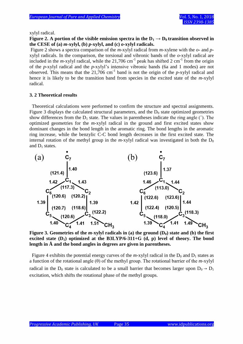
Theoretical calculations were performed to confirm the structure and spectral assignments.
Figure 3 displays the calculated structural parameters, and the D0 state optimized geometries
show differences from the D1 state. The values in parentheses indicate the ring angle (˚). The
optimized geometries for the m-xylyl radical in the ground and first excited states show
dominant changes in the bond length in the aromatic ring. The bond lengths in the aromatic
ring increase, while the benzylic C-C bond length decreases in the first excited state. The
internal rotation of the methyl group in the m-xylyl radical was investigated in both the D0
and D1 states.
Figure 3. Geometries of the m-xylyl radicals in (a) the ground (D0) state and (b) the first
excited state (D1) optimized at the B3LYP/6-311+G (d, p) level of theory. The bond
length in Å and the bond angles in degrees are given in parentheses.
Figure 4 exhibits the potential energy curves of the m-xylyl radical in the D0 and D1 states as
a function of the rotational angle (θ) of the methyl group. The rotational barrier of the m-xylyl
radical in the D0 state is calculated to be a small barrier that becomes larger upon D0 → D1
excitation, which shifts the rotational phase of the methyl groups.

European Journal of Pure and Applied Chemistry Vol. 5, No. 1, 2018 ISSN ISSN 2398-1385
Progressive Academic Publishing, UK Page 36 www.idpublications.org
Figure 4. Potential energy curves of the methyl rotation (θ) in (a) the ground state (D0)
and (b) the first excited state (D1).
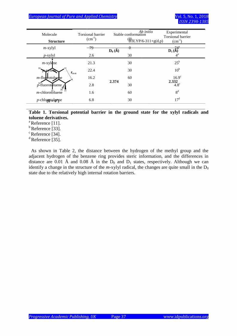
Table 1 lists the methyl torsional energy of the xylyl radicals and toluene derivatives in the
ground state. The meta-substituted toluene derivatives and p-xylyl have a small energy
barrier between 1 ~ 25 cm-1
. However, the m-xylyl radical has a relatively higher barrier at 79
cm-1
with a different stable methyl conformation (θ = 60˚). In order to explain the mechanism
of the conformation and the rotational barrier of the m-xylyl radical, we performed steric and
NBO analyses. Figure 3 shows the geometrical parameters in the D0 state and D1 state, which
were optimized using the TD-DFT calculation. The maximum changes in the bond lengths
and bond angles are 0.04 Å and 4.4°, respectively, while the average values are 0.03 Å and
2.0°, respectively.
Molecule B3LYP/6-311+g(d, p)
Experimental
torsional barrier
(cm-1
)
European Journal of Pure and Applied Chemistry Vol. 5, No. 1, 2018 ISSN ISSN 2398-1385
Progressive Academic Publishing, UK Page 37 www.idpublications.org
Table 1. Torsional potential barrier in the ground state for the xylyl radicals and
toluene derivatives. a Reference [11].
b Reference [33].
c Reference [34].
d Reference [35].
As shown in Table 2, the distance between the hydrogen of the methyl group and the
adjacent hydrogen of the benzene ring provides steric information, and the differences in
distance are 0.01 Å and 0.08 Å in the D0 and D1 states, respectively. Although we can
identify a change in the structure of the m-xylyl radical, the changes are quite small in the D0
state due to the relatively high internal rotation barriers.
Molecule Torsional barrier
(cm-1
)
Stable conformation
(θ)
Experimental
Torsional barrier
(cm-1
)
m-xylyl 79 0 74a
p-xylyl 2.6 30 4a
m-xylene 21.3 30 25b
p-xylene 22.4 30 10b
m-fluorotoluene 16.2 60 16.9c
p-fluorotoluene 2.8 30 4.8c
m-chlorotoluene 1.6 60 8d
p-chlorotoluene 6.8 30 17d
Structure
Ab initio
B3LYP/6-311+g(d,p)
D0 (Å) D1 (Å)
(θ = 0˚)
2.374 2.332
European Journal of Pure and Applied Chemistry Vol. 5, No. 1, 2018 ISSN ISSN 2398-1385
Progressive Academic Publishing, UK Page 38 www.idpublications.org
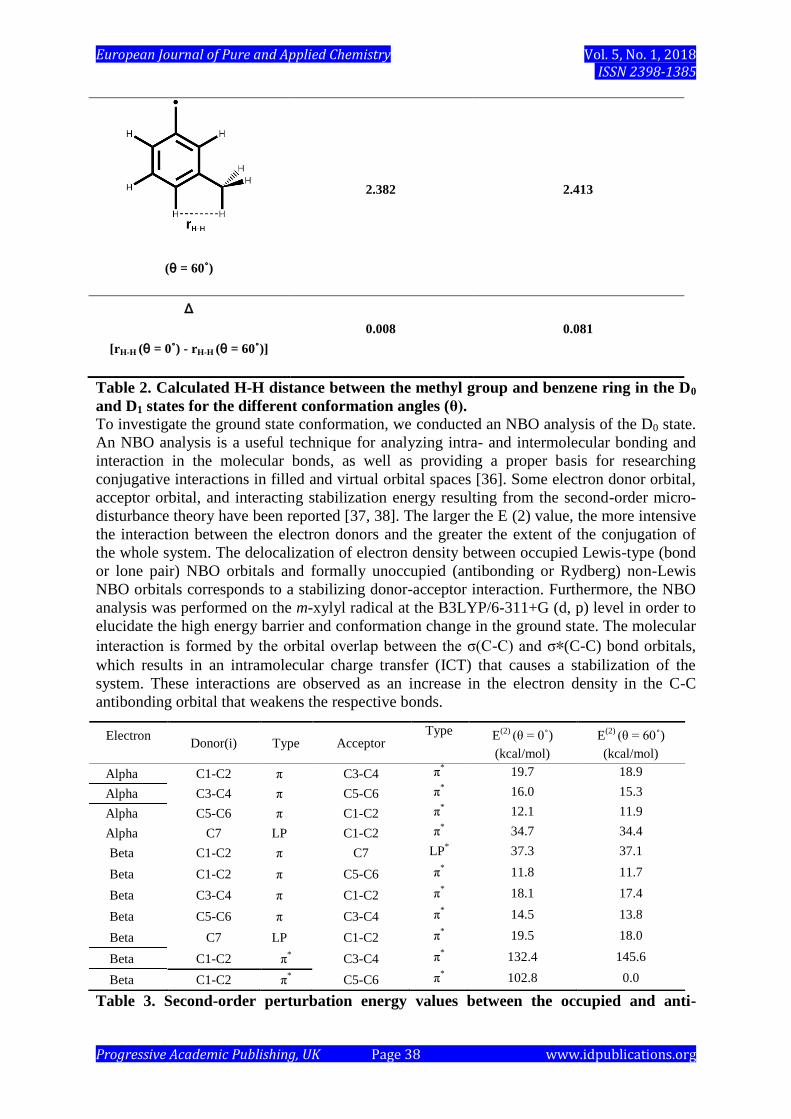
Table 2. Calculated H-H distance between the methyl group and benzene ring in the D0
and D1 states for the different conformation angles (θ). To investigate the ground state conformation, we conducted an NBO analysis of the D0 state.
An NBO analysis is a useful technique for analyzing intra- and intermolecular bonding and
interaction in the molecular bonds, as well as providing a proper basis for researching
conjugative interactions in filled and virtual orbital spaces [36]. Some electron donor orbital,
acceptor orbital, and interacting stabilization energy resulting from the second-order micro-
disturbance theory have been reported [37, 38]. The larger the E (2) value, the more intensive
the interaction between the electron donors and the greater the extent of the conjugation of
the whole system. The delocalization of electron density between occupied Lewis-type (bond
or lone pair) NBO orbitals and formally unoccupied (antibonding or Rydberg) non-Lewis
NBO orbitals corresponds to a stabilizing donor-acceptor interaction. Furthermore, the NBO
analysis was performed on the m-xylyl radical at the B3LYP/6-311+G (d, p) level in order to
elucidate the high energy barrier and conformation change in the ground state. The molecular
interaction is formed by the orbital overlap between the σ(C-C) and σ∗(C-C) bond orbitals,
which results in an intramolecular charge transfer (ICT) that causes a stabilization of the
system. These interactions are observed as an increase in the electron density in the C-C
antibonding orbital that weakens the respective bonds.
Table 3. Second-order perturbation energy values between the occupied and anti-
(θ = 60˚)
2.382 2.413
Δ
[rH-H (θ = 0˚) - rH-H (θ = 60˚)]
0.008 0.081
Electron Donor(i) Type Acceptor
Type E(2)
(θ = 0˚)
(kcal/mol)
E(2)
(θ = 60˚)
(kcal/mol)
Alpha C1-C2 π C3-C4 π* 19.7 18.9
Alpha C3-C4 π C5-C6 π* 16.0 15.3
Alpha C5-C6 π C1-C2 π* 12.1 11.9
Alpha C7 LP C1-C2 π* 34.7 34.4
Beta C1-C2 π C7 LP* 37.3 37.1
Beta C1-C2 π C5-C6 π* 11.8 11.7
Beta C3-C4 π C1-C2 π* 18.1 17.4
Beta C5-C6 π C3-C4 π* 14.5 13.8
Beta C7 LP C1-C2 π* 19.5 18.0
Beta C1-C2 π* C3-C4 π
* 132.4 145.6
Beta C1-C2 π* C5-C6 π
* 102.8 0.0

European Journal of Pure and Applied Chemistry Vol. 5, No. 1, 2018 ISSN ISSN 2398-1385
Progressive Academic Publishing, UK Page 39 www.idpublications.org
bonding orbitals of the m-xylyl radical (θ = 0˚ and 60˚) using an NBO basis.
Table 3 shows the most important interactions between the Lewis and non-Lewis orbitals,
the second-order perturbation energy values, E (2), corresponding to the conformation of θ =
0° and 60° of the methyl rotor angle. A strong interaction was observed between the p-type
orbital containing the lone electron of C7 and the π*(C1–C2), π*(C3–C4), π*(C5–C6), and
LP*(C7) anti-bonding orbitals of the benzyl structure in the alpha and beta spins. One of the
strongest interactions (102.8 kJ mol-1
) between the π*(C1–C2) and π*(C5–C6) is calculated
on an angle θ = 0° of the methyl rotor in the beta spin. However, this intensive stabilization
energy is not computed at θ = 60° of the methyl rotor. The strong intra-molecular hyper-
conjugation interaction between the π* → π* electrons in the ring leads to the stabilization of
the (θ = 0°) m-xylyl radical. This interaction confirms the conformational difference of the m-
xylyl radical in the D0 state, which is distinguishable from the p-xylyl radical and other
methyl-substituted toluene as shown in Table 2.
The theory also suggests that two m-xylyl radical conformations (θ = 0˚ and 60˚) could be
observed in the discharge as displayed in Figure 5. The TD-DFT calculation predicts the D1
→ D0 transition energy for m-xylyl (θ = 0˚) to be 232 cm-1
higher than the D1 → D0 transition
energy calculated for the m-xylyl (θ = 60˚) conformation.
3. 3 Vibronic transitions
The emission spectrum for the D1 → D0 transition in the m-xylyl radical is expected to
exhibit an intense origin band with a few vibronic transitions corresponding to aromatic ring
vibrations, since the optimized geometries for the ground and first excited state of the m-xylyl
radical are nearly identical. The intense origin band in Figure 1 is consistent with the findings
of previous studies [11, 12]. Different minimum conformations of the methyl rotor in the
ground and first excited states suggest that the emission spectra for each species should be
different and distinguishable.
European Journal of Pure and Applied Chemistry Vol. 5, No. 1, 2018 ISSN ISSN 2398-1385
Progressive Academic Publishing, UK Page 40 www.idpublications.org
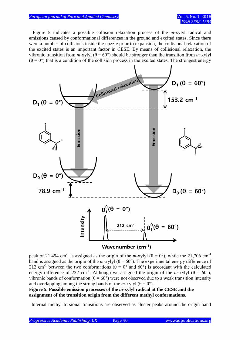
Figure 5 indicates a possible collision relaxation process of the m-xylyl radical and
emissions caused by conformational differences in the ground and excited states. Since there
were a number of collisions inside the nozzle prior to expansion, the collisional relaxation of
the excited states is an important factor in CESE. By means of collisional relaxation, the
vibronic transition from m-xylyl (θ = 60°) should be stronger than the transition from m-xylyl
(θ = 0°) that is a condition of the collision process in the excited states. The strongest energy
peak of 21,494 cm-1
is assigned as the origin of the m-xylyl (θ = 0°), while the 21,706 cm-1
band is assigned as the origin of the m-xylyl (θ = 60°). The experimental energy difference of
212 cm-1
between the two conformations (θ = 0° and 60°) is accordant with the calculated
energy difference of 232 cm-1
. Although we assigned the origin of the m-xylyl (θ = 60°),
vibronic bands of conformation (θ = 60°) were not observed due to a weak transition intensity
and overlapping among the strong bands of the m-xylyl (θ = 0°).
Figure 5. Possible emission processes of the m-xylyl radical at the CESE and the
assignment of the transition origin from the different methyl conformations.
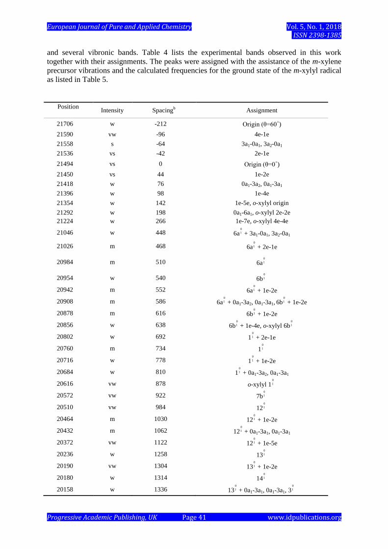
Internal methyl torsional transitions are observed as cluster peaks around the origin band
European Journal of Pure and Applied Chemistry Vol. 5, No. 1, 2018 ISSN ISSN 2398-1385
Progressive Academic Publishing, UK Page 41 www.idpublications.org
and several vibronic bands. Table 4 lists the experimental bands observed in this work
together with their assignments. The peaks were assigned with the assistance of the m-xylene
precursor vibrations and the calculated frequencies for the ground state of the m-xylyl radical
as listed in Table 5.
Position
Intensity Spacingb Assignment
21706 w -212 Origin (θ=60˚)
21590 vw -96 4e-1e
21558 s -64 3a1-0a1, 3a2-0a1
21536 vs -42 2e-1e
21494 vs 0 Origin (θ=0˚)
21450 vs 44 1e-2e
21418 w 76 0a1-3a2, 0a1-3a1
21396 w 98 1e-4e
21354 w 142 1e-5e, o-xylyl origin
21292 w 198 0a1-6a1, o-xylyl 2e-2e
21224 w 266 1e-7e, o-xylyl 4e-4e
21046 w 448 6a + 3a1-0a1, 3a2-0a1
21026 m 468 6a + 2e-1e
20984 m 510 6a
20954 w 540 6b
20942 m 552 6a + 1e-2e
20908 m 586 6a + 0a1-3a2, 0a1-3a1, 6b + 1e-2e
20878 m 616 6b + 1e-2e
20856 w 638 6b + 1e-4e, o-xylyl 6b
20802 w 692 1 + 2e-1e
20760 m 734 1
20716 w 778 1 + 1e-2e
20684 w 810 1 + 0a1-3a2, 0a1-3a1
20616 vw 878 o-xylyl 1
20572 vw 922 7b
20510 vw 984 12
20464 m 1030 12 + 1e-2e
20432 m 1062 12 + 0a1-3a1, 0a1-3a1
20372 vw 1122 12 + 1e-5e
20236 w 1258 13
20190 vw 1304 13 + 1e-2e
20180 w 1314 14
20158 w 1336 13 + 0a1-3a1, 0a1-3a1, 3
0
1
0
1
0
1
0
1
0
1
0
1
0
1
0
1
0
1
0
1
0
1
0
1
0
1
0
1
0
1
0
1
0
1
0
1
0
1
0
1
0
1
0
1
0
1
0
1
0
1
European Journal of Pure and Applied Chemistry Vol. 5, No. 1, 2018 ISSN ISSN 2398-1385
Progressive Academic Publishing, UK Page 42 www.idpublications.org
Table 4. List of the observed vibronic bands and their assignments. a
a Measured in a vacuum (cm
-1).
b Spacing from the (θ = 0˚) origin band at 21,494 cm
-1.
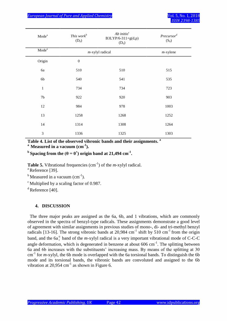
Table 5. Vibrational frequencies (cm-1
) of the m-xylyl radical. a Reference [39].
b Measured in a vacuum (cm
-1).
c Multiplied by a scaling factor of 0.987.
d Reference [40].
4. DISCUSSION
The three major peaks are assigned as the 6a, 6b, and 1 vibrations, which are commonly
observed in the spectra of benzyl-type radicals. These assignments demonstrate a good level
of agreement with similar assignments in previous studies of mono-, di- and tri-methyl benzyl
radicals [13-16]. The strong vibronic bands at 20,984 cm-1
shift by 510 cm-1
from the origin
band, and the 6a 0
1 band of the m-xylyl radical is a very important vibrational mode of C-C-C
angle deformation, which is degenerated in benzene at about 606 cm-1
. The splitting between
6a and 6b increases with the substituents’ increasing mass. By means of the splitting at 30
cm-1
for m-xylyl, the 6b mode is overlapped with the 6a torsional bands. To distinguish the 6b
mode and its torsional bands, the vibronic bands are convoluted and assigned to the 6b
vibration at 20,954 cm-1
as shown in Figure 6.
Modea This work
b
(D0)
Ab initioc
B3LYP/6-311+g(d,p)
(D0)
Precursord
(S0)
Modea
m-xylyl radical m-xylene
Origin 0
6a 510 510 515
6b 540 541 535
1 734 734 723
7b 922 920 903
12 984 978 1003
13 1258 1268 1252
14 1314 1308 1264
3 1336 1325 1303
European Journal of Pure and Applied Chemistry Vol. 5, No. 1, 2018 ISSN ISSN 2398-1385
Progressive Academic Publishing, UK Page 43 www.idpublications.org
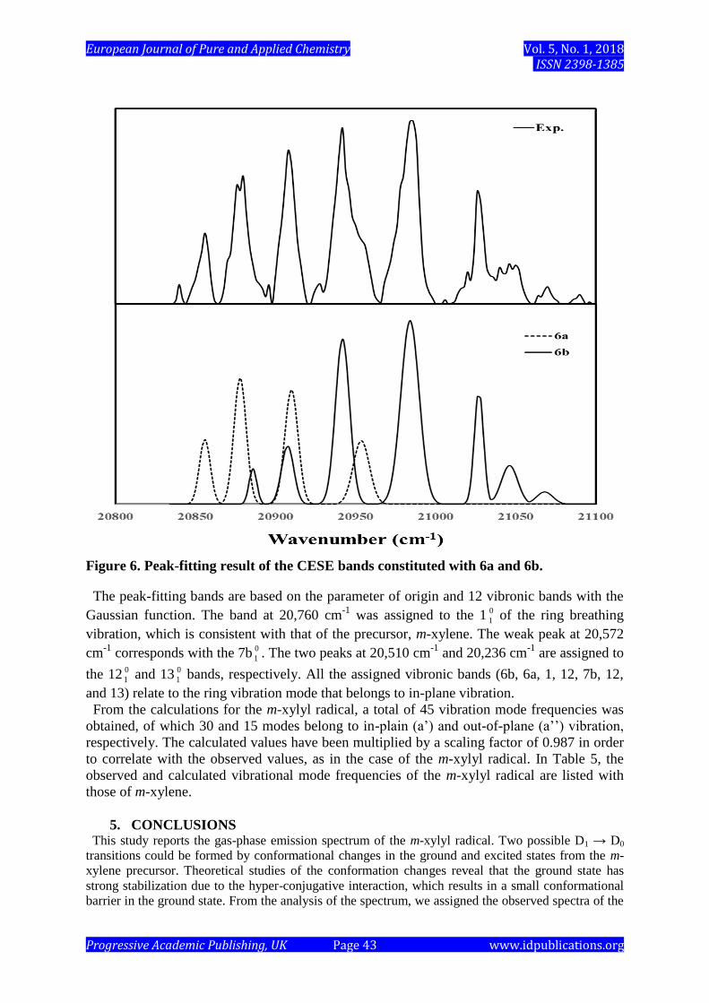
Figure 6. Peak-fitting result of the CESE bands constituted with 6a and 6b.
The peak-fitting bands are based on the parameter of origin and 12 vibronic bands with the
Gaussian function. The band at 20,760 cm-1
was assigned to the 1 0
1 of the ring breathing
vibration, which is consistent with that of the precursor, m-xylene. The weak peak at 20,572
cm-1
corresponds with the 7b 0
1 . The two peaks at 20,510 cm-1
and 20,236 cm-1
are assigned to
the 12 0
1 and 13 0
1 bands, respectively. All the assigned vibronic bands (6b, 6a, 1, 12, 7b, 12,
and 13) relate to the ring vibration mode that belongs to in-plane vibration.
From the calculations for the m-xylyl radical, a total of 45 vibration mode frequencies was
obtained, of which 30 and 15 modes belong to in-plain (a’) and out-of-plane (a’’) vibration,
respectively. The calculated values have been multiplied by a scaling factor of 0.987 in order
to correlate with the observed values, as in the case of the m-xylyl radical. In Table 5, the
observed and calculated vibrational mode frequencies of the m-xylyl radical are listed with
those of m-xylene.
5. CONCLUSIONS This study reports the gas-phase emission spectrum of the m-xylyl radical. Two possible D1 → D0
transitions could be formed by conformational changes in the ground and excited states from the m-
xylene precursor. Theoretical studies of the conformation changes reveal that the ground state has
strong stabilization due to the hyper-conjugative interaction, which results in a small conformational
barrier in the ground state. From the analysis of the spectrum, we assigned the observed spectra of the

European Journal of Pure and Applied Chemistry Vol. 5, No. 1, 2018 ISSN ISSN 2398-1385
Progressive Academic Publishing, UK Page 44 www.idpublications.org
θ = 0° and θ = 60° conformational m-xylyl radical to the D1 → D0 electronic transition. The
experimental origin reported in this study is in good agreement with the previously reported values
related to corona discharge and LIF on the m-xylyl radical. The assignment was supported by
previous methyl-substituted benzyl radicals, vibrational mode analysis, and ab initio calculations. The
experimental vibrational energies were in good agreement with the theoretical predictions.
ACKNOWLEDGEMENTS
This work was supported by a research grant from Dong-Eui University.
REFERENCES
[1] C. Cossart-Magos, S.J. Leach. (1976) Chem. Phys. 64, 4006.
[2] G.C. Eiden, J.C. Weisshaar. (1996) J. Chem. Phys. 104, 8896.
[3] J.I. Selco, P.G. Carrick. (1989) J. Mol. Spectroscopy. 137, 13.
[4] M. Fukushima, K. Obi. (1990) J. Chem. Phys. 93, 8488.
[5] H. Schuler, L. Reinbeck, A.R.Z. Kaberle. (1952) Naturforsh. 7A, 421.
[6] S. Walker, R.F. Barrow. (1954) Trans. Faraday Soc. 50, 541.
[7] T.F. Bindley, A.T. Watts, S. Watts. (1962) Trans. Faraday Soc. 58, 849.
[8] T.R. Charlton, B.A. Thrush. (1986) Chem. Phys. Lett. 125, 547.
[9] H. Hiratsuka, K. Mori, H. Shizuke, M. Fukushima, K. Obi. (1989) Chem. Phys. Lett.
157, 35.
[10] C. Cossart-Magos, D. Cossart, S. Leach. (1974) Chem. Phys. 1, 306.
[11] T.-Y.D. Lin, T.A. Miller. (1990) J. Chem. Phys. 94, 3554.
[12] J.I. Selco, P.G. Carrick. (1995) J. Mol. Spectroscopy. 173, 277.
[13] G.W. Lee, S.K. Lee. (2006) J. Chem. Phys. A 110, 1812.
[14] G.W. Lee, S.K. Lee. (2006) J. Chem. Phys. A 110, 2130.
[15] G.W. Lee, S.K. Lee. (2007) J. Chem. Phys. 126, 214308.
[16] G.W. Lee, S.K. Lee. (2007) J. Chem. Phys. A 111, 6003.
[17] Y.W. Yoon, C.S. Huh, S.K. Lee. (2012) Chem. Phys. Lett. 550, 58.
[18] Y.W. Yoon, C.S. Huh, S.K. Lee. (2012) Chem. Phys. Lett. 525, 44.
[19] C.S. Huh, Y.W. Yoon, S.K. Lee. (2012) J. Chem. Phys. 136, 174306.
[20] M.S. Han, I.S. Choi, S.K. Lee. (1996) Bull. Korean Chem. Soc. 17, 991.
[21] S.K. Lee. (2002) Chem. Phys. Lett. 358, 110.
[22] M.L. Weise, M.W. Smith, B.M. Glennon. (1966)Atomic Transition Probabilities,
NSRD-NBS4, Gaithersburg, MD.
[23] C.T. Lee, W.T. Yang, R.G. Parr. (1988) Phys. Rev. B 37, 785.
[24] A.D. Becke. (1993) J. Chem. Phys. 98, 5648.
[25] G. Scalmani, M.J. Frisch, B. Mennucci, J. Tomsi, R. Cammi, V. Barone. (2006) J.
Chem. Phys. 124, 094107.
[26] F. Furche, R. Ahlrichs. (2002) J. Chem. Phys. 117, 7433.
[27] M.E. Casida, C. Jamorski, K.C. Casida, D.R. Salahub. (1998) J. Chem. Phys. 108, 4439.
[28] R.E. Stratmann, G.E. Scuseria, M.J. Frisch. (1998) J. Chem. Phys. 109, 8218.
[29] R. Bauernschmitt, R. Ahlrichs. (1996) Chem. Phys. Lett. 256, 454.
[30] C. Van Cailie, R.D. Amos. (2000) Chem. Phys. Lett. 317, 159.
[31] E.D. Glendening, A.E. Reed, J.E. Carpenter, F. Weinhold. (1998) J. Am. Chem. Soc.
120, 12051.
[32] M.J. Frisch et al. (2009) Gaussian 09, Gaussian, Inc.: Wallingford, CT.
[33] P.J. Breen, J.A. Warren, E.R. Bernstein, J.I. Seeman. (1987) J. Chem. Phys. 87, 1917.
[34] K. Okuyama, N. Mikami, M. Ito. (1985) J. Phys. Chem. 89, 5617.
European Journal of Pure and Applied Chemistry Vol. 5, No. 1, 2018 ISSN ISSN 2398-1385
Progressive Academic Publishing, UK Page 45 www.idpublications.org
[35] H. Kojima, K. Sakeda, T. Suzuki, T. Ichimura. (1998) J. Phys. Chem. A 102, 8727.
[36] M. Sarafran, A. Komasa, E.B. Adamska. (2007) J. Mol. Struct (Theochem.) 827, 101.
[37] C. James, A. Amal Raj, R. Reghunathan, I. Hubert Joe, V.S. Jayakumar. (2007) J.
Raman Spectrosc. 37, 1381.
[38] L.J. Na, C.Z. Rang, Y.S. Fang, J. Zhejiang. (2005) Univ. Sci. 6B, 584.
[39] E.B. Wilson. (1934) Phys. Rev 45, 706.
[40] G. Varsanyi. (1974) Assignments for Vibrational Spectra of Seven Hundred Benzene
Derivatives, John Wiley & Sons, New York, NY.